Mesenchymal Stem Cell-Derived Exosomes as an Emerging Paradigm for Regenerative Therapy and Nano-Medicine: A Comprehensive Review - MDPI
←
→
Page content transcription
If your browser does not render page correctly, please read the page content below
life

Review
Mesenchymal Stem Cell-Derived Exosomes as an Emerging
Paradigm for Regenerative Therapy and Nano-Medicine: A
Comprehensive Review
Biswajit Panda † , Yashvi Sharma † , Suchi Gupta and Sujata Mohanty *

                                          DBT-Centre of Excellence for Stem Cell Research, Stem Cell Facility, All India Institute of Medical Sciences,
                                          New Delhi 110029, India; pandabiswajit22@gmail.com (B.P.); yashvi2707@gmail.com (Y.S.);
                                          gupta.s1291@gmail.com (S.G.)
                                          * Correspondence: drmohantysujata@gmail.com
                                          † These authors contributed equally to this manuscript.

                                          Abstract: Mesenchymal Stem Cells are potent therapeutic candidates in the field of regenerative
                                          medicine, owing to their immunomodulatory and differentiation potential. However, several compli-
                                          cations come with their translational application like viability, duration, and degree of expansion,
                                          long-term storage, and high maintenance cost. Therefore, drawbacks of cell-based therapy can be
                                          overcome by a novel therapeutic modality emerging in translational research and application, i.e.,
                                          exosomes. These small vesicles derived from mesenchymal stem cells are emerging as new avenues
                                          in the field of nano-medicine. These nano-vesicles have caught the attention of researchers with their
                                potency as regenerative medicine both in nanotherapeutics and drug delivery systems. In this review,
                                   we discuss the current knowledge in the biology and handling of exosomes, with their limitations
Citation: Panda, B.; Sharma, Y.;          and future applications. Additionally, we highlight current perspectives that primarily focus on their
Gupta, S.; Mohanty, S. Mesenchymal        effect on various diseases and their potential as a drug delivery vehicle.
Stem Cell-Derived Exosomes as an
Emerging Paradigm for Regenerative        Keywords: exosomes; mesenchymal stem cells; drug delivery system; therapeutics; nano-medicine;
Therapy and Nano-Medicine: A              immunomodulation
Comprehensive Review. Life 2021, 11,
784. https://doi.org/10.3390/
life11080784
                                          1. Introduction
Academic Editor:
                                                Mesenchymal stem cells (MSCs) have established their reputation as therapeutically
Prakash Gangadaran
                                          beneficial agents owing to their immunomodulatory and immunosuppressive features.
                                          They have immense regenerative capabilities and can be derived from multiple tissue
Received: 1 July 2021
Accepted: 27 July 2021
                                          sources, including bone marrow, Wharton’s jelly, adipose tissue, dental pulp, etc. The
Published: 3 August 2021
                                          potential of MSCs has been maximized in the alleviation and prevention of a plethora
                                          of diseases. Studies related to the fate of MSC injections in vivo indicate that these cells
Publisher’s Note: MDPI stays neutral
                                          migrate to, and get caught up in, the lung vasculature primarily and yet can showcase
with regard to jurisdictional claims in
                                          their healing capabilities at distant sites; this hints about the probable paracrine mechanism
published maps and institutional affil-   of action for these stem cells. Eventually, it was found that MSCs exert their therapeutic
iations.                                  effects by releasing extracellular vesicles, thereby mediating their functionality and initiat-
                                          ing contact with diseased cells. A subset of these extracellular vesicles in the 30–150 nm
                                          size range, known as exosomes, has gained extreme importance in recent times. Exosomes
                                          were previously considered the ‘dust’ or ‘garbage bags’ of cells but have recently stolen
Copyright: © 2021 by the authors.
                                          the limelight as an agent mediating intercellular contacts by virtue of their small size,
Licensee MDPI, Basel, Switzerland.
                                          ubiquitous secretion, omnipresence, and ease of their migration in vivo. The biogenesis
This article is an open access article
                                          of exosomes is of great interest because of their endosomal origin; this occurs via the
distributed under the terms and           classical ESCRT dependent pathway and the noncanonical pathway mediated by Alix
conditions of the Creative Commons        and syntenin. Therapeutic focus is thereby shifted from MSCs toward these nanovesicles
Attribution (CC BY) license (https://     due to the limitations possessed by MSCs, including viability, duration, and degree of cell
creativecommons.org/licenses/by/          expansion, long-term storage incurring the high cost of maintenance, etc. These nanosized
4.0/).                                    particles carry an intricate range of molecules, are secreted by all cells, and are present in

Life 2021, 11, 784. https://doi.org/10.3390/life11080784                                                            https://www.mdpi.com/journal/life
Life 2021, 11, 784                                                                                          2 of 26

                     all biological fluids, which potentially establishes them as a suitable candidate for drug
                     delivery and diagnostic purposes. As a result of these features, exosomes are also being con-
                     sidered for downstream clinical applications because they exhibit the MSC characteristics,
                     including an immunomodulatory nature, immune suppression, low immunogenicity, and
                     oncogenicity. MSC-derived exosomes have been applied in a wide spectrum of diseases,
                     such as cardiovascular disease, liver disease, neurological disorders, kidney diseases, etc.
                     Recently, the COVID-19 pandemic brought MSC-derived exosomes to the limelight for
                     their regenerative and healing abilities. However, research in the field of exosomes is still
                     nascent in terms of understanding its mechanics, molecular workforce, and standardized
                     handling. This review holistically discusses the biology of exosomes, their potential as a
                     drug delivery vehicle, and therapeutic applications.

                     2. A Biological and Mechanistic Approach to Confer the Potential of Exosomes: A
                     General Account
                     2.1. Biogenesis of Exosomes
                           Exosomes are nanosized membrane vesicles with sizes ranging from ~40–160 nm,
                     originating from the endosomal pathway [1–3]. The late endosomal limiting membrane
                     invaginates into multivesicular bodies (MVBs) containing intraluminal vesicles (ILVs). ILVs
                     are ultimately secreted as exosomes through the MVB fusion to the plasma membrane and
                     exocytosis [1,4,5]. Several studies have reported the importance of ESCRT machinery in
                     this process [2,6–9]. This complex is composed of ~30 types of proteins assembled into
                     four distinct complexes (numbered from ESCRT 0 to III) with some associated proteins
                     (VPS4, VTA1, ALIX) [6]. ESCRT 0 recognizes and sequesters ubiquitinated proteins in the
                     endosomal membrane and recruits ESCRT I and II [10]. Ubiquitin (Ub) acts as a signal for
                     exosomal cargo sorting on the endosome membrane. Then, ESCRT I and II initiate intralu-
                     minal membrane budding by binding to the outer surface of the endosomal membrane
                     near the ubiquitinated protein cargos, thereby selecting them to be in the newly-formed
                     intraluminal buds in the MVB and serving an important role in cargo sorting. ESCRT III
                     completes the process by sequestrating MVB proteins. After ILVs are generated, ESCRT III
                     is separated from the MVB membrane by the sorting protein VPS4 [11]. However, some evi-
                     dence shows that silencing key genes involved in the ESCRT pathway does not inhibit MVB
                     formation, suggesting the existence of an ESCRT-independent pathway [12]. For example,
                     the ubiquitous transmembrane proteins, syndecans (SDC1-4), directly regulate the ILVs
                     during exosome formation by coaccumulating with syntenin and ALIX in exosomes [13].
                     Additionally, the role of lipids in exosome biogenesis has also been reported by finding that
                     sphingolipid ceramide is required for ILV formation. Neutral sphingomyelinase (nSMase)
                     facilitates ILV formation by promoting MVB budding. In this pathway, exosomes are
                     enriched with proteolipoprotein, CD63, CD81, and TSG101 [14] (Figure 1).

                     2.2. Exosome Secretion and Internalization
                          The release of exosomes into the extracellular milieu is governed by an orchestration
                     of proteins viz. soluble N-ethylmaleimide- sensitive factor attachment protein receptors
                     (SNAREs), tethering factors, Rabs, and other Ras GTPases [15]. The SNARE proteins, R-
                     or Q-SNAREs, have been reported to affect exosome release. Fader et al. showed that the
                     R-SNARE vesicle-associated membrane protein 7 (VAMP7) is necessary for exosome release
                     in the human leukemic cell line K562 [16]. Another R-SNARE protein, YKT6, is required for
                     exosome release, as shown by two independent studies. Gross et al. showed that depletion
                     of YKT6 decreased the level of TSG101, WNT3A, and VPS26/35 in exosomes secreted
                     from human embryonic kidney HEK293 cells [17]. Further, Ruiz-Martinez et al. showed a
                     reduced level of exosome-associated TSG101 after the knockdown of YKT6 in A549 human
                     lung cancer cells [18]. Similarly, in Drosophila S2 cells, depletion of the Q-SNARE syntaxin
                     1A (Syx1A) decreased the release of EV enriched v exosomes [19]. Wei et al. reported
                     that pyruvate kinase type M2 (PKM2) phosphorylates SNAP-23, thus enabling exosome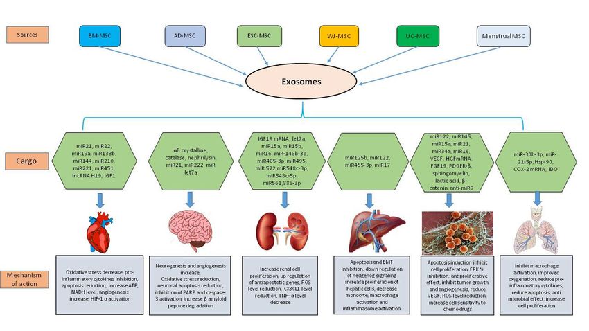
Life 2021, 11, 784 3 of 26
release [20]. Although most studies on the molecular mechanism of exosome release
Life 2021, 11, x FOR PEER REVIEW 3 of 28
are on
cancer, few (almost none) have reported on mesenchymal stem cell exosomes [21,22].
Figure 1. (A) Biogenesis, secretion, and cellular uptake of exosomes. The biogenesis of exosomes starts from the (i) early
1. (A) Biogenesis,
Figure endosomes secretion,
which mature and
into (ii) latecellular uptake
endosome, then of exosomes.
(iii) Thebodies
multivesicular biogenesis of exosomes
are formed starts fromofthe
by the invagination late(i) early
endosomes which
endosomal mature into
membrane, which(ii) late endosome,
is finally then
secreted as (iv) (iii) multivesicular
exosomes bodies
to the extracellular are in
matrix formed by the invagination
a mesenchymal stem cell. of
(B) The secreted
late endosomal exosomes
membrane, are is
which uptaken
finallybysecreted
a recipient cell exosomes
as (iv) in several ways
to theviz. (a) clathrin‐mediated
extracellular matrix inuptake, (b)
a mesenchymal
receptor‐mediated
stem cell. uptake
(B) The secreted or by the are
exosomes (c) membrane
uptaken by fusion event.
a recipient cell in several ways viz. (a) clathrin-mediated uptake,
(b) receptor-mediated uptake or by the (c) membrane fusion event.
2.2. Exosome Secretion and Internalization
RabThe release ofthe
GTPases, exosomes
largestinto the extracellular
family milieu isregulate
of small GTPases, governedmanyby an steps
orchestration
of membrane
of proteins viz. soluble N‐ethylmaleimide‐ sensitive factor attachment protein receptors
trafficking, including vesicle budding, transport of vesicles along actin and tubulin, and
(SNAREs), tethering factors, Rabs, and other Ras GTPases [15]. The SNARE proteins, R‐
membrane fusion [23], are also involved in exosome secretion. Several studies demon-
or Q‐SNAREs, have been reported to affect exosome release. Fader et al. showed that the
strated
R‐SNAREthat Rab family proteins
vesicle‐associated (Rab2b,protein
membrane Rab5a,7 Rab27a,
(VAMP7)Rab27b, Rab35,
is necessary for and Rab11) are
exosome
involved
release in the human leukemic cell line K562 [16]. Another R‐SNARE protein, YKT6, isthat the
in this process [24]. Additionally, it has also been shown by Yu et al.
tumor suppressor
required for exosomeprotein p53asmay
release, also
shown byinfluence exosome
two independent secretion
studies. Grossthrough regulating
et al. showed
transcription
that depletion genes of such
YKT6asdecreased
TSAP6 and theCHMP4C
level of [25].
TSG101,Apart from that,
WNT3A, and various
VPS26/35 stimuli
in and
changes
exosomeslike cell membrane
secreted from pH and the
human concentration
embryonic kidney ofHEK293
K+ maycellsalso trigger the secretion
[17]. Further,
Ruiz‐Martinez
of exosomes et al. showed a reduced level of exosome‐associated TSG101 after the
[26,27].
knockdown of YKT6 in A549 human lung cancer cells [18]. Similarly, in Drosophila S2
2.3.cells, depletion
Isolation of the Q‐SNARE
of Exosomes: syntaxin
The First Step towards1A Pharmaceuticalization
(Syx1A) decreased the release of EV
enriched v exosomes [19]. Wei et al. reported that pyruvate kinase type M2 (PKM2)
MSC-derived exosomes are being considered a novel tool for cell-free therapeu-
phosphorylates SNAP‐23, thus enabling exosome release [20]. Although most studies
ticson[28–31]; however,
the molecular the cardinal
mechanism step release
of exosome in evaluating the extent
are on cancer, of their
few (almost competence
none) have is
to successfully isolate and purify exosomes
reported on mesenchymal stem cell exosomes [21,22]. and obtain a good yield. Although a great deal
of experimentation
Rab GTPases, has been performed,
the largest there
family of small is stillregulate
GTPases, no uniformity
many steps in of
isolation
membrane methods;
but,trafficking,
by far, the technique
including considered
vesicle budding, best is “ultracentrifugation”
transport of vesicles along actin due to the superlative
and tubulin, and
quality of exosomes isolated within it and the ubiquity of its use [32,33]. Basic ultracen-
trifugation as an exosome isolation technique was introduced by Johnstone et al. [34] to
infer that vesicle shedding was an intermediate process during maturation to erythrocytes.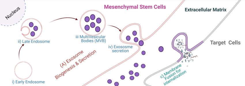
Life 2021, 11, 784                                                                                           4 of 26

                     There have been several advancements to this process, such as modulation in the number
                     of cycles of centrifugation [35] and optimization in protocols of differential ultracentrifu-
                     gation [36,37], density gradient ultracentrifugation [32,38–40], etc. Certain isolation kits
                     have also been devised to be considered a time-saving alternative showing reasonable
                     results [41–43]. The possibility of combining the beneficial effects of ultracentrifugation
                     and precipitation-based kits was explored by Ryu et al. [44]. They inferred that combining
                     the potential of both techniques was expedient for the isolation of small EVs, provided
                     a good output, and held no lags about their constitution, hence utilizable for catering to
                     massive sample-based critical clinical evaluations. Common protocols used for exosome
                     isolation are shown in Figure 2.
                           Despite abounding attempts to find a robust technique for uniform, use globally, many
                     shortcomings exist that need to be addressed, such as long duration, complicated protocols,
                     need for special equipment, lack of cost-effectiveness, limited utility, the requirement of
                     large volumes of sample, lack of specificity, truncated yield, low rate of recovery, dubious
                     purity, and risk of mechanical damage. These techniques, in their current form, are not
                     suitable for standardization. All the techniques have their advantages and drawbacks;
                     however, a technique that could satisfactorily channel the benefits of all pre-existing
                     technologies collectively while facilitating exosome isolation for downstream processing
                     at a translational level to visualize the use of exosomes for future applications like drug
                     formulation and delivery of therapeutics, is yet to be devised.

                     2.4. Characterization and Visualization of Exosomes
                           In order to entirely comprehend the functional, spatial and temporal properties of
                     exosomes, it is imperative to perceive its characterization, including labeling, imaging, and
                     visualization. As per the ISEV guidelines (2018) for EV characterization [45], EVs should
                     possess at least three positive protein markers, including at least one transmembrane/lipid-
                     bound protein, one cytosolic protein, and at least one negative protein marker, as mentioned
                     in the MISEV (2018) [45]. For characterizing individual vesicles, two different but com-
                     plementary techniques can be used [45]. Some most popular techniques include regular
                     fluorescence microscopy, SEM, TEM, Cryo-EM, AFM, NTA, Flow Cytometry [45]. Several
                     assays are performed to check the size range and distribution, concentration and spread of
                     exosomes, shape, trajectories and particle velocity, structural features including surface
                     proteins, chemical and physical properties, along with the cargo properties for the isolated
                     extracellular vesicles. Differences in these characteristics, especially in their size, shape,
                     and surface proteins, are checked to differentiate exosomes from other extracellular vesicles
                     like micro-vesicles and apoptotic bodies.
                           The basic characterization techniques standardized for the detection of exosomes
                     include nano-particle tracking analysis, which is used to assess the size and yield of the
                     exosomes. Further western blotting and flow cytometry can be used to detect exosome-
                     specific markers. The commonly used markers to identify exosomes in most experiments
                     are tetraspanins (CD9, CD63, CD81), TSG101, syntenin-1, Alix, Hsp90α, Hsp70, LAMP2,
                     cofilin, flotillin-1 [46–51]. Apart from identifying the presence of exosomes, this technique
                     is also applied to detect the expression of proteins ferried by exosomes [52,53]. Another
                     prime technique used for the characterization and visualization of exosomes is electron
                     microscopy, which provides comprehensive information about their configuration. How-
                     ever, exosomes via electron microscopy cannot be visualized in their native state due to the
                     pre-treatments required for this technique; furthermore, they appear rather cup-shaped
                     or saucer-shaped instead of their native round shape due to dehydration during sample
                     preparation [54–56]. A variation to EM is cryo-electron microscopy [57]. An advantage
                     of this technique is that exosomes can be visualized in their native round forms, thereby
                     avoiding artifacts due to fixation [58]. Extracellular vesicles can also be tracked by optical
                     microscopy within the visible light range (380–750 nm) using bioluminescence labeling
                     or fluorescence labeling by creating fusion proteins even though many alterations have
                     been experimented with in all of these techniques. Yet, no explicit tool or technology can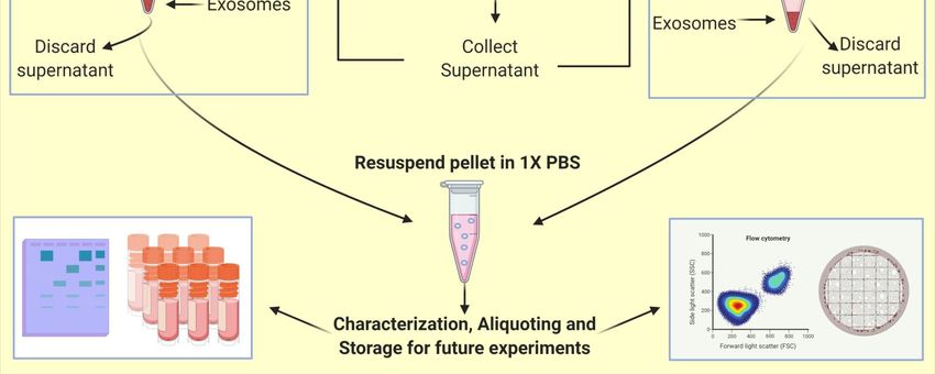
Life 2021, 11, 784                                                                                                                                5 of 26

                                      comprehensively describe all the facets of analysis of extracellular vesicles. While there are
                                       shortcomings
        Life 2021, 11, x FOR PEER REVIEW             in every technique when used individually, they may be counterweighed
                                                                                                                      5 of 28     by
                                      the benefits of another technique if explored in a synergistic manner [59,60].

              Figure 2. Isolation of Exosomes: exosomes are commonly isolated from the conditioned media. Some common
              2. Isolation ofsteps
      Figurepreprocessing      Exosomes:    exosomes
                                   are required         are
                                                 for both thecommonly
                                                              techniques,isolated  from
                                                                           including      the conditioned
                                                                                      collecting  conditionedmedia.   SomeMSCs,
                                                                                                              media from    common    preprocessing
                                                                                                                                 performing
      steps are  required  for  both  the techniques,    including   collecting  conditioned     media   from  MSCs,   performing  a
             a centrifugation round at 2000× g for 30 min to remove debris. Furthermore, the conditioned media can be subject to any centrifugation
      round ofat the two
                  2000 × gtechniques
                            for 30 min including,
                                          to remove Ultracentrifugation  (1) or Kit‐based
                                                        debris. Furthermore,                 methods (2)
                                                                                   the conditioned         for isolation
                                                                                                        media            of exosomes.
                                                                                                                 can be subject  to anyThese
                                                                                                                                         of the two
             exosomes   can be  further used for  characterization, aliquoting, and  storage  for future experiments.
      techniques including, Ultracentrifugation (1) or Kit-based methods (2) for isolation of exosomes. These exosomes can be
      further used for characterization, aliquoting, and storage for future experiments.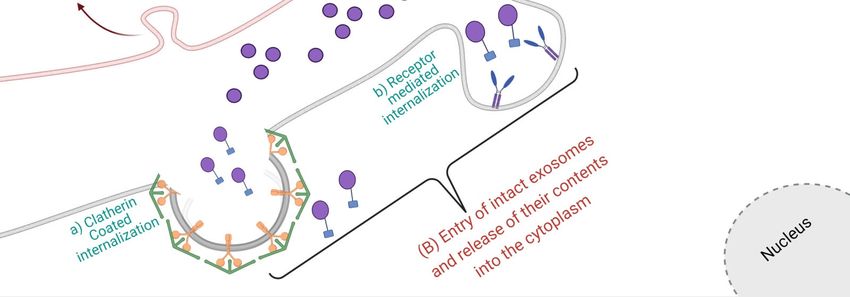
Life 2021, 11, 784                                                                                           6 of 26

                     3. The Therapeutic Nature of MSC Derived Exosomes by the Synergistic Functioning
                     of miRNAs and Proteins
                           MSCs partially function via a paracrine mechanism by the secretion of exosomes.
                     These small vesicles carry a broad range of cargo, which aids its therapeutic and regener-
                     ative capabilities in a vast spectrum of diseases and ailments. The therapeutic activities
                     of MSC-derived exosomes are mediated majorly via the horizontal transfer of its cargo
                     components, which then regulate and modulate the behavior of recipient cells by an array
                     of mechanisms [61]. MSC-derived exosomes exert some significant effects specifically
                     by virtue of the proteins and miRNAs they carry, apart from other bioactive molecules
                     like lipids, DNA, long coding mRNA, tRNA, etc. The proteins they carry may exhibit
                     any kind of functional, structural, or enzymatic activities, whereas miRNAs are small
                     noncoding sequences that may result in epigenetic modifications by the mechanism of
                     RNA silencing. These miRNAs can either cleave or destabilize the target mRNAs or result
                     in the modulation of mRNA transcription into protein, probably by reducing its efficacy.
                     The cargo carried by MSC-derived exosomes differs based on tissues from which MSCs
                     were derived [29]. The miRNAs constituted by MSC-derived exosomes can influence
                     various developmental and regulatory processes and also play a role in tumorigenesis and
                     tumor progression.
                           The RNA profile of MSC-exosomes derived from porcine adipose tissue was character-
                     ized by Eirin et al. [62] using RNA-seq technology. They claimed that vesicles from porcine-
                     derived MSCs preferentially contain discrete mRNAs and miRNAs when contrasted with
                     their parent cells and stated that several mRNAs encoding transcription factors, Golgi
                     proteins, and proteins for TGF-β signaling were found in the cargo of exosomes. These in-
                     cluded transcripts for POU3F1 (TST-1, OCT6), JARID2, p53-negative regulators- MDM434
                     & PEG3, HGF, HES1, TCF4, CEBPA, KF7, GOLGA4, ARRB1, IFT57, TGFB1, FURIN, GAS7,
                     HMGA2, LIN28B, which were involved in a diverse array of processes like stem cell func-
                     tionality, angiogenesis, splicing, cell death, adipogenesis, proteolysis, and organization of
                     genetic material. All these factors were found to be regulated by the miRNAs, like miR148a,
                     miR532-5p, miR378, let-7f, etc., which are also contained in exosomes.
                           It was also stated that cytoskeletal proteins and those related to mitochondrial and
                     calcium signaling were selectively segregated out of the vesicles. Furthermore, several
                     proteins, like EGF, FGF, and PDGF, involved in enhancing angiogenesis were found to be
                     significantly upregulated in MSCs under peripheral arterial disease-like conditions, and it
                     resulted in an enhanced angiogenic signaling profile of the exosomes. Therefore, they are
                     individually capable of inducing angiogenesis [63]. miRNAs are also known to improve
                     conditions related to heart disease. An enhanced expression of miR-29 and miR-24, and
                     downregulation of miRNAs, such as miR-21, miR-15, miR-34, miR-130, and miR-378, can
                     help in the alleviation of heart disorders by different mechanisms, such as limiting the
                     aortic vascular inflammation, inhibiting apoptosis of cardiac muscle cells, reducing the size
                     of infarction, preventing hypertrophy and preventing cardiac dysfunction, thereby also
                     reducing the risk of cardiac ischemic injuries [64].
                           The Vascular Endothelial Growth Factor (VEGF) enriched in these vesicles upregulate
                     the transmembrane ligand for Eph receptor tyrosine kinases (Ephrin-B2) and enhances
                     VEGF-induced angiogenesis, thereby inferring that these vesicles not only deliver the
                     protein VEGF but also upregulate its creation in the receiver cells [65]. This effect can also
                     be accounted for by several miRNAs contained in MSC exosomes involved in inducing
                     angiogenesis and alleviating diseases, including miR-132, miR-125a, miR-1246, miR-23a, etc.
                     Therefore, it can be stated that MSC-derived exosomes not only provide ready therapeutic
                     agents but also mechanisms for their regulation. A detailed proteomic characterization of
                     MSC-exosomes derived from bone marrow, adipose tissue, and umbilical cord revealed
                     355 proteins common to all sources. It was found that proteins in MSC-exosomes (Table 1)
                     that derived from bone marrow majorly functioned in the activation of granulocytes, in
                     regulating cell migration, and binding protein complexes and integrins. In the case of
                     exosomes derived from adipose tissue and the umbilical cord, the proteins were maximally
Life 2021, 11, 784 7 of 26
involved in activation of leukocytes and binding of cell adhesion molecules [66]. The
systematic study of miRNAs’ functionality is a very intricate process owing to the diversity
of miRNAs, including the pathways and proteins they target. However, bioinformatics
tools can help to establish miRNA landscapes that provide an “all-in-one approach” which
Life 2021, 11, x FOR PEER REVIEW 8 of 28
describe the relationship between miRNAs and their target genes and proteins. These tools
may be beneficial for enhancing means to search for miRNAs with inherent therapeutic
capabilities to formulate MSC derived exosome-based, off-the-shelf remedial agents [67].
Table 1. Comparison between MSCs and MSC derived Exosomes.
Table 1. Comparison between
MSCs MSCs and MSC derived Exosomes.
MSC Exosomes
Low stability High Stability
MSCs MSC Exosomes
High immunogenicity
Low stability Low immunogenicity
High Stability
Cannot
Highcross blood brain barrier
immunogenicity Can easily cross
Low blood brain barrier
immunogenicity
High‐cost
Cannot cross bloodstorage
brain barrier Low‐cost
Can easily storage
cross blood brain barrier
Can‐not beHigh-cost storage
readily used as off‐the‐shelf Potential forLow-cost storage
off‐the‐shelf availability
Can-not be readily used as off-the-shelf Potential for off-the-shelf availability
4. Therapeutic Potential of MSC Derived Exosomes in Various Diseases
4. Therapeutic Potential
As paracrine of MSC
effectors, Derived Exosomes
MSC‐derived exosomes havein Various
gained Diseases
much attention in the
lastAs
few years as a promising candidate for cell‐free therapeutics
paracrine effectors, MSC-derived exosomes have gained much in a wideattention
spectruminofthe
last few years as a promising candidate for cell-free therapeutics in a widelight
pathophysiological conditions. In the following paragraphs, we have shed on the of
spectrum
therapeutic potential of these exosomes in different diseases (Figure 3, Table
pathophysiological conditions. In the following paragraphs, we have shed light on the2).
therapeutic potential of these exosomes in different diseases (Figure 3, Table 2).
Figure 3. Mesenchymal stem cell exosome cargo in modulating cardiovascular diseases, neurological disorders, kidney
Figure 3. Mesenchymal stem cell exosome cargo in modulating cardiovascular diseases, neurological disorders, kidney
diseases, liver diseases, cancer, and lung diseases.
diseases, liver diseases, cancer, and lung diseases.
4.1. MSC Derived Exosomes in Cardiovascular Diseases
4.1. MSC Derived Exosomes in Cardiovascular Diseases
Several preclinical studies have demonstrated the efficacy of MSC-derived exosomes
Several preclinical studies have demonstrated the efficacy of MSC‐derived
forexosomes
CVD treatment. Lai et al. showed that the supernatant of human embryonic stem cell
for CVD treatment. Lai et al. showed that the supernatant of human embryonic
(ESC)-derived MSCs contained
stem cell (ESC)‐derived MSCssmall particles
contained (50–100
small nm in
particles diameter)
(50–100 corresponding
nm in diameter)
to corresponding
exosomes. When administered to a mouse myocardial ischemia/reperfusion
to exosomes. When administered to a mouse myocardial injury
model,
ischemia/reperfusion injury model, these exosomes remarkably reduced the infarct sizead-
these exosomes remarkably reduced the infarct size [68]. The authors also
ministered
[68]. The exosomes secreted
authors also from human
administered exosomesESC-derived MSCs
secreted from to a mouse
human modelMSCs
ESC‐derived of AMI
(Acute Myocardial Infarction), showing a reduced infarct size and improved cardiac
to a mouse model of AMI (Acute Myocardial Infarction), showing a reduced infarct size func-
and improved cardiac function [69]. Furthermore, they demonstrated that the
phosphorylation of Akt and GSK3 (possessing anti‐apoptotic effects) significantly
increased, and that c‐jun N‐terminal kinase (possessing proapoptotic effects) significantly
decreased in cardiac tissue following exosome administration. Bian et al. collected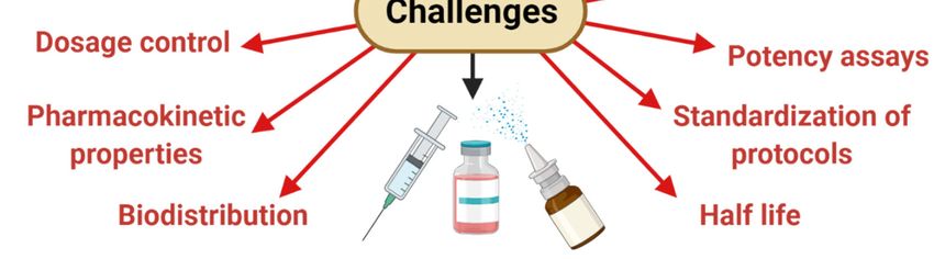
Life 2021, 11, 784                                                                                          8 of 26

                     tion [69]. Furthermore, they demonstrated that the phosphorylation of Akt and GSK3
                     (possessing anti-apoptotic effects) significantly increased, and that c-jun N-terminal ki-
                     nase (possessing proapoptotic effects) significantly decreased in cardiac tissue following
                     exosome administration. Bian et al. collected extracellular vehicles (EVs) from hypoxic
                     human BMMSCs and administered the EVs to a rat AMI model. The study showed that
                     EV administration significantly reduced infarct size, restored cardiac function, and stim-
                     ulated angiogenesis in the ischemic zone [70]. Feng et al. demonstrated that exosomes
                     secreted from mouse BMMSCs after ischemic preconditioning contained a greater amount
                     of miR-22 [71]. When administered to mice with AMI, these miR-22-enriched exosomes
                     significantly reduced infarct size and cardiac fibrosis, possibly through the downregulation
                     of methyl-CpG-binding protein 2. In another study, Yu et al. used MSCs overexpressing
                     the transcription factor GATA-4 (MSC_GATA-4) and demonstrated that the administration
                     of MSC_GATA-4-derived exosomes restored cardiac function and reduced infarct size in a
                     rat model of AMI. The study also showed that MSC_GATA-4-derived exosomes expressed
                     a greater amount of miRNAs, particularly miR-19a, which appeared to be involved in the
                     cardioprotective effect of MSC_GATA-4-derived exosomes via the downregulation of phos-
                     phatase and tensin homolog (PTEN) and subsequent activation of anti-apoptotic Akt [72].
                     Similar cardioprotective roles of MSC exosomes were also shown by Wang et al. using
                     endometrium-derived MSCs (EnMSCs). Their study suggested that miR-21 contained in
                     EnMSC-derived exosomes mediated cardioprotective effects via the downregulation of
                     PTEN and subsequent activation of Akt, resulting in the upregulation of Bcl-2 and vascular
                     endothelial growth factor [73].
                           Recently, Huang et al. showed that the therapeutic efficacy of MSC-derived exosomes
                     in AMI can be enhanced by atorvastatin (ATV), one of the most widely used lipid-lowering
                     drugs for patients with coronary heart disease. The authors showed that exosomes derived
                     from ATV-pretreated MSCs (MSCATV -Exo) significantly improved cardiac function and
                     promoted blood vessel formation compared with exosomes derived from non-pretreated
                     MSCs (MSC-Exo) via an increased level of lncRNA H19 expression. [74]. The decrease in
                     apoptosis in H9C2 cardiomyocyte cells by administration of BMMSC derived exosomes
                     enriched in miR-144 was demonstrated by Wen et al. [75]. The exosomes mediate this
                     function by targeting the PTEN/AKT pathway (decreased PTEN expression and increased
                     p-AKT expression), as evident from the study. Similarly, a study by Cheng et al. also
                     shows the efficacy of exosomes in attenuating post-infarction cardiac apoptosis. The
                     authors showed that hypoxia challenged MSC-derived exosomes enriched in miR-210,
                     reduced infarct size, and improved heart function after coronary ligation both in vitro and
                     in vivo stress [76]. Preclinical studies have also reported the beneficial effects of exosome
                     administration on neurological recovery following stroke induction. Xin et al. found
                     that the systemic administration of rat BMMSC-derived exosomes after inducing stroke
                     via ligating the middle cerebral artery significantly enhanced neurological recovery and
                     stimulated neurogenesis and angiogenesis in the ischemic boundary zone [77]. The authors
                     also demonstrated that administration of BMMSCs overexpressing miR-133b (MSCs_miR-
                     133b) in a rat stroke model enhanced the recovery of neurological function. Furthermore,
                     they showed that the expression of connective tissue growth factor (CTGF), a target for
                     miR-133b, was significantly reduced in the ischemic boundary zone after MSCs_miR-
                     133b administration; this suggests that exosome-derived miR-133b was implicated in the
                     MSC-mediated recovery of neurological function in the model. However, Doeppner et al.
                     showed that improvement in neurological function and stimulation of neurogenesis and
                     angiogenesis at the ischemic boundary remained the same in both BMMSC administration
                     and BMMSC-derived EV administration [78].

                     4.2. MSC Derived Exosomes in Neurodegenerative Diseases
                         The ability of exosomes to cross the BBB (Blood–Brain Barrier) establishes them as a
                     potential candidate for drug delivery to the brain in various neurodegenerative diseases.
                     Several studies support this concept. For instance, it has been suggested that exosomes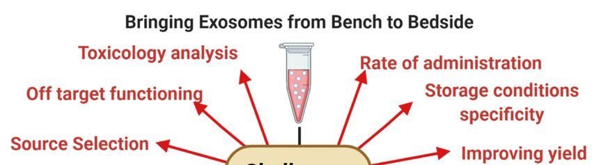
Life 2021, 11, 784                                                                                         9 of 26

                     enter CNS via two mechanisms: uptaken by endothelial cells and crossing into the cell
                     through transcytosis, or crossing intercellular junctions between endothelial cells and
                     entering the CNS [79,80]. It has been shown that exosome-associated miR-105 can down-
                     regulate the expression of ZO-1, a critical molecular component of tight junctions, hence
                     demolishing the barrier action of endothelial cells [81]. Exosomes have produced beneficial
                     effects in a variety of models for neurodegenerative diseases, such as Parkinson’s disease.
                     Jarmalavičiūtė et al. reported that exosomes obtained from human dental pulp stem cells
                     could suppress apoptosis of dopaminergic neurons following treatment with 6-OHDA
                     (6-Hydroxydopamine) in a Parkinson’s disease model [82]. 6-OHDA induces apoptosis
                     through the generation of reactive oxygen species (ROS), suggesting that exosomes can
                     decrease the sensitivity of dopaminergic neurons to oxidative stress [83]. Furthermore, it
                     has been shown that chaperone αB crystalline, a pigment produced by the human retinal
                     epithelium, is found inside exosomes, which may play a protective role against oxidative
                     stress in retinal cells [84]. Haney et al. showed that mouse macrophage-derived exosomes
                     remarkably enhanced cell survival against 6-OHDA-induced injury [85]. Interestingly,
                     exosomes caused a reduction in ROS levels in activated macrophages regardless of whether
                     they were loaded with catalase, suggesting that exosomes might function similarly in
                     microglial cells under neuroinflammatory conditions. Furthermore, in vivo studies have
                     suggested that exosomes loaded with catalase causes a decrease in microgliosis and im-
                     proves the survival of dopaminergic neurons in mice treated with 6-OHDA [86]. Hence,
                     it can be suggested that stem cell-derived exosomes produce neuroprotection through
                     reduced oxidative stress.
                           Several studies have suggested a neurotherapeutic behavior of exosomes obtained
                     from adipose tissue-derived MSCs (ADMSCs). ADMSCs can secrete neprilysin-bound
                     exosomes [87]. As a type II membrane-associated metalloendopeptidase, neprilysin is
                     reportedly a critical proteolysis product in the cleavage of β-amyloid. It was demonstrated
                     that the expression and function of neprilysin were reduced in patients with Alzheimer’s
                     disease [88]. ADMSC-derived exosomes display neprilysin-related enzyme activity and
                     are involved in reducing β- amyloid levels in neuroblastoma cells. Additionally, these
                     exosomes express greater amounts of neprilysin compared with BMMSCs, emphasizing
                     functional and activity differences in exosomes obtained from different sources [89]. It was
                     also indicated that exosomes obtained from murine adipose tissue-derived MSCs improved
                     the survival of human neuroblastoma cells and protected the murine hippocampal neurons
                     from oxidative damage [90]. Furthermore, the authors of this study showed that exosomes
                     obtained from murine ADMSCs enhanced remyelination and stimulated the progression
                     of oligodendroglial progenitors [90]. These neuroprotective effects of exosomes are also
                     supported by Bonafede et al. in an in vitro model of ALS (Amyotrophic lateral sclerosis).
                     The study showed that administration of murine ADMSC-derived exosomes in a motor-
                     neuron-like cell line expressing a high amount of SOD1, hence under oxidative stress,
                     protects the motor-neuron-like cells from oxidative injury [91]. These results highlight
                     the potential of applying MSC-derived exosomes as a therapeutic tool to treat motor
                     neuron disorders.

                     4.3. MSC Derived Exosomes in Kidney Diseases
                          Recent studies on the therapeutic potentials of MSC-derived EVs suggest their ability
                     to regenerate injured renal cells in experimental acute kidney injury (AKI) and chronic
                     kidney disease (CKD) models. Results from these studies suggest that EVs exert their
                     trophic and reparative effects by shuttling their cargo of genes, microRNAs, and proteins
                     to recipient cells in the kidney, attenuating renal injury and improving its recovery compe-
                     tence. In an in vitro model of cisplatin-induced AKI, Tomasoni et al. demonstrated that
                     coincubation of damaged proximal renal tubular epithelial cells with MSC-derived EVs,
                     which are selectively enriched with IGF1R mRNA, enhanced cell proliferation and repair,
                     suggesting that the transfer of this gene to tubular cells is an important mechanism by
                     which MSCs confer renoprotective effects in experimental AKI [92]. Bruno et al. reported
Life 2021, 11, 784 10 of 26
that human adult MSC-derived microvesicles, which include exosomes, mimicked the pro-
tection against AKI as provided by intravenously administered MSC [93]. RNase treatment
of EVs abrogated EV-induced in vitro proliferation and resistance to apoptosis, indicating
that the mRNAs shuttled by EVs activate a transcriptional program of repair in recipient
cells. In line with this observation, EVs released from kidney-derived MSCs preincubated
with RNase failed to ameliorate TGF-β1-induced peritubular capillary rarefaction and tubu-
lointerstitial fibrosis in mice with unilateral ureteral obstruction (UUO) [94]. Bruno et al.
further showed that a single intravenous administration of MSC-derived EVs improved
mouse survival after injecting a lethal dose of cisplatin, whereas multiple EV injections fur-
ther decreased mortality and preserved renal structure and function [95]. Administration
of MSC-derived EVs upregulated the expression of the anti-apoptotic genes BCLX, BCL2,
and BIRC8, but downregulated the expression of the pro-apoptotic genes CASP1, CASP8,
and LTA in cisplatin-treated human tubular epithelial cells, suggesting that modulation
of apoptosis may contribute to MSC-derived EV-induced renal repair. Using an in vitro
model of ischemia-reperfusion injury (IRI) induced by ATP depletion in renal proximal
tubular epithelial cells, Lindoso et al. found that incorporation of MSC-EVs in damaged
cells modulated several microRNAs related to important processes in renal recovery [96].
Renal oxidative stress and inflammation are also known to be modulated by MSC-derived
EVs. Renal expression of the NADPH oxidase (NOX)-2 is upregulated in rats with IRI but
not in those treated with intravenous MSC-derived EVs [97]. Interestingly, this intervention
not only alleviates oxidative stress but also reduces apoptosis and enhances renal cell
proliferation, suggesting that post-transcriptional regulation of NOX2 in renal recipient
cells may be implicated in MSC-derived EVs-induced renal repair. In rats with IRI, MSC-
derived EVs alleviated renal inflammation and improved renal function by suppressing
the expression of C-X3-C motif ligand-1 (CX3CL1), a potent chemo-attractant protein for
macrophages that also promotes interstitial fibrosis [98]. Interestingly, MSC-derived EVs
were enriched with miR-16, miR-15b, and miR-15a, all of which target CX3CL1, suggesting
that post-transcriptional modulation of CX3CL1 is an important mechanism by which
MSC-derived EVs mitigate inflammation and renal injury in ischemic AKI. Promising
results from these experimental studies provided the impetus to apply MSC-derived EVs
to address the clinical needs of patients with renal disease. However, only a few clinical
trials investigated the safety and therapeutic efficacy of MSC-derived EVs in patients with
kidney diseases. Nassar et al. published their results of a phase II/III clinical trial using
cord tissue MSC-derived EVs to ameliorate the progression of chronic kidney disease
(CKD) [99]. In this study, 20 patients who had been diagnosed for more than 6 months
with chronic kidney disease (eGFR 15–60 mg/mL) were treated with two doses (1 week
apart) of MSC-derived EVs (100 µg/kg/dose). Patients treated with MSC-derived EVs
exhibited improved eGFRs and urinary albumin creatinine ratio, as well as significant
decreases in BUN and creatinine after 1 year. In addition, the patients showed a signifi-
cant increase in plasma levels of TGF-B and IL-10 with persistent, significant decreases
in TNF-α. Similarly, Ingato et al. showed their results from a single-center, randomized,
placebo-controlled, phase II/III clinical pilot study that recruited 40 patients with stage
III-IV CKD (eGFR between 15–60 mg/mL/min), who were randomized to receive either
placebo or two doses (first intravenous and second intraarterial) of MSC-derived EVs, one
week apart [100]. After a 12-month follow-up, EV-treated patients exhibited a significant
improvement in renal function (improved eGFR and decreased serum creatinine, BUN,
and albuminuria). Clinical improvement paralleled changes in plasma levels of several
immune-inflammatory markers, including TNF-α, TGF-β1, and IL-10. These observations
suggest that MSC-derived EVs are safe and can ameliorate the inflammatory immune
reaction and improve the overall kidney function in CKD patients.
4.4. MSC Derived Exosomes in Liver Diseases
MSC administration in animal models of liver fibrosis/cirrhosis has been shown to
ameliorate the disease [101]. Similar results are also found using the MSC-conditionedLife 2021, 11, 784 11 of 26
media [102], suggesting that MSC might achieve their role in vivo through their secreted
exosomes. Using a carbon tetrachloride (CCl4 )-induced liver injury model in Kunming
mice, Li et al. showed that the exosomes derived from human umbilical cord MSCs amelio-
rate liver fibrosis by inhibiting both the epithelial-mesenchymal transition of hepatocytes
and collagen production. The exosomes were found to significantly restore the serum
aspartate aminotransferase activity and inactivate the TGF-β1/Smad signaling pathway
by decreasing collagen type I/III and TGF-β1 and the phosphorylation of Smad2 [103].
Another study showed that chorionic plate-derived MSCs can release exosomes containing
miR-125b, mediate miR-125b transfer between MSCs and target cells, such as Hedge-
hog (Hh)-responsive hepatic stellate cells (HSCs), and thus alleviate hepatic fibrosis in
CCl4 -treated Sprague–Dawley rats by impeding the activation of Hh signaling via the
inhibition of Smo expression [104]. Lou et al. showed that exosomes produced by adi-
pose tissue-derived MSCs (AD-MSC-122) expressing miR-122 were more effective than
those expressing scramble miRNA or naive exosomes in reducing the proliferation and
activation of the human HSC cell line LX2 or primary HSCs from C57BL/6 mice [105].
AD-MSC-122-derived exosomes could transfer miR-122 into HSCs cells and then regulate
the expression of miR-122-target genes, such as P4HA1 and IGF1R, which are involved
in the proliferation and collagen maturation of HSCs [106]. These studies indicate that
MSCs show their therapeutic efficacy via miR-122 present in the exosomes, thereby repre-
senting a new strategy for treating liver fibrosis. The therapeutic effects of MSC-derived
exosomes/EVs have been reported in several experimental models of acute kidney, cardiac,
and lung injury. However, only a few studies are currently available on the therapeutic
effects of MSC exosomes in acute liver injury. Tan et al. found that HuES9.E1 MSC-
derived exosomes elicit hepatoprotective effects in both in vitro models of acetaminophen
or H2 O2 -induced hepatocyte injury and a C57BL/6 mouse model of CCl4 -induced acute
liver injury, through an increase in hepatocyte proliferation, demonstrated by elevated
proliferating cell nuclear antigen and high cell viability. The increased survival rate is
associated with the upregulation of genes involved in the priming phase liver regeneration,
which subsequently leads to high expression of proliferation proteins (proliferating cell
nuclear antigen and Cyclin D1), the anti-apoptotic gene Bcl-xL, and the signal transducer
and activator of transcription 3 (STAT3) [107]. Recently, a study by Shao et al. showed that
administration of human umbilical cord mesenchymal stem cells (hUC-MSCs)-derived
miR-455-3p-enriched exosomes suppressed monocyte/macrophage activation and alle-
viated acute liver injury by inhibiting IL-6 signaling (by targeting the PIK3r1 gene) in a
carbon tetrachloride (CCl4 )-induced liver injury in a mice model [108]. Furthermore, a
study on a lipopolysaccharide/d-galactosamine-induced acute liver failure mice model by
Liu et al. showed that adipose tissue-derived MSC (AMSC) exosomes alleviate acute liver
failure (ALF) by reducing serum alanine aminotransferase and aspartate aminotransferase
levels and hepatic inflammasome activation. The miR-17, which can suppress NLRP3
inflammasome activation by targeting TXNIP expression, is abundant in AMSC-Exo cargo;
this clearly indicates that AMSC-Exo-based therapy may be a promising approach for
treating TXNIP/NLRP3 inflammasome-related inflammatory liver diseases [109].
4.5. MSC Derived Exosomes in Cancer
The role of MSCs in cancer is a debatable topic. This area of cancer research, in the
light of exosomes, has been gaining momentum in the past few years. Several studies have
shown that MSCs act as a double-edged sword in both tumor suppression, or progression
in different tumor models [110–113]. However, the mechanisms remain elusive. The EVs
secreted by MSCs contain paracrine factors through which they mediate their effects on
tumor progression [114]. For instance, exosomes released from multiple myeloma patient
BMMSCs promote multiple myeloma tumor growth in SCID-beige mice. BMMSC derived
exosomes promote gastric or colon tumor growth in BALB/c nu/nu mice by enhancing the
expression of vascular endothelial growth factor (VEGF) in tumor cells [115,116]. They fa-
cilitate nasopharyngeal carcinoma progression and migration in non-obese diabetic/severeLife 2021, 11, 784 12 of 26
combined immunodeficient (NOD/SCID) mice by activating the FGF19-FGFR4-dependent
ERK signaling cascade and epithelial-mesenchymal transition [117]. Vallabhaneni et al.
demonstrate that the exosomes secreted from hMSCs are rich in miR-21 and 34a, supporting
breast cancer cell proliferation and metastasis [118].
However, antitumor effects are also exhibited by exosomes [119]. Bruno et al. found
that exosomes from human BM-MSCs inhibit the growth and survival of three different
human tumor cell lines. Similar results were observed in NOD/SCID mouse models [120].
Furthermore, exosomes derived from MSCs overexpressing the TRAIL gene-induced
apoptosis in a range of cancer cell lines. In another study, mouse BMMSC-derived exosomes
were found to suppress tumor progression and angiogenesis in the mouse breast cancer
cell line 4T1 by downregulating VEGF expression in vitro and in vivo via shuttling miR-16,
which is a known effector of VEGF enriched in MSC-derived exosomes [121].
A different study showed that exosomes derived from menstrual stem cells suppress
the secretion of pro-angiogenic factors in prostate tumor cell line PC3 in a reactive oxygen
species-dependent manner and inhibit prostate tumor angiogenesis in PC3-bearing NOD
SCID gamma mice [122]. Exosomes derived from BMMSCs inhibit cell cycle progression
and induce apoptosis in HepG2 cells. Ko et al. showed that AD-MSC-derived exosomes
in rat N1S1 cells, an orthotopic HCC model, can promote NKT cell antitumor responses
in rats, thereby facilitating hepatocellular carcinoma (HCC) suppression and low-grade
tumor differentiation [123].
In addition to modulating tumor development, MSCs-derived exosomes have been
shown to influence tumor chemosensitivity. Exosomes from human umbilical cord MSCs
significantly induce the resistance of gastric cancer cells to 5-fluorouracil in a BALB/c
nu/nu mice subcutaneous xenograft tumor model by antagonizing 5-fluorouracil-induced
apoptosis and enhancing the expression of multidrug resistance-associated proteins [124].
In another study, exosomes from anti-miR-9-transfected BMMSCs delivered anti-miR-9 into
temozolomide-resistant glioblastoma multiforme cells and reversed their chemoresistance
by affecting the expression of the multidrug transporter P-glycoprotein [125]. Lou et al.
demonstrated that exosomes from miR-122-modified AD-MSC (122-Exo) can mediate
miR-122 transfer between AD-MSCs and HCC cells, thereby enhancing cell sensitivity to
chemotherapeutic agents by regulating miR-122-target gene expression in HCC cells [105].
The use of MSC-derived exosomes in cancer therapy must be conducted with caution
because their role in tumor growth remains elusive. At the same time, their role in tumor
suppression also establishes them as promising candidates in cell-free therapy for cancer.
A better understanding of the mechanisms involved in regulating MSC-derived exosomes
is important to determine their true role in cancer progression and use as a possible
therapeutic agent in cancer treatment.
4.6. MSC Derived Exosomes in Lung Diseases
Several studies have shown the efficacy of MSC-derived exosomes as immunosup-
pressive, anti-inflammatory agents in lung disease [126–130]. The potential of exosomes
from BMMSC has also been shown by Khatri et al. in influenza virus-induced acute lung
injury in a pig model. The exosomes express cyclooxygenase (COX)-2 mRNA, the enzyme
that induces prostaglandin E2 (PGE2) synthesis, which in turn reprogram proinflammatory
monocyte-macrophages (M1) to the anti-inflammatory (M2) type. Additionally, MSC exo-
somes interact with immune cells and cause the production of transforming growth factor
(TGF)β and T-regulatory cells (Tregs). Tregs cause a decrease in the haemagglutination
activity of influenza viruses and virus replication [131]. Similarly, Yi et al. also reported the
inhibition of Serum amyloid A3 by exosomal miR-30b-3p from bone marrow [132]. Fur-
thermore, the authors showed increased cell proliferation and reduction in the apoptosis in
type II alveolar epithelial cells of lungs after BMMSC exosome treatment. The role of MSC
exosome miRs was also demonstrated by Wei et al. In a lung ischemia/reperfusion injury
murine model, the authors showed a reduction of edema and dysfunction in lungs by
miR-21-5p [133]. The miR-21-5p further decreases the level of proinflammatory cytokinesLife 2021, 11, 784 13 of 26
in the lungs. Heat shock proteins (hsp-90) present in MSC exosomes also play a role in E.
coli-induced acute lung injury, as demonstrated by Varkouhi et al. [134]. Exosomes derived
from MSC in the human umbilical cord are rich in hsp-90, causing a reduced alveolar
protein leak and reduction in alveolar TNFα concentrations. Additionally, some reports
showed the antimicrobial effect of bone marrow MSC exosomes in E. coli-induced acute
lung injury [135,136].
MSC Derived Exosomes in COVID-19
COVID-19 is the most recent pandemic lung disease to shake the world with its highly
infectious and deadly nature. Several clinical trials have employed MSCs and their exo-
somes against the pathophysiology of COVID-19, showing astonishing outcomes, includ-
ing the alleviation of symptoms, faster recovery, and phenomenal regeneration [137–139].
COVID-19 is caused by SARS-CoV-2 coronavirus, which creates a storm of cytokines in
the lungs by invoking major proinflammatory factors, such as CCL-2, CXCL-10, IL-2, IL-6,
IL-7, IL-1β, IFN, and TNFα, which attract immune cells and lead to extreme inflammatory
conditions [140–144]. It results in deadly damage to the lungs and other organs. In order
to combat such a situation, the small size, specificity, and immunosuppressive features of
MSC-exosomes can be used. In fact, in a nonrandomized open-label cohort study, Sengupta
et al. showed that intravenous administration of BMMSC exosomes improved patients’
clinical status and oxygenation, as evident by the improvements in absolute neutrophil and
lymphocyte counts [145]. Additionally, they also showed that these exosomes reduced the
level of C-reactive protein, ferritin, and D-dimer in the lungs. However, future randomized
controlled trials (RCTs) are needed to determine its therapeutic potential.
Table 2. A summary of the mechanism of action of MSC-derived exosomes against different diseases.
Disease Cell Source Exosome Content Mechanism of Action Reference
Out of the complex mixture of
nutrients, growth factors,
microvesicles etc. in the
Myocardial
conditioned media, exosomes
ischemia/reperfusion hESC derived MSC Not given [68]
are specifically responsible for
injury
tissue repair and
cardioprotective effects in case
of ischemia/reperfusion injury
Peroxiredoxins and Increased levels of ATP and
glutathione NADH, decreased oxidative
Acute Myocardial
hESC derived MSC S-transferase, stress, increased [69]
Infarction
enzymatically phosphorylated-Akt and
active CD73 phosphorylated-GSK-3β
Sonic hedgehog, Increased angiogenesis, HIF-1
Myocardial Infarction hBMMSC [70]
PDGFR alpha activation
Targeting the methyl CpG
Ischemic heart Mice BMMSC miR22 [71]
binding protein 2 (Mecp2)
Anti-apoptotic effect,
MSC overexpressing miR-19a, miR-451, reduction in PTEN and BIM
Myocardial Infarction [72]
GATA-4 miR-221, IGF-1 expression, Akt/ERK
signalling pathway
Human
Myocardial Infarction Endometrium-derived miR-21 PTEN/Akt pathway [73]
MSC (EnMSC)
Increased angiogenesis,
Acute Myocardial Atorvastatin treated
lncRNA H19 inhibited the elevation of IL-6 [74]
Infarction MSC
and TNF-α
H9C2 cardiomyocyte Mice BMMSC miR-144 PTEN/Akt pathway [75]Life 2021, 11, 784 14 of 26
Table 2. Cont.
Disease Cell Source Exosome Content Mechanism of Action Reference
Reduce apoptosis of
cardiomyocytes, AIFM3/p53
Myocardial Infarction Mice BMMSC miR-210 [76]
and PI3K/Akt
signaling pathways
Enhanced neurological
Stroke Rat BMMSC miR-133b recovery, stimulated [77]
neurogenesis and angiogenesis
Stimulated neurogenesis
Stroke hBMMSC Not given [78]
and angiogenesis
Suppressed 6-OHDA-induced
Human dental pulp
Parkinson’s disease Not given apoptosis in [82]
stem cells
dopaminergic neurons
Age-related macular Retinal pigment Inhibition of caspase 3 and
αB crystallin [84]
degeneration epithelial cells PARP activation
Mouse macrophage cell
Parkinson’s disease Catalase Reduced Oxidative stress [85]
line
β-amyloid peptide
Alzheimer’s disease hADMSC Neprilysin [86]
degradation
SH-SY 5Y human Reduction of
murineADMSC Not given [88]
neuroblastoma cells neuronal apoptosis
Amyotrophic lateral miR21, miR222, Apoptosis-inhibiting pathway,
murineADMSC [87]
sclerosis miRlet7a cell cycle progression
Increased proximal renal
Acute kidney injury hBMMSC IGF-1R tubular epithelial [92]
cell proliferation
Induced de-differentiation of
Acute kidney injury hBMMSC mRNA mature cells, [93]
triggered proliferation
Upregulated anti-apoptotic
Acute kidney injury hMSC Not given genes Bcl-xL, Bcl2 and BIRC8 [96]
in tubular epithelial cells
let7- a, miR-148b-3p,
Downregulation of apoptotic
375, 410, 451, 485-3p,
Renal proximal tubular genes, SHC1 mediated
hBMMSC 495, 522, 548c-3p, [97]
epithelial cells inhibition of
548c-5p, 561, and
EGFR-Ras-ERK pathway
886-3p
Renal
Supress expression of NOX2,
ischemia/reperfusion hWJMSC Not given [98]
ROS level reduction
injury
Renal
miR-15a, miR-15b and
ischemia/reperfusion hWJMSC Downregulation of CX3CL1 [99]
miR-16
injury
Increase in TGF-β1 and IL-10
Chronic kidney disease hCBMSC Not given levels, decrease in [100]
TNF-α levels
Inhibition of hepatocellular
Chronic liver fibrosis murineBMMSC Not given apoptosis, inhibition of [98]
proliferation of LX-2
Inhibition of EMT, inactivation
Liver fibrosis hUCMSC Not given of the [103]
TGF-β1/Smad signallingLife 2021, 11, 784 15 of 26
Table 2. Cont.
Disease Cell Source Exosome Content Mechanism of Action Reference
chorionic plate-derived
Downregulation of
Liver fibrosis mesenchymal stem miR-125b [104]
hedgehog signaling
cells (CP-MSCs)
proliferation and collagen
Liver fibrosis ADMSC miR-122 [106]
maturation of HSCs
Up regulation of PCNA and
hESC-derived cyclin D1, inhibition of the
Liver Injury Not given [107]
HuES9.E1 MSC APAP- and H2 O2 -induced
hepatocytes apoptosis
PI3K signaling, inhibition of
IL-6-related signaling
Acute liver Injury hUC-MSC miR-455-3p pathways, suppress [108]
monocyte/macrophage
activation
Suppress NLRP3
Acute liver failure miceADMSC miR-17 [109]
inflammasome activation
Inhibit cell proliferation,
Prostate cancer hADMSC miR-145 [113]
inducing apoptosis
Inhibited the growth of
Multiple Myeloma hBMMSC miR-15a [114]
MM cells
Activation of AKT and
Renal cancer hWJMSC HGF mRNA ERK1/2 signaling pathways, [115]
reduction of HGF expression
Human gastric Inhibition of
hBMMSC VEGF [116]
carcinoma ERK1/2 activation
Nasopharyngeal FGF19-FGFR4-dependent
hBMMSC FGF19 [117]
carcinoma ERK signaling
miR-21, miR-34a,
PDGFR-β, TIMP-1, and
Breast cancer cell line Inhibited cell death,
hSDMSC TIMP-2, [118]
(MCF-7) anti-proliferative effect
sphingomyelin, lactic
acid, glutamic acid
Osteosarcoma (MG63)
and gastric cancer hBMMSC Not given Hedgehog signaling pathway [120]
(SGC7901) cells
Inhibition of tumor growth
Mouse breast cancer
miceBMMSC miR-16 and angiogenesis, reduces the [121]
cell line (4T1)
VEGF expression
Prostate Reduction in VEGF secretion
Menstrual MSC Not given [122]
Adenocarcinoma PC3 and NF-κB activity, lower ROS
Promoted NKT-cell antitumor
Hepatocellular
ratADMSC β-catenin responses, low-grade tumor [123]
carcinoma
differentiation
Glioblastoma Reduced miR-9, cell surface
hBMMSC anti-miR-9 [125]
multiforme P-gp
Hepatocellular miR-122-modified Enhancing cell sensitivity to
miR-122 [106]
carcinoma AD-MSC chemotherapeutic agentsYou can also read