Control of the Antitumor Immune Response by Cancer Metabolism
←
→
Page content transcription
If your browser does not render page correctly, please read the page content below
cells
Review
Control of the Antitumor Immune Response by
Cancer Metabolism
Charlotte Domblides 1,2 , Lydia Lartigue 3,†       and Benjamin Faustin 1,4, *,†
 1    Bordeaux University, CNRS, UMR 5164, ImmunoConcEpT, 33000 Bordeaux, France;
      charlotte.domblides@chu-bordeaux.fr
 2    Department of Medical Oncology, Hôpital Saint-André, Bordeaux University Hospital-CHU,
      33000 Bordeaux, France
 3    Curematch, Inc., 6440 Lusk Bvld, San Diego, CA 92121, USA; llartigue@curematch.com
 4    Cellomet, CGFB, 146 Rue léo Saignat, F-33000 Bordeaux, France
 *    Correspondence: bfaustin@immuconcept.org; Tel.: +33-(0)557-571-470
 †    These authors contributed equally to this work.

 Received: 22 December 2018; Accepted: 28 January 2019; Published: 31 January 2019

 Abstract: The metabolic reprogramming of tumor cells and immune escape are two major hallmarks
 of cancer cells. The metabolic changes that occur during tumorigenesis, enabling survival and
 proliferation, are described for both solid and hematological malignancies. Concurrently, tumor
 cells have deployed mechanisms to escape immune cell recognition and destruction. Additionally,
 therapeutic blocking of tumor-mediated immunosuppression has proven to have an unprecedented
 positive impact in clinical oncology. Increased evidence suggests that cancer metabolism not only
 plays a crucial role in cancer signaling for sustaining tumorigenesis and survival, but also has wider
 implications in the regulation of antitumor immune signaling through both the release of signaling
 molecules and the expression of immune membrane ligands. Here, we review these molecular events
 to highlight the contribution of cancer cell metabolic reprogramming on the shaping of the antitumor
 immune response.

 Keywords: cancer; metabolism; immunity

1. Introduction
      Both the innate and adaptive immune systems have now established roles in the host defense
against cancers through various mechanisms, which are raising an unprecedented development of
modern cancer immunotherapies. Several cancers (i.e., from early neoplasia to advanced stages)
are immunogenic because of their high mutational rate [1], leading to the expression of neoantigens
recognized by immune lymphoid cells for further elimination. However, immunoediting (a hallmark
of cancer cells) [2] is a dynamic process by which a tumor changes its immunogenicity in order to
escape the immune response. It was shown to have three phases: elimination, equilibrium and escape.
The elimination leads to the rejection of tumor cells by the intervention of innate and adaptive immune
systems. The equilibrium state means that the immune systems have not permitted the complete
elimination of the tumor cells. This is due to the genomic instability of cancer cells that can generate
clones with reduced immunogenicity. During this phase, immune systems can control the tumor
growth. The tumor cells enter a dormant state, sometimes for several years. These first two phases are
not clinically visible, but new tumor cell variants can emerge, selecting the less immunogenic cells or
cells that harbor immune resistance mutations. This leads to the final step of immune escape, where
tumor cells can proliferate and overtake host immune defenses. Immune cell response depends on the
expression of genetic programs, leading to the transition between quiescent, activated and memory

Cells 2019, 8, 104; doi:10.3390/cells8020104                                    www.mdpi.com/journal/cells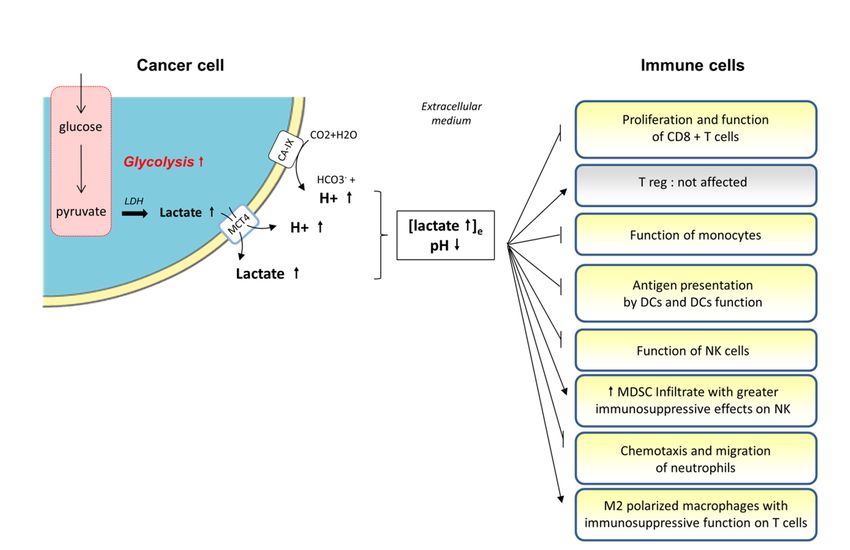
Cells 2019, 8, 104                                                                                  2 of 30

states, and associated with profound differences in energy requirements. The immune response is
associated with dramatic modifications in tissue metabolism, including the depletion of nutrients,
increased oxygen consumption, and the generation of reactive nitrogen and oxygen intermediates.
These modifications are, in part, due to the recruitment of many inflammatory and immune cells [3].
      The most used nutrient is glucose. Glycolysis converts glucose into pyruvate in the cytoplasm.
Under normoxic conditions, pyruvate is transformed into acetyl-CoA in the mitochondria, by oxidative
decarboxylation. Acetyl-CoA enters the tricarboxylic acid (TCA) cycle and is oxidized into CO2,
permitting the reduction of (i) nicotinamide adenine nucleotide (NAD) into NADH and/or (ii) flavin
adenine dinucleotide into FADH2. These two redox cofactors can be used by the electron transport
chain (ETC) for oxidative phosphorylation (OXPHOS), to create a proton gradient strong enough to
permit the phosphorylation of ADP into ATP by the ATP synthase. The ETC is the most efficient way
to produce energy for cell metabolism. Indeed, OXPHOS produces 36 molecules of ATP from one
molecule of glucose [4]. Cells can metabolize other substrates, such as glutamine via glutaminolysis or
fatty acids in β-oxidation, to replenish the TCA cycle and OXPHOS.
      Cells can also produce ATP through the fermentation. Under hypoxic conditions, pyruvate
remains in the cytoplasm and is converted into lactate rather than acetyl-CoA. This reaction is catalyzed
by the lactate dehydrogenase (LDH). This pathway is quicker but less efficient than OXPHOS. Indeed,
it produces only two molecules of ATP per molecule of glucose. Fermentation can also be done under
normoxic conditions; this is the Warburg effect. Many cells, including cancer cells, prefer to use aerobic
glycolysis because it is quicker and it generates precursors for the chemical constituents that form the
macromolecules essential for cell division, such as nucleotides, lipids, proteins and nucleic acids [5].

2. Metabolism of Tumor Cells
     Tumor cells modify their metabolism in order to supply energy for cell growth and proliferation.
Metabolic reprogramming is now an established hallmark of cancer [2] and is targeted in the clinic.
In most solid tumors, cancer cell metabolism is characterized by the Warburg effect, which means
that, even in the presence of oxygen and fully functioning mitochondria, tumor cells preferentially
use aerobic glycolysis and lactate fermentation as opposed to oxidative phosphorylation (Figure 1).
This metabolic reprogramming permits the production of energy, but also nutrients as lipids, proteins
or nucleic acids, necessary for the generation of daughter cells. This reprogramming leads to several
modifications in nutrient uptake, the activation of metabolic pathways and in interactions with the
microenvironment, modifying the fate of tumor cells and components of the microenvironment.
     Because aerobic glycolysis is less efficient for the production of ATP, tumor cells increase their
nutrient uptake from the microenvironment [6]. The two main nutrients used by cancer cells are glucose
and glutamine (Figure 1). These nutrients permit the maintenance of the pool of carbon intermediates
in the cells, to generate NADH and FADH2 for ATP production, NADPH for biosynthetic reactions or
to maintain redox balance in cells. Aerobic glycolysis has low efficiency and cancer cells upregulate
transporters in order to increase glucose uptake, notably that of GLUT1 [7]. This increased glucose
uptake is normally regulated by extracellular growth signals and by the interactions between cells
and the matrix; however, cancer cells become insensitive to these external signals due to numerous
genomic alterations (activation of Pi3k/Akt/mTOR pathway or Ras) [7]. The Pi3K pathway plays a
central role in this metabolic reprogramming as a master regulator of glucose uptake and metabolism.
This pathway is induced by hypoxic conditions, through the production of Hypoxia-inducible
factor 1-alpha (HIF-1α). mTOR upregulates the translocation of GLUT1 at the cell surface [8,9].
Furthermore, hypoxia conditions also induce the overexpression of several glycolytic enzymes such as
hexokinase and phosphofructokinase activities, through Akt activation [10]. Cancer cells also inhibit
the mitochondrial TCA cycle through the upregulation of pyruvate dehydrogenase kinase (PDK)
expression and mutations of fumarate hydratase (FH) and succinate dehydrogenase (SDH), while
promoting glycolysis.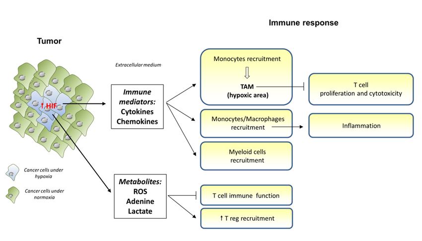
2018, 8,
Cells 2019, 7, 104
x FOR PEER REVIEW 3 of 30
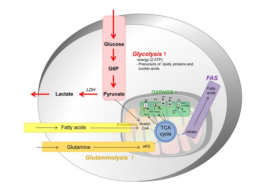
Figure 1. Cancer
Figure 1. Cancer cell
cell metabolism. Most cancer
metabolism. Most cancer cells
cells depend
depend onon aa higher
higher glycolytic
glycolytic metabolism
metabolism toto
proliferate and disseminate, regardless of the presence of oxygen. This is made possible
proliferate and disseminate, regardless of the presence of oxygen. This is made possible through the through
the overexpression
overexpression of GLUT1,
of GLUT1, a glucose
a glucose cell cell surface
surface transporter.
transporter. ThisThis metabolic
metabolic switch,
switch, known
known as
as the
the Warburg effect, occurs at the expense of the oxidative phosphorylation (OXPHOS) and
Warburg effect, occurs at the expense of the oxidative phosphorylation (OXPHOS) and provides, in a provides,
in a short
short amount
amount of time,
of time, the the energy,
energy, as well
as well as many
as many precursors
precursors of the
of the macromolecules,
macromolecules, required
required to
to sustain a high rate of proliferation. In cancer cells, pyruvate is largely directed to produce lactate
sustain a high rate of proliferation. In cancer cells, pyruvate is largely directed to produce lactate
instead of being metabolized within mitochondria. Glutamine and fatty acids constitute two other
instead of being metabolized within mitochondria. Glutamine and fatty acids constitute two other
substrates used preferentially by tumor cells, and which provide intermediates of the tricarboxylic acid
substrates used preferentially by tumor cells, and which provide intermediates of the tricarboxylic
cycle (TCA) cycle. They are further used to produce other building blocks for these demanding cells
acid cycle (TCA) cycle. They are further used to produce other building blocks for these demanding
and to maintain some mitochondrial activity and functioning.
cells and to maintain some mitochondrial activity and functioning.
Glutamine also plays an important role in cancer cell metabolism and proliferation. It is involved
Glutamine also plays an important role in cancer cell metabolism and proliferation. It is involved
in cell proliferation through its transformation into α-ketoglutarate, which enters the TCA cycle for
in cell proliferation through its transformation into α-ketoglutarate, which enters the TCA cycle for
the production of amino acids, nucleotides and fatty acids [5,11]. Furthermore, glutamine is also
the production of amino acids, nucleotides and fatty acids [5,11]. Furthermore, glutamine is also
important for the production of glutathione, involved in redox homeostasis. C-Myc is the principal
important for the production of glutathione, involved in redox homeostasis. C-Myc is the principal
driver of glutamine metabolism [12], which increases the expression of glutamine transporters and
driver of glutamine metabolism [12], which increases the expression of glutamine transporters and
their metabolic enzymes [13]. Furthermore, the loss of the retinoblastoma (Rb) tumor suppressor is also
their metabolic enzymes [13]. Furthermore, the loss of the retinoblastoma (Rb) tumor suppressor is
involved in increased glutamine uptake and metabolism as it blocks the uptake of glutamine [14,15].
also involved in increased glutamine uptake and metabolism as it blocks the uptake of glutamine
It is worthwhile to note that glucose and glutamine are also two metabolites required for the activation
[14,15]. It is worthwhile to note that glucose and glutamine are also two metabolites required for the
of naive T cells [16,17]. Upon stimulation, T cells undergo a Warburg-like metabolic reprogramming,
activation of naive T cells [16,17]. Upon stimulation, T cells undergo a Warburg-like metabolic
enhancing glucose uptake and glycolysis to promote proliferation [18]. They also require glutamine
reprogramming, enhancing glucose uptake and glycolysis to promote proliferation [18]. They also
to generate various intermediates as well as precursors of protein and lipid biosynthesis to complete
require glutamine to generate various intermediates as well as precursors of protein and lipid
differentiation and fulfill their tasks [19]. These common requirements create a metabolic competition
biosynthesis to complete differentiation and fulfill their tasks [19]. These common requirements
within the tumor microenvironment (TME), where oncogene-driven cancer cells eventually take control
create a metabolic competition within the tumor microenvironment (TME), where oncogene-driven
and deplete surrounding nutrients upon proliferation, at the expense of the immune cell’s fitness and
cancer cells eventually take control and deplete surrounding nutrients upon proliferation, at the
survival, thereby blunting immune responses [19].
expense of the immune cell’s fitness and survival, thereby blunting immune responses [19].
In addition to glucose and glutamine addiction, cancer cells also rely on the so-called
In addition to glucose and glutamine addiction, cancer cells also rely on the so-called ‘one-
‘one-carbon metabolism’ to sustain their high rate of proliferation [20]. One-carbon units are indeed
carbon metabolism’ to sustain their high rate of proliferation [20]. One-carbon units are indeed
indispensable for nucleotide production, methylation processes and NADH/NADPH pool renewal.
indispensable for nucleotide production, methylation processes and NADH/NADPH pool renewal.
The serine-to-glycine pathway constitutes one major source of one-carbon units and, as such, is often
The serine-to-glycine pathway constitutes one major source of one-carbon units and, as such, is often
diverted and over-activated in tumors to favor their growth. Cancer cells generally increase their
3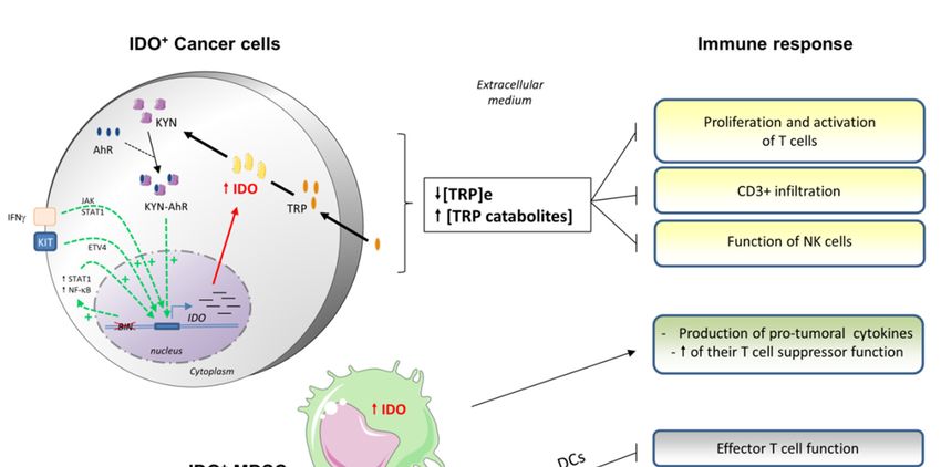
Cells 2019, 8, 104                                                                                  4 of 30

serine supply, by up-regulating the intracellular de novo serine synthesis pathway or by augmenting
its uptake from the local environment [21–23]. This specific metabolic dependency is of therapeutic
interest as serine deprivation has been shown to inhibit cancer cell proliferation. However, recent
advances have also revealed the importance of serine in T cell expansion upon activation [24], thereby
demonstrating a metabolic competition between tumor and immune cells for this important amino
acid. It is therefore crucial to further improve knowledge of this pathway to target tumors without
impairing T cell function and immune response.
     Finally, cancer cells also undergo a modified lipid metabolism that is mostly characterized to a
lipogenic phenotype. Indeed, they overexpress enzymes involved in the synthesis of saturated lipids.
     As previously described, profound metabolic changes occur within immune cells upon activation
as part of their response to invading pathogens or injury [3]. Lymphoid cells generally switch
from mitochondrial OXPHOS to aerobic glycolysis to become fully active and proliferate when
they encounter a danger signal. As soon as the clearing is finished, the remaining memory cells
undergo a reversed metabolic switch, correlating to their reacquisition of a quiescent non-proliferative
state. Dendritic cells also rely on higher glycolytic flux to be activated, as do proinflammatory
macrophages upon M1 polarization. As for anti-inflammatory M2 polarized macrophages, they are
mostly dependent on mitochondrial OXPHOS and fatty acid oxidation (FAO). All these metabolic
changes are key to an efficient immune response in normal or disease conditions, including cancer,
as detailed in [3]. Although further details on the immune cells metabolic requirements won’t
be discussed here, it is important to consider that immune cells are permanently competing with
tumor cells for nutrient resources (glucose, glutamine, serine, tryptophan) in order to proliferate and
function properly [16,25]. Metabolic competition is therefore poised as one crucial step in the immune
hypo-responsiveness observed in cancer [26].
     Aside from immune cells, the whole tumor niche influences the metabolic status of cancer cells
and vice versa [27]. Intricate relationships exist between a cancer cell and its microenvironment, usually
including non-tumor cells such as fibroblasts, adipocytes, endothelial cells and immune cells, as well
as multiple soluble secreted factors. This complex network guides and shapes the metabolic balance of
the tumor and, in turn, its behavior. Although the global metabolic cooperation/competition between
the various components of the TME are not the point of this review, it is worth mentioning that,
ultimately, these cells are organized to promote cancer growth and facilitate dissemination [27]. Indeed,
as mentioned, competition between cancer cells and T cells for certain metabolites leads to immune
suppression, while also supporting the polarization of pro-tumorigenic M2 macrophages that rely on
OXPHOS and FAO [16]. Highly glycolytic cancer-associated fibroblasts (CAF) build up a favorable
milieu for cancer cell development, releasing lactate, acidifying the milieu and providing high energy
metabolites intermediates to fuel proliferation. Additionally, adipocytes turn into cancer-associated
adipocytes within the TME, offering a vital source of fatty acids for the quickly dividing and energy
demanding tumor cells [28].

3. Effect of Cancer Metabolism on Infiltrating Immune Cells
     Immune escape is a major hallmark of cancer cells [2]. Tumor cells disable immune components
that are activated to eliminate them. Several mechanisms can lead to this escape: reduced expression of
antigens at the surface of tumor cells, reduced expression of major histocompatibility (MHC) molecules
by antigen-presenting cells (APC), impaired co-localization of T-cell receptor (TCR) and co-stimulatory
receptors, secretion of inhibitory cytokines and activation of inhibitory receptors on T cell surface.
This leads to a selection of tumor cells that are resistant to the immune response and that generate an
immunosuppressive and pro-tumoral microenvironment. Immune cells acquire functional defects and
enter a hyporesponsive (or anergic) reversible state, with impaired effector capacities favoring tumor
progression. Next, we discuss how cancer metabolism triggers immune escape mechanisms and we
review the implications of this in current immunotherapy strategies.
Cells 2019, 8, 104                                                                                   5 of 30

3.1. Medium Acidification and Lactate Accumulation

3.1.1. The Production of Lactate by Tumor Cells
      As discussed above, tumor cells derive their energy from a number of nutrients, mainly glucose
and glutamine. Both pathways produce significant amounts of lactate because of the overexpression of
the lactate dehydrogenase (LDH) and the greater rate of glucose consumption. Lactic acid accumulates
in the cytoplasm and extracellular medium because of high rates of production and insufficient vascular
clearing [29]. During this process, acidification can occur, based on the co-transport of protons with
lactate export, through the Mono-Carboxylate Transporter 4 (MCT4). MCT4 controls intracellular
pH. It has a low affinity for lactic acid and a high capacity for transport in order to meet the high
rates of production and utilization of lactate [30,31]. Tumor cells release lactic acid in the extracellular
medium since intracellular pH variation can alter cell survival and metabolism, motility, apoptosis,
and biosynthesis [32,33].
      Furthermore, tumor cells overexpress membrane Carbonic Anhydrase IX (CA-IX), contributing to
further acidification of the tumor microenvironment [34]. This enzyme catalyzes the transformation of
carbon dioxide into bicarbonate and protons. Its expression is controlled by the HIF pathway and is
associated with poor prognosis and a higher rate of metastasis [35]. This enzyme preserves intracellular
pH and ATP levels in vitro and in vivo, leading to the promotion of cell survival and growth [34].
Tumor cells are able to control the extracellular pH to promote their proliferation and survival. They
modulate their own metabolism to regulate lactate efflux [36]. Indeed, Warburg metabolism seems to
be a dynamic negative feedback loop permitting the regulation of the microenvironment pH by actively
modifying the balance between OXPHOS and glycolysis. Under extracellular acidification, tumor cells
can attenuate glucose consumption, reduce lactate production and increase OXPHOS gene activation
in order to limit further acidification. Therefore, tumor cells can control their microenvironment and
maintain an aggressive phenotype.
      Lactic acid is involved in the tumor phenotype. First, high levels of lactate can induce the
degradation of the extracellular milieu, promoting migration and more frequent metastasis in a
dose-dependent manner in vitro [37,38]. The knockdown of LDH inhibits the migration of glioma
cells in vitro and this effect is reversed by supplying lactic acid in the medium [39]. The acidification
of the extracellular environment also modifies tumor cell migration. Pharmacological or genetic
inhibition of sodium/proton antiport was shown, for instance, to reduce the migration potential of
hepatocarcinoma cell lines (without effecting their proliferation), through a reduced expression of
metalloproteinase MMP-2 [40]. Furthermore, lactic acid induces the expression of protumor molecules,
such as hyaluronic acid from tumor-associated fibroblasts and TGF-β [41,42], that are also involved in
invasive phenotypes. Indeed, in vitro melanoma models showed that tumor cells overexpressed CD44
under acidic conditions (through lactate response elements in CD44-promoter) to retain hyaluronic
acid on their surface and to favor their aggressive phenotype [29].
      In addition, lactic acid has an angiogenic role, through the coupling between lactate and pyruvate
via LDH, by inducing the expression of HIF-target genes [43]. The acidification leads to the elevation
of IL-8 (endothelial cells) and VEGF (macrophages and endothelial cells) amounts, which are both
proangiogenic factors involved in the metastatic process [44]. IL-8 inhibition through RNA interference
leads to the inhibition of angiogenesis in tumors [45]. Finally, acidification increases tumor cell
survival [46], and is associated with radioresistance in vitro and in vivo [47], possibly because of its
antioxidant properties [48], which protect cancer cells from damage caused by radiation.

3.1.2. Role of Lactate and Extracellular Medium Acidification on Immune Cells
      The effects of acidosis on immune functions are associated with immunodeficiency in non-cancer
diseases such as sepsis [49]. In cancer, acidification also provides a growth advantage to tumor
cells at the expense of immune cells [38,50,51] (Figure 2). The tumor releases lactic acid in order to
acidify the medium, leading to higher concentrations and a lower pH in the extracellular medium.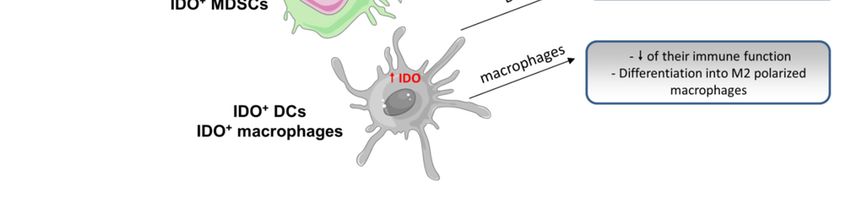
Cells 2018, 7, x FOR PEER REVIEW 6 of 30
Cells 2019, 8, 104 6 of 30
This pH gradient induces the blockade of lactate efflux from T cells and its accumulation in the T
cell’s intracellular medium [50]. Medium acidification leads to CD8+ TILs (Tumor Infiltrating
This pH gradient
Lymphocytes) induces
anergy the blockade
in human and mouse of models
lactate efflux
[50,52].from
The Taccumulation
cells and itsofaccumulation in the
lactic acid induces
T cell’s intracellular medium [50]. Medium acidification leads to CD8 + TILs (Tumor Infiltrating
a diminution of T cell proliferation, after antigen stimulation, in a dose-dependent and time-
Lymphocytes)
dependent manneranergy in This
[50]. human and mouse
inhibition models
occurs [50,52]. The
immediately afteraccumulation
the contact of of Tlactic
cellsacid
withinduces
lactate.a
diminution ofalso
Acidification T cell proliferation,
impairs after antigen
their function stimulation,
(reduced cytokine in a dose-dependent
secretion and cytolytic andactivity
time-dependent
through
manner [50].
reduced This inhibition
expression of granzymeoccursB)immediately
and decreases aftertheir
the contact
activationof T(reduced
cells withTCR lactate. Acidification
expression and
also impairs
STAT5 and ERK their function (reduced
activations) cytokine secretion
[52]. Furthermore, prolongedand cytolytic
exposure (at activity
least 24 through reduced
h) to lactic acid
expression
induces of granzyme
apoptosis of moreB)thanand60%decreases their
of T cells, activation
whereas (reduced
a shorter TCR expression
exposure leads to only and STAT5 and
a diminished
ERK activations)
function of T cells [52]. Furthermore,
without impairmentprolonged exposure
of their (at These
survival. least 24effects
h) to lactic
wereacid not induces apoptosis
found when the
of more thanof
acidification 60%theofmedium
T cells, whereas
was done a shorter exposure leads
with hydrochloric acid,tomeaning
only a diminished
that lactic function of T to
acid is toxic cells
T
without impairment of their survival. These effects were not found when
cells with a mechanism other than the sole acidification of the medium. This anergy is a reversible the acidification of the
medium
state, was done
because with hydrochloric
treatment of mice withacid, meaning that
a proton-pump lactic acid
inhibitor (PPI)is toxic to T cells
restored theirwith a mechanism
immune effector
other than
function andthe sole acidification
increased the efficacy ofof
the medium.
adoptive This anergy is[52].
immunotherapy a reversible state,of
These effects because
lactate treatment
on T cells
of mice
are withdue
possibly a proton-pump
to the impairment inhibitor
of the (PPI) restored their
TCR-triggered immune
activation of effector function and
c-Jun N-terminal increased
kinase (JNK),
the efficacy
c-Jun and p38of[53].
adoptive immunotherapy
Mitogen-activated [52].kinase
protein These(MAPK)
effects ofandlactate
mTOR on pathways
T cells areare possibly due to
not affected.
thecontrast,
In impairment of the T
regulatory TCR-triggered
cells (Treg) doactivation
not seemof toc-Jun N-terminal
be impaired kinaseand
by lactate (JNK), c-Jun and because
acidification, p38 [53].
Mitogen-activated
of protein based
a different metabolism, kinaseon (MAPK) and oxidation
fatty acid mTOR pathways are not affected. In contrast, regulatory
(FAO) [54].
T cells (Treg) do not seem to be impaired by lactate and acidification, because of a different metabolism,
based on fatty acid oxidation (FAO) [54].
Figure 2.
Figure Cancer cells
2. Cancer cells induced-lactate
induced-lactate and and-proton
-protonrelease
releasewithin
withinthetheperitumoral
peritumoral environment
environment andandthe
consequences for immune cell function. The metabolic switch towards aerobic
the consequences for immune cell function. The metabolic switch towards aerobic glycolysis observed glycolysis observed
inmost
in mostcancer
cancercells
cells(i.e.,
(i.e.,Warburg’s
Warburg’seffect)
effect)produces
producessignificant
significantamounts
amountsof ofpyruvate
pyruvatethatthatare
aremostly
mostly
metabolized into lactate. The intracellular increase of lactate, potentially toxic for the cancer
metabolized into lactate. The intracellular increase of lactate, potentially toxic for the cancer cell, cell, is then
is
released into the extracellular medium together with protons (thanks to transporters
then released into the extracellular medium together with protons (thanks to transporters such as such as MCT4),
inducing
MCT4), an acidification
inducing of the local
an acidification environment.
of the The frequent
local environment. The up-regulation of carbonicofanhydrase
frequent up-regulation carbonic
anhydrase IX (CA-IX) in cancer cells also contributes to the local increase of H+ inneighborhood
IX (CA-IX) in cancer cells also contributes to the local increase of H+ in the immediate the immediate of
cancer cells. Both,
neighborhood the accumulation
of cancer of lactate
cells. Both, the and the of
accumulation decrease
lactate in thethe
and pHdecrease
of the extracellular
in the pH of milieu
the
create a detrimental environment for the antitumoral immune response, (i)
extracellular milieu create a detrimental environment for the antitumoral immune response, (i) decreasing the function
of the CD8+
decreasing theT function
cells, dendritic cells (DCs),
of the CD8+ T cells,monocytes, natural
dendritic cells (DCs),killer (NK) cells
monocytes, and/or
natural (ii) (NK)
killer increasing
cells
the myeloid-derived suppressor cells (MDSC) infiltrate or M2-polarized macrophages with greater
and/or (ii) increasing the myeloid-derived suppressor cells (MDSC) infiltrate or M2-polarized
immunosuppressive functions on NKs and T cells, respectively.
macrophages with greater immunosuppressive functions on NKs and T cells, respectively.
6Cells 2019, 8, 104 7 of 30
Lactate accumulation in the extracellular medium also alters monocyte function and metabolism.
Indeed, it impairs the tumor necrosis factor (TNF) secretion of monocytes in vitro without effecting their
viability [55]. This effect is due to an increased uptake of lactate and the co-transport of protons through
MCT-1 and MCT-4, because of the pH gradient. There is also an inhibition of reactive oxygen species
(ROS) production, which is important to the cytotoxic activity. All these effects were reversible after the
administration of the lactate inhibitor oxamic acid. Lactic acid reduces the expression of genes involved
in the inflammatory effector functions of monocytes, such as in the expression of cytokines (IL-23 and
TNF mostly) and chemokines (CCL2 and CCL7). It also delays the phosphorylation and activation of
Akt (involved in glycolysis), and reduces nuclear-factor (NF)-κB accumulation in monocytes (which
regulates the expression of cytokines and chemokines) [56]. Extracellular lactate was shown to be
sufficient to switch inflammatory M1 macrophage polarization towards immunosuppressive M2; the
latter being evidenced to accumulate in the tumor microenvironment [57].
Lactic acid does not alter monocyte differentiation into dendritic cells (DCs) in the in vitro
models. However, lactate-mediated acidification induces an alteration of antigen presentation by
DCs, and of their functional activity. This leads to the acquisition of tumor-associated DC phenotypes,
and the blockade of lactate restores their normal phenotype [58]. These tumor-associated DCs have
a particular pattern for the presentation of antigens, which favor tumor progression. These effects
are probably due to the secretion, by tumor-favoring DCs and tumor cells, of IL-6 and growth factors
such as macrophage colony-stimulating factor (M-CSF). Furthermore, lactate increased the production
of IL-23 in human lung adenocarcinoma cell lines [59]. IL-23 is overexpressed by macrophages
and DCs in tumors and promotes tumor growth [60]. It is an important molecule that upregulates
IL-17, IL-6 and TNF-α secretion, with an autocrine and paracrine effect. These cytokines have a
proinflammatory effect, without increasing immune infiltration, because of reduced Th1-dependent
interferon (IFN)-γ secretion. Furthermore, IL-23 increases the expression of the matrix metalloprotease
9, the angiogenesis, and reduces CD8+ T cell infiltration in the tumor microenvironment [59]. Indeed,
models of a tumor-xenograft mouse which harbor a deletion of IL-23 have restricted tumor growth [60].
Lactic acid also acts on other immune cells. Indeed, through its induction of HIF expression,
it induces the expression of arginase I and iNOS in M2-polarized macrophages [61], harboring an
immunosuppressive function on T cells. On natural killer (NK) cells and myeloid-derived suppressor
cells (MDSC), mouse models have shown that lactic acid decreases the function of NK cells, through
a diminution of the expression of granzyme B and perforin and a diminution of the expression of
the activating receptor NKp46 (which plays an important role in their cytotoxicity) [62]. Lactic acid
also enhances the infiltration of MDSCs in a peritumoral environment, with greater suppressive
effects on NK cells. Finally, lactic acid leads to the inhibition of chemotaxis and the migration of
polymorphonuclear neutrophils [63–65].
3.1.3. Impact in Clinical Routines
High levels of lactate in primary tumors are correlated with a poor outcome and an increased
rate of distant metastasis in several cancers [66–68]. Targeting lactate production and its repercussions
remains, therefore, attractive through various therapeutic strategies, as described below.
This can be done through alkalization of the medium. PPI, used in gastro-intestinal diseases,
inhibits the vacuolar-ATPase, whose expression is enhanced in several tumors. This ATPase is
a proton pump involved in medium acidification. Its inhibition decreases proton extrusion from
cells and leads to extracellular pH alkalization. It induces cytotoxicity in melanoma cells in vitro,
through the acidification of the intracellular medium (pH gradient), leading to the activation of
caspases and the apoptosis of tumor cells, independently of the mutational profile [29]. In mouse
models, PPI permitted reduced tumor growth and increased survival in melanoma-bearing animals.
The extracellular alkalization can also favor the immune cell efflux of lactic acid, and restore their
effector functions. The administration of PPI is safe because its activation depends on a medium pH.
Indeed, PPI is only activated under acidic conditions, targeting its activation mainly in tumor tissues.Cells 2019, 8, 104 8 of 30
One Phase 2 clinical trial has been completed (NCT01069081), assessing the efficacy and safety
profile of concomitant administration of docetaxel and cisplatin chemotherapy with high doses of PPI in
metastatic breast cancer. Results showed an improvement of the antitumor effect of the chemotherapies,
inciting a Phase 3 study [69]. Another way to inhibit acidification and restore immune response is the
use of sodium bicarbonate [70]. Thus, treatment of tumor-bearing mice with oral sodium bicarbonate
lead to an increase in the pH and to lower metastasis, but not in all tumor models. Preliminary
investigations showed that alkalization reduced the release of cathepsin B in the extracellular medium,
an important matrix remodeling protease.
Next was the inhibition of lactate production. Oxamate is a pyruvate analogue that competitively
inhibits LDH isoform A (LDH-A). In a xenograft mouse model, it was reported that the inhibition
of LDH-A activity leads to the inhibition of aerobic glycolysis in tumor cells, whereas normal cells
were not impaired [71]. In vitro, oxamate was shown to have a proapoptotic effect via the activation
of caspase 3 and the expression of Bax [72]. It suppresses the proliferation of lung cancer cell lines,
with a lower effect on normal cells [73]. This effect was due to a reduction of ATP and ROS and to a
phase G0/G1 arrest. Others strategies for inhibiting LDH-A, such as siRNA, have been assessed [74].
No ongoing trials are currently assessing these molecules in cancer.
3.2. Indoleamine 2,3-Dioxygenase (IDO) and Tryptophan Dioxygenase (TDO)
3.2.1. IDO Characteristics
The tryptophan dioxygenase (TDO) was first described as the only enzyme capable of
metabolizing L-tryptophan, an essential amino acid. Later, however, indoleamine 2,3-dioxygenase
(IDO) was discovered in rabbits and shown to be able to metabolize both L- and D-tryptophan [75].
There are three isoforms existing, IDO1, IDO2, and TDO2. These enzymes have different inducers
as well as different tissue expression patterns [76]. Indeed, IDO1 is induced by proinflammatory
cytokines such as IFN-γ, LPS and pathogens like influenza viruses [77,78]. This is widely expressed
across most organs, in plasmacytoid DCs of lymph nodes and the spleen, macrophages, endothelial
cells, stromal mesenchymal cells and fibroblasts [79,80]. TDO2 is induced by tryptophan itself and
glucocorticoids, and is expressed in the liver, brain and placenta, and IDO2 is expressed in hepatocytes,
neuronal cells and DCs [81].
IDO is encoded by the IDO1 and IDO2 genes in human chromosome 8p11 and TDO2 gene in
chromosome 4q32. It was the first IFN-activated gene identified in the 1970s [82]. It is a cytosolic
enzyme which catalyzes the first step of the tryptophan catabolism in the kynurenine pathway
(catabolism of tryptophan into N-formyl-kynurenine). Tryptophan metabolism is important for the
production of the energy cofactor NAD+ . The enzyme is a 407 amino acid heme-containing protein.
In mice, IDO was described as a protein that prevents fetal rejection [83]. In humans, IDO modulates
antigen-dependent activation of immune cells on the mucosal surfaces of lungs and the digestive
intestine [80]. Furthermore, it prevents excessive cytotoxic immune response leading to tissue damage.
The IDO1 promoter contains ISREs (IFN-stimulated response elements) and GASs (IFNγ-activated
sites). Several transcription factors can translocate into the nucleus in order to enhance the expression
of IDO1. IFN-γ is the most potent IDO1 inducer. Similar to LPS, it activates the Janus kinase/signal
transducer and activator of transcription (JAK/STAT) pathway, which leads to the expression of STAT1
or STAT3 [84]. Kynurenine, its metabolite, through its interaction with the aryl hydrocarbon receptor
(AhR), can also induce IDO1 expression through the STAT3 pathway. Others transcription factors
can also activate IDO1 transcription: IRF1 (IFN regulator 1) [85], the NF-κB pathway and ETV4 (ETS
variant 4) [84].
3.2.2. IDO Expression in Tumor Cells
IDO is associated with numerous immune diseases, as diverse as cancer, allergies, autoimmune
and inflammatory diseases. IDO1 can have two expression patterns. In some tumors, IDO1-expressingCells 2019, 8, 104 9 of 30
tumor cells are in lymphocyte-rich areas, meaning that IDO-expression can be the consequence of
IFN-γ expression and a resistance mechanism. In other cancers, IDO1 expression is constitutive and
IDO1 expressing tumor cells are surrounded by less lymphocytes. In vitro, several cell lines can
constitutively overexpress IDO, despite the absence of IFN-γ, with variable levels of activity according
to cell lines [86,87]. This is explained by Bin1 mutations [88]. Bin1 is a tumor suppressor gene encoding
an adaptor protein, the Bin1/amphiphysin/Rvs167 (BAR). It is found to be attenuated in several
cancers, promoting proliferation, motility and survival [79]. In vivo studies have shown that the main
consequence of Bin1 inactivation is the increase of intracellular amounts of STAT1 and NF-κB, leading
to the upregulation of IDO expression. Its expression was also found in peritumoral cells, but not in
distant stroma.
IDO activity can also be induced by several factors, such as the oncogene Kit that is commonly
altered in several cancers. Once activated, Kit induces ETS variant 4 (ETV4) in cytoplasm. Furthermore,
IDO1 can sustain its own expression through an autocrine loop [89]. Indeed, the IDO1 gene can be
activated by the binding of kynurenine-AhR on its response elements, activating STAT3. STAT3 can
induce expression of IDO1 and IL-6, which exerts an autocrine/paracrine feedback loop based on the
interaction between IL-6 and its receptor that enhances expression of STAT3.
IDO acts at multiple levels of tumorigenesis, all associated with inflammation: metastatic
process, immune escape, invasion and angiogenesis [79]. IDO seems to be an integral component of
chronic inflammation, required to support tumor development in chronic inflammatory models [90].
There is probably an interconnection between inflammation and immune escape programs, because
IDO is expressed only until some degree of inflammation occurs in the tumors [87]. IDO acts at
different stages by favoring tumor progression and metastatic evolution [79], by maintaining a
proinflammatory and protumor microenvironment. Indeed, IDO1 deficient mice are resistant to
tumorigenesis [91], develop less lung metastasis, have a lower IL-6 amount and have better survival
rates [92]. Furthermore, these deficient mice have impaired angiogenesis in the lungs, even in the
absence of cancer. IDO seems to display a more complicated role, beyond its immunomodulatory,
pro-metastatic and angiogenic functions.
IDO can be overexpressed either in tumor cells or in tumor-associated cells such as dendritic
cells, macrophages, or endothelial cells (Figure 3). Indeed, its overexpression leads to tryptophan
deprivation, which can impair the immune cell functions, creating a de novo local tolerance, due to
the anti-inflammatory effect [93]. IDO1 overexpression in tumor cells also leads to reduced plasmatic
amounts of tryptophan, meaning that the immunosuppressive effect of IDO1 is not locally restricted
but is systemic [94].
Next we review the effects of tryptophan metabolism on lymphocytes. The overexpression of
IDO by tumor cells leads to a tryptophan deprivation in the extracellular medium (Figure 3). T cells
are very sensitive to the variation of tryptophan abundance in their microenvironment. Thus, this
deprivation has effects as soon as the level decreases lower than 0,5–1 µM [95,96]. This deprivation
leads to anergy and to a reduced proliferation in vitro and in vivo [97].
IDO overexpression can block TILs proliferation through two mechanisms: by direct tryptophan
deprivation in the microenvironment (GCN2 activation and mTOR inhibition), and by indirectly
induced toxicity of tryptophan catabolites [86,98]. The accumulation of uncharged tryptophan-tRNA,
due to tryptophan deprivation, induces the enhancement of stress signals, mediated by GCN2 (general
control nonderepressible 2) which phosphorylates the transcription factor EIF2α [99]. GCN2 activation
leads to the polarization of CD4+ T cells into Tregs cells from Th17 [99,100]. Tryptophan deprivation
also induces an impairment of their antigen-dependent activation [101], downregulation of the TCR
ζ-chain [100], suppression of factors needed for the downstream signaling of the TCR through the
MAPK pathway [102], and activation of autophagy through the inhibition of mammalian target of
rapamycin (mTORC1) and protein kinase C (PKC-Θ) [103]. IDO expression also leads to the lower
infiltration of CD3+ cells [104]. Finally, tryptophan deprivation increases the sensibility of T cells toCells 2019, 8, 104 10 of 30
Fas-mediated apoptosis
Cells 2018, 7, x FOR [105], but this proapoptotic effect is still controversial [86]. Several studies
PEER REVIEW 10 of 30
have shown that its supply restores T cell function.
Figure 3. Consequences of indoleamine 2,3 dioxygenase (IDO) overexpression on the antitumoral
Figure 3.response.
immune Consequences
Cancer of indoleamine
cells 2,3 dioxygenase
frequently overexpress IDO as(IDO) overexpression
a result on theupregulation.
of its transcriptional antitumoral
immune
This can be response.
triggeredCancer cells frequently
by (i) cytokines overexpress
such as IFNγ, as longIDO as a result are
as lymphocytes of present
its transcriptional
within the
tumor, (ii) the oncogene
upregulation. This can be Kit, or (iii) mutations
triggered withinsuch
by (i) cytokines the tumor
as IFNγsuppressor
, as long asgene Bin. IFNγare
lymphocytes promotes
present
IDO
withinoverexpression
the tumor, (ii)by theactivating
oncogenethe Kit,Janus
or (iii)kinase/signal
mutations within transducer
the tumor andsuppressor
activator ofgene
transcription
Bin. IFNγ
(JAK/STAT)
promotes IDO pathway, while Kit by
overexpression andactivating
Bin stimulate the ETV4
Janus (ETS variant 4),transducer
kinase/signal and STAT1and and/or nuclear
activator of
factor (NF)-κB,(JAK/STAT)
transcription respectively.pathway,
IDO expression
while levels
Kit and canBin
alsostimulate
be regulated
ETV4 by(ETS
an autocrine
variant loop,
4), andimplying
STAT1
the complex
and/or nuclearKYN-AhR. Once in the
factor (NF)-κB, cytosol, IDO
respectively. IDOmediates
expressionthe transformation
levels can also of betryptophan
regulated (TRP)
by an
into
autocrine loop, implying the complex KYN-AhR. Once in the cytosol, IDO mediatesleads
N-formylkynurenine that is then transformed into kynurenine (KYN). IDO overexpression the
to a decrease in the
transformation of extracellular
tryptophan level (TRP)of into
TRP, which limits the actionthat
N-formylkynurenine of immune
is thencells: T cells are into
transformed not
proliferating
kynurenine (KYN). and can’tIDO beoverexpression
activated; and CD3+ leads to cells infiltrate in
a decrease less
thefrequently
extracellularandlevel
the function
of TRP, of NK
which
cells
limitsis the
reduced.
actionCells other than
of immune cancer
cells: cells
T cells arecannotoverexpress
proliferatingIDO,andincluding some immune
can’t be activated; cells such
and CD3+ cells
as MDSCsless
infiltrate and frequently
DCs/macrophages. The overexpression
and the function of NK cellsofisIDO in suchCells
reduced. cells restrains
other than their own immune
cancer cells can
function (macrophages), suppresses T cells activity (DCs), and
overexpress IDO, including some immune cells such as MDSCs and DCs/macrophages. induces protumoral cytokines (MDSCs).The
As a result, in IDO
overexpression overexpressing
of IDO in such cells tumors,
restrainsthetheir
antitumoral
own immuneimmune response
function is largely compromised.
(macrophages), suppresses
T cells activity (DCs), and induces protumoral cytokines (MDSCs). As a result, in IDO overexpressing
Furthermore, tryptophan catabolism produces soluble metabolites directly toxic to several
tumors, the antitumoral immune response is largely compromised.
immune cells. Kynurenine is one of these immunosuppressive metabolites [106,107], probably by its
interaction with aryl hydrocarbon
IDO overexpression can blockreceptors (AhR) [108].
TILs proliferation AhR has
through twoamechanisms:
role in the regulation
by direct of immune
tryptophan
response,
deprivationinflammation and carcinogenesis.
in the microenvironment (GCN2 After its binding
activation to AhR,
and mTORkynurenine-AhR
inhibition), andtranslocates
by indirectly to
the nucleus and induces proinflammatory gene expression. The binding between kynurenine
induced toxicity of tryptophan catabolites [86,98]. The accumulation of uncharged tryptophan-tRNA, and AhR
favors
due topathologic
tryptophan inflammation in induces
deprivation, the microenvironment,
the enhancement which
of has a protumoral
stress role and by
signals, mediated facilitates
GCN2
escape to immune surveillance. Kynurenine-AhR has an effect on CD8 + TILs and CD4 + T 1 cells,
(general control nonderepressible 2) which phosphorylates the transcription factor EIF2α [99]. GCN2 H
through the induction of caspase 8 and cytochrome c [109,110], but not on T 2 cells
activation leads to the polarization of CD4 T cells into Tregs cells from Th17 H[99,100]. Tryptophan
+ [111]. Other
catabolites,
deprivationsuch
alsoas 3-hydroxyanthranilic
induces an impairment acid (3HAA),
of their also block T cell
antigen-dependent activation,
activation inducing
[101], apoptosis
downregulation
through
of the TCRthe ζ-chain
depletion of intracellular
[100], suppressionglutathione and thefor
of factors needed inhibition of NF-κBsignaling
the downstream activationof[105,112].
the TCR
Catabolites seem to have the same effects on B cells and NK cells in vitro
through the MAPK pathway [102], and activation of autophagy through the inhibition [113]. Indeed, in NK cells,
of mammalian
target of rapamycin (mTORC1) and protein kinase C (PKC-Θ) [103]. IDO expression also leads to the
lower infiltration of CD3+ cells [104]. Finally, tryptophan deprivation increases the sensibility of T
10Cells 2019, 8, 104 11 of 30
IDO induces the impairment of their cell-mediated killing function by the down-regulation of the
activation of several receptors such as NKG2D and NKp46, without effect on the other receptors [114].
Resting Tregs must be activated to become immunosuppressive T cells. When tolerogenic
IDO-expressing DCs activate resting Tregs through the TCR, the activation of the Akt pathway can
favor its phenotype into helper T cells [93]. Tregs inhibit these signals via different mechanisms in
order to preserve the immunosuppressive phenotype. First, they maintain durable Akt inhibition,
which is a key element for Treg activation [115,116]. Indeed, they inhibit mTOR activation through
amino acid deprivation, GCN2 activation, and upregulation of PTEN, thanks to the expression of PD1
and neuropilin-1. Catabolites of tryptophan also promote the expansion of some T cells into Tregs,
depending on TGF-β and FoxP3 expression [100,117]. There is a positive feedback loop between DCs
and Tregs. Indeed, IDO-expressing tolerogenic DCs induce Treg differentiation. In turn, Tregs express
CTLA-4 that enhance IDO-expression in tolerogenic DCs [118,119]. As a consequence, in clinical
practice, CTLA-4 blocking can significantly inhibit IDO expression [120].
Here we discuss the effects of tryptophan metabolism on DCs and macrophages. IDO-expressing
DCs induce a tolerogenic phenotype on naive DCs and favor the IDO-expression. The binding of
kynurenine on AhR [85] enables them to suppress effector T cell responses [121,122]. Indeed, this
interaction inhibits the production of IL-12 and favors the secretion of IL-10 and TGF-β, leading to
the modification of their antigen-presenting ability and favoring a tolerogenic phenotype [90,123,124].
The inhibition of IDO with 1-MT or siRNA in DCs leads to the diminution of the expression of
costimulatory molecules [125]. It is also involved in their maturation and migration in vitro [125].
IDO expression in DCs is induced through several pathways. First, DC-secreted TGF-β activates
the non-canonical NF-κB pathway and the phosphorylation of IKKα. This leads to the enhancement of
IDO expression and, also, to TGF-β, with an autocrine/paracrine effect on APC. Then, IL-6 secreted
by tumor or immune cells induces expressions of IDO1, such as IL-1β. PGE2 or TNF-α also induces
PKA which favors IDO expression. IDO expression can also be induced by negative coregulatory
membrane molecules such as CTLA-4, CD200, and GITR through IRF-1 or STAT1 [118,119,126,127],
thereby amplifying antigenic tolerance. In particular, CTLA-4 induces an IFNγ-dependent expression
of IDO [119].
In macrophages, one tryptophan metabolite, 3-HAA, suppresses NO secretion due to the
inhibition of NF-κB and iNOS [81]. Because NO is crucial for its ability to eliminate tumor cells,
3HAA has an immunosuppressive effect on macrophages. Cells expressing IDO can co-express iNOS
in response to IFN-γ, which produces NO that inhibits, in turn, IDO. Thus, several cells can induce IDO
and factors that also inhibit IDO activity, in order to regulate its expression. Furthermore, IDO favors
their differentiation into the M2 phenotype [128]. Tryptophan deprivation also plays an important role
because it regulates cytokine secretion and favors inflammation [129].
Finally, here we discuss the effects of tryptophan metabolism on MDSCs. Some studies have
found that high levels of IDO expression in MDSCs and IDO-expressing MDSCs highly inhibit the
antitumor immune response [130]. IDO1 induces the expression of pro-tumoral cytokines such as
IL-6 [92], which recruits MDSCs at the tumor site. As discussed above, IL-6 induces the expression
of IDO1 by MDSCs through an autocrine loop [89]. The deficit of IL-6 leads to a diminution of the
generation and the function of MDSCs, a diminution of their T cell suppressor function, leading to a
diminution of tumor progression and to its metastatic potent. These IL-6 amounts were associated,
in patients, with more frequent relapses [131]. Furthermore, local IDO recruits and activates MDSC
through IDO-activated Tregs [132].
In conclusion, IDO is upregulated in tumor cells, either constitutively or upon IFN-γ response.
This leads to a deprivation of tryptophan in the microenvironment, and the release of toxic catabolites.
Both induce an immunosuppression on DCs and cytotoxic T cells that become incompetent for an
efficient antitumor immune response [91]. Furthermore, IDO induces a tolerogenic phenotype on APCs
and the generation of immunosuppressive cells such as Tregs and MDSCs. This immunosuppression
can be local as well as systemic due to plasmatic tryptophan deprivation.Cells 2019, 8, 104 12 of 30
3.2.3. Clinical Targeting of IDO
IDO overexpression and elevated amounts of AhR in tumors have a negative prognosis in several
cancers (ovarian, leukemia, colorectal, cervical and endometrial) [80,86,104,133]. Furthermore, high
IDO1 expression is associated with more frequent metastasis [104]. IDO targeting is very interesting
because several tumors overexpress this enzyme, even in the absence of IFN-γ stimulation. Three
strategies can be developed to limit IDO effects: blockade of its expression (inhibition of NF-κB,
Jak/STAT or Bin1), blockade of its activity with enzymatic inhibitors, or blockade of its downstream
signaling (indoximod).
First, we present the indoximod or 1-Methyl-D-tryptophan strategy. The 1-Methyl-D-tryptophan
(1-MT) is a competitive inhibitor and a tryptophan analogue [134]. Indoximod suppresses the
downstream effects of IDO activation, through the mTOR pathway [103]. The treatment of IDO+
mice with 1-MT leads to more efficient tumor rejection than that of non-treated mice [86]. The 1-MT
is well tolerated in mice models, because it exerts no inhibition on TDO [86]. Thus, TDO is a liver
enzyme that regulates plasmatic levels of tryptophan, avoiding tryptophan deprivation in non-tumor
cells [135]. One of the expected side effects is the auto-immune phenomenon and the activation of
latent diseases by the inhibition of the negative feedback on immune cells.
A Phase 1 clinical study (NCT00567931) was recently published assessing indoximod in 48 patients
with metastatic solid malignancies [136]. The response rates were modest, with only five stabilizations,
but a good safety profile was observed. Further investigations are needed to optimize the biomarkers
selection of patients that could benefit from indoximod. Furthermore, the modest activity of this
single-agent can be explained by its cytostatic activity. The next trials must assess combinations of
indoximod with others antitumor therapies. Indeed, it could drastically increase the efficiency of
chemotherapies [88], as shown in the murine models of spontaneous breast tumors. Furthermore, in
glioma mouse models, concomitant administration of temozolomide and indoximod led to a significant
decrease in tumor proliferation [137]. In an ex vivo model of breast cancer, its association with paclitaxel
led to reduced tumor growth and IDO expression [138]. This efficacy is not associated with increased
bioavailability of the molecules, but an immunomodulatory effect via the relief of the T cell blockade.
Furthermore, in mouse glioma models, the association of inhibition of IDO, CTLA-4 and PD-L1
led to the enhancement of the long-term survival of 100% of the mice and to a decrease of Treg in
the tumors [139,140]. The immunosuppression of T cells before 1-MT treatment reduced its efficacy,
meaning that its effect is probably dependent on the T lymphocyte response. Indeed, the indoximod
is actually evaluated in association with adoptive cell therapy, immunotherapies like ipilimumab, or
chemotherapies, in several kind of cancers (lung, prostate, breast, pancreas, brain tumors), with Phases
1 and 2 currently recruiting (see clinicaltrials.gov).
Other enzymatic inhibitors include two competitive inhibitors of IDO, INCB024360 and
NLG919, which were developed in order for more specific crystallography assessment. INCB024360
(Epacadostat) is a hydroxyamidine compound which selectively inhibits IDO1, diminishing plasmatic
kynurenine in mouse and dog models [141]. A Phase 12 , in combination with pembrolizumab (anti-PD1),
showed a positive response rate (57%) and disease control rate (86%), with a suggestive safety profile
in 19 advanced cancer patients [142]. It is actually assessed in several trials in association with
chemotherapies or others immunotherapies.
IDO is spontaneously recognized by CD8+ T cells, leading to the activation of cytotoxic
activity [143,144]. Thus, small parts of IDO peptides can be used for vaccinotherapy. A Phase 1
study has recently shown a long-time stabilization and partial response in patients with metastatic
non-small cell lung cancer (NSCLC) [145].
COX2 induces the production of prostaglandin as PGE2, which stimulates IDO activity [146].
Celecoxib, a COX2 inhibitor in association with adoptive therapy, enhances the antitumor effect of
the vaccinotherapy with an increased survival rate, linked to the increased cytotoxicity of T cells and
the decreased amounts of tumor-associated IDO [147]. Another model of lung cancer showed thatYou can also read