Carbohydrate Immune Adjuvants in Subunit Vaccines - MDPI
←
→
Page content transcription
If your browser does not render page correctly, please read the page content below
pharmaceutics

Review
Carbohydrate Immune Adjuvants in Subunit Vaccines
Sahra Bashiri 1 , Prashamsa Koirala 1 , Istvan Toth 1,2,3, * and Mariusz Skwarczynski 1, *
 1   School of Chemistry and Molecular Bioscience, The University of Queensland, St Lucia, Brisbane,
     Queensland 4072, Australia; s.bashiri@uq.edu.au (S.B.); p.koirala@uq.edu.au (P.K.)
 2   Institute for Molecular Bioscience, The University of Queensland, St Lucia, Brisbane, Queensland 4072,
     Australia
 3   School of Pharmacy, The University of Queensland, St Lucia, Brisbane, Queensland 4072, Australia
 *   Correspondence: i.toth@uq.edu.au (I.T.); m.skwarczynski@uq.edu.au (M.S.)

 Received: 8 September 2020; Accepted: 12 October 2020; Published: 14 October 2020

 Abstract: Modern subunit vaccines are composed of antigens and a delivery system and/or
 adjuvant (immune stimulator) that triggers the desired immune responses. Adjuvants mimic
 pathogen-associated molecular patterns (PAMPs) that are typically associated with infections.
 Carbohydrates displayed on the surface of pathogens are often recognized as PAMPs by receptors
 on antigen-presenting cells (APCs). Consequently, carbohydrates and their analogues have been
 used as adjuvants and delivery systems to promote antigen transport to APCs. Carbohydrates are
 biocompatible, usually nontoxic, biodegradable, and some are mucoadhesive. As such, carbohydrates
 and their derivatives have been intensively explored for the development of new adjuvants.
 This review assesses the immunological functions of carbohydrate ligands and their ability to
 enhance systemic and mucosal immune responses against co-administered antigens. The role of
 carbohydrate-based adjuvants/delivery systems in the development of subunit vaccines is discussed
 in detail.

 Keywords: carbohydrates; peptide/protein subunit vaccines; immunostimulation; adjuvants

1. Introduction
      Subunit vaccines built from fragments of pathogens, such as proteins, are becoming
extensively popular in vaccine design [1]. In contrast to classical vaccines based on whole
organisms (live and attenuated pathogen), subunit vaccines avoid the use of redundant pathogen
components, greatly reducing the likelihood of triggering autoimmunity or allergic responses [2,3].
Synthetic peptide/protein subunit vaccines can be designed to induce only the desired immune
responses. However, peptides/proteins as antigens, alone, are poorly immunogenic as they are not
easily recognized by immune cells. They have a low permeability and oral absorption due to their high
molecular weight and hydrophilic character [4]. In addition, both proteins and peptides are susceptible
to enzymatic degradation, conferring short half-lives in vivo. Consequently, subunit vaccines need the
assistance of adjuvants and/or delivery systems [2].
      Carbohydrates are natural and anabolic products present in all living organisms, including animals,
plants, fungi, and bacteria. They exist in a variety of forms, such as monosaccharides, oligosaccharides,
polysaccharides, and glycoconjugates (e.g., glycoproteins, glycolipids). While they are responsible for
critical biological functions in the healthy human body, they also have roles in pathogenesis, such as
microbial adherence, colonization, biofilm formation, and virulence [5].
      Carbohydrates on bacterial cell surfaces are capable of adhering to human tissue, preventing bacteria
from desiccation, protecting them against complement deposition, and providing protection against
innate defense mechanisms. Certain structures of carbohydrates are also associated with pathogen
invasion. However, pathogen-associated carbohydrates are often recognized by the host’s immune

Pharmaceutics 2020, 12, 965; doi:10.3390/pharmaceutics12100965             www.mdpi.com/journal/pharmaceutics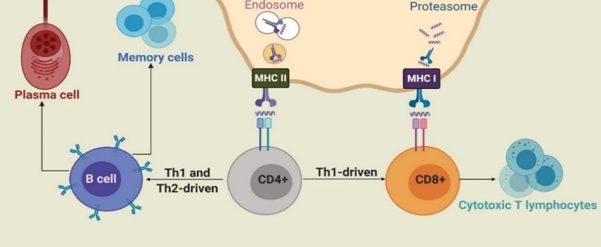
Pharmaceutics 2020, 12, 965                                                                          2 of 33

system and signal the pathogen as a foreign invader [6]. As most vaccine strategies are designed
to mimic the key features of pathogens, carbohydrates can be used as natural and relatively safe
vaccine adjuvants/immune stimulators, recognized by receptors on the surface of antigen-presenting
cells (APCs). Following recognition, they can initiate a cascade of innate and adaptive immune
responses [7–11]. Moreover, carbohydrate (e.g., chitosan, glucan, hyaluronic acid)-based delivery
vehicles can transport antigens to desired sites in the body, such as local lymph nodes. They are
especially efficient for mucosal antigen delivery because of their mucoadhesive properties [9,12]. Due to
the generally inherent immunomodulating properties, biocompatibility, biodegradability, and low
toxicity of carbohydrates, they are becoming intensively studied as adjuvants and delivery systems [13].
      Since the antigenic function of carbohydrates is already extensively covered elsewhere [6,8,14],
this review focuses on the receptor-based recognition of carbohydrates and their ability to enhance
systemic and mucosal immunity against co-administered antigens. Similarly, the gene-based
(DNA/mRNA) subunit vaccines have been recently reviewed [15,16], and therefore this review
emphasizes the protein/peptide-based subunit vaccines.

2. Immunostimulation

2.1. Innate and Adaptive Immunity
      To design an effective vaccine, an understanding of how the vaccine will interact with the immune
systems, as well as the nature of the immune systems itself, is crucial. Innate and adaptive immunity
are the two main components of immune systems. Innate immunity refers to the nonspecific defense
mechanisms of the body that are activated immediately after pathogen attack. Innate immunity
keeps foreign particles out of the body or limits the ability of the foreign material to spread and
move throughout the body. This occurs through a variety of mechanisms, including physiological
and anatomical barriers (e.g., skin), inflammatory responses, secretion of antimicrobial peptides, and
phagocytosis [17]. The innate immune system is also responsible for activating the adaptive immunity.
Adaptive immunity provides antigen (pathogen)-specific responses, including long-lasting memory
immune responses. Adaptive immune responses are divided into cell-mediated and humoral immunity.
      During infection or following vaccine administration, antigens are first taken up by immature
APCs, such as macrophages and dendritic cells (DCs), with the help of pattern recognition receptors
(PRRs), such as toll-like receptors (TLRs) and C-type lectin receptors (CLRs). APCs then mature
and the captured antigens are degraded to peptide fragments in endosomes or by proteasome and
recognized by major histocompatibility complex (MHC) class II or class I molecules, respectively.
The MHCs/peptide complexes are then transported on the surface of APCs, where they can be
recognized by T-cell antigen receptors (TCRs). CD8+ and CD4+ T-cells are stimulated by antigen-MHC
I and II complexes, respectively. Upon activation, CD8+ cells change into cytotoxic T-lymphocytes
(CTLs), which directly kill infected cells. Activated CD4+ cells differentiate into either T-helper 1 (Th1)
or T-helper 2 (Th2) cells [2,18]. Th1 cells produce cytokines, such as interferon-γ (IFN-γ), tumor necrosis
factor-α (TNF-α), and interleukin (IL-2 and IL-12) and stimulate mainly cell-mediated immunity (CD8+
cells) against intercellular pathogens [3]. Cytokines produced by Th2 include IL-1, IL-4, IL-5, IL-6,
IL-10, and IL-13. In addition, upon antigen (B-cell epitopes) recognition, B-cells are stimulated by Th2
cells to differentiate into memory and plasma cells and produce antibody-based humoral immune
responses (Figure 1) [19]. As such, B-cells are part of the adaptive immune response and contribute to
immunological memory, the process whereby immune cells respond more quickly and efficiently to an
antigen that they have encountered previously [20].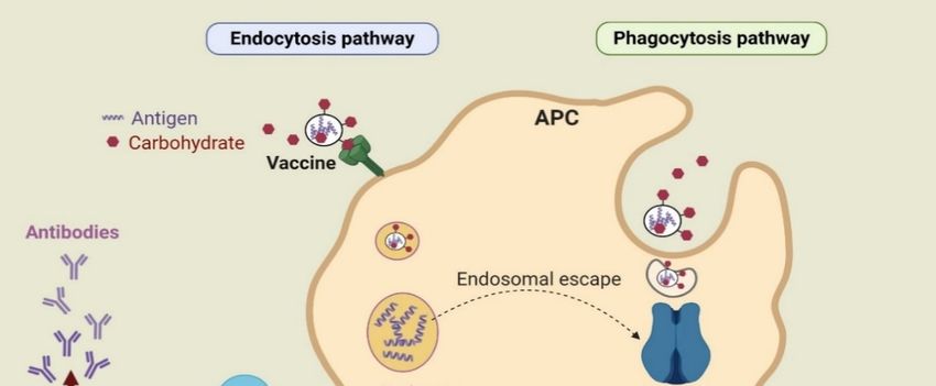
of antigens through phagocytosis, endosome escape of antigen and lysosomal processing, and finally
the MHC I pathway [26]. Antigens alone are usually unable to activate the immune system due to
low capacity, to be taken up by DCs and to reach the MHC I processing pathway [27]. However,
sufficient uptake of antigens by APCs facilitated by carbohydrate-based adjuvants initiates
processing 2020,
Pharmaceutics and 12,
presentation
965 by MHC I and triggers the physiological processes involved in cross-
3 of 33
presentation and CTL priming [28]. These responses are essential for triggering antitumor immunity.
Figure 1.
Figure 1. TheThe
basic uptake
basic pathways
uptake of antigens
pathways (e.g., endocytosis
of antigens and phagocytosis)
(e.g., endocytosis by antigen-
and phagocytosis) by
presenting cells (APCs) to induce immune responses.
antigen-presenting cells (APCs) to induce immune responses.
2.2. Mucosal and Systemic
Carbohydrates can beImmunity
recognized by receptors present on the surface of APCs and, when associated
with Human
an antigen, can enhance
immune responsesuptake
can be viadivided
endocytosis/phagocytosis.
into systemic and mucosal For example, mannose
immunity. The can be
sterile
recognized by mannose receptor [21]; chitosan can bind to different receptors, including
interior cavity of the body is known as the “systemic” environment, and it contains all of the organs TLR2 and
TLR4
except[22]; β-glucan
for the canmucosal
skin and be recognized by receptors
surfaces. Injection consisting
into the bodyof dectin-1,
is needed TLR2, 4, and 5 the
to stimulate [23];systemic
lipid A,
aimmune
lipidated system and result in the production of protective antibodies and T-cells. Therefore, the [25].
disaccharide, can be taken up by TLR4 [24]; and saponin can bind to DC-SIGN receptors first
Furthermore,
interaction between carbohydrate
an infectiousincorporation
agent and within
a hostnanovaccines
usually occurs canonactivate cross-presentation
a mucosal surface. Thus, of
antigens through phagocytosis,
mucosal immunity can prevent the endosome
entranceescape of antigen
of pathogens intoand
the lysosomal
body and stop processing,
resultingand finally
infection.
the MHC I pathway [26]. Antigens alone are usually unable to activate
The mucosal immune system consists of an integrated network of tissues, lymphoid and constitutive the immune system
due
cells,to
andlow capacity,
effector to be (antibodies,
molecules taken up bycytokines,
DCs andand to chemokines)
reach the MHC [29].IAsprocessing pathway
such, mucosal [27].
infection,
However, sufficient uptake of antigens by APCs facilitated by carbohydrate-based
or a mucosal-administered vaccine, can result in the activation of protective B- and T-cells in both adjuvants
initiates
mucosal processing
and systemic and presentation[30].
environments by MHC I and triggers the physiological processes involved
in cross-presentation
The mucosa-associated and lymphoid
CTL priming tissue[28].
includesThese responses are lymphoid
the nasal-associated essential tissue
for triggering
(NALT),
antitumor immunity.
the bronchus-associated lymphoid tissue (BALT), and the gut-associated lymphoid tissue (GALT)
[31]. All these tissues serve as the principal mucosal inductive and effector sites. The inductive sites
2.2. Mucosal and Systemic Immunity
Human immune responses can be divided into systemic and mucosal immunity. The sterile
interior cavity of the body is known as the “systemic” environment, and it contains all of the
organs except for the skin and mucosal surfaces. Injection into the body is needed to stimulate
the systemic immune system and result in the production of protective antibodies and T-cells.
Therefore, the first interaction between an infectious agent and a host usually occurs on a mucosal
surface. Thus, mucosal immunity can prevent the entrance of pathogens into the body and stop
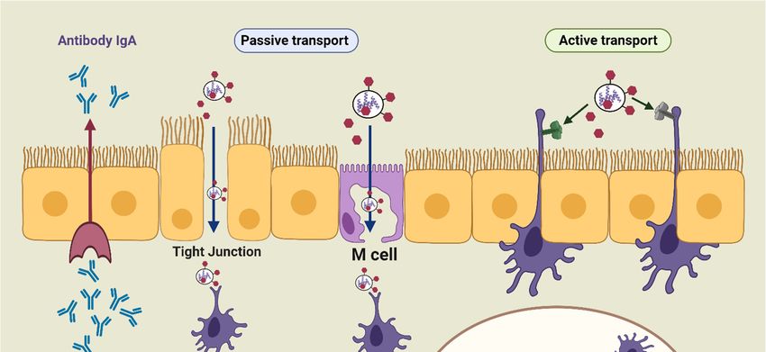
resulting infection. The mucosal immune system consists of an integrated network of tissues, lymphoid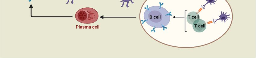
include mucosa-associated follicles such as intestinal Peyer’s patches (PPs), isolated lymphoid
follicles, and lymphoid aggregation in the bronchial tract [32]. In addition, these sites comprise a
complex network of innate and adaptive immune components, such as DCs, macrophages, T-cells,
B-cells, and natural killer (NK) cells, which are overlayered with epithelial cells and specialized
microfold
Pharmaceuticscells (M-cells)
             2020, 12, 965   [33]. M-cells offer functional openings in the epithelial barrier through 4 of 33
vesicular transport activity, and the ability of antigens to release the intraepithelial space, facilitating
antigen contact with T and B lymphocytes and macrophages [30,34]. Besides M-cells, the antigen can
and constitutive cells, and effector molecules (antibodies, cytokines, and chemokines) [29]. As such,
be collected by the receptors present on DC transepithelial dendrites (active transport). By taking up
mucosal infection, or a mucosal-administered vaccine, can result in the activation of protective B- and
the antigen, the antigen-specific T- and B-cells undergo activation and differentiation into T and B
T-cells in both mucosal and systemic environments [30].
effector cells, which results in secretion of immunoglobulin A (IgA). The effector cells and their
      The mucosa-associated lymphoid tissue includes the nasal-associated lymphoid tissue (NALT),
actions (e.g., antibody production, cytokine secretions, and cytotoxicity) migrate from inductive sites
the bronchus-associated lymphoid tissue (BALT), and the gut-associated lymphoid tissue (GALT) [31].
to effector sites (e.g., lamina propria and surface epithelium). Therefore, the immune responses of
All these tissues serve as the principal mucosal inductive and effector sites. The inductive sites
mucosal systems operate on the same general principle of systemic immunity: capturing the antigen
include mucosa-associated follicles such as intestinal Peyer’s patches (PPs), isolated lymphoid follicles,
with DCs or macrophages, then inducing T-cell responses and/or generating antibodies (Figure 2).
and lymphoid aggregation in the bronchial tract [32]. In addition, these sites comprise a complex
      Targeting the mucosal routes (e.g., oral, nasal, pulmonary, ocular, rectal, vaginal, etc.) has
network of innate and adaptive immune components, such as DCs, macrophages, T-cells, B-cells,
several advantages over parenteral vaccination, including: (a) injection can be avoided, reducing the
and natural killer (NK) cells, which are overlayered with epithelial cells and specialized microfold
risk of needle-stick injury and cross-contamination [35]; (b) full sterilization of the vaccine is not
cells (M-cells) [33]. M-cells offer functional openings in the epithelial barrier through vesicular
required; (c) the vaccine can be self-delivered; (d) vaccine storage conditions are usually less
transport activity, and the ability of antigens to release the intraepithelial space, facilitating antigen
restrictive [36]; (e) immunization of one mucosal site often induces immune responses in other
contact with T and B lymphocytes and macrophages [30,34]. Besides M-cells, the antigen can be
mucosal effector tissues [37]; and (f) most importantly, the production of mucosal antibodies (IgA)
collected by the receptors present on DC transepithelial dendrites (active transport). By taking up
can prevent systemic infection [30,38].
the antigen, the antigen-specific T- and B-cells undergo activation and differentiation into T and B
      Nanoparticles based on polysaccharides can interact with M-cells and undergo endocytosis and
effector cells, which results in secretion of immunoglobulin A (IgA). The effector cells and their actions
transcytosis [39,40]. Carbohydrates, for example, chitosan-based carriers, can open tight junctions to
(e.g., antibody production, cytokine secretions, and cytotoxicity) migrate from inductive sites to effector
directly transfer antigens through epithelial barriers [30]. These carriers also provide a prolonged
sites (e.g., lamina propria and surface epithelium). Therefore, the immune responses of mucosal
interaction between associated antigens and epithelial cells due to chitosan’s mucoadhesive
systems operate on the same general principle of systemic immunity: capturing the antigen with DCs
properties. Consequently, they elicit strong systemic immunoglobulin (IgG) and mucosal IgA
or macrophages, then inducing T-cell responses and/or generating antibodies (Figure 2).
responses [41].

                           Figure
                           Figure 2.
                                  2. The
                                     Thesimplified
                                         simplified pathway
                                                    pathway of
                                                            of mucosal
                                                               mucosal immunity.
                                                                        immunity.

      Targeting the mucosal routes (e.g., oral, nasal, pulmonary, ocular, rectal, vaginal, etc.) has several
advantages over parenteral vaccination, including: (a) injection can be avoided, reducing the risk of
needle-stick injury and cross-contamination [35]; (b) full sterilization of the vaccine is not required;
(c) the vaccine can be self-delivered; (d) vaccine storage conditions are usually less restrictive [36];
(e) immunization of one mucosal site often induces immune responses in other mucosal effector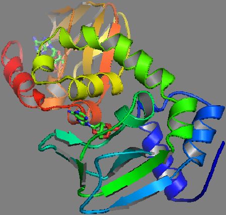
Pharmaceutics 2020, 12, 965                                                                      5 of 33

tissues [37]; and (f) most importantly, the production of mucosal antibodies (IgA) can prevent systemic
infection [30,38].
     Nanoparticles based on polysaccharides can interact with M-cells and undergo endocytosis and
transcytosis [39,40]. Carbohydrates, for example, chitosan-based carriers, can open tight junctions to
directly transfer antigens through epithelial barriers [30]. These carriers also provide a prolonged
interaction between associated antigens and epithelial cells due to chitosan’s mucoadhesive properties.
Consequently, they elicit strong systemic immunoglobulin (IgG) and mucosal IgA responses [41].

3. Adjuvants
      Adjuvants are defined as any substances, compounds, or even strategies that result in the
enhancement of immune responses, particularly adaptive immune responses, when delivered together
with an antigen [42,43]. An adjuvant is primarily used to mimic the danger signal and is recognized by
PRRs to facilitate antigen uptake via major target cell types (e.g., monocytes, macrophages, and DCs),
thus enhancing immunogenicity. In addition, adjuvants can activate and maturate macrophages,
T lymphocytes, B lymphocytes, and NK cells [13,42], and inflammasomes, pro- and anti-inflammatory
signals, inducing inflammatory chemokine and cytokine responses [5,44–46].
      The key features of a successful adjuvant include not only its immune-stimulating potency to
reduce the dose of antigens and the number of boost immunizations needed to trigger long-lasting
protective immunity, but also its safety and stability. Salt-based adjuvants such as aluminum have
been introduced as an adjuvant for human use since the 1930s [47]. Although it stimulates effective
humoral immune responses against whole pathogen-based vaccines, its stimulating potency against
subunit vaccines is limited. Alum is a poor immune stimulator of CTLs and induces side reactions such
as indurations, erythemas, cutaneous nodules, swellings, irritation, and redness [48]. More recently,
several other adjuvants have been developed; however, they have been approved only for the use in
particular vaccines and countries [49]. Therefore, the development of a safe and effective adjuvant is
still of high priority.
      Carbohydrates are promptly recognized APCs, can protect antigen against degradation,
have mucoadhesive properties and most importantly are usually biodegradable and safe for human
use. Therefore, carbohydrates, both natural and synthetic, are one of the most promising and most
investigated immune stimulators.

4. Carbohydrate-Based Adjuvants
     Carbohydrates enhance the immunogenicity of a vaccine by binding to specific glycan-binding
receptors on the surface of APCs (e.g., TLRs, nucleotide-binding oligomerization domain 2 (NOD2)-like
receptors (NLRs), and CLRs). This encourages carbohydrate and associated antigen uptake by
phagocytosis and endocytosis [50].
     Specifically, CLRs, including mannose receptors, DEC-205, dectin-1, DNGR-1, DC-SING, Mincle,
and single CD22, have the potential to recognize carbohydrate (sugar) moieties of glycosylated
antigens [9,25,51,52]. In addition to CLRs, T-cell receptors, such as TLR2 and TLR4, can recognize
pathogen-derived polysaccharides and glycoconjugates [43,53,54]. Carbohydrate interaction with
CLRs triggers intracellular signaling cascades mediating APC maturation, which is necessary for naive
B- and T-cell activation [55,56]. In addition, carbohydrates can also be taken up by APCs based on
charged interactions [57]. Cumulatively, this triggers a variety of signaling pathways resulting in the
stimulation of innate and adaptive immune responses [9]. Importantly, carbohydrates can stimulate
balanced pro- and anti-inflammatory responses generating improved potency and safety compared to
other adjuvants [55].
     Carbohydrates with adjuvating activities are generally divided into two classes: saccharides,
such as mannose and mannan; and saccharide derivatives, which are sugars modified with lipids,
peptides, etc.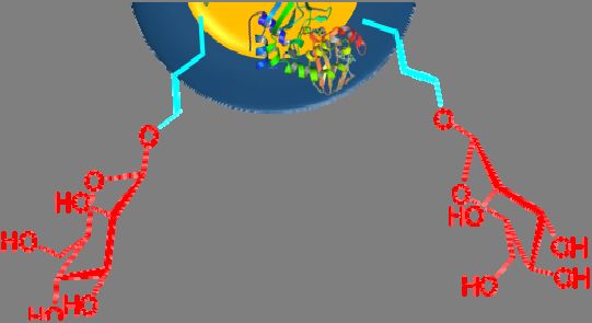
Pharmaceutics 2020, 12, 965                                                                      6 of 33

4.1. Saccharides

4.1.1. Mannose
     Mannose is commonly found on the surface of bacteria, fungi, and viruses [58]. A variety of
immune receptors can recognize mannose, including different CLRs (e.g., mannose-binding lectin,
mannose receptors, Mincle [59], and DC-SIGN receptors [21]) and TLR4, which binds O-linked
mannosylated ligands [60]. Binding can induce complement activation and phagocytosis [5,11],
triggering innate immune responses [61]. Furthermore, stimulation of these receptors can also
induce receptor-mediated endocytosis and influence TLR signaling cascades, activating the adaptive
immune system [42,62,63]. For example, mannose receptors CD206 and CD209, which are C-type
lectins expressed on macrophages and DCs, recognized mannosylated antigen-bearing constructs
and delivered antigens onto MHC I and MHC II receptors. These were further recognized by T-cells,
triggering adaptive immunity [64]. Activation of mannose receptors enhances CD4+ and CD8+ T-cell
responses, inducing Th1 and Th2-type immunity, which in turn induces IgG production and long-lasting
immunity [62,63,65]. The activation of mannose receptors in tumor-associated macrophages has also
been shown to improve innate and adaptive antitumor immunity [11]. Therefore, mannose is often
used in vaccine design to enhance the immunogenicity of co-administered antigens.
     Glaffig et al. developed anticancer vaccines where two mannosyl glycolic acids were linked to
both amino groups of the N-terminal lysine of a peptide derived from mucin 1 protein (which is
overexpressed by several cancers) [66]. The mannosylated antigens were internalized via endocytosis
by DCs and macrophages 2–4 orders of magnitude faster than non-mannosylated vaccine candidates.
The mannosylated compounds also triggered an increase in the number of macrophages, DCs, and CD4+
T-cells in the local lymph organs of mice. The vaccine induced the production of high titers of IgG
specific to tumor-associated glycopeptide antigens. Apostolopoulos and coworkers reported that
mucin 1 conjugated to mannose under oxidizing conditions caused the endosomal escape of antigens
into the cytoplasm, presented mucin 1 peptides into the MHC I pathway, generated a Th1 response,
and yielded a high level of CTL and a low level of IgG2a antibodies compared to other subtypes [67,68].
The vaccine candidate also increased the level of IL-12 and IFN-γ cytokines. Overall, the mannosylated
antigen enhanced cross-presentation, a pathway where tumor-associated antigens can induce cytotoxic
CD8+ T-cell activation and resulted in induction of stronger antitumor immunity [69].
     To enhance the immunogenicity of ovalbumin (OVA), a combination of TLR7 ligand and
mannose-targeting moiety was tested [70]. The mixture of imidazoquinolines, TLR7 agonists,
and mannose was conjugated to OVA through a self-immolative linker via a metal-free cycloaddition
reaction. This model vaccine amplified antigen presentation to T-cells and generated greater humoral
and cellular immunity than vaccine candidates lacking either a mannose-receptor targeting moiety or
a TLR7 ligand. The same strategy was also used for the delivery of Plasmodium falciparum-derived
circumsporozoite protein, with the result of high IgG production and the inhibition of sporozoite
invasion into hepatocytes in mice.
     Nanoparticles have recently become one of the most popular and effective vaccine delivery
systems [71], and mannose has been employed to modify some of these vaccine constructs (Figure 3a,b).
Coating the surface of nanoparticles and nanocapsules with mannose has been found to enhance antigen
uptake by APCs [72]. For example, alginate was conjugated to OVA (model antigen) via a pH-sensitive
Schiff base bond [61]. Tetrabutylammonium-alginate and 4-aminophenyl d-mannopyranoside were
also used to synthesize mannosylated alginate. Nanoparticles were then formed by cross-linking two
types of modified alginates with the help of CaCl2 . The nanoparticle size and zeta potential were
310 nm and −46 mV, respectively. These nanoparticles were promptly taken up by mouse bone marrow
DCs (BMDCs) in vitro. Uptake of mannose-free nanoparticles was 3-fold lower, and subsequent
activation of APCs was 2-fold lower (when measured by APC CD40 marker overexpression).
Furthermore, cross-presentation of antigen and CTL activation were significantly enhanced when
mannosylated nanoparticles were administered to mice. In another study, poly d-l-lactide-co-glycolide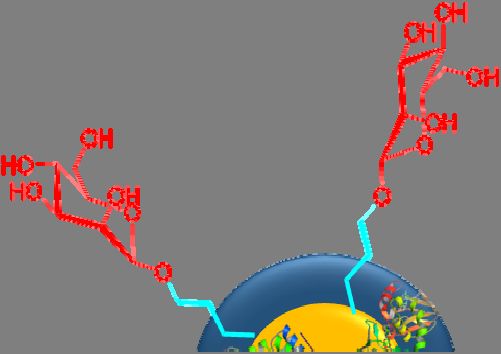
Pharmaceutics 2020, 12, 965                                                                                    7 of 33

 nanoparticles were loaded with imiquimod as a TLR7 agonist, coated with cancer cell membrane as
 an antigen, and mannose was linked to the nanoparticles via a lipid-based anchoring moiety [73].
 The mannosylated vaccine candidates enhanced nanoparticle DC uptake and stimulated DC maturation.
 Compared to mice treated with free nanoparticles, mannosylated nanoparticles effectively migrated to
 draining lymph nodes and triggered tumor-specific immune responses. The lymph node retention
 of the nanovaccine was also enhanced with the help of mannose. The treatment of mice with an
 anti-programmed death-1 (anti-PD-1) checkpoint blockade failed to cure mice in tumor challenge
 experiments. However, once both strategies, mannosylation and anti-PD-1 blockage, were combined,
 half of the2020,
Pharmaceutics mice12,survived theREVIEW
                      x FOR PEER  tumor challenge.                                            7 of 33

     Figure
      Figure3.3. Mannosylated
                  Mannosylatedpeptide/protein-based
                                 peptide/protein-basedvaccine
                                                        vaccineconstructs:
                                                                constructs:(a)(a)antigenic
                                                                                  antigenicpeptide/protein
                                                                                            peptide/protein
     conjugated  to mannose; (b) mannose  conjugated to a polymer/lipid-based    nanocapsule.
      conjugated to mannose; (b) mannose conjugated to a polymer/lipid-based nanocapsule.

      The
       Thepolysaccharide,
             polysaccharide,chitosan,
                                chitosan,isisused
                                               usedwidely
                                                     widelyfor
                                                             forvaccine
                                                                 vaccinedelivery
                                                                            deliverydue
                                                                                     duetotoits
                                                                                              itsmucoadhesive
                                                                                                 mucoadhesiveand   and
adjuvating
  adjuvatingproperties
                properties[40,74–77].
                             [40,74–77].Following
                                           Following subcutaneous
                                                         subcutaneousadministration    in mice,
                                                                           administration        nanoparticles
                                                                                            in mice,             (120
                                                                                                       nanoparticles
nm,
  (120−12
       nm,mV)−12formed    by electrostatic
                  mV) formed                 interactions
                                 by electrostatic           between
                                                    interactions       chitosan
                                                                  between        and mannosylated
                                                                             chitosan  and mannosylatedalginate  and
                                                                                                            alginate
loaded    with  tumor   cell lysates  were   efficiently  taken  up  by   DCs  in the draining    lymph
  and loaded with tumor cell lysates were efficiently taken up by DCs in the draining lymph node [78].    node  [78].
Moreover,
  Moreover,the themannosylated
                    mannosylatednanoparticles
                                      nanoparticlesenhanced
                                                       enhancedthe theexpression
                                                                         expressionofofAPC
                                                                                         APCmaturation
                                                                                                maturationmarkers,
                                                                                                            markers,
including
  including MHC I and MHC II, CD40, CD80, and CD86, and stimulated higher TNF-α and IL-12levels
             MHC   I and  MHC    II, CD40,  CD80,    and CD86,   and  stimulated   higher  TNF-α    and IL-12   levels
ininserum.
     serum.TheTheactivity
                   activityofofCTLs
                                CTLswaswasanalyzed
                                            analyzedusing
                                                       usingthe
                                                              theCD107
                                                                  CD107marker
                                                                            markerofofCTL
                                                                                        CTLdegranulation
                                                                                             degranulationandandthe the
level
  levelofofIFN-γ
            IFN-γexpression.
                  expression.ThisThiswas
                                       wassignificantly
                                             significantlyhigher
                                                            higherininthe
                                                                        thelymph
                                                                            lymphnode
                                                                                    nodeand
                                                                                          andspleen
                                                                                                spleenfollowing
                                                                                                       followingthe the
administration of mannosylated nanoparticles, in comparison to non-mannosylated nanoparticles
and free antigens. The nanoparticles delayed the growth of early-stage tumor cells; however, when
mice were challenged with 4T1 mouse breast tumor cells and then immunized, mannosylated vaccine
candidates failed to provide antitumor activity.
      Liposomes are another popular vaccine delivery strategy [79], and carbohydrates have similarly
Pharmaceutics 2020, 12, 965 8 of 33
administration of mannosylated nanoparticles, in comparison to non-mannosylated nanoparticles and
free antigens. The nanoparticles delayed the growth of early-stage tumor cells; however, when mice
were challenged with 4T1 mouse breast tumor cells and then immunized, mannosylated vaccine
candidates failed to provide antitumor activity.
Liposomes are another popular vaccine delivery strategy [79], and carbohydrates have similarly
been utilized within these formulations. Mannose can be easily anchored to liposomal surfaces
by lipidation. For example, mannosylated lipidated peptide was anchored to the surface of
liposomes prepared by lipid film hydration [80]. The mannosylated liposomes efficiently increased
antigen uptake by APCs and up-regulated expression of MHC II and co-stimulatory molecules,
including CD80 and CD86 on both DCs and macrophages. In contrast, corresponding non-mannosylated
liposomes were poor activators of APCs. Mannose-modified liposomal vaccines can improve
vaccine efficacy, not only by inducing and promoting the desired immune response, but also by
decreasing adverse effects by minimizing interaction with irrelevant cells and tissues. OVA-loaded
1,2-dioleoyl-3-trimethylammoniumpropane (DOTAP) liposomes and DOTAP-polyethylene glycol-poly
caprolactone (PEG)-mannose liposomes have both been studied. The liposomes with mannose
showed dynamic lymphatic trafficking in vivo with accumulation in draining lymph nodes in
a short period, indicating accelerated drainage from the injection site into lymph nodes [81].
Furthermore, the mannosylated liposomes improved cross-presentation and cytokine production,
stimulated greater lymphocyte activation, CD4+ , and CD8+ T-cell response, effector cytokine secretion,
and induced Th1-biased humoral responses compared to non-mannosylated liposomes [82]. Zhu and
coworkers produced hybrid liposomes, named polymersomes [62]. Polycaprolactone (PCL)-PEG-PCL
polymersomes bearing 1,2-distearoyl-sn-glycero-3-phosphoethanolamine (DSPE)-PEG-mannose
(mannose functionalized lipid hybrid polymersome), which was synthesized by covalent binding
with DSPE-PEG-NH2 and d-mannopyranosylphenyl isothiocyanate, were formed. In addition,
positively charged lipid (DOTAP), TLR4 agonist (lipid A), and TLR7/8 agonists (imiquimod) were
incorporated into the hydrophobic layer of polymersomes. The negatively charged model antigen OVA,
was encapsulated electrostatically into polymersomes (220 nm and −1.6 mV) and the construct was
found to be non-toxic to mouse BMDCs. The polymersomes were taken into BMDC twice as efficiently
as OVA alone, and OVA mixed with lipid A. The liposomes were recognized by mannose receptors,
facilitating antigen uptake via endocytosis into a distinct endosome subpopulation. The antigens
were also protected from lysosomal protease degradation and were allowed to escape into the
cytoplasm [83]. Therefore, mannosylated polymersomes effectively cross-presented antigens and
produced antigen-specific CTL responses, which were further verified by a mouse tumor challenge with
EG7-OVA cells. The mannosylated polymersomes prolonged tumor-free time, effectively suppressed
tumor growth, and greatly extended the median survival time of mice [62].
4.1.2. Oligo- and Polysaccharides of Mannose
The oligo- and polymerized forms of mannose have also been assessed and found to be
promptly recognized by PRRs on the surface of human immune cells (e.g., DCs, macrophages,
epithelial cells, and endothelial cells) [60]. Mannan, the polymerized form of mannose, can
be recognized by a wide range of receptors, including mannose receptors [84], dectin-2 [85],
dectin-3 [86], Mincle, DC-SIGN [87,88], galectin-3 [89], FcγR [90], TLR2, TLR4 [91], and TLR6 [92,93].
Therefore, mannan serves as a promising APC-targeting agent that can enhance the uptake and
processing of co-administered antigens in APCs. Although it is recognized by the immune system
in a similar manner to mannose [92], mannan is a more potent adjuvant. It binds more effectively
to receptors consisting of multiple carbohydrate-recognizing domains as it bears multiple ligands
(mannose moieties) [94]. Mannan comprises linear and branched polymers of mannose sugars linked
via α-1,2, α-1,3, α-1,4, α-1,6, and β-1,2 glycoside bonds (Figure 4) [95]. The ligand–receptor interaction
and the stimulation of immunity depends on the conformation of mannan (and other polysaccharides,
too), the types of glycosidic bonds in the molecule, and the degree of branching, charge and molecularPharmaceutics 2020, 12, 965 9 of 33
Pharmaceutics 2020, 12, x FOR PEER REVIEW 9 of 33
weight
more [96]. Interestingly,
selectively coating nanoparticles
to DCs. Interestingly, the higherwith mannan
oligomers enhances (Tetra-Man
of mannose not only vaccine delivery to
and Penta-Man)
APCs, but also prevents nanoparticle aggregation [97].
were not taken up by APCs any more efficiently than Tri-Man.
Figure 4.
Figure Thebranched
4. The branched and
and linear
linear forms
forms of
of mannan.
mannan.
Nanoparticles
The conjugation chemically modified
strategy used or coated
to combine with oligomannose
mannan with antigensand alsopolymannose are popularly
plays an important role in
applied for
receptor vaccine delivery
recognition. Mannan [9]. For example,
conjugated a seriescancer
to the breast of diether
antigen, lipids
mucinwere1, conjugated
under either to
mannose (Man), trimannose (Tri-Man), branched trimannose (Man-Tri),
oxidation or reducing conditions, was injected intraperitoneally into mice [68]. The oxidative tetramannose (Tetra-Man) or
pentamannose
conjugation led(Penta-Man) through and
to CTL activation “clickTh1 chemistry”
cytokinebetween
(IFN-γ)azido mannosewhile
production, oligosaccharide
stimulationand of
the diether lipid (propargyl-PEG8-diether) [21]. Mannosylated lipids
antibody (predominantly IgG2a) production was poor. In contrast, the reductive coupling were anchored to liposomes
betweenand
their ability
mannan andtothe be taken
antigen upstimulated
by mannose receptors
strong or DC-SIGN
humoral immunity wasandanalyzed using DC2.4
no cellular response. cellsMice
and
DC-SIGN HEK293
immunized cells, respectively.
with oxidized mannan coupled Man-Tri withwas thepreferentially
antigen weretaken protectedup byinDC-SIGN receptors,
a tumor challenge
while linear Tri-Man and Man were recognized preferentially by mannose
with mucin-1 3T3 tumor cells, whereas tumor growth was not inhibited in mice treated with reduced receptors. In contrast to
mannose receptors, which are expressed in a variety of cells, DC-SIGN
mannan-antigen conjugate. It has been suggested that the oxidizing conditions allowed mannan is expressed predominantly on
DCs. Therefore,
aldehyde groupsthe to branched
form Schiff form
basesof the
not mannosylated
only with antigens, vaccine can
but be with
also deliveredAPCs,more selectively
inducing strongto
DCs. Interestingly, the higher oligomers of mannose (Tetra-Man and Penta-Man)
antitumor immunity by targeting the antigen to the intracellular processing pathway for presentation were not taken up by
APCsMHC
with any more efficiently
I molecules. than Tri-Man.
However, this was contradicted by another study that showed high antibody
The conjugation strategy
responses against vaccines bearing used to combinemannan
oxidized mannan[98]. withAantigens also plays anvaccine
nanoparticle-based important role in
candidate
receptor recognition. Mannan conjugated to the breast cancer antigen,
(200–800 nm) against porcine circovirus type 2 (PCV2) containing oxidized mannan attached to mucin 1, under either oxidation
or reducing
PCV2 conditions, was injected intraperitoneally into mice [68]. The oxidative conjugation led to
ΔCap42-233 protein via an acid-sensitive Schiff base reaction was developed. When subcutaneously
administered, itand
CTL activation showed Th1 cytokine (IFN-γ)
a higher level production,
of IL-4 while stimulation
(which regulates of antibody
antibody-mediated (predominantly
humoral immune
IgG2a) production was poor. In contrast, the reductive coupling
responses), and consequently higher antibody production, than non-mannanylated vaccine between mannan and the antigen
stimulated strong
candidates. humoral
The level immunity
of IFN-γ was notandsignificantly
no cellular response.
higher inMicemiceimmunized
treated with witha oxidized mannan
mannan-bearing
coupled with the antigen were protected in a tumor challenge with mucin-1
vaccine. The conflicting outcome of the two studies can be related to different abilities of the vaccines3T3 tumor cells, whereas
tumor
to formgrowth was notand/or
nanoparticles, inhibited
thein mice structural
varied treated with reduced
nature mannan-antigen
(branching, molecularconjugate.
weight, even It has been
purity)
suggested that the oxidizing conditions allowed mannan aldehyde
of the mannans used. Thus, despite the mannan’s advantage of being able to stimulate stronger groups to form Schiff bases not
only with antigens, but also with APCs, inducing strong antitumor immunity
immune responses, the systems are less defined and show greater variability in immune responses by targeting the antigen
to themannose.
than intracellular processing pathway for presentation with MHC I molecules. However, this was
contradicted by another study ability
The immune-stimulating that showed high antibody
of reductive responses
and oxidative against vaccines
mannan-antigen bearing oxidized
conjugates can also
mannan [98]. A nanoparticle-based vaccine candidate (200–800 nm) against
be affected by the route of administration. For example, mannan was coupled to secreted listeriolysin porcine circovirus type 2
O (LLO) protein, the immunodominant antigen of Listeria monocytogenes, or to the 19-kDa proteinSchiff
(PCV2) containing oxidized mannan attached to PCV2 ∆Cap42-233 protein via an acid-sensitive (19-
basesecreted
FP) reactionbywas developed. tuberculosis
Mycobacterium When subcutaneously
under eitheradministered, it showed
oxidation or reductive a higher level
conditions. Theseofwere
IL-4
(which
each regulates antibody-mediated
administered humoral immune[99].
intranasally or intraperitoneally responses), and consequently
The intranasally higheroxidative
administered antibody
mannan-LLO conjugate induced greater humoral immune responses, especially LLO-specific IgA,
IgG1, and IgG2a titers, in comparison to the conjugate-administered intraperitoneally. IgA antibodyPharmaceutics 2020, 12, 965 10 of 33
production, than non-mannanylated vaccine candidates. The level of IFN-γ was not significantly
higher in mice treated with a mannan-bearing vaccine. The conflicting outcome of the two studies
can be related to different abilities of the vaccines to form nanoparticles, and/or the varied structural
nature (branching, molecular weight, even purity) of the mannans used. Thus, despite the mannan’s
advantage of being able to stimulate stronger immune responses, the systems are less defined and
show greater variability in immune responses than mannose.
The immune-stimulating ability of reductive and oxidative mannan-antigen conjugates can also
be affected by the route of administration. For example, mannan was coupled to secreted listeriolysin O
(LLO) protein, the immunodominant antigen of Listeria monocytogenes, or to the 19-kDa protein (19-FP)
secreted by Mycobacterium tuberculosis under either oxidation or reductive conditions. These were
each administered intranasally or intraperitoneally [99]. The intranasally administered oxidative
mannan-LLO conjugate induced greater humoral immune responses, especially LLO-specific IgA,
IgG1, and IgG2a titers, in comparison to the conjugate-administered intraperitoneally. IgA antibody
titers from vaginal washings induced by LLO- or 19-FP-mannan conjugates administered through
the intranasal route were greater than those induced by a physical mixture of mannan and LLO
or 19-FP. Oxidized mannan-LLO induced significantly higher LLO-specific IgA, IgG1, and IgG2
antibodies than the reduced mannosylated antigen. This suggested that the mucosal adjuvanticity of
mannan and the induced immunity of both oxidized and reductive mannan-antigen conjugations were
administration route-dependent.
4.1.3. Glucan
Glucan, the polymerized form of glucose and a natural component of the cell wall of yeast and
some bacteria, has been found to have immune-stimulating activity [100]. The polysaccharide is linked
by a variety of glycosidic bonds, such as α and β-1,3, and 1,4 glycosides bonds. Moreover, glucan can
be isolated (e.g., from Saccharomyces cerevisiae) as hollow spherical particles (GPs) [101]. All of the
homopolymer forms of glucose are recognized by immune cells (e.g., neutrophils, macrophages,
and DCs) [102]. Glucan binds to various PRRs, such as dectin-1, TLR2, TLR6, and TLR9, and therefore
stimulates phagocytosis and endocytosis of antigens leading to a proliferation of CD4+ and CD8+
cells, differentiation of Th1 and Th17, the up-regulation of cytokines IL-4 and IL-3, and the production
of high levels of IgG and IgA antibodies [56,60,103,104]. Furthermore, glucan is non-toxic and has
anticoagulant, antithrombotic, and antioxidant properties [105]. Glucan and related GPs have been
used for vaccine delivery. For example, β-glucan was conjugated to three hollow silica particles,
which were produced on the following templates: (a) Escherichia coli particles (rod, 900 nm × 1.2–3.2 µm),
(b) Staphylococcus aureus particles (spherical, 900 nm), and (c) polystyrene (spherical, 220 nm) [56].
The particles were then loaded with OVA. All glucan-conjugated particles were taken up by dectin-1,
complement receptors, and TLR-2 on APCs. This successfully induced APC maturation, overexpression
of MHC II, and strong IgG antibody responses in comparison to plain particles. All glucan particles
induced Th1 and Th2 responses with only a minor difference in Th1/Th2 specificity between particles.
The high solubility of GPs and their strong capacity to entrap antigenic protein and peptides into
the inner hollow cavity of the particles make GPs interesting vaccine delivery vehicles. In addition,
owing to their ability to interact with different human epithelial cell lines, glucan-based delivery
systems can enhance mucosal immunity and elicit humoral responses. For example, GPs were loaded
with OVA by dissolving OVA in hydrated GPs followed by lyophilization (particle size = 3.7 µm,
zeta potential = −6.5 mV, and entrapment efficacy = 98%) [106]. OVA-loaded GPs enhanced the
expression of dectin-1, TLR2, proinflammatory cytokines, and chemokines. GPs were recognized
and internalized by M-cells in PPs. Finally, the GPs promoted Th17 responses and OVA-specific IgA
production; however, they did not trigger higher IgG production than OVA alone following oral
vaccination in mice.
Various types of glucan homopolymers exist in nature, and their biological and immunological
properties depend on their molecular and structural features, such as polymer length, branchingPharmaceutics 2020, 12, 965 11 of 33
degree, type of glycosidic bond, and solubility. The immune-stimulating properties of different glucans
were reviewed by Moreno-Mendieta et al. [45]. Curdlan sulfate (β-1,3 glucan) recognized by dectin-1,
cellulose (β-1,4-glucan), lentinan (β-1,3-linked glucan with a single β-1,6 branch every 5 residues)
recognized by TLR4, and phycarine or laminarin (β-1,3-linked glucan with a single β-1,6 branch) binding
to complement receptor 3, induced activation of different immune pathways. For example, sparan,
a six-branched β-1,3 glucan obtained from the medicinal mushroom Sparassis crispa, was recognized
through TLR4 and led to the activation of mitogen-activated protein kinase and NF-κB signaling
pathways [107]. It induced DC maturation as measured by the overexpression of MHC I and II and
co-stimulatory molecules (CD40, CD80, and CD86). Additionally, sparan enhanced the production of
IL-12, IL-1, TNF-α, IFN-γ, and IL-2.
Glucan-rich bacterial extracts are also used as adjuvants. Zymosan is an insoluble cell wall extract
from Saccharomyces cerevisiae that is composed of glucan (55%), mannan, proteins, lipids, and chitin [108]
and it is recognized by dectin-1 and TLR2 [109,110]. Co-activation of dectin-1 and TLR2-signaling
pathways, mediated by zymosan, results in a synergistic increase in the production of IL-12 and
TNF-α. Influenza vaccines adjuvanted with polyinosinic:polycytidylic acid (poly (I:C)), zymosan,
and A/PR8 antigens have been examined in BALB/c mice [111]. Poly (I:C) induced TLR3-mediated
signaling pathways and zymosan induced dectin-1 and TLR-2-mediated signaling pathways in BMDCs.
Secretory IgA and serum IgG levels were increased significantly in mice immunized with the vaccine
candidates carrying both adjuvants (zymosan and poly I:C) compared to those with vaccine candidates
bearing a single adjuvant. In addition, mice immunized with a vaccine carrying both adjuvants
(zymosan and poly I:C) developed a protective response to the influenza virus, and had an increased
survival rate and a reduced weight loss.
4.1.4. Chitosan
Chitosan is a linear amino polysaccharide composed of β-1,4-linked monomers of d-glucosamine
and N-acetyl-d-glucosamine (Figure 5). It is usually produced by deacetylation of chitin
(β-1,4-N-acetyl-d-glucosamine) isolated from animals (e.g., crayfish, shrimp waste, crabs, and lobsters)
or fungal resources (e.g., Agaricus, Hydnum, and Boletus species) [13,112]. However, the low solubility
of chitosan hinders its applicability to drug and vaccine delivery. To increase its solubility and stability,
chitosan is often chemically modified to produce glycated chitosan, thiolated chitosan, monomethyl
chitosan, and trimethyl chitosan (TMC) [76,95]. TMC bearing a permanent positive charge (Figure 5) is
the most hydrophilic compound among these derivatives. Moreover, TMC can be easily produced
in a cost-effective manner. It has high antimicrobial activity, high absorption through biological
membranes, and it is mucoadhesive and non-toxic [113]. Furthermore, TMC has immune-stimulating
properties, which typically exceed those of anionic polysaccharides [114]. Its mucoadhesive properties
retain vaccine formulations on mucosal surfaces, so chitosan/TMC is often used for intranasal
vaccine delivery [115]. Chitosan/TMC can also open the tight junctions between epithelial cells,
further improving vaccine uptake [116]. Consequently, chitosan/TMC-based nanoparticles easily reach
M-cells [115], penetrate epithelial cells, deliver antigens to immune competent cells, and induce both
systemic and mucosal immunities [117]. For example, a lipopeptide-based vaccine bearing J14 peptide
epitopes derived from group A Streptococcus (GAS) M-protein was loaded into polymeric nanoparticles.
These nanoparticles (100–300 nm) were prepared from dextran, poly d,l-lactic-acid-co-glycolic acid
(PLGA), and/or TMC via a two-stage double centrifugation technique [114]. Negatively charged
polymers (PLGA and dextran) were used to formulate polyelectrolyte nanoparticles with the positively
charged lipopeptide [118]. Dextran and dextran/TMC nanoparticles were both efficiently taken
up by DCs, but only dextran/TMC significantly enhanced the maturation of DCs, as measured by
overexpression of all tested co-stimulatory molecules (i.e., CD40, CD80, and CD86). Dextran/TMC
nanoparticles were also the most effective in the induction of antigen-specific systemic (IgG) and
mucosal (IgA) antibody titers following intranasal immunization, and produced antibodies showing a
high opsonic activity against tested clinical GAS isolates in vitro. In other strategies, TMC was used toPharmaceutics 2020, 12, 965 12 of 33
form nanoparticles (~200 nm, +36 mV) with GAS peptide epitope-polyglutamic acid (PGA) conjugates
via electrostatic interaction between PGA and TMC [119]. These nanoparticles induced DCs and
macrophage maturation (overexpression of MHC II and CD80); while either PGA or TMC mixed with
antigens did not. Moreover, the nanoparticles induced significantly higher IgG and IgA antibody titers
than antigens adjuvanted with cholera toxin B subunit (CTB). Mice immunized with the nanoparticles
showed a reduced bacterial load when challenged with M1 GAS, while the CTB-adjuvanted vaccine
did not. This system was further improved by antigen lipidation and the replacement of crustacean
TMC with better-defined, low molecular weight, highly deacetylated fungi-derived TMC [120,121].
Pharmaceutics 2020, 12, x FOR PEER REVIEW 12 of 33
Comparison of the chemical structures of chitin, chitosan, and TMC.
Figure 5. Comparison
In addition to
In addition to its
itsmucoadhesive
mucoadhesiveproperties,
properties,chitosan
chitosanalso
alsoacts
actsasasa receptor
a receptor ligand
ligand andand binds
binds to
to NLRs, dectin-1, leukotriene B4 receptor [122], and TLR2, TLR4, and
NLRs, dectin-1, leukotriene B4 receptor [122], and TLR2, TLR4, and TLR5 on APCs [123]. Moreover, TLR5 on APCs [123].
Moreover, the glucosamine
the glucosamine units in units in chitosan
chitosan are recognized
are recognized by mannose
by mannose receptors
receptors [124].Chitosan
[124]. Chitosanactivates
activates
macrophages [13,125], induces cytokine secretion from NK cells, and stimulates
macrophages [13,125], induces cytokine secretion from NK cells, and stimulates NALT to produce NALT to produce mucosal
secretory IgA, IgG, TNF-α,
mucosal secretory IgA, IgG, IL-6, and IFN-γ
TNF-α, IL-6,[43].
andPCV2IFN-γsubunit vaccine
[43]. PCV2 basedvaccine
subunit on the PCV2
basedcapsid
on theprotein
PCV2
was conjugated with chitosan oligosaccharide and OVA-based carriers
capsid protein was conjugated with chitosan oligosaccharide and OVA-based carriers [123]. The[123]. The chitosan in the
vaccine formulation increased cell proliferation, phagocytosis activity in macrophages,
chitosan in the vaccine formulation increased cell proliferation, phagocytosis activity in TLR2 and TLR4
expression
macrophages,on macrophages,
TLR2 and TLR4 and expression
subsequently onincreased antigen
macrophages, presentation.
and subsequently Moreover,
increasedthe antigen
vaccine
candidate consisting of chitosan conjugated with PCV2 increased RAW 264.7 macrophage
presentation. Moreover, the vaccine candidate consisting of chitosan conjugated with PCV2 increased proliferation,
nitric
RAW oxide production (playing
264.7 macrophage a critical
proliferation, roleoxide
nitric in theproduction
activation of immune
(playing cells, such
a critical roleas inmacrophages),
the activation
and proinflammatory
of immune cells, suchcytokine production. and
as macrophages), It also significantly increased
proinflammatory cytokine the levels of TNF-α,
production. It also
IFN-γ,
significantly increased the levels of TNF-α, IFN-γ, IL-1β, IL-6, and IL-8 in comparison to PCV2 PCV2,
IL-1β, IL-6, and IL-8 in comparison to PCV2 alone, protein-based carrier loaded alone,
and chitosan-mixed
protein-based carrierPCV2.
loaded PCV2, and chitosan-mixed PCV2.
Chitosan and TMC
Chitosan and TMC particles can disrupt
particles can disrupt lysosomes
lysosomes using
using thethe “lysozyme
“lysozyme escapeescape pathway”.
pathway”.
Antigen presentation is then possible through the MHCI pathway, which primes CD8 ++ and evokes
Antigen presentation is then possible through the MHCI pathway, which primes CD8 and evokes
strong
strong CTL
CTL responses
responses [126]. OVA has
[126]. OVA has been
been entrapped
entrapped in in nanosheets
nanosheets formed
formed by by catechol-modified
catechol-modified
chitosan/calcium phosphate via coprecipitation (thickness = 200–300 nm, width = 5–20 µm) [127]. The
in vitro cross-presentation of OVA by BMDCs was evaluated by a LacZ antigen presentation assay.
The antigen uptake, antigen endo/lysosomal escape, and antigen cross-presentation was facilitated
by the nanosheets and they enhanced the expression of MHC I complexes and CD8+ T-cell activation.Pharmaceutics 2020, 12, 965 13 of 33
chitosan/calcium phosphate via coprecipitation (thickness = 200–300 nm, width = 5–20 µm) [127].
The in vitro cross-presentation of OVA by BMDCs was evaluated by a LacZ antigen presentation assay.
The antigen uptake, antigen endo/lysosomal escape, and antigen cross-presentation was facilitated by
the nanosheets and they enhanced the expression of MHC I complexes and CD8+ T-cell activation.
4.1.5. Hyaluronic Acid
Hyaluronic acid (HA), known as hyaluronan, is a linear anionic and hydrophilic polysaccharide
based on (β-1,4 and β-1,3) d-glucuronic acid and N-acetyl-d-glucosamine [128] (Figure 6). This polymer
is produced via extraction from rooster combs and through bacterial fermentation. It is recognized by
a variety of receptors, including the receptor for hyaluronic acid-mediated motility (RHAMM) [129],
Pharmaceutics 2020, 12, x FOR PEER REVIEW 13 of 33
transmembrane protein layilin, hyaluronic acid receptor for endocytosis (HARE), lymphatic vessel
endocytic receptors
lymphatic (LYVE-1)receptors
vessel endocytic [130], and intracellular
(LYVE-1) [130], HA-binding proteins,
and intracellular includingproteins,
HA-binding CDC37,
RHAMM/IHABP, P-32, and IHABP4 [131]. Moreover, it interacts with dermal
including CDC37, RHAMM/IHABP, P-32, and IHABP4 [131]. Moreover, it interacts with dermal DCs DCs and epidermal
Langerhans
and epidermalcellsLangerhans
through HAcellsreceptors,
throughTLR2,
HA and TLR4 present
receptors, in immune
TLR2, and cells [132].
TLR4 present HA receptors
in immune cells
are expressed in keratinocytes, which are the most abundant cells in the epidermis,
[132]. HA receptors are expressed in keratinocytes, which are the most abundant cells in the and fibroblasts in
the dermis. Upon activation by low molecular HA, keratinocytes produce an innate
epidermis, and fibroblasts in the dermis. Upon activation by low molecular HA, keratinocytes immune response
(release β-defensins)
produce an innate immune [133]. response (release β-defensins) [133].
Figure
Figure 6.
6. The
Thechemical
chemicalstructure
structure of
of hyaluronic
hyaluronic acid.
acid.
HA has
HA has been
beenexplored
explored as asaapotential
potentialadjuvant
adjuvantfor forantigen
antigendelivery
deliveryduedueto toits
itsimmune-stimulating
immune-stimulating
properties and
properties and its
its ability
ability toto form
form nanoparticles
nanoparticles via via electrostatic
electrostatic interaction
interaction with
with cationic
cationic components
components
(cationic polymers, or some lipidic formulations). HA-coated nanoparticles
(cationic polymers, or some lipidic formulations). HA-coated nanoparticles are able to promote are able to promote DC
DC activation
activation and and maturation,
maturation, enhance
enhance co-stimulatory
co-stimulatory molecules
molecules such
such asasCD40
CD40and andCD86,
CD86, induce
induce
antigen-specific CD4+ +and
antigen-specificCD4 and CD8CD8 + T-cell responses, stimulate the secretion of cytokines, facilitate
+ T-cell responses, stimulate the secretion of cytokines, facilitate robust
robust antigen-specific IgG antibody
antigen-specific IgG antibody production, production,
and and enhance
enhance memory
memory T-cellgeneration
T-cell generation[129,134].
[129,134].
Nanoparticles were
Nanoparticles were formed
formed by by ionic
ionic interaction
interaction between TMC and HA (321 nm, +13 +13 mV).
mV). Thiolated
Thiolated
TMC and
TMC and thiolated
thiolated HA HA also
also formed
formed nanoparticles
nanoparticles (TMC-S-S-HA)
(TMC-S-S-HA) via via ionic
ionic gelation
gelation followed
followed by by
spontaneous disulfide formation (338 nm, +7 mV). A third type of nanoparticles
spontaneous disulfide formation (338 nm, +7 mV). A third type of nanoparticles was formed by the was formed by the
conjugation of
conjugation of maleimide
maleimide PEG PEG to to the
the remaining
remaining thiol
thiol groups
groups of of TMC-S-S-HA
TMC-S-S-HA (352 (352 nm, +4mV)
nm, +4 mV)[135].
[135].
OVA
OVA was loaded into the nanoparticles and these vaccine candidates were administered
into the nanoparticles and these vaccine candidates were administered by nasal and by nasal and
transdermal routes
transdermal routes inin mice.
mice. The
The non-PEG
non-PEG (TMC-S-S-HA)
(TMC-S-S-HA) nanoparticles
nanoparticles induced
induced significantly
significantly higher
higher
OVA-specific IgG
OVA-specific IgG antibody
antibody titers
titers compared
compared to to PEGlyated
PEGlyated TMC-S-S-HA
TMC-S-S-HA upon upon nasal
nasal administration.
administration.
However, plain
However, plain OVA
OVA induced higher higher OVA-specific
OVA-specific IgG IgG titers
titers than
thanthethemixed
mixedTMC/HA
TMC/HAnanoparticles.
nanoparticles.
In addition,
In addition, mice
mice immunized
immunized transdermally
transdermally with with TMC-S-S-HA
TMC-S-S-HA vaccine vaccine candidates
candidates (either
(either with
with oror
without PEG)
without PEG) and
and mixed
mixed TMC/HA
TMC/HA induced induced significantly
significantly higher
higher OVA-specific
OVA-specific IgG IgG antibody
antibody titers
titers
than plain
than plain OVA.
OVA. AllAll three nanoparticle
nanoparticle compounds
compounds induced induced higher
higher IgG1
IgG1 titers
titers than
than IgG2
IgG2 titers;
titers;
however, TMC-S-S-HA
however, TMC-S-S-HA(either (eitherwithwith ororwithout
withoutPEG)PEG)elicited significantly
elicited higher
significantly IgG1 IgG1
higher titers than
titersmixed
than
TMC/HA nanoparticles.
mixed TMC/HA nanoparticles.
HA-coated cationic
HA-coated cationic nanoparticles
nanoparticles can can be
be taken
taken up up byby APCs
APCs via via HA-CD44
HA-CD44 receptor-mediated
receptor-mediated
endocytosis, and
endocytosis, andatatthe same
the sametime undergo
time an endosomal
undergo an endosomal escapeescape
throughthrough
the hyaluronidases-catalyzed
the hyaluronidases-
degradation
catalyzed of HA in
degradation of the
HA endosome
in the endosome or lysosome
or lysosome [136].[136].ForForexample,
example, DOTAP/PLGA
DOTAP/PLGA hybrid hybrid
nanoparticles encapsulating
nanoparticles encapsulating OVA OVA were coated with with HAHA (HA-DOTAP-PLGA)
(HA-DOTAP-PLGA) (236 nm, +22 +22mV)
mV)[134].
[134].
HA-DOTAP-PLGA nanoparticles
HA-DOTAP-PLGA nanoparticleswere weretaken
takenupupbyby
HA-CD44
HA-CD44 receptor-mediated
receptor-mediated endocytosis, and OVA
endocytosis, and
OVA encapsulated in the HA-DOTAP-PLGA partially escaped from the lysosomes into the cytosolic
space. Thus, the antigen was presented by both MHC I and MHC II pathways. The level of co-
stimulatory molecules CD40 and CD86 were also increased significantly in mice immunized with
HA-DOTAP-PLGA. Moreover, Th1 polarizing cytokine (TNF-α and IL-12) and Th2 polarizing
cytokine (IL-6) levels were notably higher in mice immunized with HA-DOTAP-PLGA inYou can also read