Aurora B-dependent Ndc80 degradation regulates kinetochore composition in meiosis - Genes Dev
←
→
Page content transcription
If your browser does not render page correctly, please read the page content below
Downloaded from genesdev.cshlp.org on December 13, 2020 - Published by Cold Spring Harbor Laboratory Press

Aurora B-dependent Ndc80 degradation
regulates kinetochore composition
in meiosis
Jingxun Chen,1 Andrew Liao,1 Emily N. Powers,1 Hanna Liao,1 Lori A. Kohlstaedt,2 Rena Evans,3
Ryan M. Holly,1 Jenny Kim Kim,4 Marko Jovanovic,4 and Elçin Ünal1
1
 Department of Molecular and Cell Biology, University of California at Berkeley, Berkeley, California 94720, USA; 2UC Berkeley
QB3 Proteomics Facility, University of California at Berkeley, Berkeley, California 94720, USA; 3Fred Hutchinson Cancer Research
Center, Seattle, Washington 98109, USA; 4Department of Biology, Columbia University, New York City, New York 10027, USA

The kinetochore complex is a conserved machinery that connects chromosomes to spindle microtubules. During
meiosis, the kinetochore is restructured to accommodate a specialized chromosome segregation pattern. In budding
yeast, meiotic kinetochore remodeling is mediated by the temporal changes in the abundance of a single subunit
called Ndc80. We previously described the regulatory events that control the timely synthesis of Ndc80. Here, we
report that Ndc80 turnover is also tightly regulated in meiosis: Ndc80 degradation is active in meiotic prophase, but
not in metaphase I. Ndc80 degradation depends on the ubiquitin ligase APCAma1 and is mediated by the proteasome.
Importantly, Aurora B-dependent Ndc80 phosphorylation, a mark that has been previously implicated in correcting
erroneous microtubule–kinetochore attachments, is essential for Ndc80 degradation in a microtubule-independent
manner. The N terminus of Ndc80, including a 27-residue sequence and Aurora B phosphorylation sites, is both
necessary and sufficient for kinetochore protein degradation. Finally, defects in Ndc80 turnover predispose meiotic
cells to chromosome mis-segregation. Our study elucidates the mechanism by which meiotic cells modulate their
kinetochore composition through regulated Ndc80 degradation, and demonstrates that Aurora B-dependent regu-
lation of kinetochores extends beyond altering microtubule attachments.
[Keywords: meiosis; kinetochore; Aurora B; Ndc80; chromosome; proteolysis; APC]
Supplemental material is available for this article.
Received October 18, 2019; revised version accepted December 12, 2019.

Reproduction is a fundamental feature of life and depends                   either part can have a profound impact on kinetochore ac-
on the accurate segregation of chromosomes from one                         tivity and genome inheritance, with potentially deleteri-
generation to the next. In eukaryotes, a conserved protein                  ous consequences. For example, overexpression of the
complex known as the kinetochore mediates chromo-                           centromeric histone CENP-A, a component of the inner
some segregation. Research over the past three decades                      kinetochore, in yeast, flies, and human cells causes chro-
has identified at least 40 different proteins that constitute               mosome mis-segregation and genomic instability (Heun
the core of this essential machinery (reviewed extensively                  et al. 2006; Au et al. 2008; Shrestha et al. 2017). Addition-
in Biggins 2013). While the function of individual kineto-                  ally, overexpression of the outer kinetochore subunits,
chore components has been well established, much less is                    such as Hec1 (also known as Ndc80) or SKA1, has been ob-
understood about how the levels of specific subunits are                    served in many types of cancers and implicated in tumor-
regulated under varying cellular states and how these                       igenesis (Chen et al. 1997, 2018; Hayama et al. 2006;
changes affect kinetochore function.                                        Li et al. 2014; Shen et al. 2016).
  The kinetochore is composed of two distinct parts: in-                       Aside from these pathological states, changes to kinet-
ner and outer kinetochore. The inner kinetochore sub-                       ochore composition also occur in physiological con-
units associate with the chromosome at the centromere,                      texts. In various organisms, the kinetochore undergoes
while the outer kinetochore components interact with
spindle microtubules. It has been shown that changes in
                                                                            © 2020 Chen et al. This article is distributed exclusively by Cold Spring
                                                                            Harbor Laboratory Press for the first six months after the full-issue publi-
                                                                            cation date (see http://genesdev.cshlp.org/site/misc/terms.xhtml). After
Corresponding author: elcin@berkeley.edu                                    six months, it is available under a Creative Commons License (Attribu-
Article published online ahead of print. Article and publication date are   tion-NonCommercial 4.0 International), as described at http://creative-
online at http://www.genesdev.org/cgi/doi/10.1101/gad.333997.119.           commons.org/licenses/by-nc/4.0/.

                  GENES & DEVELOPMENT 34:1–17 Published by Cold Spring Harbor Laboratory Press; ISSN 0890-9369/20; www.genesdev.org                   1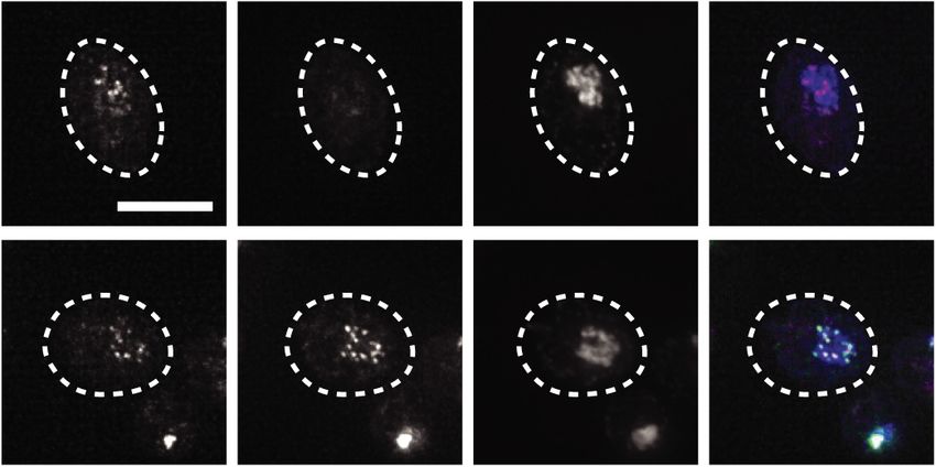
Downloaded from genesdev.cshlp.org on December 13, 2020 - Published by Cold Spring Harbor Laboratory Press

Chen et al.

extensive remodeling during meiotic differentiation (Asa-                 complex Ime1–Ume6 after meiotic entry and cannot
kawa et al. 2005; Sun et al. 2011; Miller et al. 2012; Kim                be translated into Ndc80 protein. Instead, NDC80 LUTI
et al. 2013; Meyer et al. 2015), which is the developmental               expression acts to interfere with the transcription of
program that generates reproductive cells through two                     the canonical, protein-coding NDC80 mRNA isoform.
consecutive nuclear divisions. Specifically in budding                    As a result, in meiotic prophase, a stage when NDC80 LUTI
yeast, the Ndc80 complex disassembles in early meiosis                    is highly expressed, Ndc80 protein synthesis is turned off.
and reassembles during the meiotic divisions, thereby re-                 After cells exit from meiotic prophase, transcription of the
stricting kinetochore activity in a temporal fashion (Asa-                coding NDC80 isoform is induced by another transcrip-
kawa et al. 2005; Miller et al. 2012; Meyer et al. 2015;                  tion factor called Ndt80, leading to resynthesis of Ndc80
Chen et al. 2017). This dynamic kinetochore behavior is                   and kinetochore activation (Chen et al. 2017). Thus, the
driven by the fluctuating Ndc80 levels, which are barely                  developmentally coordinated toggling between these
detectable in meiotic prophase but become highly abun-                    two functionally distinct mRNA isoforms controls
dant during the meiotic divisions. Failure to temporally                  Ndc80 production in meiosis.
regulate Ndc80 protein levels and kinetochore activity                       The LUTI-based regulation explains how meiotic
causes defects in meiotic chromosome segregation and                      cells can effectively repress Ndc80 protein synthesis.
gamete inviability (Miller et al. 2012; Chen et al. 2017),                However, since Ndc80 is clearly detected at meiotic entry
highlighting the importance of Ndc80 regulation.                          (Asakawa et al. 2005; Miller et al. 2012; Meyer et al. 2015;
   One way to regulate Ndc80 protein levels occurs                        Chen et al. 2017), regulated Ndc80 synthesis alone
through controlling Ndc80 synthesis. Ndc80 production                     cannot fully explain kinetochore inactivation in meiotic
is relatively high during the meiotic divisions, but is                   prophase. Additional mechanisms must be in place to
completely shut down in meiotic prophase (Fig. 1A;                        clear the existing pool of Ndc80 such that the kineto-
Chen et al. 2017; Chia et al. 2017). This repression in                   chores can disassemble in a timely manner. Interestingly,
synthesis requires the expression of a meiosis-specific,                  the human homolog of Ndc80, Hec1, undergoes degra-
5′ extended mRNA expressed from an alternate NDC80                        dation in a cell-cycle-dependent manner, but the turn-
promoter. This transcript, called LUTI (long undecoded                    over mechanism remains elusive (Ferretti et al. 2010).
transcript isoform), is induced by the transcription factor               More generally, little is known about the factors that

 A                                                    B                             Figure 1. Ndc80 degradation is temporally regulated
                                                                                    during meiosis. (A) LUTI-based regulation of Ndc80 pro-
                                                                                    tein synthesis in budding yeast meiosis. In meiotic pro-
                                                                                    phase, the Ime1–Ume6 transcription factor complex
                                                                                    induces a long undecoded transcript isoform of
                                                                                    NDC80 called NDC80 LUTI, which cannot produce
                                                                                    Ndc80 protein due to the upstream open reading frames
                                                                                    in its 5′ extension. NDC80 LUTI represses transcription
                                                                                    of a protein-coding isoform of NDC80, NDC80 ORF.
                                                                                    Through this combined act of transcriptional and trans-
C                                                       D
                                                                                    lational repression, NDC80 LUTI inhibits Ndc80 protein
                                                                                    synthesis. In the meiotic divisions, NDC80 ORF is in-
                                                                                    duced by a second meiotic transcription factor, Ndt80.
                                                                                    URS1 (upstream regulatory sequence 1), a DNA-binding
                                                                                    motif for Ume6. MSE (mid-sporulation element), a
                                                                                    DNA-binding motif for Ndt80. (B) The lexO-LUTI sys-
                                                                                    tem induces NDC80 LUTI expression upon β-estradiol
                                                                                    addition, thus conditionally inhibiting NDC80 ORF ex-
E                                                                                   pression and Ndc80 protein synthesis. (Top) Regulatory
                                                                                    elements of the NDC80 gene. (Bottom) The lexO-LUTI
                                                                                    system. mse, a mutant MSE site defective in Ndt80
                                                                                    binding. (C ) Ndc80 turnover in early or late meiotic pro-
                                                                                    phase. The strain carrying the lexO-LUTI (UB14883)
                                                                                    was transferred to the sporulation medium (SPO) at 0
                                                                                    h to induce meiosis, and β-estradiol was added at either
1.5 or at 4 h after meiosis induction. The strain was halted in meiotic prophase using an ndt80Δ block. Here and throughout, Ndc80 levels
were determined by anti-V5 immunoblot. Hxk2, loading control. Unless specified, the numbers below the immunoblots were calculated
by first normalizing Ndc80 levels to Hxk2 levels in each lane, and then dividing the ratio to the 0-h time point. All the experiments in this
study were performed at least twice, and one representative biological replicate is shown. (D) Induction levels of NDC80 LUTI mRNA for
the experiment in C, measured by reverse transcription followed by quantitative PCR (RT-qPCR). For all RT-qPCR experiments,
NDC80 LUTI signals were normalized to that of PFY1. (a.u.) Arbitrary unit. The mean from three independent experiments, along with
the standard error of the mean, is displayed. The P-values were calculated by a two-tailed Student’s t-test. (E) Ndc80 turnover in late mei-
otic prophase or in metaphase I arrest. The ndt80Δ (UB19616) and the ndt80Δ cdc20-mn (UB19618) strains were cultured in SPO for 4 h
before β-estradiol addition. Both strains were halted in meiotic prophase with an ndt80Δ block. The cdc20 meiotic null mutant (cdc20-mn,
UB19678) was cultured in SPO for 5 h before β-estradiol addition and subsequently halted in metaphase I for 3 h.

2      GENES & DEVELOPMENT
Downloaded from genesdev.cshlp.org on December 13, 2020 - Published by Cold Spring Harbor Laboratory Press

                                                                                         Aurora B regulates Ndc80 proteolysis in meiosis

mediate kinetochore subunit degradation in a develop-               polymerase chain reaction (RT-qPCR) (Fig. 1D), suggest-
mental context.                                                     ing that Ndc80 synthesis was successfully repressed.
   Here, we describe the mechanism by which Ndc80 deg-              This result suggests that Ndc80 turnover can occur
radation is controlled in budding yeast meiosis. We found           throughout meiotic prophase.
that the degradation of Ndc80 is temporally regulated. Its            To determine whether Ndc80 is degraded beyond mei-
proteolysis in meiotic prophase requires Aurora B/Ipl1              otic prophase, we monitored Ndc80 levels during a meta-
kinase-dependent phosphorylation, which has been previ-             phase I arrest induced by Cdc20 depletion (cdc20-mn).
ously linked to correcting erroneous microtubule–kineto-            Cdc20 is an activator of the anaphase-promoting complex,
chore attachments (for review, see Biggins 2013). The N             APC/C, a ubiquitin ligase necessary for metaphase-to-
terminus of Ndc80, including a 27-residue sequence and              anaphase transition (Visintin et al. 1997; Hwang et al.
Ipl1 phosphorylation sites, is both necessary and suffi-            1998; Yu 2007). For this experiment, we mutated the
cient for kinetochore protein degradation. In addition to           Ndt80 binding site (also known as mid-sporulation ele-
phosphorylation, Ndc80 degradation depends on the                   ment [MSE]) at the NDC80 promoter (Chen et al. 2017).
ubiquitin ligase APCAma1 and proteasome activity. Fail-             This alteration is required because the second burst of
ure to degrade Ndc80 causes premature kinetochore                   Ndc80 synthesis, which depends on the MSE site, occurs
assembly in meiotic prophase and predisposes cells to               after cells exit meiotic prophase. Mutating this site en-
meiotic chromosome segregation defects. Our results                 sures that Ndc80 synthesis can be repressed by β-estradiol
provide mechanistic insight into how cells can develop-             addition even after meiotic prophase. We found that while
mentally modulate kinetochore composition through                   Ndc80 was degraded in meiotic prophase, it remained re-
subunit proteolysis and highlight the importance of time-           markably stable during the metaphase I arrest induced by
ly Ndc80 degradation in promoting accurate meiotic chro-            cdc20-mn (Fig. 1E). The level of NDC80 LUTI induction
mosome segregation.                                                 was ∼40% lower in cdc20-mn cells than in wild type
                                                                    (Supplemental Fig. S1B). In principle, this reduction of
                                                                    NDC80 LUTI could cause an increase in Ndc80 synthesis,
Results                                                             leading to higher protein levels. To exclude this possi-
                                                                    bility, we used cycloheximide to globally inhibit protein
Ndc80 degradation is temporally regulated in meiosis
                                                                    synthesis. Ndc80 was still stable during the metaphase
In meiotic prophase, the residual Ndc80 protein from the            I arrest and degraded in late prophase I under these
premeiotic cell cycle is turned over by an unknown                  conditions (Supplemental Fig. S1C), suggesting that the
mechanism (Chen et al. 2017). To study Ndc80 degrada-               stability of Ndc80 protein differed between the two states.
tion without a confounding effect from its synthesis regu-          While it is possible that APCCdc20 may regulate Ndc80
lation, we took advantage of a previously established               degradation in metaphase I, we found that Cdc20 was dis-
method, which allowed us to turn off Ndc80 synthesis                pensable for Ndc80 degradation in meiotic prophase
in a conditional manner (Chia et al. 2017). Specifically,           (ndt80Δ cdc20-mn) (Fig. 1E). We conclude that Ndc80 deg-
we used a strain in which the endogenous NDC80 LUTI                 radation is temporally regulated, occurring in a meiotic
promoter was replaced with an inducible promoter con-               prophase-specific manner.
trolled by an array of eight lex operators (8lexO) (Fig.
1B). The same strain carries a chimeric lexA-B112
                                                                    Proteasome activity and Aurora B/Ipl1 regulate Ndc80
transcription factor fused to an estradiol-binding domain
                                                                    degradation
(lexA-B112-ER), which allows inducible transcription
from the 8lexO promoter in the presence of β-estradiol              To identify the regulators of Ndc80 degradation, we sur-
(Ottoz et al. 2014). Without β-estradiol (uninduced),               veyed the key cellular and proteolytic events in meiotic
the coding NDC80 transcript (hereafter referred to as               prophase. We found that synapsis, recombination, and
NDC80 ORF) is expressed and Ndc80 is synthesized. After             DNA replication were all dispensable for Ndc80 degra-
β-estradiol addition, NDC80 LUTI is expressed, resulting in         dation. Ndc80 degradation was normal in spo11Δ cells
repression of Ndc80 synthesis. In comparison with wild-             despite the lack of recombination and synapsis (Fig. 2A;
type cells, this induction system led to similar kinetics           Giroux et al. 1989; Cao et al. 1990; Keeney et al. 1997).
of Ndc80 degradation following meiotic entry (Supple-               Ndc80 degradation also occurred normally in cells deplet-
mental Fig. S1A).                                                   ed of the DNA replication factor Cdc6 (cdc6-mn) (Hoch-
   Using this system, we examined Ndc80 turnover at dif-            wagen et al. 2005; Brar et al. 2009; Blitzblau et al. 2012),
ferent stages of meiosis to determine the specific time             consistent with a previous report (Fig. 2A; Meyer et al.
window of Ndc80 degradation. We treated cells with                  2015). These results suggest that Ndc80 degradation,
β-estradiol either close to meiotic entry (1.5 h after meiot-       and thus kinetochore remodeling, is independent of major
ic induction) or later (4 h after meiotic induction). Mean-         meiosis-specific changes to chromosomes.
while, the cells were held in meiotic prophase by deletion            Next, we tested the ubiquitin-proteasome system, a key
of NDT80, which encodes a transcription factor required             protein degradation pathway in the cell (Finley et al.
for meiotic progression. We found that Ndc80 was degrad-            2012). In meiotic prophase, proteasomes are localized
ed with similar kinetics in either condition (Fig. 1C). The         to chromosomes (Ahuja et al. 2017) and could mediate
levels of NDC80 LUTI induction were also similar, as mea-           Ndc80 degradation. We treated meiotic prophase cells
sured by reverse transcription followed by quantitative             with the proteasome inhibitor MG132. Compared with

                                                                                                     GENES & DEVELOPMENT              3
Downloaded from genesdev.cshlp.org on December 13, 2020 - Published by Cold Spring Harbor Laboratory Press
Chen et al.
A B abundance was increased upon meiotic depletion of Ipl1
(ipl1-mn) (Fig. 2C). This increase was not the result of
elevated Ndc80 synthesis, as shown by three obser-
vations. First, the level of NDC80 LUTI mRNA was not
C D altered in ipl1-mn mutants (Supplemental Fig. S2C).
Second, Ipl1 depletion increased Ndc80 levels additively
with a mutant that fails to repress Ndc80 synthesis
(ΔNDC80LUTI) (Fig. 2C). Finally, the expression of the
protein-coding NDC80 ORF isoform did not significantly
change in the double mutant (ΔNDC80LUTI ipl1-mn) com-
pared with the single mutant (ΔNDC80LUTI), as shown by
single molecule RNA fluorescence in situ hybridization
(smFISH) (Fig. 2D; Supplemental Fig. S2D). Based on these
Figure 2. Proteasome and the Aurora B/Ipl1 kinase regulate
Ndc80 degradation. (A) Dependency of Ndc80 protein degrada- data, we conclude that Ipl1 regulates Ndc80 turnover
tion on recombination, synapsis, or DNA replication. The rather than synthesis.
wild-type (UB1338), spo11Δ (UB11793), and cdc6 meiotic null
(cdc6-mn, UB13656) strains were transferred to SPO at 0 h and
Ndc80 degradation requires Ipl1-mediated
halted in meiotic prophase using the pGAL-NDT80 GAL4.ER
system until 6 h in SPO. The numbers below the immunoblots phosphorylation
were calculated by first normalizing Ndc80 levels to Hxk2 lev- How does Ipl1 regulate Ndc80 abundance mechanistical-
els in each lane, and then dividing the ratio to the 0-h time ly? In one model, Ipl1 depletion may alter Ndc80 abun-
point. (B) Dependency of Ndc80 degradation on active protea-
dance indirectly by affecting microtubule behavior in
somes. Cells (pdr5Δ, UB2405) were induced to sporulate at 0
meiotic prophase. At this meiotic stage, the yeast centro-
h. After 3 h in SPO, cells were split and treated with either
DMSO or the proteasome inhibitor MG132 (100 µM). Thirty somes, known as the spindle pole bodies (SPBs), are dupli-
minutes later, cycloheximide (CHX) was added (0.2 mg/mL). cated but prevented from separating to form spindle
For either condition (DMSO or MG132), all of the time points microtubules. Meanwhile, the kinetochores become
were normalized to the time point immediately before the cy- dispersed from the SPBs, presumably due to the lack of
cloheximide addition (3.5 h). (C ) The effects on Ndc80 levels microtubule–kinetochore interactions (Kim et al. 2013;
when the meiotic depletion of IPL1 (ipl1-mn) was combined Meyer et al. 2013, 2015). Both kinetochore dispersion
with a mutant that fails to repress Ndc80 synthesis and inhibition of spindle formation in meiotic prophase
(ΔNDC80LUTI). The wild-type (UB1338), ipl1-mn (UB1013), require Aurora B/Ipl1 activity (Kim et al. 2013; Meyer
ΔNDC80LUTI (UB2932), and ΔNDC80LUTI ipl1-mn (UB3948)
et al. 2013, 2015). ipl1-mn mutants prematurely separate
cells were sporulated as in A. (D) smFISH quantification of the
the duplicated SPBs and form long microtubules (Shirk
NDC80 ORF and NDC80 LUTI mRNA levels in the ΔNDC80LUTI
cells (UB2932) and ΔNDC80LUTI ipl1-mn (UB3948) cells in mei- et al. 2011; Kim et al. 2013). These microtubules interact
otic prophase. Samples were taken at 5 h in SPO. The relative with the kinetochores, leading to kinetochore recluster-
frequency histograms of the cells with a given number of ing (Meyer et al. 2013, 2015). In this context, microtubules
NDC80 LUTI and NDC80 ORF transcripts per cell were graphed could shield Ndc80 from degradation. Accordingly, the
using the data pooled from two independent experiments. effect of ipl1-mn would depend on the presence of micro-
(Dashed line) The median number of transcripts per cell. A total tubules. However, when we depolymerized microtubules
of 218 cells was counted for ΔNDC80LUTI and 208 cells for in ipl1-mn cells using a microtubule poison cocktail
ΔNDC80LUTI ipl1-mn. Two-tailed Wilcoxon rank sum test was (benomyl and nocodazole), Ndc80 remained stable in
performed.
meiotic prophase (Supplemental Fig. S3A–C). Thus, it is
unlikely that Ipl1 indirectly regulates Ndc80 degradation
by affecting microtubule behavior.
the vehicle control, Ndc80 levels were modestly stabi- Since Ipl1 is a kinase, it may promote Ndc80 turnover
lized within 1 h of MG132 treatment (Supplemental Fig. by phosphorylating factors that turn over Ndc80, or by
S2A). During this time, the expression of NDC80 LUTI phosphorylating Ndc80 itself. Previous work has shown
did not change significantly, but it had decreased by that Ndc80 is a direct substrate of Ipl1 (Cheeseman et al.
∼50% by the second hour of treatment (Supplemental 2002; Akiyoshi et al. 2009). Ndc80 has seven known Ipl1
Fig. S2B). To exclude the confounding effects from the consensus sites ([KR]-X-[ST]-[ILVST]) (Cheeseman et al.
differences in Ndc80 synthesis, we performed a cyclo- 2002), which are required for Ipl1 to phosphorylate
heximide chase experiment. The levels of Ndc80 were Ndc80 in vitro (Akiyoshi et al. 2009). To test whether
stabilized in the first hour of cycloheximide treatment Ndc80 is degraded through a phosphorylation-dependent
although they eventually declined (Fig. 2B). This result in- mechanism, we first asked whether Ndc80 is phosphory-
dicates that the proteasome contributes, at least partially, lated at the time of its degradation. We immunoprecipitat-
to Ndc80 degradation. ed Ndc80 from meiotic prophase cells 1 h after MG132
Previously, it has been shown that the decline of Ndc80 treatment and analyzed the posttranslational modifica-
levels in meiotic prophase requires the kinase Aurora B/ tions by mass spectrometry. We found two recurring,
Ipl1 (Meyer et al. 2015), raising the possibility that Ipl1 high-confidence phosphorylation sites T54 and T248 on
regulates Ndc80 degradation. We confirmed that Ndc80 Ndc80 (Fig. 3A). The first site has been previously
4 GENES & DEVELOPMENTDownloaded from genesdev.cshlp.org on December 13, 2020 - Published by Cold Spring Harbor Laboratory Press
Aurora B regulates Ndc80 proteolysis in meiosis
A 1 100 200 300 400 500 600 691 B h in SPO: 0 3 4 5 6
Figure 3. Ndc80 degradation requires Ipl1-mediated
Ndc80 phosphorylation. (A) Phosphorylation sites detected on
NDC80
N C Hxk2 wild-type Ndc80 proteins and Ndc80(Δ2–28) proteins
N-term CH Hair- loop 1.0 0.5 0.5 0.4 0.4
tail domain pin 0 3 4 5 6 in meiotic prophase by mass spectrometry (MS). The
Ndc80
Phospho (# pT peptide/ #Total peptide) wild-type (pdr5Δ, UB2405) cells were treated with
NDC80-
Hxk2 7A
site Peptide sequences wild type ∆2-28 MG132 at 3 h after transfer to SPO, and samples were
1.0 1.2 1.0 1.0 0.8
R1 R2 R1 R2
T54 N(PT)ISGTGIPTGGINK 1/8 2/13 5/21 3/7 Ndc80
0 3 4 5 6 collected 1 h after treatment. For the Δ2–28 cells
NDC80-
T248 SLINQN(PT)QEITILSQPLK 0/6 3/17 3/39 0/7 Hxk2 (UB5662), samples were collected after 5 h in SPO.
14A
% Ndc80 coverage 55% 57% 71% 59% 1.0 1.1 0.9 1.0 0.9
(Top) Schematic of Ndc80 and the in vivo phosphoryla-
C Mtw1- Ndc80-7A
D Dispersed Partial Clustered E Ndc80 negative tion sites detected by MS (pink circles). (Gray bars)
mCherry -eGFP DAPI Merge
100
Ndc80 positive The 7 Ipl1 consensus sites; (N-term) N-terminal; (CH)
Percentage of cells with
dispersed kinetochores
Clustered calponin homology. (Bottom) Detected Ndc80 phospho-
Percentage of cells
100
80
peptides. The detected number of phosphopeptides and
75
Dispersed 60 total peptides (phosphorylated and unmodified com-
40 50 bined), as well as the overall sequence coverage of
Partial
20 25 Ndc80, are reported for two biological replicates. (R1) re-
peat 1; (R2) repeat 2. (B) Ndc80 protein level in meiotic
h in 0 0 3 4 5 0 3 4 5 h in
0
Inset
SPO:
3 4 5 prophase for the wild-type (UB4074), NDC80-7A
NDC80 NDC80-7A SPO: NDC80-7A
(UB13658), and NDC80-14A (UB17707) cells. Samples
were taken and processed as in Figure 2A. The numbers
below the immunoblots were calculated by first normalizing Ndc80 levels to Hxk2 levels in each lane, and then dividing the ratio to the 0-
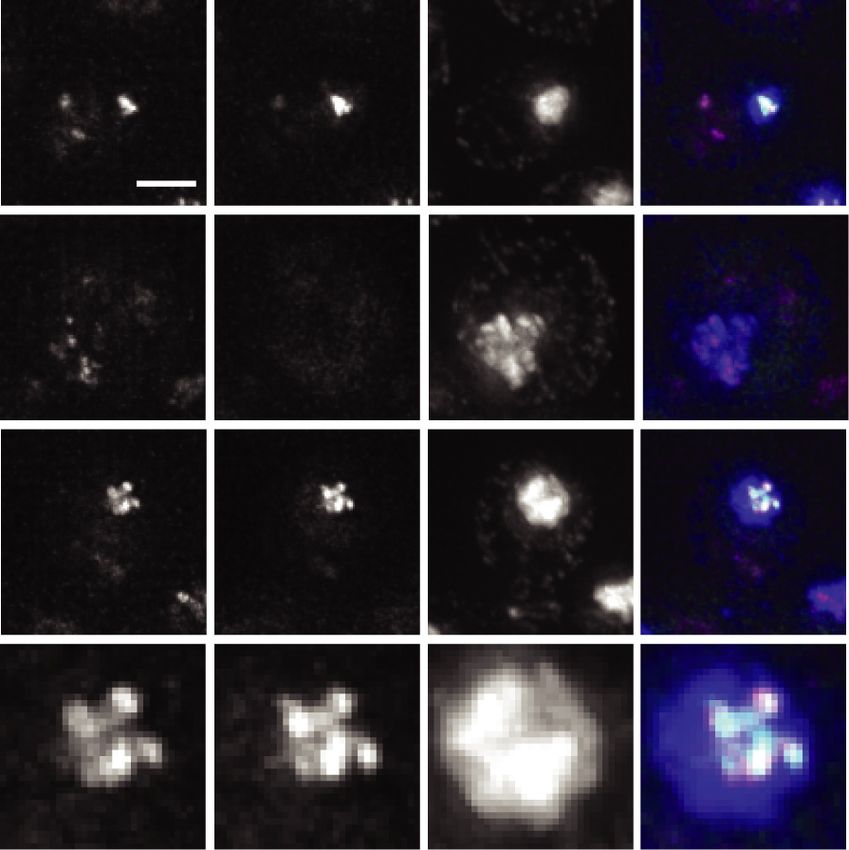
h time point. (C) Representative images of the clustered, dispersed, and partially clustered kinetochores in NDC80-7A (UB15701) cells,
which were fixed at 5 h after transfer to SPO. Mtw1 was tagged with mCherry and Ndc80-7A tagged with eGFP. DNA was stained with
DAPI. Scale bar, 2 µm; inset scale bar, 1 µm. (D) The percentage of cells with clustered, partially clustered, or dispersed kinetochores in
meiotic prophase. The wild-type (UB1083) and NDC80-7A (UB15701) cells were fixed immediately after transfer to SPO (0 h) and at 3, 4,
and 5 h later. One-hundred cells were counted per time point. The mean and the range of the percentage for two biological replicates are
graphed. (E) The percentage of cells with dispersed kinetochores that contained Ndc80-7A-eGFP signal on at least one kinetochore at the
indicated time points.
characterized and contains an Ipl1 consensus site (Cheese- had partially clustered kinetochores (Fig. 3C, partial and
man et al. 2002; Akiyoshi et al. 2009), whereas the second inset), all of which had Ndc80-7A-eGFP on them. By
site lacks the Ipl1 consensus sequence. 5 h, >50% of the NDC80-7A cells had dispersed kineto-
Next, we mutated the serine or threonine in all seven chores (Fig. 3D), and about half of these cells had Ndc-
Ipl1 consensus sites, generating the allele (NDC80-7A) 80-7A-eGFP on at least one kinetochore (Fig. 3E). These
known to greatly reduce Ndc80 phosphorylation by Ipl1 results demonstrate that the stabilized Ndc80-7A proteins
in vitro (Akiyoshi et al. 2009). The mutations did not af- localize to the kinetochore and affect the timing of kinet-
fect NDC80 LUTI expression (Supplemental Fig. S3D), rul- ochore dispersion in meiotic prophase.
ing out the possibility that the synthesis repression of
Ndc80 was disrupted. We found that Ndc80-7A was high-
Ndc80 degradation requires a specific sequence at its
ly stable in meiotic prophase (Fig. 3B). Mutating addition-
N terminus
al serine and threonine residues around T54 and T248
(NDC80-14A) did not enhance Ndc80 stabilization (Fig. Since the seven Ipl1 consensus sites are located in the 112-
3B). Thus, we conclude that the seven Ipl1 consensus sites residue N-terminal tail of Ndc80, we systematically trun-
at the N-terminal region of Ndc80 are required for its turn- cated this region to narrow down the sites necessary for
over in meiotic prophase. Ndc80 degradation. We found residues 2–28 to be neces-
We further asked whether the stabilized Ndc80-7A pro- sary for the decline of Ndc80 levels (Fig. 4A, top panel).
teins localized to the kinetochores and affected kineto- Within this segment, residues 11–19 were the most criti-
chore behavior. We fused Ndc80-7A to the enhanced cal (Fig. 4A, bottom panel). To our surprise, the decline
green fluorescent protein (eGFP), and generated a of Ndc80 levels was not altered when the 11 serines
mCherry-tagged allele for the inner kinetochore protein and threonines in the first 30 residues of Ndc80 were
Mtw1, which remains chromosome-associated through- mutated (NDC80-11A) (underlined in Fig. 4B), indicat-
out meiotic prophase. For both the wild-type and ing that Ndc80 degradation does not depend on the poten-
NDC80-7A mutant, over 85% of the cells had clustered tial phosphorylation sites within this region. Ndc80
kinetochores at meiotic entry (0 h in SPO), and all of the turnover was also normal when the four histidines in
kinetochore clusters had Ndc80-eGFP signal (Fig. 3C,D). the 11–19 region (green letters in Fig. 4B) were mutated
When the wild-type cells progressed into meiotic pro- to either alanines (NDC80-4A) or leucines (NDC80-4L)
phase (3–5 h in SPO), the kinetochores became dispersed (Supplemental Fig. S4A).
in >70% of the cells, and none of the dispersed kineto- How does the 2–28 region regulate Ndc80 abundance?
chores had Ndc80-eGFP signal (Fig. 3D). In contrast, fewer We first confirmed that the 2–28 region regulates Ndc80
than 30% of the NDC80-7A cells had dispersed kineto- degradation rather than Ndc80 synthesis. The wild-type,
chores after 3 h in SPO (Fig. 3D). Instead, ∼40% of the cells Δ2–28, and Δ11–19 cells had similar NDC80 LUTI levels
GENES & DEVELOPMENT 5Downloaded from genesdev.cshlp.org on December 13, 2020 - Published by Cold Spring Harbor Laboratory Press
Chen et al.
A B Figure 4. Ndc80 degradation requires a short sequence
at its N terminus. (A) Truncation analysis of Ndc80 to
identify the residues necessary for Ndc80 degradation.
Strains harboring the deletions of residues 2–28 (Δ2–
28, UB5662), residues 29–56 (Δ29–56, UB15972), resi-
dues 2–10 (Δ2–10, UB7039), residues 11–19 (Δ11–19,
UB7029), and residues 20–28 (Δ20–28, UB7031) were
sporulated along with wild-type cells (UB4074) as in
C D Figure 2A. The vegetative samples (V) were taken while
each strain was growing exponentially in rich medium.
The numbers below the immunoblots were calculated
by first normalizing Ndc80 levels to Hxk2 levels in
each lane, and then dividing the ratio to the 0-h time
point. Note: Hxk2 level declined slightly as meiotic
prophase progressed. Thus, the normalized level of
Ndc80 became >1.0 in the later stages of meiotic pro-
phase. (B) An abridged schematic of Ndc80. The se-
quence of the first 30 residues is displayed. (Gray
bars) The seven Ipl1 consensus sites. The underlined
E F G
residues are the 11 serines and threonines mutated in
the 11A mutant, and the green residues are the four
histidines mutated in the 4(H to A) and 4(H to L) mu-
tants. (C) The effects on Ndc80 levels when the Δ11–19
mutant was combined with a mutant that fails to re-
press Ndc80 synthesis (ΔNDC80LUTI). The wild-type
(UB4074), Δ11–19 (UB7029), ΔNDC80LUTI (UB11797),
and ΔNDC80LUTI Δ11–19 (UB11799) cells were sporu-
lated as described in Figure 2A. (D) In vitro kinase assay for the wild-type Ndc80 and Ndc80(Δ2–28) proteins purified from UB16284
and UB19957, respectively. The Ndc80 proteins were phosphorylated in vitro by either 1 µM recombinant AurB∗ (fusion of the C-ter-
minal activation box of INCEPSli15 to Aurora BIpl1) or 1 µM kinase-dead AurB∗ (KD) and γ-32P-ATP. All reactions were performed for
either 1 or 5 min at room temperature, analyzed by SDS-PAGE, and then visualized by silver staining and autoradiography. (wt) Wild-
type Ndc80; (Δ) Ndc80(Δ2–28); (∗ ) AurB∗ ; (^) background bands. (E) The percentage of the wild-type or Δ2–28 cells with dispersed, par-
tially clustered, and clustered kinetochores after 3 h in SPO. The average, as well as the range, of two independent biological replicates
are displayed. Over 100 meiotic cells were counted. (F ) Representative images of the wild-type Ndc80 and Ndc80(Δ2–28) proteins in
meiotic prophase. The wild-type (UB1083) and Δ2–28 (UB15619) cells were fixed after 5 h in SPO. Mtw1 was tagged with mCherry
and Ndc80(Δ2–28) tagged with eGFP. DNA was stained with DAPI. Scale bar, 5 µm. (G) The average percentages of meiotic prophase
cells (identified by the pachytene DAPI morphology) with colocalized Ndc80-eGFP and Mtw1-mCherry signals, as well as the range, of
two independent biological replicates are displayed. At least 70 meiotic prophase cells were counted.
in meiotic prophase (Supplemental Fig. S4B). Also, the ly in the Δ2–28 mutant. Thus, we conclude that the
Ndc80 protein levels were additively increased when the 2–28 region is required for Ndc80 degradation at a step
Δ11–19 mutant was combined with the mutant that fails downstream from or in parallel to Ipl1-dependent
to inhibit Ndc80 synthesis (ΔNDC80LUTI) (Fig. 4C). De- phosphorylation.
spite the difference in protein levels, the expression of We attempted to understand the role of the 2–28 resi-
the coding NDC80 ORF mRNA was not significantly differ- dues in Ndc80 degradation by looking for changes in the
ent between the double mutant (ΔNDC80LUTI Δ11–19) binding partners of the wild-type or Ndc80(Δ2–28) protein
and the single mutant (ΔNDC80LUTI) (Supplemental during meiotic prophase. By quantitative mass spectro-
Fig. S4C,D), suggesting that the 2–28 residues regulate metry using tandem mass tags, we found that Sis1, a
Ndc80 stability. J domain protein that regulates heat-shock protein activi-
Since Ndc80 degradation requires Ipl1-dependent ty (Kampinga and Craig 2010), interacted with the wild-
phosphorylation, we next asked whether such phosphory- type but not the Ndc80(Δ2–28) protein (Supplemental
lation depends on the 2–28 residues. We immunopre- Fig. S4E). However, Sis1 did not appear to be required for
cipitated Ndc80(Δ2–28) protein from meiotic prophase Ndc80 degradation since even the mutants that displayed
and performed mass spectrometry. Both phosphorylation normal Ndc80 degradation (e.g., Δ29–56) were still defec-
sites (T54 and T248) observed in the wild-type Ndc80 tive in Sis1 binding (Supplemental Fig. S4F). It remains un-
protein were detected (Fig. 3A). In addition, we perfor- clear how the 2–28 segment mediates Ndc80 degradation.
med an in vitro kinase assay using a recombinant Ipl1 Despite acting through an unknown mechanism, the
protein variant (AurB∗ ) (de Regt et al. 2018) and found 2–28 residues of Ndc80 are evidently important for
no noticeable difference in the degree of Ndc80 phos- clearing Ndc80 from the kinetochores and facilitating ki-
phorylation between the wild-type and Ndc80(Δ2–28) netochore dispersion in meiotic prophase. For the Δ2–28
protein (Fig. 4D). These results strongly suggest that mutant, we observed an increased proportion of cells
the Ipl1-depedent Ndc80 phosphorylation occurs normal- with partially clustered kinetochores after 3 h in SPO
6 GENES & DEVELOPMENTDownloaded from genesdev.cshlp.org on December 13, 2020 - Published by Cold Spring Harbor Laboratory Press
Aurora B regulates Ndc80 proteolysis in meiosis
(∼25%) (Fig. 4E), although the percentage was lower than tor of the APC/C, Ama1, to be required for Ndc80 turn-
that of the NDC80-7A mutant (∼40%) (Fig. 3D). By 5 h in over (Fig. 6A). APCAma1 regulates Ndc80 degradation
SPO, most of the Δ2–28 cells had dispersed kinetochores rather than synthesis, because the levels of NDC80 LUTI
in meiotic prophase, and Ndc80(Δ2–28)-eGFP signal was were not significantly altered in the ama1Δ mutant (Sup-
detected on all the dispersed kinetochores (Fig. 4F,G), plemental Fig. S6A). A cycloheximide chase experiment
demonstrating that the stabilized Ndc80(Δ2–28) protein further supported that APCAma1 acts at a posttranslational
localizes to the kinetochore. In addition, our observations step in regulating Ndc80 levels (Fig. 6C,D). In addition to
suggest that while Ndc80 degradation is not required for Ndc80, the Ame1-tail fusion was also stabilized in an
kinetochore dispersion, it can affect dispersion kinetics APCAma1-dependent manner (Fig. 6B).
(given the accumulation of cells with partially clustered It has been shown previously that AME1 deletion
kinetochores), potentially by acting in conjunction with causes a mitotic-like cellular state in meiotic prophase
Ipl1-dependent phosphorylation. (Okaz et al. 2012). This occurs due to stabilization of
two APCAma1 substrates: Ndd1, a transcription factor re-
quired for the mitotic expression of B-type cyclins and
The N terminus of Ndc80 is sufficient to induce Clb4, a B-type cyclin itself. Therefore, it is possible that
proteolysis such a mitotic-like state in the ama1Δ mutant could
Both the 2–28 region of Ndc80 and Ipl1-mediated phos- mimic a condition in which Ndc80 is stable, such as meta-
phorylation are required for Ndc80 degradation. Are phase I. However, we found no evidence in support of this
these two features sufficient to induce protein degrada- model. In our strains, premature spindle formation, a
tion in meiotic prophase? We tested this idea by fusing phenotype associated with elevated cyclin/CDK activity,
the N-terminal region of Ndc80 (residues 2–112), includ- was not apparent in the ama1Δ mutant (Supplemental
ing both the 2–28 region and the Ipl1 consensus sites, to Fig. S6B). As an additional test, we removed Ndd1 and
an inner kinetochore protein, Ame1 (Fig. 5A). The protein Clb4 from the ama1Δ mutant and found that Ndc80 was
levels of Ame1 were stable in meiotic prophase, as shown still stable (Fig. 6C,D; Supplemental Fig. S6C), suggesting
by a cycloheximide chase experiment (Supplemental Fig. that AME1 deletion does not stabilize Ndc80 levels
S5). We placed this AME1-tail construct under the pro- through creating an alternative cellular state in meiotic
moter of NDC80, which leads to synthesis repression prophase.
in meiotic prophase. Addition of the N-terminal tail of Next, we tested whether Aurora B/Ipl1 activity is dis-
Ndc80 caused Ame1 to become unstable in meiotic pro- rupted in the ama1Δ mutant, as Ipl1 down-regulation
phase in an Ipl1-dependent manner (Fig. 5B). Therefore, would lead to Ndc80 stabilization. We found that the
the N-terminal tail of Ndc80 is sufficient to induce prote- wild-type and ama1Δ cells had comparable levels of serine
olysis in meiotic prophase. 10 phosphorylated histone H3 (Fig. 6E), which is an estab-
lished Aurora B/Ipl1 substrate (Hsu et al. 2000). Thus, Ipl1
activity is unaffected by AME1 deletion. These observa-
APCAma1 regulates Ndc80 degradation tions led us to conclude that APCAma1 acts downstream
from Ipl1-dependent phosphorylation to mediate Ndc80
Since proteasome activity contributes to Ndc80 degrada- degradation.
tion, we posited that Ndc80 is degraded via a system me-
diated by ubiquitin/proteasome, which requires one or
Defects in Ndc80 degradation predispose meiotic cells to
more E3 ligases to ubiquitinate the substrate for protea-
chromosome segregation errors
some recognition (Finley et al. 2012). Thus, we surveyed
a candidate list of E3 ubiquitin ligases for their roles in How does Ndc80 degradation impact kinetochore activity
Ndc80 degradation. We found the meiosis-specific activa- and function? We used the Δ2–28 mutant to address this
A Figure 5. The N terminus of Ndc80 is sufficient to in-
duce proteolysis. (A) Schematic of the Ndc80 tail-
Ame1 fusion. The tail of Ndc80 (residues 2–112), in-
cluding both the 27-residue sequence and the Ipl1 phos-
phorylation sites, is fused to the C terminus of Ame1.
The fusion construct is controlled by the NDC80 pro-
B moter, which allows synthesis repression in meiotic
prophase. (B) Ame1-tail is degraded in meiotic prophase
in an Ipl1-dependent manner. The IPL1 AME1
(UB20358), IPL1 AME1-tail (UB20354), and ipl1-mn
AME1-tail (UB22397) cells were sporulated as de-
scribed in Figure 2A. In all of the strains, Ndc80 was
tagged with 3V5, the same epitope tag for Ame1, as
an internal control. The relative Ndc80 levels were cal-
culated as described in Figure 1C. The relative Ame1
levels were calculated by normalizing Ame1 levels to Hxk2 levels in each lane, and then dividing the normalized values to that of
time 0 h.
GENES & DEVELOPMENT 7Downloaded from genesdev.cshlp.org on December 13, 2020 - Published by Cold Spring Harbor Laboratory Press
Chen et al.
h in Cu
B Figure 6. APCAma1 regulates Ndc80 degradation. (A)
A SPO: 2 3 3.5 4 4.5 5 AME1 tail
Ndc80
Dependency of Ndc80 levels on Ama1 in meiotic pro-
pNDC80LUTI pNDC80ORF
h in SPO: 0 1 2 3 4 5 6 0 1 2 3 4 5 6
phase. Sporulation of the wild-type (UB6598) and
Hxk2
Ndc80 ama1Δ (UB22499) cells was synchronized with the
1.0 0.8 0.6 0.5 0.3 0.3
wild type pCUP-IME1/pCUP-IME4 system. Meiotic entry was in-
h in Cu Ame1-tail
SPO: 2 3 3.5 4 4.5 5 duced by CuSO4 addition after cells were incubated in
Ndc80 Hxk2 SPO for 2 h. The numbers below the immunoblots
Hxk2 rel. Ndc80: 1.0 0.7 0.6 0.6 0.5 0.4 0.3 1.0 1.2 1.2 1.5 1.4 2.0 2.6 were calculated by first normalizing Ndc80 levels to
rel. Ame1: 1.0 0.7 0.6 0.7 0.6 0.5 0.4 1.0 1.3 1.4 1.4 1.4 1.6 1.5
1.0 0.9 0.8 1.0 1.1 1.0
AME1-tail AMA1 AME1-tail ama1∆
Hxk2 levels in each lane, and then dividing the ratio to
ama1∆
C CHX CHX
the 0-h time point. (B) Dependency of Ame1-tail fusion
min: 0 15 30 60 90 120 150 0 15 30 60 90 120 150 D levels on Ama1 in meiotic prophase. The AME1-tail
1.4 ama1∆, ndd1-mn, clb4∆
Ndc80 AMA1 (UB20354) and AME1-tail ama1Δ (UB23001)
Ndc80 levels relative
1.2 ama1∆ ∆2-28 wild type
Hxk2 1.0 strains were sporulated as in Figure 2A. Quantification
to time 0
E ama1∆ ama1∆ ndd1-mn clb4∆ 0.8 of Ndc80 and Ame1 were performed as in Figure 5B.
0.6
h in SPO: 0 2 3 4 5 0 2 3 4 5 0 2 3 4 5
0.4
(rel) Relative. (C ) Assessing the requirement of Ndd1
H3S10-P 0.2 and Clb4 in the Ndc80 stabilization that was induced
Hxk2 0.0 by ama1Δ. The ama1Δ (UB20223) and pCLB2-NDD1
0 15 30 60 90 120 150
1.0 3.3 4.8 6.8 9.1 0.4 0.3 0.4 0.4 0.6 1.4 3.4 6.1 7.2 7.8 Minutes after CHX addition (ndd1-mn) clb4Δ ama1Δ (UB22850) cells were cultured
wild type ipl1-mn ama1∆
in SPO for 3 h before a final concentration of 0.2 mg/
mL cycloheximide was added. During the entire CHX
experiment, both strains were halted in meiotic prophase. (D) The Ndc80 protein levels relative to the time point immediately before
CHX addition. The average and the standard errors of the mean for five independent experiments are graphed. (E) Ipl1 activity in the
ama1Δ mutant. The wild-type (UB3954), ipl1-mn (UB1013), and ama1Δ (UB20223) cells were sporulated as in Figure 2A. The levels of
H3 serine 10 phosphorylation (H3S10-P) were detected by a phospho-specific antibody. The number below each lane was first normalized
to Hxk2 levels and then to the 0-h time point of the wild-type strain.
question. We first tested whether this mutant had growth their chromosomes, as shown by tracking the segrega-
defects at a higher temperature (37°C) or on plates con- tion of the homozygous CENV-GFP dots (Fig. 7D; Supple-
taining the microtubule depolymerizing drug benomyl. mental Fig. S7A). Both the wild-type and Δ2–28 cells had
These two phenotypes are often exhibited by mutants >95% sporulation efficiency (Fig. 7E) and >96% spore
with defects in kinetochore function (Spencer et al. viability (Supplemental Fig. S7B,C), a metric that would
1990; Wigge et al. 1998; Hyland et al. 1999) and spindle as- be lowered if chromosomes missegregated. These results
sembly checkpoint (e.g., mad2Δ) (Li and Murray 1991). suggest Ndc80 degradation is dispensable for correcting
We found that the Δ2–28 mutant grew similarly to the erroneous microtubule–kinetochore interactions in
wild-type cells in both conditions (Fig. 7A), demonstrating meiosis.
that these amino acids in Ndc80 are dispensable for nor- Instead, we found that Ndc80 degradation modulated
mal kinetochore function in mitotic cells. kinetochore activity by changing kinetochore composi-
To examine whether Ndc80 degradation affects kineto- tion. Remodeling kinetochore composition is crucial for
chore function in meiosis, we monitored chromosome proper meiosis I. In meiotic prophase, the outer kineto-
movements using the centromeric TetR/TetO GFP dot chore dissociates from the inner kinetochore to down-reg-
assay (Michaelis et al. 1997), where both homologs of ulate kinetochore activity, which prevents premature
chromosome V contained TetO arrays that were integrat- microtubule–kinetochore interactions and is crucial to es-
ed near the centromere and were bound by the TetR-GFP tablish a meiosis I-specific chromosome segregation pat-
fusion protein (CENV-GFP). We performed our experi- tern (Asakawa et al. 2005; Miller et al. 2012; Meyer et al.
ment in the spo11Δ background because (1) these cells 2015; Chen et al. 2017). When the Δ2–28 or Δ11–19
have long spindles that help resolve chromosome move- mutant were combined with a mutant that prematurely
ments (Shonn et al. 2000) and (2) the homologous chro- forms spindle microtubules in meiotic prophase (CUP-
mosomes fail to recombine (Klapholz et al. 1985), which CLB3), we observed that sister chromatids, instead of ho-
renders the spo11Δ cells unable to establish biorientation. mologous chromosomes, segregated in meiosis I (Fig. 7E).
As a result, multiple rounds of microtubule–kinetochore In this sensitized background, the NDC80-7A mutant also
attachments and detachments occur for an extended caused precocious sister chromatid segregation, albeit
period, as part of the error correction mechanism. Ipl1 modestly (Fig. 7E), likely because the level of Ndc80-7A
inactivation is known to cause defects in error correc- eventually reduced in prolonged prophase arrest (Fig. 3B,
tion, which is manifested by a reduced frequency of 6 h). The same phenotype occurs when Ndc80 is over-
kinetochore detachments in the spo11Δ cells (Meyer expressed in meiotic prophase or when the LUTI-based
et al. 2013). We found a similar distribution of kineto- repression of Ndc80 synthesis is disrupted (Miller et al.
chore detachments between the wild-type and Δ2–28 cells 2012; Chen et al. 2017). Therefore, the lack of Ndc80 turn-
(Fig. 7B,C), suggesting that the frequency of error correc- over in meiotic prophase prematurely activates the kinet-
tion (and, by inference, the kinetochore function) is unal- ochore by providing a high abundance of Ndc80, which is
tered upon Ndc80 stabilization. Consistently, >95% of the limiting subunit of the kinetochore activity in meiotic
the wild-type and the Δ2–28 tetrads correctly segregated prophase, and predisposes cells to meiotic chromosome
8 GENES & DEVELOPMENTDownloaded from genesdev.cshlp.org on December 13, 2020 - Published by Cold Spring Harbor Laboratory Press
Aurora B regulates Ndc80 proteolysis in meiosis
A B Figure 7. Ndc80 degradation regulates kinetochore
80 NDC80
30 ˚C 37 ˚C benomyl composition in meiosis. (A) Growth phenotype asso-
∆2-28
Percentage of cells
ndc80-1 60 ciated with the various truncations of the Ndc80 N-
∆mad2
40
terminal tail. Tested strains included the tempera-
NDC80
ture-sensitive NDC80 allele (ndc80-1, UB494), the
∆2-112
∆29-56
20 spindle assembly checkpoint mutant Δmad2
∆2-28 0 (UB700), wild-type NDC80 (UB3262), and the
0 1 2 3 3+ NDC80 mutants that carry the following deletion:
SPB split Numbers of detachments
residues 2–112 (Δ2–112, UB3275), residues 29–56
C 0 4.5’ 9’ 13.5’ 18’ 22.5’ 27’ 31.5’ 33’
(Δ29–56, UB4695), and residues 2–28 (Δ2–28,
NDC80 UB5015). Cells were serially diluted and grown on
plates containing nutrient-rich medium (YPD) at 30°
SPB CENV C or 37°C, as well as on a benomyl plate (15 µg/mL)
∆2-28 at 23°C. (B) Frequency distribution of the cells with
a given number of kinetochore detachments and re-at-
D E F 60 + bipolar spindle in meiotic tachments for the experiment in C. The mean per-
prophase (pCUP-CLB3)
100 100 centage and the standard deviation for three
abnormal meiosis I (%)
segregated chr V (%)
normal abnormal
biological replicates are displayed. (C ) Kinetochore at-
cells correctly
meiosis I meiosis I
tri/tetrads (%)
40
tachments and detachments for the NDC80
50 50
(UB15905) and Δ2–28 (UB15873) strains in the
20
homologs sisters spo11Δ background. Each strain carries the TetR-
separate separate GFP fusion protein, and both homologs of chromo-
0 0 0
wild ∆ wild ∆
wild NDC80 pCUP- ∆11-19 ∆2-28 7A some V have the centromeric TetO array (homozy-
type 2-28 type 2-28 type NDC80 gous CENV-GFP, marked in green). (Magenta) Spc42
fused with mCherry, which marks the spindle pole
body (SPB). White arrowhead marks the kinetochore that would undergo detachment. The time when the SPBs were split is marked as
0. Scale bar, 5 µm. (D) The percentage of cells correctly segregated chromosome V for the wild-type (UB21757) and Δ2–28 (UB21758)
strains. These strains carry the TetR-GFP fusion proteins, and both homologs of chromosome V are marked by the centromeric TetO re-
peats (CENV-GFP). Strains were sporulated for 7 h before formaldehyde fixation. At least 90 cells were counted for each strain. For panels D
and E, the mean and the range of two biological replicates are graphed. (E) Spore formation for the meiosis I of wild-type (UB21756) and Δ2–
28 (UB21758) strains. (F ) Sister chromatid segregation in wild-type (UB2942), pCUP-CLB3 NDC80 (UB877), pCUP-CLB3 pCUP-NDC80
(UB880), pCUP-CLB3 NDC80(Δ11-19) (UB16561), pCUP-CLB3 NDC80(Δ2-28) (UB16565), and pCUP-CLB3 NDC80-7A (UB16872). All
strains carry pGAL-NDT80 and GAL4.ER, which allows reversible arrest in meiotic prophase. A pair of the chromosome V sister chroma-
tids is marked by the centromeric TetO repeats. A schematic for normal and abnormal meiosis I is shown at the right. In an abnormal
meiosis I, two separated GFP dots were observed in binucleates, one in each nucleus. Cells were sporulated for 5 h before addition of
CuSO4 to induce cyclin Clb3 expression. Immediately after the induction, cells were released from the prophase arrest using β-estradiol,
which induces NDT80. Samples were taken 1 h 45 min after the release. The average fraction of binucleates that displayed sister segre-
gation in meiosis I, as well as the range of two biological replicates, is graphed. One-hundred cells were counted per strain per experiment.
segregation errors. Lastly, disrupting both Ndc80 synthe- netochores, and predisposes meiotic cells to chromosome
sis repression (ΔLUTI) and Ndc80 degradation (Δ11–19) segregation defects. All of these observations highlight the
synergistically enhanced this segregation phenotype (Sup- importance of regulating Ndc80 turnover in meiosis.
plemental Fig. S7D), consistent with the idea that both
modes of regulation contribute to kinetochore inactiva- Phosphorylation-mediated Ndc80 degradation
tion in meiotic prophase. in meiotic prophase
Multiple lines of evidence support a model in which Au-
Discussion rora B/Ipl1-dependent phosphorylation of Ndc80 triggers
its degradation in meiotic prophase. First, Ipl1 is required
In this study, we showed that Ndc80 degradation in mei- for Ndc80 degradation. Second, phosphorylation of an Ipl1
osis is a temporally regulated process. The proteolysis of consensus site on Ndc80 is detected by mass spectrome-
Ndc80 in meiotic prophase is independent of major chro- try in meiotic prophase, a stage when Ndc80 degradation
mosome remodeling events, but requires the ubiquitin li- occurs. Third, mutating the seven Ipl1 consensus sites
gase APCAma1 and proteasome activity. Furthermore, leads to Ndc80 stabilization in meiotic prophase, suggest-
Ndc80 degradation is coupled to Aurora B/Ipl1-mediated ing that these sites are necessary for Ndc80 degradation.
phosphorylation of Ndc80, a posttranslational modi- Altogether, these results are consistent with a phosphory-
fication known for correcting erroneous microtubule– lation-dependent degradation mechanism.
kinetochore attachments. The N terminus of Ndc80, Given that Ndc80 phosphorylation is not disrupted
which includes a 27-residue sequence and the Aurora B/ when the 2–28 residues are deleted, we posit that this
Ipl1 consensus sites, is both necessary and sufficient to 2–28 segment may act in parallel to or downstream from
drive the degradation of kinetochore proteins. The failure the Ipl1-dependent phosphorylation to promote Ndc80
to degrade Ndc80 in meiotic prophase alters the kinetics of degradation. These residues may serve as the binding
kinetochore dispersion, causes premature activation of ki- site for protein factors that mediate Ndc80 proteolysis.
GENES & DEVELOPMENT 9Downloaded from genesdev.cshlp.org on December 13, 2020 - Published by Cold Spring Harbor Laboratory Press
Chen et al.
It is possible that such factors are recruited by Ndc80 phosphorylation would cause its degradation at a time
phosphorylation, or that phosphorylation alters the local when the presence of Ndc80 is essential. This problem
conformation around the 2–28 residues to expose this seg- could be reconciled by the fact that Ndc80 protein is up-
ment for protein factor binding. regulated as cells exit from meiotic prophase (Chen
Our data are consistent with the idea that the ubiquitin et al. 2017), which may provide extra proteins to com-
ligase APCAma1 and the proteasome act downstream from pensate for the loss due to degradation. In addition, the ac-
the Ipl1-dependent phosphorylation to mediate Ndc80 tivity of the ubiquitin ligase APCAma1 is repressed by Clb-
proteolysis. We excluded the possibility that the ama1Δ CDK activity, which rises in prometaphase I (Oelschlae-
mutation stabilizes Ndc80 by increasing cyclin/CDK ac- gel et al. 2005; Tsuchiya et al. 2011). Since APCAma1
tivity or by down-regulating Ipl1 activity (Fig. 6C–E). It is required for Ndc80 degradation, the degradation mech-
is currently unknown whether APCAma1 directly ubiquiti- anism of Ndc80 could be turned off after meiotic pro-
nates Ndc80 or regulates other player(s) in the degradation phase, even before error correction is completed.
pathway to promote Ndc80 turnover. Interestingly, the
APC/C degron repository (Davey laboratory and Morgan
Protein turnover as a mechanism to create meiosis-
laboratory) predicts that Ndc80 has two D-box motifs
specific kinetochores
and three KEN motifs, which are known recognition sites
for the APC/C (Glotzer et al. 1991). However, these sites In vegetative growth, the yeast kinetochores transiently
are located outside of the 112-residue N-terminal region, disassemble during DNA replication when the centro-
which is both necessary and sufficient for Ndc80 degrada- meric DNA replicates, but they remain bound to centro-
tion in meiotic prophase. Furthermore, mutating the D- meres for the rest of the cell cycle. It has been shown
boxes does not block Ndc80 degradation (Supplemental that the subunit stoichiometry of the kinetochore is regu-
Fig. S7E). The D-box motif is also not required for the lated during the cell cycle (Dhatchinamoorthy et al. 2017,
APCAma1-dependent proteolysis of Ssp1 (Maier et al. 2019). However, it is unclear whether the protein levels
2007; Diamond et al. 2009). Future work to characterize of the kinetochore subunits are regulated in vegetative
the substrates of APCAma1, as well as the mechanism of growth. Degradation of a few kinetochore proteins has
substrate recognition, will be important to fully grasp been reported in yeast. These include Dsn1 (Akiyoshi
the meiotic regulation of Ndc80 turnover. et al. 2013), Cbf13 (Kaplan et al. 1997), and the centromer-
ic histone Cse4 in budding yeast (Herrero and Thorpe
2016; Ohkuni et al. 2016; Cheng et al. 2017), as well as
Interplay between error correction and Ndc80
Spc7 in fission yeast (Kriegenburg et al. 2014). The degra-
degradation
dation pathways of these subunits have been proposed to
It is well established that Ndc80 phosphorylation by Au- be quality controls that remove nonfunctional or excess
rora B/Ipl1 helps correct microtubule–kinetochore attach- proteins, rather than direct means to alter kinetochore
ments that do not result in tension (Biggins 2013). Ndc80 function.
phosphorylation is thought to weaken microtubule bind- In contrast, regulating protein abundance is the
ing, resulting in microtubule detachment and thus provid- key mechanism of controlling kinetochore activity in
ing the opportunity for the kinetochore to reattach in the meiosis. The outer kinetochore disassembles in meiotic
correct orientation. How is the error correction process prophase and reassembles in prometaphase I. This disas-
interrelated with Ndc80 degradation given that both pro- sembly is triggered by a reduction in Ndc80 levels, which
cesses require Ndc80 phosphorylation by Aurora B? We occurs as a result of two separate mechanisms: LUTI-
found that disrupting Ndc80 degradation alone (Δ2–28) based repression of new Ndc80 synthesis and degradation
does not appear to affect meiotic chromosome segregation of the existing Ndc80 pool. In meiosis, Ndc80 degradation
(Fig. 7B–E; Supplemental Fig. S7A–C). This observation is not a passive consequence of DNA replication but a tar-
suggests that Ndc80 degradation is not required for error geted process mediated by Aurora B/Ipl1, which has also
correction. been implicated in preventing untimely spindle formation
Instead, we propose that the completion of error correc- in meiotic prophase (Shirk et al. 2011; Kim et al. 2013). We
tion may repress Ndc80 degradation in metaphase I, a stage propose that Ipl1 prevents premature microtubule–kinet-
when Ndc80 is stable (Fig. 1E; Supplemental Fig. S1B,C). ochore interactions through two independent pathways:
Once the chromosomes attach to spindles properly, one by altering microtubule behavior, and the other by
Ndc80 phosphorylation is removed by phosphatases to sta- triggering Ndc80 proteolysis. This dual mechanism en-
bilize the microtubule attachments (Biggins 2013). Thus, sures that the kinetochore interacts with spindles only af-
it is possible that the Ndc80 levels become stable after ter meiotic prophase. This delay in microtubule–
the removal of Ndc80 phosphorylation in metaphase kinetochore interaction is required for proper meiotic
I. We note that it is unlikely that microtubule binding chromosome segregation (Miller et al. 2012).
directly prevents Ndc80 degradation through steric effect
because, at least in meiotic prophase, microtubule depoly-
Cells customize kinetochore activity by controlling
merization cannot destabilize Ndc80 in ipl1-mn cells.
kinetochore assembly or disassembly
Our model presents a puzzle, however. As the major mi-
crotubule-binding site, Ndc80 is required to build new at- Examples from various organisms indicate that regulating
tachments during error correction. In our model, Ndc80 kinetochore assembly or disassembly is a common way to
10 GENES & DEVELOPMENTYou can also read