Mitochondrial Targeting Involving Cholesterol-Rich Lipid Rafts in the Mechanism of Action of the Antitumor Ether Lipid and Alkylphospholipid ...
←
→
Page content transcription
If your browser does not render page correctly, please read the page content below
pharmaceutics

Review
Mitochondrial Targeting Involving Cholesterol-Rich Lipid
Rafts in the Mechanism of Action of the Antitumor Ether Lipid
and Alkylphospholipid Analog Edelfosine
Faustino Mollinedo *                  and Consuelo Gajate

                                          Centro de Investigaciones Biológicas Margarita Salas, Consejo Superior de Investigaciones Científicas (CSIC),
                                          Laboratory of Cell Death and Cancer Therapy, Department of Molecular Biomedicine, C/Ramiro de Maeztu 9,
                                          E-28040 Madrid, Spain; cgajate@cib.csic.es
                                          * Correspondence: fmollin@cib.csic.es

                                          Abstract: The ether lipid edelfosine induces apoptosis selectively in tumor cells and is the prototypic
                                          molecule of a family of synthetic antitumor compounds collectively known as alkylphospholipid
                                          analogs. Cumulative evidence shows that edelfosine interacts with cholesterol-rich lipid rafts, endo-
                                          plasmic reticulum (ER) and mitochondria. Edelfosine induces apoptosis in a number of hematological
                                          cancer cells by recruiting death receptors and downstream apoptotic signaling into lipid rafts, whereas
                                          it promotes apoptosis in solid tumor cells through an ER stress response. Edelfosine-induced apop-
                                          tosis, mediated by lipid rafts and/or ER, requires the involvement of a mitochondrial-dependent
                                          step to eventually elicit cell death, leading to the loss of mitochondrial membrane potential, cy-
                                tochrome c release and the triggering of cell death. The overexpression of Bcl-2 or Bcl-xL blocks

                                          edelfosine-induced apoptosis. Edelfosine induces the redistribution of lipid rafts from the plasma
Citation: Mollinedo, F.; Gajate, C.
                                          membrane to the mitochondria. The pro-apoptotic action of edelfosine on cancer cells is associated
Mitochondrial Targeting Involving
                                          with the recruitment of F1 FO –ATP synthase into cholesterol-rich lipid rafts. Specific inhibition of the
Cholesterol-Rich Lipid Rafts in the
                                          FO sector of the F1 FO –ATP synthase, which contains the membrane-embedded c-subunit ring that
Mechanism of Action of the
Antitumor Ether Lipid and
                                          constitutes the mitochondrial permeability transcription pore, hinders edelfosine-induced cell death.
Alkylphospholipid Analog                  Taking together, the evidence shown here suggests that the ether lipid edelfosine could modulate
Edelfosine. Pharmaceutics 2021, 13,       cell death in cancer cells by direct interaction with mitochondria, and the reorganization of raft-
763. https://doi.org/10.3390/             located mitochondrial proteins that critically modulate cell death or survival. Here, we summarize
pharmaceutics13050763                     and discuss the involvement of mitochondria in the antitumor action of the ether lipid edelfosine,
                                          pointing out the mitochondrial targeting of this drug as a major therapeutic approach, which can
Academic Editor: Joanna Kopecka           be extrapolated to other alkylphospholipid analogs. We also discuss the involvement of cholesterol
                                          transport and cholesterol-rich lipid rafts in the interactions between the organelles as well as in the
Received: 12 April 2021
                                          role of mitochondria in the regulation of apoptosis in cancer cells and cancer therapy.
Accepted: 11 May 2021
Published: 20 May 2021
                                          Keywords: mitochondria; cholesterol; lipid raft; mitochondrial permeability transition pore; alkylphos-
                                          pholipid analog; edelfosine
Publisher’s Note: MDPI stays neutral
with regard to jurisdictional claims in
published maps and institutional affil-
iations.
                                          1. Introduction
                                                The ether lipid edelfosine (1-O-octadecyl-2-O-methyl-rac-glycero-3-phosphocholine,
                                          ET-18-OCH3 ) (Figure 1) is considered as the prototype of a family of synthetic antitumor
Copyright: © 2021 by the authors.
                                          drugs collectively known as alkylphospholipid analogs (APLs) or antitumor ether lipids
Licensee MDPI, Basel, Switzerland.
                                          (AELs) [1–3]. Among the distinct APLs, it is worth highlighting miltefosine, perifosine,
This article is an open access article
                                          erucylphosphocholine and erufosine, in addition to edelfosine (Figure 1). Miltefosine
distributed under the terms and           (hexadecyl 2-(trimethylazaniumyl)ethyl phosphate, also known as hexadecylphospho-
conditions of the Creative Commons        choline) represents the minimal structural requirement for the antitumor activity of APLs
Attribution (CC BY) license (https://     and has become the first oral drug in the treatment of visceral leishmaniasis [4–6], being
creativecommons.org/licenses/by/          commercialized under the trademark name of Impavido® (oral solid human pharmaceu-
4.0/).                                    tical product; Zentaris, Frankfurt, Germany). Miltefosine is also used in the clinic as a

Pharmaceutics 2021, 13, 763. https://doi.org/10.3390/pharmaceutics13050763                             https://www.mdpi.com/journal/pharmaceutics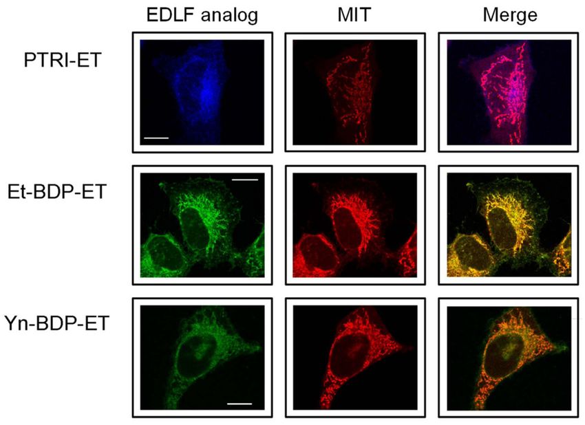
Pharmaceutics 2021, 13, 763                                                                                          2 of 28

                              topical treatment for cutaneous metastases of breast cancer [7], and commercialized under
                              the trademark name of Miltex® (topical liquid human pharmaceutical product; Baxter,
                              Newbury, UK). Miltefosine is also used under the trademark of Milteforan® for the treat-
                              ment of canine leishmaniasis (oral liquid veterinary pharmaceutical product for dogs;
                              Virbac, Carros, France) [8]. Perifosine (octadecyl-[1,1-dimethyl-piperidino-4-yl]phosphate),
                              where the choline moiety of miltefosine is replaced by a heterocyclic piperidine group,
                              shows a promising orally active antitumor APL [9,10] that is currently used in clinical
                              trials [11–15]. Erucylphosphocholine ([13Z]-docos-13-en-1-yl 2-(trimethylammonio)ethyl
                              phosphate, ErPC), an APL-derivative with a 22 carbon atom chain and a cis-13, 14 dou-
                              ble bond, shows distinctive reduced hemolytic activity, thereby allowing intravenous
                              injection, and holds promise for the treatment of human brain tumors [16–19]. The ErPC
                              closely related congener erufosine (erucylphosphohomocholine, or erucylphospho-N,N,N-
                              trimethylpropylammonium, ErPC3) [20,21], a member of the latest generation of APLs, can
                              be applied intravenously and can cross the blood–brain barrier [22–25].

                              Figure 1. Chemical structures of some clinically relevant alkylphospholipid analogs.

                                   However, edelfosine remains as the most active antitumor APL, and is the golden
                              standard and prototype for other APLs and for studies on the mechanism of action of this
                              family of compounds. Furthermore, our in vitro and in vivo results have revealed that
                              edelfosine, orally administered, is the most potent APL in killing different Leishmania spp.,
                              showing higher anti-Leishmania activity than miltefosine, and is less prone to generate drug
                              resistance than miltefosine [26].
                                   A major feature of the above APLs is that they target cell membranes, particu-
                              larly lipid rafts, affecting several biochemical processes, ion transport and signaling
                              pathways [1,2,27–30]. Edelfosine shows a high affinity for both model and cell membranes,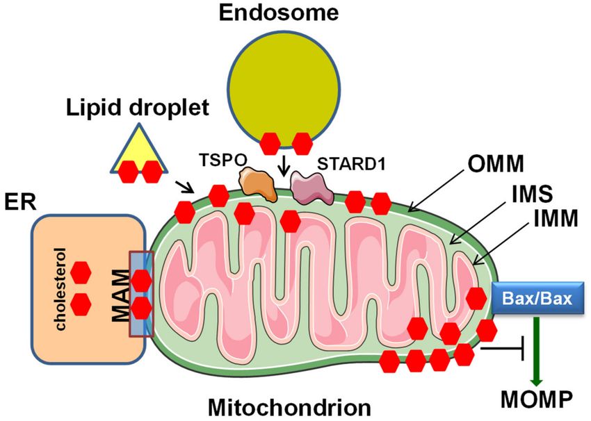
Pharmaceutics 2021, 13, 763                                                                                             3 of 28

                              but weak detergent activity [31]. A remarkable characteristic of the ether lipid edelfosine
                              is its selectivity in inducing apoptosis in cancer cells, whereas non-transformed cells are
                              spared [1,2,9,27,32]. This selectivity is due to the preferential drug uptake by cancer cells by
                              a not yet fully understood mechanism [1,2,27,33–35]. Edelfosine targets the cell membrane
                              and, depending on the cell type, leads to the onset of different types of cell death [36], rang-
                              ing from apoptosis, which is predominantly triggered in most cancer cells [1,2,27,33–35], to
                              necrosis/necroptosis [37,38], with mitochondria playing a key role in the irreversible onset
                              of the cell death process [9,39,40].

                              2. The Alkylphospholipid Analog Edelfosine Induces Apoptosis Selectively in
                              Cancer Cells
                                    A direct antitumor action of edelfosine on cancer cells was already reported in the late
                              1970s and 1980s [41–44], but it was not until the 1990s and 2000s that the molecular mecha-
                              nism underlying the antitumor activity of this drug started to be unveiled, showing that the
                              induction of apoptosis by edelfosine was the main effect that explained the direct antitumor
                              action of APLs [45,46]. Then, a number of findings in the Faustino Mollinedo’s and Con-
                              suelo Gajate’s laboratory, first in Valladolid (Spain) and then in Salamanca (Spain) in the
                              late 1990s and early 2000s, respectively, demonstrated the selective pro-apoptotic effect of
                              edelfosine on cancer cells, following the preferential drug uptake in tumor cells [32,33,47]
                              and the reorganization of membrane lipid raft platforms [27,48]. These data provided
                              the first evidence for a selective pro-apoptotic drug and for the involvement of lipid rafts
                              in cancer chemotherapy. Edelfosine (Figure 1) is an oral drug showing potent antitumor
                              activity against different kinds of tumors in cancer animal models [34,35,40,49], and lacks
                              toxicity in rats after edelfosine oral treatment at pharmacological relevant doses, with no
                              cardiotoxicity, hepatotoxicity or renal toxicity [50].
                                    In general, apoptosis can be mainly induced either by an extrinsic pathway, mediated
                              through the activation of death receptors, or by an intrinsic pathway or mitochondria-
                              mediated process, which permeabilizes the outer mitochondrial membrane (OMM), leading
                              to the release of cytochrome c, located in the mitochondrial intermembrane/intercristae
                              spaces where it functions as an electron shuttle in the respiratory chain. Mitochondria-
                              mediated apoptosis is characterized by mitochondrial outer membrane permeabilization
                              (MOMP) and the subsequent release of mitochondrial cytochrome c into the cytoplasm to
                              activate caspases. Once in the cytosol, cytochrome c binds the adaptor molecule APAF-1
                              (apoptosis protease-activating factor-1), causing it to oligomerize through a conformational
                              change, and form a heptameric structure called the apoptosome complex, made up of
                              cytochrome c and APAF-1. The apoptosome recruits and potentiates the activation of
                              procaspase-9, which in turn cleaves and activates downstream effector caspases, such as
                              caspase-3 and -7 [51]. MOMP is regulated by the Bcl-2 family of proteins [51].

                              3. Edelfosine Accumulates in Lipid Rafts and the Endoplasmic Reticulum of Cancer
                              Cells, and the Generated Apoptotic Signals Converge on Mitochondria to
                              Elicit Apoptosis
                                    A major milestone in the study of the mechanism of action of APLs was achieved
                              in 2001 when the apoptosis induced by the ether lipid edelfosine was first found to be
                              mediated by lipid rafts [48]. Edelfosine accumulates in the lipid rafts of a wide array of
                              hematological cancer cells [9,27,34,52–54], leading to apoptosis through the reorganiza-
                              tion of these membrane domains, especially by promoting co-clustering of lipid rafts and
                              Fas/CD95 death receptor signaling [9,27,34,53,54]. These seminal reports identified lipid
                              rafts as a novel and promising target in cancer therapy [9,27,34,53–57], and paved the way
                              for future studies in raft-targeted cancer therapy [29,30,56,58–63]. Edelfosine induced the
                              clustering and recruitment of the Fas/CD95 death receptor, as well as other death receptors
                              and downstream signaling molecules in lipid rafts, thus triggering apoptosis in a variety of
                              cancer cells, including myeloid and lymphoid cancer cells [9,27,33,48]. This mechanism
                              of action involved a raft-mediated activation of apoptosis via Fas/CD95, independently
                              of its physiological FasL/CD95L ligand, which could be pharmacologically modulated,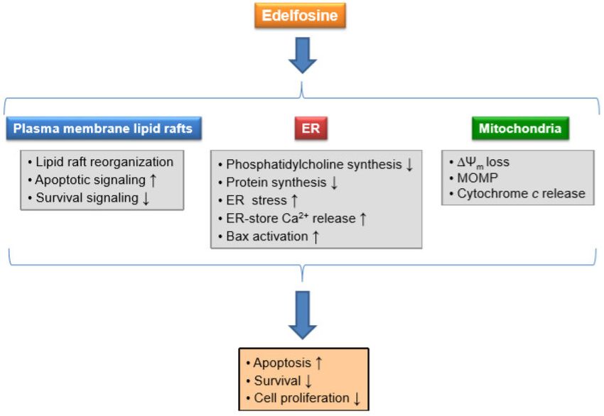
Pharmaceutics 2021, 13, 763                                                                                            4 of 28

                              thus opening a new therapeutic approach in cancer therapy [1,27,34,35,53,59,64]. Interest-
                              ingly, edelfosine prompted the recruitment of death receptors and downstream signaling
                              molecules in lipid rafts, whereas Akt survival signaling was displaced from the rafts [30,65].
                                    Yeast cells show different raft domains that contain transporters and proteins involved
                              in the control of Na+ , K+ and pH homeostasis, required for the proper function of yeast,
                              and that modulate yeast growth and death [66]. The active transport of ions and nutrients
                              in yeast relies on the existence of an electrochemical gradient of protons across the plasma
                              membrane, and this electrochemical gradient is mainly generated in Saccharomyces cerevisiae
                              by the H+ -ATPase gene pma1, an essential H+ pump for yeast growth [67] and a resident
                              raft protein [68]. We found that edelfosine treatment in S. cerevisiae displaced Pma1p from
                              lipid rafts [69–71], and induced its internalization as well as of the plasma membrane
                              arginine/H+ symporter Can1p (arginine permease) and the uracil/H+ symporter Fura4p
                              (uracil permease), two nutrient H+ -symporters associated with yeast lipid rafts [66,72,73].
                              Our studies on the mechanism of action of edelfosine in S. cerevisase showed that the
                              ether lipid displaces the essential proton pump Pma1p from the lipid rafts, inducing
                              its internalization into the vacuole, the yeast equivalent to the mammalian endosome–
                              lysosome system, and subsequent degradation, thus leading to altered pH homeostasis
                              and cell death [69–71]. The displacement of Pma1p from the rafts following edelfosine
                              treatment was preceded by the rapid movement of the yeast sterol ergosterol out of the
                              plasma membrane and into the cell [69–71].
                                    Taking together, edelfosine induces cell death through the reorganization of lipid
                              rafts by modifying the balance of apoptotic versus survival signaling molecules in these
                              membrane domains. Thus, the recruitment of apoptotic signaling molecules into lipid rafts
                              and the displacement of survival molecules from these membrane domains is critical in the
                              mechanism of action of this ether lipid.
                                    Biophysical studies have shown that edelfosine has a high affinity for cholesterol, in-
                              creases membrane thickness, and alters raft organization [74]. The high affinity of edelfosine
                              for cholesterol is easily and visually explained on the basis of the complementarity of the
                              molecular geometries of edelfosine and sterols in general [75]. The combination of “cone-
                              shape” sterols and “inverted cone-shape” edelfosine leads to a more stable bilayer [75].
                                    Studies on solid tumor cells, including pancreatic adenocarcinoma, lung adenocarci-
                              noma, cervix epithelioid carcinoma and Ewing’s sarcoma cells, have shown that edelfosine
                              accumulated mainly in the endoplasmic reticulum (ER), triggering an ER stress response
                              that eventually led to apoptosis [39,40,49,76]. Interestingly, edelfosine accumulated first
                              in plasma membrane lipid rafts and subsequently in the ER of S. cerevisiae, used as a
                              eukaryotic model organism [70].
                                    However, although edelfosine has been found to accumulate in the membrane
                              rafts [9,27,34,53,54] and the ER [40,49] of human tumor cells, as assessed by using radioac-
                              tive edelfosine and fluorescent analogs of the ether lipid, all the apoptotic signals generated
                              from either the plasma membrane rafts or the ER converge on the mitochondria to eventu-
                              ally trigger apoptosis (Figure 2). Thus, the overexpression of Bcl-2 or Bcl-xL totally prevents
                              the apoptotic response induced by edelfosine in cancer cells, either from a hematological
                              or solid tumor origin [9,32,40,77]. These data highlight the critical role of mitochondria as
                              a meeting and convergence point of different apoptotic signaling pathways, irreversibly
                              leading to apoptosis.
Pharmaceutics 2021, 13, 763 5 of 28
Figure 2. Schematic model of the involvement of plasma membrane lipid rafts, ER and mitochondria in edelfosine-induced
apoptosis in cancer cells. Protection of mitochondria by Bcl-2 or Bcl-xL overexpression, or by Bax/Bak double knock-out
(Bax-/- /Bak-/- ), prevents cell death, indicating that the apoptotic signals derived from plasma membrane and ER converge
on mitochondria. See text for details. MOMP, mitochondrial outer membrane permeabilization.
Edelfosine-induced apoptosis involved mitochondria as assessed by the disruption of the
mitochondrial transmembrane potential (∆Ψm ), measured using 3,30 -dihexyloxacarbocyanine
iodide [DiOC6(3)] fluorescence, and the production of reactive oxygen species (ROS), detected
using the conversion of dihydroethidium into ethidium, in both leukemic [47] and solid
tumor cells [49]. Edelfosine also induced Bax activation, cytochrome c release, caspase-9
activation, and DNA fragmentation in both leukemic [9,47] and solid tumor cells [40,49],
and Bcl-2 or Bcl-xL overexpression prevented the above-mentioned mitochondria-related
responses [9,40,77].
4. Localization of Edelfosine in the Mitochondria of Cancer Cells Using
Fluorescent Analogs
In 2004, we synthesized the first fluorescent edelfosine analog, which preserved
pro-apoptotic activity comparable to that of the parent drug [27,78], as an excellent tool
to unveil the mechanism of action of this drug. To this aim, we tried to synthesize a
fluorescent edelfosine analog with a minimum modification of the chemical structure. Our
previous structure–activity relationship studies at the time showed that some modifications
preserved the apoptotic activity, including the presence of a double bond in the O-octadecyl
chain at the C1 of edelfosine [32]. In this regard, a conjugated pentaene group appeared as
a convenient candidate, considering that this fluorophore had led to the development of
useful fluorescent probes for lipid membranes [79,80]. On these grounds, we reasoned that
the replacement of the C18 aliphatic chain by a lipophilic fluorescent group of similar length
could preserve the unique properties of this drug regarding its activity and selectivity.
This led to the synthesis of the first fluorescent analog, containing the conjugated all-(E)-
phenyltetraene blue-emitting chromophore, which was coined as PTE-ET [27] (Figure 3).
This PTE-ET fluorescent analog, as well as the subsequently synthesized PTRI-ET (Figure 3),Pharmaceutics 2021, 13, 763 6 of 28
containing the all-(E)-phenyltrienyne blue-emitting chromophore, were the first fluorescent
edelfosine analogs [27,70,78,81]. These fluorescent edelfosine analogs largely preserved the
chemical structure of edelfosine (Figure 3), and shared analogous fluorescence traits with a
poor fluorescence yield and photostability under intense near-UV laser excitation [28,78,81].
In order to improve the fluorescence yield and provide a more stable fluorescent signal, we
synthesized a second generation of fluorescent analogs by adding a BODIPY (4,4-difluoro-
4-bora-3a,4a-diaza-s-indacene; boron-dipyrromethene) fluorochrome attached to the alkyl
chain of edelfosine, leading to the green-emitting Et-BDP-ET and Yn-BDP-ET fluorescent
edelfosine analogs [28,81] (Figure 3). These two compounds had a higher fluorescence
yield and resistance to photodegradation than the first generation fluorescent edelfosine
analogs, and allowed a thorough analysis through confocal microscopy [28,81]. The use
of all the above fluorescent edelfosine analogs, either first or second generation, allowed
to localize edelfosine in the mitochondria of cancer cells (Figure 4) [81,82], in addition to
the subcellular localizations of this drug in the ER [40,49,76] and lipid rafts [9,27,34,35] in
solid tumor cells and hematological cancer cells, respectively. Interestingly, mitochondrial
localization of edelfosine was also found in Leishmania parasites [82].
Figure 3. Chemical structures of fluorescent edelfosine analogs.
Polyene lipids (linear hydrocarbons containing a conjugated double-bond system)
display a unique structural similarity to natural lipids, which results in minimal effects on
the lipid properties. The above PTE-ET fluorescent analog could be included in this type
of lipids. In this regard, polyfosine (Figure 3), a polyene fluorescent analog of edelfosine
containing five conjugated double bonds, was also found to accumulate in the mitochondria
and to induce morphological changes and apoptosis in COS7 cells [83].Pharmaceutics 2021, 13, 763 7 of 28
Figure 4. Colocalization of fluorescent edelfosine analogs and mitochondria in cancer cells. HeLa
cells were incubated for 12 h with 10 µM of the indicated fluorescent edelfosine (EDLF) analogs (PTRI-
ET, blue fluorescence; Et-BDP-ET, green fluorescence; Yn-BDP-ET, green fluorescence) to visualize
edelfosine subcellular localization. Mitochondrial location was examined using MitoTracker Red
probe (MIT, red fluorescence). Areas of colocalization between edelfosine analogs and mitochondria
in the merge panels are purple (for PTRI-ET) or yellow (for Et-BDP-ET and Yn-BDP-ET). Bar, 10 µm.
Image taken from [81], Springer Nature, 2011.
5. Cholesterol in Mitochondria
A major question raises from the above subcellular localization experiments. How
does edelfosine accumulate in the mitochondrial membrane? We reason that a putative
explanation for this accumulation could lie in the above-stated high affinity of edelfosine
for cholesterol.
Lipids are not randomly distributed among biological membranes, but their relative
content is characteristic for each organelle, affecting their shape, structure and function [84].
Lipids constitute approximately 50% of the mass of most cell membranes (e.g., plasma
membrane), although this proportion is highly dependent on the type of membrane (e.g.,
mitochondrial inner membrane contains 75% protein as a result of the abundance of protein
complexes involved in electron transport and oxidative phosphorylation). However, one
must bear always in mind that there are many more lipid molecules than protein molecules
in membranes because lipid molecules are small compared with proteins. On these grounds,
it might be estimated the presence of about 50 lipid molecules for each protein molecule in
the plasma membrane.
Among the distinct lipids, cholesterol (a major sterol component in animal cell mem-
branes, making up about 30% of the lipid bilayer on average) has attracted much attention
since its first isolation from gallstones in the eighteen century. The French doctor and
chemist François-Paul Poulletier de la Salle (1719–1788) first identified cholesterol in gall-
stones in about 1758–1769, albeit his work was never published [85–87]. Later on, choles-
terol was rediscovered in 1815 by the French chemist Michel Eugène Chevreul (1786–1889)
who named it “cholesterine” [85–87].
Cholesterol is an essential building block of the plasma membrane, having diverse
structural and functional roles [88,89], and playing pleiotropic actions in normal andPharmaceutics 2021, 13, 763 8 of 28
cancer cells [30]. Cholesterol plays a unique and pivotal role among the different lipids
in maintaining the structural integrity and regulating the fluidity of the mammalian cell
membranes [90,91]. As compared to other lipids, cholesterol moves rapidly as a monomer
across membranes and between membrane organelles on protein carriers [89]. However,
cholesterol is not uniformly distributed within biological membranes and across different
cellular compartments. Cholesterol has been suggested to be enriched in the cytosolic
(inner) leaflet of the plasma membrane [92]. Recent imaging methods, using tunable
orthogonal cholesterol sensors, have revealed a marked transbilayer asymmetry of plasma
membrane cholesterol in mammalian cells, with the cholesterol concentration in the inner
leaflet being ~12-fold lower than in the outer leaflet [93]. Cellular cholesterol, derived from
low-density lipoprotein receptor-mediated endocytosis or synthesized de novo in the ER,
is mainly (up to 90%) located in the plasma membrane, constituting 10–45% (mol%) of
the total plasma membrane lipids [93–95]. Cholesterol plays major roles in the structural
and functional modulation of integral membrane proteins [96], and in the formation of
cholesterol-rich membrane domains, such as the so-called lipid or membrane rafts. Lipid
rafts are membrane microdomains enriched in cholesterol and sphingolipids, involved in
the lateral compartmentalization of molecules at the cell surface, and can coalesce to form
membrane raft platforms [30,97].
Mitochondrial membranes are cholesterol-poor, particularly the inner mitochondrial
membrane, as compared to other subcellular membranes in mammalian cells [98,99]. The
relative proportion of phospholipid/cholesterol in the rat liver plasma membrane is 5.25,
whereas this rate increases up to 58.3 in the rat liver mitochondria [98,99]. The sterol-to-
protein ratio in mitochondria is low compared to other subcellular fractions (rat liver),
as follows: mitochondria (0.003 mg sterol/mg protein); ER (0.014 mg sterol/mg protein);
lysosomes (0.038 mg sterol/mg protein); Golgi (0.038 mg sterol/mg protein); and plasma
membrane (0.128 mg sterol/mg protein) [98,99].
The mitochondria are made up of an OMM, an inner mitochondrial membrane (IMM),
an inter-membrane space (IMS) in between, and the mitochondrial matrix enclosed by the
IMM (Figure 5). The IMM shows a number of invaginations, called cristae, thus making
the surface of the IMM significantly larger than that of the OMM. The whole machinery
of oxidative phosphorylation, including the electron transport chain (ETC) complexes
(complexes I–IV) as well as the F1 FO –ATP synthase (complex V), resides in the IMM. The
OMM separates the mitochondrion from the cytosol. The OMM forms a smooth lipid-rich
surface with high membrane fluidity, whereas the IMM is highly folded and shows an
elevated protein level and lower lipid content [98,99]. In this regard, cholesterol is enriched
in the OMM compared to the IMM (0.04 mg sterol/mg protein in OMM versusPharmaceutics 2021, 13, 763 9 of 28
Figure 5. Import and transfer of cellular cholesterol into mitochondria. Cholesterol is transported
to the mitochondria through vesicular and non-vesicular trafficking, involving the ER, lipid droplets,
endosomes, TSPO and STARD1. Elevated mitochondrial cholesterol levels in cancer cells affect mito-
chondrial membrane and impair Bax/Bak oligomerization in OMM and subsequent MOMP formation,
representing a mechanism of cell death resistance in tumor cells. See text for details. ER, endoplasmic
reticulum. IMM, inner mitochondrial membrane. IMS, inter-membrane space. MAM, mitochondria-
associated membrane. OMM, outer mitochondrial membrane. STARD1, steroidogenic acute regulatory
protein-related lipid transfer domain containing protein 1. TSPO, translocator protein.
Cholesterol critically influences membrane fluidity, permeability, curvature and mem-
brane protein interactions in biological cell membranes [30,60,100–102], affecting the cell
surface distribution of membrane proteins, modulation of cellular signaling transmission
and intracellular trafficking. A major feature of cholesterol is its ability to modulate the
physicochemical properties of cellular membranes. Cholesterol orients in a phospholipid
bilayer with its polar hydroxyl group towards the aqueous phase and the hydrophobic
steroid ring oriented parallel to the hydrocarbon chains of the phospholipids, thus interact-
ing with the membrane phospholipids and sphingolipids and being a critical contributor
to lipid raft assembly [103].
6. Cholesterol Transport to Mitochondria
The insertion of cholesterol into the membrane lends rigidity and promotes the for-
mation of protein-tethering platforms, such as lipid rafts [30,97]. In the mitochondria,
cholesterol plays a number of major roles, some are as follows [104]: (a) a structural compo-
nent of the OMM and IMM, providing the appropriate fluidity, curvature and biophysical
properties; (b) a precursor of steroidogenesis, by which cholesterol is converted to bio-
logically active steroid hormones, with the first biochemical reactions taking place in the
mitochondrial matrix [105]; (c) the core of membrane platforms interacting with the ER,
lysosomes and other vesicles or intracellular compartments; and (d) a tethering element
for mitochondrial DNA.
Cholesterol is transported to the mitochondria through vesicular and non-vesicular
trafficking. Some critical proteins and organelles/vesicles involved in mitochondrial
cholesterol delivery to the mitochondria are schematically displayed in Figure 5, and
indicated below.Pharmaceutics 2021, 13, 763 10 of 28
STARD1 (30 kDa steroidogenic acute regulatory protein-related lipid transfer domain
containing protein 1) acts at the OMM to mediate the import of cholesterol and transports
cholesterol from the OMM to the IMM [106].
Translocator protein (18 kDa), TSPO (formerly known as the peripheral-type benzo-
diazepine receptor), is a ubiquitous mitochondrial protein, localized to the OMM, and
involved in several biological functions, including mitochondrial cholesterol transport and
steroid hormone biosynthesis [107,108]. TSPO is a five transmembrane domain protein
found as a monomer, dimer and polymer, and is highly abundant in the OMM [107,108].
TSPO has been shown to interact with STARD1 and the voltage-dependent anion-selective
channel 1 (VDAC1), the latter being the most abundant VDAC of the three isoforms
VDAC1-3 [109,110]. TSPO is a high-affinity cholesterol-binding protein that oligomer-
izes to form a cholesterol transporting channel and prompts cholesterol transfer to the
IMM [111,112]. TSPO has been shown to associate with different cytosolic and mitochon-
drial proteins as part of a large multiprotein complex involved in mitochondrial cholesterol
transport [108,109].
Cholesterol transfer to the mitochondria is mediated by a series of direct interactions
between the mitochondria and a series of intracellular organelles, such as ER, lipid droplets
and endosomes (Figure 5). The mitochondria and the ER interact through the so-called
mitochondria-associated membranes (MAMs) [113], which are involved in the transfer of
cholesterol and other lipids between the ER and mitochondria [114]. Wide-field fluores-
cence microscopy combined with digital deconvolution has revealed that mitochondria
form a largely interconnected dynamic network, and by expressing different fluorescent
markers targeted to the mitochondria and ER, 5–20% of the mitochondrial surface was esti-
mated to be in close apposition to (10–30 nm distance) or in association with the ER [115].
In fact, a large body of evidence demonstrates that mitochondria interact and communicate
directly with the ER through MAMs to modulate several cellular responses [115–121].
Lipid droplets, originating from the ER, are dynamic structures able to interact with
most other cellular organelles, are critical to buffer the levels of toxic lipid species [122,123]
and are involved in lipid storage and mobilization [123]. Lipid droplets have been en-
visaged to interact directly with mitochondria to facilitate lipid transfer [124,125]. These
lipid droplet–mitochondria interactions have been suggested to be mediated by SNAP-23
(23-kDa synaptosome-associated protein) [126], a protein that plays a major role in general
membrane fusion processes, and serves as an important regulator of transport vesicle
docking and fusion in all mammalian cells [127–130].
A major source of cholesterol is derived from the endocytosis of exogenous lipopro-
teins, transferring cholesterol from the lipoproteins and plasma membrane to the endo-
somes and multivesicular late endosomes [131–133]. The subsequent transport of choles-
terol out of late endosomes requires the so-called Niemann-Pick type C1 (NPC1) and NCP2
proteins. Niemann-Pick disease type C (NP-C) is a rare neurodegenerative disorder of
autosomal recessive inheritance, with an estimated incidence of 1 in 120,000–150,000 live
births, and affects cholesterol trafficking [134,135]. NP-C is characterized by endosomal
accumulation of unesterified cholesterol and glycolipids in various tissues, including the
brain, leading to progressive central nervous system neurodegeneration and death [135].
This disease is caused by mutations of the NPC1 (accounting for 95% of NP-C cases) or
the NPC2 gene (5% of NP-C cases). Currently, there is no cure for NP-C and patients
usually die before adulthood (frequently in the second decade of life), but adult forms of
NP-C are being increasingly recognized, having a more insidious onset and slower progres-
sion [136,137]. NP-C is characterized by impaired cholesterol efflux from late endosomes
and lysosomes, and secondary accumulation of lipids due to mutations in the NPC1 or
NPC2 proteins, which act in coordination to mediate the efflux of unesterified cholesterol
from lysosomes or late endosomes. Human NPC1 encodes a 1278 amino acid (170–190 kDa)
glycoprotein, found in late endosomes and lysosomal membranes, with 13 transmembrane
domains, which binds both cholesterol and oxysterol [138]. NPC2 (18 kDa) is a soluble
lysosomal glycoprotein containing 132 amino acids and is found in the lumen of latePharmaceutics 2021, 13, 763 11 of 28
endosomes/lysosomes [139,140]. NPC1 binds cholesterol with nanomolar affinity, whereas
NPC2 binds cholesterol with micromolar affinity [138,141]. In relation to this review, it is
interesting to note that resistance to the ether lipid drug edelfosine represents the first phe-
notype caused by the deletion of the NCR1 gene in S. cerevisiae [142], further supporting the
strong relationship between the ether lipid edelfosine and cholesterol. NCR1 is the S. cere-
visiae homolog of the human NCP1, and the Ncr1p protein localizes to the vacuole [142].
Under normal circumstances, NPC2, as a soluble sterol transfer protein in the late endo-
some, transfers cholesterol from the internal vesicle to the membrane-bound NPC1, which
mediates cholesterol egress from the late endosomes to the ER and plasma membrane.
Putative transport from the late endosome to the mitochondria could be mediated through
STARD3 (also known as metastatic lymph node 64 protein (MLN64)), a 50.5 kDa protein
(containing 445 amino acids) that localizes in the membrane of late endosomes, and is
involved in cholesterol transport [143,144]. However, the direct transport from endosomes
to mitochondria or through an ER-mediated step is still a matter of controversy.
7. Mitochondrial Cholesterol in Cancer
Mitochondria are considered cholesterol-poor organelles, with estimates ranging
from 0.5–3% of the content found in the plasma membrane [145]. However, increased
mitochondrial cholesterol levels have been reported in a number of diseases or patho-
physiological conditions, including some types of cancer, steatohepatitis, cardiac ischemia,
aging and neurodegenerative diseases [146]. When it comes to neurodegenerative diseases,
Alzheimer’s disease and the lysosomal disorder NP-C call particular attention [147]. The
functions of mitochondria are altered in all of the above conditions, and it is tempting
to suggest the existence of an interplay between the abnormally increased mitochondrial
cholesterol levels, mitochondria dysfunction and disease pathology [146,147]. The accumu-
lation of intracellular cholesterol alters mitophagy and reduces the clearance of defective
mitochondria in neurodegenerative diseases [148]. Regarding cancer, larger amounts of
mitochondrial cholesterol have been found in solid tumors as compared to their normal
counterparts, and this correlates with tumor growth and malignancy [149]. About 2- to
5-fold higher levels of mitochondrial cholesterol were found in the tumors from Buffalo
rats containing transplanted Morris hepatomas, when compared to the content found in
the mitochondria prepared from a host liver [150,151]. The mitochondrial cholesterol levels
in H35 rat hepatoma cells and HepG2 human hepatoma cells were 3- to 10-fold higher than
the corresponding cholesterol levels in normal rat and human liver mitochondria [152]. The
high levels of mitochondrial cholesterol contribute to chemotherapy resistance [149,152].
As stated above, cholesterol level tends to be high in cancer cells, the meaning of which
is currently controversial [30,153,154]. A number of studies have shown elevated mito-
chondrial cholesterol levels in cancer cells, being associated with chemotherapy resistance,
low mitochondrial proton leak, and altered patterns of the Krebs cycle metabolism, which
might affect the activity of certain mitochondrial enzymes [150–152,155,156]. An increased
cholesterol level in the OMM, and its subsequent decrease in membrane fluidity, inhibits
Bax oligomerization and activation (Figure 5), thus impairing MOMP and contributing to
the resistance to apoptosis-inducing agents [149,157].
8. Mitochondrial Permeability Transition Pore (mPTP) and Regulation of Cell Death
Mitochondria are critical subcellular structures that control cellular life through energy
production as well as cell death through the induction of apoptosis and necrosis. Different
death signaling pathways converge on mitochondria, and the so-called mitochondrial
permeability transition pore (mPTP) acts as a key nodal point in mediating cell death.
Mitochondrial permeability transition is defined as the process whereby the IMM shows
an increased permeability to solutes with a molecular mass ofPharmaceutics 2021, 13, 763 12 of 28
suggested to be part of the mPTP complex or closely related to its function as regulators of
mPTP activity. These proteins include the following: the adenine nucleotide translocator
(ANT) [163], a 32 kDa protein located in the IMM responsible for the import of ADP into
the mitochondrial matrix in exchange for ATP; VDAC1, the most abundant protein in the
OMM with a molecular weight of ~32 kDa through which metabolites and nucleotides
traverse the OMM [164]; the translocator protein (TSPO) (also known as the peripheral
benzodiazepine receptor) [165], a 18 kDa transmembrane protein mainly found on the
OMM [166], which is required for the mitochondrial cholesterol import that is essential for
steroid hormone production [167]; the mitochondrial phosphate carrier (PiC) (also known
as SLC25A3; solute carrier family 25, member 3), a ~40 kDa IMM solute carrier that is the
primary transporter of inorganic phosphate (Pi) into the mitochondrial matrix [168]; and
cyclophilin D (CypD), a 18.9 kDa matrix peptidyl-prolyl cis-trans isomerase that resides in
the mitochondrial matrix, associates with the inner mitochondrial membrane during the
mitochondrial membrane permeability transition [161], and interacts with and modulates
F1 FO –ATP synthase [169,170].
However, subsequent genetic studies showed the mPTP opening in the absence of
ANT, VDAC, TSPO and PiC, suggesting that these proteins are not an integral component
of the mPTP structure, but rather may play regulatory roles in pore formation [171–175].
The evidence accumulated in the last ten years has brought a new player to the scene
that provides the key to the solution of the elusive and long-lasting enigma in mPTP biology.
This new player is F1 FO –ATP synthase, the ubiquitous and universal enzyme that provides
cellular energy in the form of ATP by oxidative phosphorylation and photophosphorylation
in animals, plants and microorganisms, thus leading to a dual and critical role of F1 FO –ATP
synthase in the regulation of cell life and death, playing major roles in energy generation
and apoptosis regulation [176].
In mitochondria, oxidative phosphorylation has the following two critical parts: the
ETC and chemiosmosis. The ETC includes a series of protein complexes (complex I, II,
III and IV) bound to the IMM, through which electrons pass through in a series of redox
reactions, leading to the translocation of protons from the mitochondrial matrix to the IMS,
and thus forming an electrochemical gradient. This proton gradient increases the acidity in
the IMS, generating an electrical difference with a positive charge outside and a negative
charge inside. F1 FO –ATP synthase (also known as complex V) uses the ETC-generated
proton gradient across the IMM to form ATP through a chemiosmotic process. This enzyme
is made up of two mechanical rotary motors, each driven by ATP hydrolysis or proton
flux down the membrane potential of the protons. These two molecular motors, connected
by a common rotor shaft, interconvert the chemical energy of ATP hydrolysis and proton
electrochemical potential through mechanical rotation of the rotary shaft.
Mitochondrial F1 FO –ATP synthase can undergo a Ca2+ -dependent transformation
to form channels with properties matching those of the mPTP, as a key player in cell
death [177]. The catalytic site of the F1 FO –ATP synthase β subunit constitutes the Ca2+
trigger site, involved in the induction of a conformational change and transition of the F1 FO –
ATP synthase to a channel, behaving as an mPTP [177]. F1 FO –ATP synthase is a complex
enzyme with a molecular weight of >500 kDa, made up of two sectors, the inner membrane
bound FO region (indicating that it can be inhibited by the antibiotic oligomycin) and the
matrix-exposed F1 region, acting as rotary motors (Figure 6). The F1 sector (~380 kDa) is
the hydrophilic water-soluble part of the complex, which acts as an ATP-driven motor and
is composed of three copies of each of the subunits α and β (catalytic subunit), forming the
catalytic head of the complex, and one each of the subunits γ, δ and ε, which constitute
the central stalk of the complex. F1 faces the mitochondrial matrix, and conformational
changes in the F1 subunits catalyze the formation of ATP from ADP and Pi. The FO sector
(~120 kDa) is hydrophobic and embedded in the IMM. FO contains a proton corridor that
is protonated and deprotonated repeatedly as H+ ions flow down the gradient from the
IMS to the matrix, causing rotation, which in turn alters the orientation and conformation
of the F1 subunits, thus driving ATP synthesis. FO consists of several copies of subunit cPharmaceutics 2021, 13, 763 13 of 28
(8 to 10 copies in mammalian mitochondria) [178–180], which form a ring complex, and
one copy each of the following subunits: b, the oligomycin sensitivity-conferring protein
(OSCP); d and F6, which constitute the FO peripheral stalk; f, the 6.8-kDa mitochondrial
proteolipid (6.8PL), diabetes-associated protein in insulin-sensitive tissues (DAPIT); and
g e, a and A6L, which act as FO supernumerary subunits [180] (Figure 6). The c-ring is
critical for the transport of protons through the FO region [180]. The two FO and F1 sectors
of the F1 FO –ATP synthase complex push each other in the opposite direction [181], thus
transforming a proton electrochemical potential into the synthesis of ATP from ADP and
Pi . However, this complex can also act in the reverse direction, hydrolyzing ATP to pump
protons and form an electrochemical potential. When the electrochemical potential of
the protons is large enough to surpass the free energy of ATP hydrolysis, it drives the
FO sector to generate a rotary torque upon proton translocation through the c-ring to
produce ATP synthesis in the F1 sector. Conversely, when the electrochemical potential
is small, the F1 sector, acting as an F1 –ATPase (hydrolyzing ATP) induces FO to rotate the
c-ring in the reverse direction to pump protons against the electrochemical potential [181].
Thus, the proton electrochemical potential can drive the complex to synthesize ATP and,
conversely, ATP hydrolysis (as an ATP-driven motor) can lead to the transfer of protons
in the opposite way. Dimers of the F1 FO –ATP synthase complex have been shown to be
distributed along the inner folds of the mitochondrial cristae by high-resolution transmis-
sion electron microscopy [182,183]. This dimerization and localization of the F1 FO –ATP
synthase at the tips of the cristae induces a strong curvature to the membrane, leading to
the characteristic folded morphology of the mitochondrial cristae [183,184]. The F1 FO –ATP
synthase would act as a sink of protons [182], generating a H+ gradient higher at the cristae
than in the rest of the intermembrane space. Compelling evidence has led to the novel
concept that the IMM-embedded c-subunit ring of the membrane-spanning component
FO of the human mitochondrial ATP synthase complex, forms the mPTP [185–188], and
this pore is also functional in the ATP synthase monomer, not requiring ATP synthase
dimerization [189,190]. The purified reconstituted c-subunit ring of the F1 FO– ATP syn-
thase forms a voltage-sensitive channel, the persistent opening of which leads to the rapid
and uncontrolled depolarization of the IMM in cells [187]. Depletion of the c-subunit
hinders Ca2+ -induced IMM depolarization as well as Ca2+ - and ROS-induced cell death,
whereas the overexpression of the c-subunit favors cell death [187]. Genetic manipulation
of c-subunit expression levels by siRNA in HeLa cervical cancer cells affected the mPTP
activity [185]. Knockdown of the c-subunits of the F1 FO –ATP synthase reduced the mPTP
opening in response to ionomycin or hydrogen peroxide and their overexpression enhanced
the mPTP opening [185].
The current view of the mPTP includes a c-subunit channel embedded in the IMM
(Figure 6). Mnatsakanyan and Jonas [190] have proposed a model of the F1 FO –ATP syn-
thase c-subunit channel, in which there are physiological reversible and pathological
non-reversible openings of the c-subunit ring pore (Figure 6). It has been envisaged that
the F1 sector of the F1 FO –ATP synthase can act as an inhibitor of the c-subunit ring pore
and it can be reversibly tilted, through a conformational change, to release the close contact
between the F1 sector and the c-subunit pore in the FO sector. This conformational change
pulls away F1 from the mouth of the c-subunit pore to open the channel from the matrix
side (Figure 6). Under certain circumstances, including during long-lasting openings of the
c-subunit channel, F1 dissociates from FO , thus leading to a permanent opening, mitochon-
dria swelling, OMM rupture, and cell death. These conformational changes can be induced
by the mPTP inducers CypD and Ca2+ through their binding to the OSCP and β subunit of
the F1 FO –ATP synthase, respectively, thus inducing a conformational change in the ATP
synthase peripheral stalk subunits, promoting the removal of the F1 sector from the top of
the c-ring.Pharmaceutics 2021, 13, 763 14 of 28
F6 F1 sector is tilted
β through a
α α conformational
change to release
F1 sector β F1-c subunit ring
d contact
b
γ
Matrix
ε δ
f g e
6.8PL IMM
DAPIT
a A6L
FO sector
c8-10 ring IMS
H+
(mPTP) H+ H+
Figure 6. Organization of subunits in F1 FO –ATP synthase in mammalian mitochondria and opening
of the mPTP. OSCP, F6, d, and b constitute the FO peripheral stalk. Under certain circumstances, the
FO peripheral stalk and F1 sector are tilted to free the c-subunit ring channel from the side facing the
matrix, thus opening the mPTP (see [190]). See text for details. IMM, inner mitochondrial membrane.
IMS, inter-membrane space. mPTP, mitochondrial permeability transition pore.
The pro-apoptotic Bcl-2 family members Bax and Bak have been suggested to function
as the OMM component of the mPTP in regulating cell death [191]. The mitochondria
from Bax and Bak double-deleted mouse embryonic fibroblasts (MEFs) were resistant to
Ca2+ -induced swelling, and displayed reduced OMM permeability and conductance as
well as cell death [191]. In contrast, Bcl-2 (~26 kDa) and its homologue Bcl-xL (~27 kDa),
as well as other anti-apoptotic Bcl-2 family members, protect mitochondria by interacting
with pro-apoptotic Bcl-2 members and hence prevent MOMP and subsequent apoptosis.
The anti-apoptotic Bcl-2 members can also mediate the activity of the mPTP by direct
interactions with regulatory components [192,193]. In addition, Bcl-xL has been found
to interact directly with the β-subunit of the F1 FO –ATP synthase, regulating metabolic
efficiency [194]. Thus, Bcl-xL , once thought to be present exclusively on the OMM, is
now accepted to be an F1 FO –ATP synthase regulator in the IMM that stabilizes the inner
membrane potential [194,195].
Oligomycin has been recognized as a potent inhibitor of the mitochondrial ATP
synthase since the late 1950s and 1960s [196,197], with the FO sector being responsible to
confer oligomycin sensitivity [197,198]. In fact, the high-resolution (1.9 Å) crystal structure
of oligomycin bound to the subunit c10 ring of the yeast mitochondrial ATP synthase has
been reported [199].
OSCP is located on top of the catalytic F1 sector (Figure 6), connecting F1 and the
peripheral stalk, and ensuring the structural and functional coupling between FO and F1 ,
which is disrupted by oligomycin [200].
The Bcl-2 family of proteins have the capacity to regulate the permeability of intracel-
lular membranes to ions and proteins. The pro-apoptotic members of the Bcl-2 family (e.g.,
Bax and Bid) are able to form channels in the membranes and regulate preexisting channels,
whereas the anti-apoptotic Bcl-2 members have the opposing effects on membrane channel
formation [201].Pharmaceutics 2021, 13, 763 15 of 28
9. Edelfosine Induces Indirect and Direct Effects on Mitochondria
The inhibitor of the mPTP cyclosporin A [202] inhibited [47,82], whereas Bcl-2 or
Bcl-xL overexpression totally prevented [9,40,47] edelfosine-induced apoptosis in cancer
cells. These results, together with the edelfosine-induced mitochondrial-mediated changes
depicted in Figure 2, provide strong evidence for the major role of mitochondria in the
apoptotic response triggered by edelfosine in cancer cells.
The fact that edelfosine induces Bid cleavage, generating the 15 kDa cleaved form of
truncated Bid (tBid), as well as BAP31 cleavage into the p20 fragment [39,40,49], further
supports the involvement of mitochondria in the pro-apoptotic action of the ether lipid.
Taken together, this suggests a complex interplay between the plasma membrane, ER and
mitochondria in edelfosine action. Bid is a potent pro-apoptotic Bcl-2 family member
which, upon proteolytic activation by caspases 8 or 10, translocates onto mitochondria and
promotes the activation of Bax/Bak, thus contributing to cytochrome c release [203]. BAP31
is an integral membrane protein of the ER that modulates ER-mediated apoptosis through
its caspase-8-mediated cleavage into a 20 kDa fragment. This p20–BAP31 fragment prompts
the discharge of Ca2+ from the ER and its concomitant uptake into the mitochondria, thus
directing pro-apoptotic signals between the ER and mitochondria [204]. Edelfosine induces
all these changes, namely, caspase-8 and -10 activation, Bax activation, cytochrome c release,
∆Ψm loss, depletion of ER-stored Ca2+ , and the generation of the p20–BAP31 fragment,
leading to cell death [9,27,28,39,40] (Figure 2). In addition, bax-/- bak-/- double-knockout SV-
40-transformed MEFs were resistant to edelfosine [39], further supporting the involvement
of mitochondria in the ether lipid-induced apoptosis response. These data strongly suggest
the mitochondrial involvement in the pro-apoptotic effect of edelfosine, through signals
generated from the death receptor-mediated extrinsic apoptotic signaling in membrane
lipid rafts and from an ER stress response.
The accumulation of edelfosine in the mitochondria also raises the possibility that the
ether lipid could have a direct effect on the mitochondria during the onset of apoptosis.
In fact, we found that edelfosine accumulates in the mitochondria in cancer cells [81,82]
and affects the mitochondria in a direct way [81,205]. Edelfosine induced swelling in
isolated mitochondria from adult rat livers, indicating an increase in the mitochondrial
membrane permeability [81,205]. This mitochondrial swelling was independent of ROS
generation [81,205]. Furthermore, edelfosine was also found to inhibit mitochondrial respi-
ration and decrease transmembrane electric potential on the isolated mitochondria [205].
These latter effects were also observed with the APL perifosine, together with its ability
to induce mitochondrial permeability transition [205], suggesting that the above actions
constitute a rather general feature of APLs. Interestingly, preincubation with the cholesterol-
depleting agent methyl-β-cyclodextrin (MCD) [9,206], which disrupts membrane rafts,
inhibited edelfosine-induced mitochondrial swelling in the isolated mitochondria [81],
suggesting that the action of edelfosine on isolated mitochondria seems to be dependent
on mitochondrial lipid rafts.
10. Edelfosine-Induced Apoptosis Involves F1 FO –ATPase and Its Recruitment to
Lipid Rafts
Oligomycin is a highly selective inhibitor of the membrane-embedded FO sector
(proton channel) of the F1 FO –ATP synthase that binds to the rotating c-ring within the
membrane and inhibits the enzyme complex [199,207,208]. We found that oligomycin
prevented edelfosine-induced ∆Ψm dissipation and DNA degradation in cancer cells and
Leishmania parasites [82], suggesting a major role of the FO component of the F1 FO –ATP
synthase in the antitumor and anti-Leishmania activity of the ether lipid. In fact, recent data
indicate that the accumulation of edelfosine in the kinetoplast-mitochondrion, leading to
∆Ψm loss and to the successive breakdown of mitochondrial and nuclear DNA, underlies
the potent action of this alkylphosphocholine analog against Leishmania parasites [82].
Oligomycin also attenuated apoptosis and ∆Ψm loss induced by erufosine in glioblastoma
cells [209]. Erufosine was found to interact with the 18 kDa translocator protein (TSPO),Pharmaceutics 2021, 13, 763 16 of 28
leading to the activation of the mitochondrial apoptosis cascade [20]. Furthermore, the
Saccharomyces cerevisiae ATP7∆ mutant, with a deletion in the gene encoding for subunit d
of the stator stalk of mitochondrial F1 FO –ATP synthase, which is conserved in mammalian
cells [210], was resistant to edelfosine [82]. This edelfosine-resistant phenotype could be
reverted by transformation with the wild-type ATP7 gene [82]. The above evidence strongly
supports the involvement of F1 FO –ATPase in the killing activity of edelfosine and in the
onset of APL-induced apoptosis in general.
Edelfosine affects membrane lipid organization, making membranes more fluid [211,212].
On these grounds, edelfosine could be hypothesized to make the OMM more porous and
permeable, thus favoring the leakage of H+ ions. This would lead to the dissipation of
the proton gradient, and therefore the F1 FO –ATP synthase could run in reverse, that is,
hydrolyzing ATP and alkalinizing the matrix by proton extrusion. Matrix alkalinization
causes the mPTP opening [213], and then it could be envisaged that the F1 FO –ATP synthase
could promote the onset of cell death by this F1 FO –ATP synthase-mediated increase in the
matrix pH. This mechanism has been previously proposed for the inhibition of Bax-induced
apoptosis by oligomycin in yeast and mammalian cells [214].
By proteomic analyses in lipid rafts, isolated from different hematological cancer cells
through discontinuous sucrose gradient centrifugation [215,216], we found that edelfosine
treatment in hematological cancer cells led to a dramatic recruitment of mitochondrial
F1 FO –ATP synthase to the rafts [82]. This remarkable F1 FO –ATP synthase translocation into
the rafts could suggest that the enzyme is either translocated to lipid rafts present at the cell
surface or in the mitochondria. Several studies have reported the presence of raft-located
F1 FO –ATP synthase at the plasma membrane of different normal and tumor cells, having
been proposed to act as a proton channel, a modulator of extracellular ATP level, or as a
regulator of intracellular Ca2+ levels, involved in numerous biological processes, including
cell migration and intracellular pH modulation [217–224].
11. Edelfosine Promotes Raft Translocation to Mitochondria and Presence of Raft-Like
Domains in Mitochondria
The present evidence cannot discern between the translocation of F1 FO –ATP synthase
to lipid rafts at the cell surface or to raft domains present in the mitochondria. However,
edelfosine has been shown to promote the redistribution of lipid rafts from the plasma mem-
brane to the mitochondria, suggesting a raft-mediated plasma membrane–mitochondria
link [81]. In this context, we have found a lipid raft-mediated connection between the
extrinsic and intrinsic apoptotic pathways in human multiple myeloma MM144 cells [225].
The fact that edelfosine can interact with plasma membrane lipid rafts and mitochon-
dria could led us to suggest that the ether lipid could be translocated from the plasma
membrane to the mitochondria, where it would ultimately exert its pro-apoptotic activity
promoting the accumulation of F1 FO –ATP synthase into mitochondrial rafts, ∆Ψm dissi-
pation, cytochrome c release, leading eventually to cell demise. Lipid rafts were mainly
located at the plasma membrane in untreated HeLa cells, as assessed by the raft marker flu-
orescein isothiocyanate-cholera toxin B subunit that binds ganglioside GM1 [226], mainly
found in these domains [227]. Mitochondria (stained with MitoTracker Red) were observed
as a widespread network in the interior of the cell. Following edelfosine treatment, the
membrane rafts were gradually internalized into the cell, and colocalized with mitochon-
dria at the time of apoptosis onset, thus unveiling a link between plasma membrane rafts
and mitochondria driven by edelfosine [81]. This suggests the presence of cholesterol-rich
raft-like domains in mitochondria that could be involved in edelfosine-induced apoptosis.
It is interesting to note that the GD3 raft component can proceed from the cell plasma
membrane to the mitochondria via a microtubule-dependent mechanism, which could be
regulated by CLIPR-59, a new CLIP-170-related protein involved in microtubule dynam-
ics [228–230]. Although the presence of lipid rafts in mitochondria remains a controversial
issue [231–234], there is increasing evidence favoring the presence of raft-like domains in
theses organelles. Lipid raft-like domains enriched in ganglioside GD3 have been found
in mitochondria and are involved in apoptosis regulation [229,232,235]. It is temptingYou can also read