An IDH1-vitamin C crosstalk drives human erythroid development by inhibiting pro-oxidant mitochondrial metabolism - CNRS
←
→
Page content transcription
If your browser does not render page correctly, please read the page content below
Article

An IDH1-vitamin C crosstalk drives human erythroid
development by inhibiting pro-oxidant mitochondrial
metabolism
Graphical Abstract                                                   Authors
                                                                     Pedro Gonzalez-Menendez,
                                                                     Manuela Romano, Hongxia Yan, ...,
                                                                     Narla Mohandas, Naomi Taylor,
                                                                     Sandrina Kinet

                                                                     Correspondence
                                                                     pedro.gonzalez-menendez@igmm.cnrs.
                                                                     fr (P.G.-M.),
                                                                     taylorn4@mail.nih.gov (N.T.),
                                                                     kinet@igmm.cnrs.fr (S.K.)

                                                                     In Brief
                                                                     Gonzalez-Menendez et al. show that
                                                                     terminal erythroid differentiation is
                                                                     dependent on the suppression of
                                                                     mitochondrial metabolism. Disturbances
                                                                     in levels of the a-ketoglutarate and IDH1
                                                                     TCA-cycle regulators result in ineffective
                                                                     erythropoiesis, with generation of
                                                                     morphologically abnormal erythroblasts.
                                                                     Vitamin C restores redox homeostasis
                                                                     and rescues late-stage erythropoiesis by
Highlights                                                           scavenging reactive oxygen species.
d   Glutamine-dependent OXPHOS drives early erythroid
    differentiation

d   OXPHOS-induced ROS inhibit erythroblast enucleation

d   IDH1 downregulation augments ROS, leading to pathological
    erythroid differentiation

d   Vitamin C rescues erythroid differentiation under conditions
    of oxidative stress

           Gonzalez-Menendez et al., 2021, Cell Reports 34, 108723
           February 2, 2021
           https://doi.org/10.1016/j.celrep.2021.108723                                              ll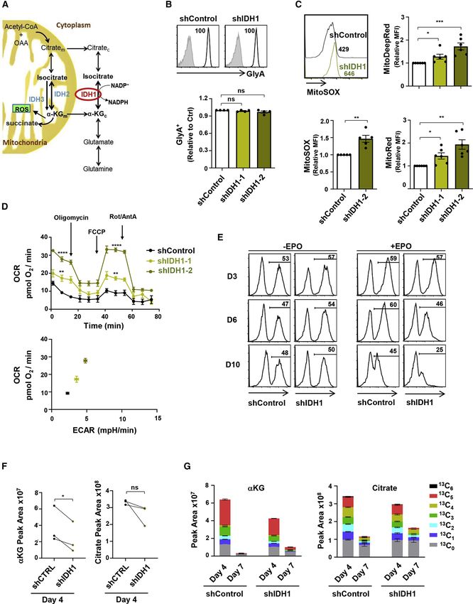
ll
                                                                                                                         OPEN ACCESS

Article
An IDH1-vitamin C crosstalk drives
human erythroid development by inhibiting
pro-oxidant mitochondrial metabolism
Pedro Gonzalez-Menendez,1,2,10,* Manuela Romano,1,2,10 Hongxia Yan,1,3 Ruhi Deshmukh,4 Julien Papoin,5
Leal Oburoglu,1,2 Marie Daumur,1,2 Anne-Sophie Dumé,1,2 Ira Phadke,1,2,6 Cédric Mongellaz,1,2 Xiaoli Qu,3
Phuong-Nhi Bories,7 Michaela Fontenay,2,7 Xiuli An,3 Valérie Dardalhon,1,2 Marc Sitbon,1,2 Valérie S. Zimmermann,1,2
Patrick G. Gallagher,8 Saverio Tardito,4,9 Lionel Blanc,5 Narla Mohandas,3 Naomi Taylor,1,2,6,11,* and Sandrina Kinet1,2,*
1Institut de Génétique Moléculaire de Montpellier, Univ. Montpellier, CNRS, Montpellier, France
2Laboratory   of Excellence GR-Ex, Paris 75015, France
3New York Blood Center, New York, NY, USA
4Cancer Research UK Beatson Institute, Glasgow G61 1BD, UK
5The Feinstein Institute for Medical Research, Manhasset, NY, USA
6Pediatric Oncology Branch, NCI, CCR, NIH, Bethesda, MD, USA
7Service d’Hématologie Biologique, Assistance Publique-Ho    ^ pitaux de Paris, Institut Cochin, Paris, France
8Departments of Pediatrics and Genetics, Yale University School of Medicine, New Haven, CT, USA
9Institute of Cancer Sciences, University of Glasgow, Glasgow G61 1QH, UK
10These authors contributed equally
11Lead contact

*Correspondence: pedro.gonzalez-menendez@igmm.cnrs.fr (P.G.-M.), taylorn4@mail.nih.gov (N.T.), kinet@igmm.cnrs.fr (S.K.)
 https://doi.org/10.1016/j.celrep.2021.108723

SUMMARY

The metabolic changes controlling the stepwise differentiation of hematopoietic stem and progenitor cells
(HSPCs) to mature erythrocytes are poorly understood. Here, we show that HSPC development to an
erythroid-committed proerythroblast results in augmented glutaminolysis, generating alpha-ketoglutarate
(aKG) and driving mitochondrial oxidative phosphorylation (OXPHOS). However, sequential late-stage eryth-
ropoiesis is dependent on decreasing aKG-driven OXPHOS, and we find that isocitrate dehydrogenase 1
(IDH1) plays a central role in this process. IDH1 downregulation augments mitochondrial oxidation of aKG
and inhibits reticulocyte generation. Furthermore, IDH1 knockdown results in the generation of multinucle-
ated erythroblasts, a morphological abnormality characteristic of myelodysplastic syndrome and congenital
dyserythropoietic anemia. We identify vitamin C homeostasis as a critical regulator of ineffective erythropoi-
esis; oxidized ascorbate increases mitochondrial superoxide and significantly exacerbates the abnormal
erythroblast phenotype of IDH1-downregulated progenitors, whereas vitamin C, scavenging reactive oxygen
species (ROS) and reprogramming mitochondrial metabolism, rescues erythropoiesis. Thus, an IDH1-vitamin
C crosstalk controls terminal steps of human erythroid differentiation.

INTRODUCTION                                                            burst-forming unit cells, followed by erythroid colony-forming
                                                                        unit cells and proerythroblasts. Terminal erythroid differentiation
Hematopoietic stem cell (HSC) numbers as well as their differen-        begins with proerythroblasts differentiating in a stepwise manner
tiation state are tightly regulated throughout the lifetime of an in-   to early then late basophilic erythroblasts, polychromatic eryth-
dividual, allowing the sustained production of all mature blood         roblasts, and orthochromatic erythroblasts that enucleate to
lineages under physiological conditions. Circulating mature             become reticulocytes.
erythrocytes are a terminally differentiated product of HSCs               Erythropoiesis is a metabolically daunting process when eval-
that have undergone a series of lineage-fate engagements that           uated at the level of cell numbers. In healthy adults, committed
gradually restrict their potential, resulting in a commitment to        erythroid progenitors support the production of 2.4 million eryth-
the erythroid lineage. Commitment defines the beginning of              rocytes per second via a synchronized regulation of iron,
erythropoiesis; a three-stage process characterized by early            glucose, fatty acid, and amino acid metabolism. Iron is indis-
erythropoiesis, terminal erythroid differentiation, and reticulo-       pensable for heme biosynthesis in erythroblasts (Oburoglu
cyte maturation. Early erythropoiesis is characterized by               et al., 2016). The utilization of both glutamine and glucose in de
commitment of multilineage progenitor cells into erythroid pro-         novo nucleotide biosynthesis is a sine qua non for erythroid dif-
genitor cells, with proliferation and development into erythroid        ferentiation (Oburoglu et al., 2014), and glutamine-derived

                                                                                           Cell Reports 34, 108723, February 2, 2021 1
                      This is an open access article under the CC BY-NC-ND license (http://creativecommons.org/licenses/by-nc-nd/4.0/).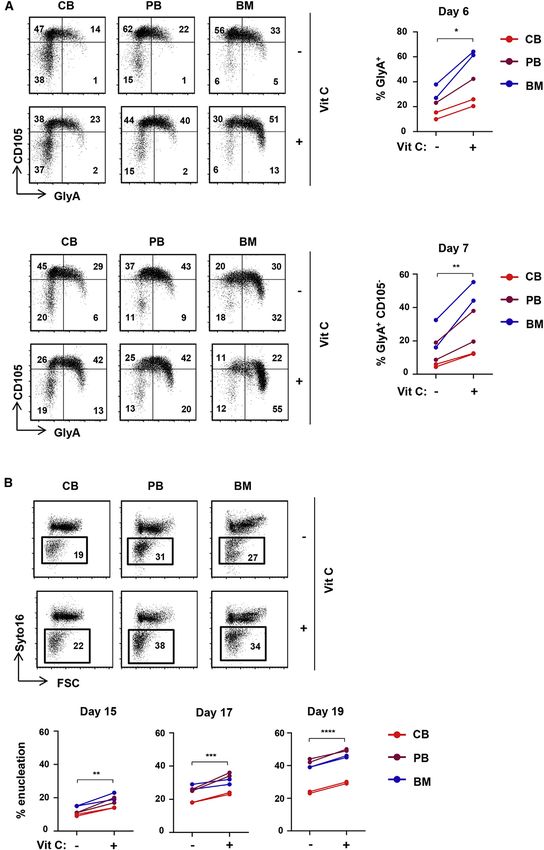
ll
     OPEN ACCESS                                                                                                               Article

production of succinyl-coenzyme A (succinyl-CoA) is also                rate (aKG) TCA-cycle intermediate, generated by the anaplerotic
required for the production of heme (Burch et al., 2018). Further-      utilization of glutamine, and a directly augmented OXPHOS.
more, amino acids regulate mTOR signaling (Chung et al., 2015)          However, upon terminal differentiation of erythroblasts, mito-
as well as lipid metabolism (Huang et al., 2018) during                 chondrial biomass, OXPHOS, mitochondrial ROS, and superox-
erythropoiesis. The critical nature of metabolism in erythroid dif-     ide production were all markedly decreased. Supraphysiological
ferentiation is further highlighted by the recent identification of     levels of aKG markedly attenuated terminal erythroid differentia-
‘‘metabolic regulators’’ as an erythropoietin (EPO)-induced             tion and enucleation. The impact of aKG was directly coupled to
phosphorylation target set (Held et al., 2020).                         its role in mitochondrial metabolism, as enucleation was similarly
   It is important though to note that terminal erythroid               inhibited by mitochondrial ROS and superoxide, induced by Mi-
differentiation is a distinctive process; each mitosis results in       toParaquat (MitoPQ) or DHA. Moreover, enucleation was
the production of daughter cells that differ, morphologically           rescued by ROS scavengers, including vitamin C, glutathione
and functionally, from their parent cells. This sequential matura-      (GSH), N-acetylcysteine (NAC), and vitamin E.
tion is tightly regulated at each stage of erythroid differentiation,      We identified the cytoplasmic isocitrate dehydrogenase 1
associated with decreased cell size, enhanced chromatin                 (IDH1) enzyme, catalyzing the interconversion between aKG
condensation, progressive hemoglobinization, and changes in             and isocitrate, as a critical enzyme in this process. Lentiviral-
membrane organization and transcriptome specificities (An               mediated downregulation of IDH1, by significantly increasing
et al., 2014; Hu et al., 2013; Li et al., 2014; Ludwig et al., 2019;    mitochondrial metabolism and mitochondrial superoxide pro-
Schulz et al., 2019). Moreover, during the late stages of mamma-        duction, dramatically inhibited enucleation and resulted in the
lian terminal erythroid differentiation, erythroblasts expel their      generation of abnormal multinucleated erythroblasts. Notably,
nuclei and lose all organelles, including mitochondria (Moras           both aKG and DHA markedly exacerbated the negative impact
et al., 2017). Thus, the constraints of a late-stage terminally         of IDH1 downregulation, while the vitamin C antioxidant rescued
differentiating erythroblast differ significantly from that of an       erythroid differentiation. Indeed, the vitamin-C-mediated
erythroid-committed progenitor early in erythroid development.          quenching of ROS in erythroblasts accelerated human erythroid
Indeed, in mice, early-stage erythroid progenitors have been            differentiation. This effect was specific to an IDH1-vitamin C axis;
found to require mTORC1-mediated mitochondrial biogenesis               inhibition of TET2 in late-stage erythroblasts did not attenuate
and reactive oxygen species (ROS) production (Liu et al., 2017;         enucleation. Thus, our data identify IDH1 as a critical regulator
Luo et al., 2017; Zhao et al., 2016), while terminal erythropoiesis     of redox homeostasis, promoting late-stage erythropoiesis.
requires that cells be protected from oxidative stress (Case et al.,    Furthermore, these results highlight the therapeutic potential of
2013; Filippi and Ghaffari, 2019; Hyde et al., 2012; Xu et al., 2019;   vitamin C in human erythropoiesis, especially under pathological
Zhao et al., 2016).                                                     conditions where ROS are increased and vitamin C levels are not
   Significant differences in murine versus human erythroid             sufficient to combat oxidative damage.
development have been documented, especially as relates to
the diversity of erythroid progenitor subpopulations in human           RESULTS
bone marrow (BM) (Gautier et al., 2020; Schulz et al., 2019;
Yan et al., 2018). Furthermore, regarding oxidative stress, it is       Early erythropoiesis is associated with augmented
important to note that murine and human erythropoiesis differ           glutamine utilization and OXPHOS
dramatically as a function of vitamin C/ascorbate plasma con-           As glutaminolysis is required for erythroid lineage specification
centrations and expression of the SLC2A1/GLUT1 transporter,             (Oburoglu et al., 2014), we evaluated the utilization of glutamine
shuttling dehydroascorbic acid (DHA) across the membrane                during early stages of human erythropoiesis at day 4 following re-
and rapidly reducing it to ascorbate (reviewed by May et al.,           combinant erythropoietin (rEPO) stimulation, a time point at which
2001). Indeed, humans exhibit a deficiency in vitamin C produc-         the erythroid markers CD36, CD71 (transferrin receptor), and gly-
tion, due to inactivation of L-gulono-g-lactone oxidase (GLO),          cophorin A (GlyA) are upregulated (Figure S1A). The anaplerotic
the enzyme that catalyzes the terminal step of L-ascorbic acid          contribution of glutamine can be evaluated by tracing glutamine
(AA) biosynthesis (Burns, 1957). While human erythrocytes ex-           labeled with heavy stable carbons and nitrogens (13C515N2). As
press the highest level of the GLUT1 transporter, harboring             shown in Figure 1A, the portion of glutamate derived from
                                                                        13
greater than 200,000 molecules per cell (Helgerson and Car-                C515N2 glutamine, a measure of glutaminase activity, increased
ruthers, 1987; Mueckler, 1994), we found that erythroid GLUT1           significantly following erythropoietin-induced erythroid differentia-
expression is a specific feature of mammals that have lost the          tion of human CD34+ progenitors (p < 0.05; Figure 1A). Addition-
ability to synthesize AA from glucose (Montel-Hagen et al.,             ally, the contribution of labeled glutamine to the pool of glutamate
2008a, 2008b). This potential evolutionary compensation, and            (13C515N1 glutamate/13C0 glutamate) as well as the portion of 13C5
subsequent alterations in the ability of plasma vitamin C to scav-      labeled aKG increased significantly upon erythroid differentiation.
enge ROS/reactive nitrogen species (RNS), make it critical to           This glutamate-derived generation of aKG was critical for OX-
evaluate the stepwise changes that regulate the progression of          PHOS in erythroid progenitors, as the oxygen consumption rate
human erythropoiesis.                                                   (OCR) was abrogated by the aminooxyacetic acid (AOA) transam-
   Here, we demonstrate that during the early stages of human           inase inhibitor, blocking conversion of glutamate to aKG, and was
red blood cell development, erythroid progenitors exhibit               completely restored by aKG supplementation (Figure 1B).
increased oxidative phosphorylation (OXPHOS) activity. This                These data show that glutamine supports the tricarboxylic acid
correlated with the increased generation of the alpha-ketogluta-        (TCA) cycle and downstream OXPHOS via the cataplerotic

2 Cell Reports 34, 108723, February 2, 2021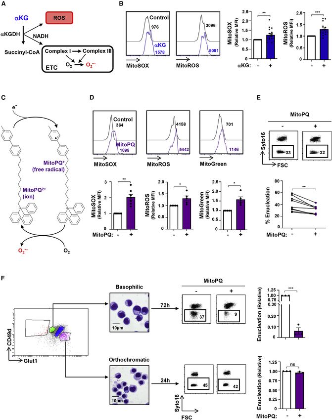
ll
Article                             OPEN ACCESS

                                   (legend on next page)

          Cell Reports 34, 108723, February 2, 2021 3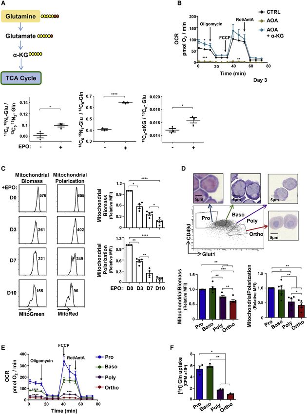
ll
      OPEN ACCESS                                                                                                                                    Article

utilization of aKG. As each stage of terminal erythroid differentia-                we evaluated whether aKG, the anaplerotic product of glutamine
tion is defined by a decreased cell size with the final stage of eryth-             that enters into the TCA cycle, directly contributed to the eryth-
ropoiesis resulting in enucleation and reticulocyte maturation, we                  roblast’s respiratory capacity. Importantly, we found that aKG
assessed whether mitochondrial biomass and transmembrane                            directly regulates the metabolic state of progenitors, as the injec-
polarization decrease following commitment of progenitors to an                     tion of a cell-permeable aKG immediately augmented the
erythroid lineage fate. Indeed, both mitochondrial biomass and                      maximal respiratory capacity of erythroblasts at both day 3
polarization, evaluated by MitoGreen and MitoRed staining,                          and day 7 of EPO induction (p < 0.001 and p < 0.0001; Figure 2A).
respectively, decreased massively upon erythroid differentiation,                   This increase in OXPHOS did not alter the generation of
monitored as a function of day of differentiation (days 0–10) as                    CD34CD36+ committed erythroid colony-forming unit (CFU-E)
well as on erythroblast subsets fluorescence-activated cell sorting                 progenitors or GlyA+ erythroblasts or the upregulation of CD36
(FACS) sorted on the basis of their CD49d/GLUT1 staining profiles                   or CD71 erythroid markers (Figures 2B and S2A). While aKG
(p < 0.05 to p < 0.0001; Figures 1C, 1D, and S1B).                                  did not alter mitochondrial biomass or polarization at early
   These changes in mitochondrial biomass and polarization had                      stages of development (day 3; Figure S2B), it substantially
functional consequences on terminal erythroid differentiation                       affected these parameters when it was added only at later stages
(Figure 1D). Erythroblasts at distinct stages of differentiation ex-                (from days 7 to 10). Mitochondrial biomass and polarization were
hibited significant differences in their levels of OXPHOS, as esti-                 significantly higher in aKG-supplemented late-stage erythro-
mated by their OCRs; basal OCR decreased from 146.9 ± 15.5 to                       blasts than in control erythroblasts (p < 0.01 and p < 0.001; Fig-
17.5 ± 0.6 pmol O2/min between the proerythroblast and ortho-                       ure 2C). Furthermore, progression from the CD49d+GLUT1+
chromatic erythroblast stages of differentiation (p < 0.0001; Fig-                  basophilic erythroblast to the CD49dGlut1+ orthochromatic
ure 1E). Notably, the respiratory capacities of both polychromatic                  stage was significantly inhibited by aKG (p < 0.05 and p <
and orthochromatic subsets were minimal; neither of these termi-                    0.01; Figure 2D). This phenomenon was also associated with a
nal subsets exhibited a spare respiratory capacity, as measured                     significantly attenuated level of enucleation, decreasing by
by the ability of the cell to increase its respiration in response to an            32% ± 7% and 52% ± 7% in the presence of aKG at days 10
Carbonyl cyanide-4-(trifluoromethoxy)phenylhydrazone (FCCP)-                        and 14 of erythroid differentiation, respectively (p < 0.001 and
mediated disruption of mitochondrial membrane potential (Fig-                       p < 0.01; Figure 2D). Notably, this effect was specific to aKG
ure 1E). Similarly, these differences were also detected with                       and was not recapitulated by other TCA-cycle intermediates
increasing time after EPO induction (Figure S1C). Consistent                        such as succinate or citrate (Figure S2C).
with a critical role for glutamine in the oxidative potential of the
erythroblast, glutamine uptake was significantly higher in proery-                  Augmented mitochondrial superoxide generation at the
throblasts and basophilic erythroblasts than in polychromatic/                      basophilic erythroblast stage abrogates terminal
orthochromatic erythroblasts (p < 0.01; Figure 1F). Thus, in                        erythroid differentiation and enucleation
marked contrast with the high-energy state of early erythroid pro-                  Generation of ROS within mitochondria is associated with the
genitors, terminal maturation results in a significant attenuation of               oxidative function of these organelles (Adam-Vizi and Tretter,
glutamine uptake and an associated decrease in OXPHOS.                              2013; Murphy, 2009), and aKG can directly contribute to ROS
                                                                                    generation through the catalytic activity of aKG dehydrogenase
aKG increases mitochondrial function while negatively                               (aKGDH) (Starkov et al., 2004; Tretter and Adam-Vizi, 2004).
regulating terminal erythroid differentiation and                                   NADH produced by aKGDH also stimulates superoxide produc-
enucleation                                                                         tion by complex I of the electron transport chain (ETC; Figure 3A)
As decreased mitochondrial function of terminally differentiated                    (Adam-Vizi and Tretter, 2013; Murphy, 2009). We therefore eval-
erythroblasts was associated with decreased glutamine uptake,                       uated whether addition of aKG, the substrate of aKGDH, alters

Figure 1. Early erythropoiesis results in increased metabolism followed by decreased mitochondrial activity during terminal erythroid
differentiation
(A) Schematic representation of glutamine conversion to a-ketoglutarate (aKG), showing the metabolism of [13C515N2] heavy-labeled glutamine (Gln) carbons
(yellow) and nitrogens (orange; top). Peak area ratios of heavy atoms from [13C515N2]Gln into glutamate ([13C515N1]Glu), the relative level of [13C515N1]Glu, and the
portion of [13C5]aKG at day 4 of differentiation in the absence or presence of rEPO are shown (±SEM). Horizontal lines represent mean levels. Each dot represents
an individual technical replicate, representative of one of two independent experiments.
(B) Oxygen consumption rates (OCRs), a measure of OXPHOS, were monitored by sequential injection of oligomycin, FCCP, and rotenone/antimycin A (Rot/AntA)
as indicated. Mean basal oxygen consumption, maximal consumption following FCCP, and minimal levels following inhibition of the electron transport chain
(ETC) Rot/AntA (bottom panel) are presented at day 3 of erythroid differentiation in control conditions (CTRL), in the presence of the AOA transaminase inhibitor
(1 mM) or AOA + aKG (3.5mM). Means ± SEM are presented (n = 3).
(C) Mitochondrial biomass and polarization were evaluated by MitoTracker Green and MitoTracker Red staining, respectively, at days 0, 3, 7, and 10 of differ-
entiation (n = 4).
(D) Pro-erythroblasts (Pro) and basophilic (Baso), polychromatic (Poly), and orthochromatic (Ortho) erythroblasts were sorted as a function of their GLUT1/CD49d
profile (top panel). H&E staining of sorted erythroblasts is shown. Mitochondrial biomass and polarization were evaluated as in (C), and means ± SEM are
presented (n = 5).
(E) OCR was monitored 15 h post-sorting and means ± SEM are presented (n = 6).
(F) Glutamine uptake was monitored 18 h following sorting using L-[3,4-3H (N)]glutamine (0.5 mCi) for 10 min at room temperature (RT). Uptake is expressed as
mean counts per minute (CPM) for triplicate samples ± SEM.
Statistical significance was determined by a one-way ANOVA with Tukey’s post hoc test (*p < 0.05; **p < 0.01; ***p < 0.001; ****p < 0.0001).

4 Cell Reports 34, 108723, February 2, 2021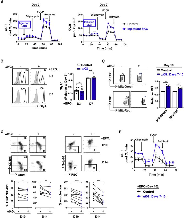
ll
Article                                                                                                                                          OPEN ACCESS

Figure 2. aKG increases mitochondrial function while significantly inhibiting terminal erythroid differentiation and enucleation
(A) CD34+ HSPCs were differentiated with EPO, and OXPHOS was evaluated at days 3 (left) and 7 (right) directly following injection of cell-permeable aKG
(3.5 mM, blue line) into the Seahorse analyzer. Mean OCR ± SEM are presented for the indicated conditions (n = 5–6 per time point).
(B) Progenitors were stimulated with rEPO in the absence () or presence (+) of aKG. Expression of GlyA was monitored at days 3 and 7, and representative
histograms (left) are shown (gray histograms, isotype controls; black lines, specific staining). Mean levels of GlyA+ cells were quantified in the different conditions
(right), with levels at day 7 arbitrarily set at 1 (means ± SEM of five independent experiments; ns, nonsignificant).
(C) Mitochondrial biomass and mitochondrial membrane potential (MMP) were monitored following erythroid differentiation in the absence () or presence (+) of
aKG (between days 7 and 10). Representative dot plots showing the percentages of cells with high mitochondrial biomass and MMP at day 10 are presented (left).
Mean biomass and MMP (±SEM) are presented (right; n = 5).
(D) CD49d/Glut1 profiles of GlyA+ erythroblasts differentiated in the absence or presence of aKG (starting at day 7) were monitored at days 10 and 14 of dif-
ferentiation, and representative dot plots are shown (top left). Percentages of Glut1+CD49d orthochromatic erythroblasts are shown for eight individual donors
(bottom left). Erythroblast enucleation was evaluated as a function of Syto16 nucleic acid staining, and representative dot plots indicating the percentages of
Syto16 enucleated cells are presented (top right). Percentage enucleation in nine individual donors is presented (bottom right).

                                                                                                                                  (legend continued on next page)

                                                                                                               Cell Reports 34, 108723, February 2, 2021 5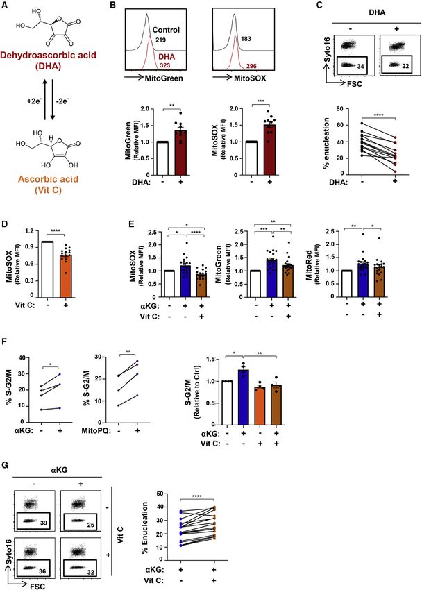
ll
      OPEN ACCESS                                                                                                                                 Article

the redox state of the cell. Direct measurement of mitochondrial                  but its activity is countered by the oxidized form of vitamin C,
superoxide with the fluorescent MitoSOX indicator showed that                     DHA. The rapid reduction of intracellular DHA to vitamin C (Fig-
aKG induced a 1.3-fold increase in superoxide (p < 0.01) as well                  ure 4A) results in concomitant increases in endogenous ROS in
as an overall increase of 1.3-fold in all mitochondrial ROS (p <                  tumor cell lines (Kc et al., 2005; Yun et al., 2015), but its role in
0.001), monitored as a function of DHR123 fluorescence (Mi-                       erythroid cells is not known. Of note, DHA is transported via the
toROS) (Figure 3B).                                                               GLUT1 glucose transporter, the most highly expressed trans-
   These data suggested that the negative impact of aKG on                        porter on human erythrocytes (Bianchi and Rose, 1986; Helger-
erythroid maturation was linked to its role in altering the eryth-                son and Carruthers, 1987; May, 1998; Montel-Hagen et al.,
roblast redox state. We therefore assessed whether directly                       2008b, 2009). We found that the addition of DHA significantly
increasing mitochondrial superoxide would inhibit erythroid                       increased mitochondrial biomass and superoxide production
maturation. Erythroblasts were treated with MitoPQ, a newly                       in differentiating erythroblasts (Figure 4B). The DHA-induced in-
synthesized paraquat compound that is targeted to the mito-                       crease in superoxide production was associated with a signifi-
chondria by conjugation to the lipophilic tri-phenylphospho-                      cant decrease in the enucleation of late-stage erythroblasts
nium cation (Robb et al., 2015). This compound selectively in-                    (from a mean of 40% ± 3% to 24% ± 3%; p < 0.0001; Fig-
creases superoxide production within mitochondria at 1,000-                       ure 4C). Thus, under all tested conditions of superoxide pro-
fold higher efficiency than untargeted paraquat (PQ) (Robb                        duction, enucleation of late-stage erythroblasts was severely
et al., 2015); within the mitochondria, mitoPQ2+ is reduced                       attenuated.
to the radical monocation at the flavin site of complex I and                        Based on these data, it was important to evaluate whether
the monocation then reacts with O2 to generate superoxide                         vitamin C would have opposing effects on the redox state of
(O2) (Figure 3C). MitoPQ dramatically increased mitochon-                        erythroblasts and, subsequently, on erythroid maturation. As
drial superoxide (MitoSOX), MitoROS, and mitochondrial                            vitamin C is extremely labile, undergoing oxidation to DHA in cul-
biomass (MitoGreen) in differentiating erythroblasts (p < 0.01                    ture media with a half-life of ~70 min (Yun et al., 2015), we
and p < 0.05; Figure 3D). The impact of both MitoPQ and                           evaluated the effect of AA-2-phosphate, which is resistant to
aKG was specific to mitochondrial ROS, as total intracellular                     spontaneous oxidation (Frikke-Schmidt and Lykkesfeldt, 2010;
ROS levels were not significantly changed (Figures S3A and                        Hata and Senoo, 1989; Manning et al., 2013). This phosphory-
S3B). Notably, short-term MitoPQ treatment significantly                          lated vitamin C molecule significantly enhanced the antioxidant
altered late erythropoiesis, with a 25% ± 5% decrease in                          capacity of the developing erythroblast, with a 23% ± 3%
erythroblast enucleation (p < 0.01; Figure 3E).                                   decrease in mitochondrial superoxide production (p < 0.0001;
   To assess whether mitochondrial superoxide production has                      Figures 4D and S3C). To evaluate whether vitamin C can alter
similar effects on erythroblasts at different stages of differentia-              an imbalanced high oxidative state in erythroblasts, we tested
tion, we FACS-sorted early basophilic and late orthochromatic                     its impact on aKG-induced metabolic changes in differentiating
erythroblasts on the basis of their GLUT1/CD49d profiles. Repre-                  erythroblasts. Vitamin C significantly diminished mitochondrial
sentative cytospins are shown in Figure 3F. Notably, MitoPQ                       superoxide production as well as mitochondrial biomass and po-
dramatically decreased the ability of basophilic erythroblasts to                 larization (p < 0.0001, p < 0.01, and p < 0.05; Figures 4E and
differentiate to reticulocytes, attenuating enucleation by 94% ±                  S3D). Moreover, we found that while both aKG and MitoPQ
3% (p < 0.001; Figure 3F). In contrast, the maturation of ortho-                  significantly increased the percentages of erythroblasts in the
chromatic erythroblasts was not affected by MitoPQ treatment                      S-G2/M phases of the cell cycle (with cell death remaining
(Figure 3F). These data show that a cell type that exhibits only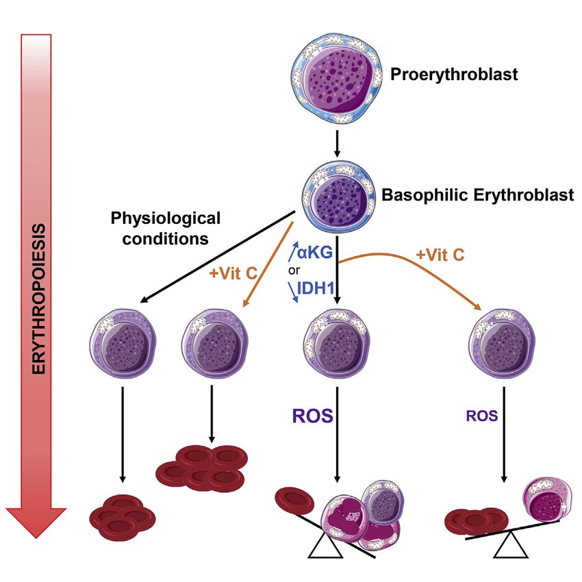
ll
Article                             OPEN ACCESS

                                   (legend on next page)

          Cell Reports 34, 108723, February 2, 2021 7
ll
OPEN ACCESS Article
The ensemble of these data strongly suggests that the impact IDH1 activity regulates mitochondrial activity in
of vitamin C is due to its alteration of the redox state of the eryth- differentiating erythroblasts
roblast. However, as autophagy is also a critical regulator of The data presented above demonstrated that conditions that in-
enucleation (Keerthivasan et al., 2011), we evaluated the impact crease mitochondrial activity in human erythroblasts, including
of vitamin C following Spautin1-mediated inhibition of autophagy ectopic aKG and MitoPQ, inhibit terminal erythroid differentia-
(Liu et al., 2011). As expected, Spautin significantly inhibited tion. Under physiological conditions, aKG is mobilized in the
enucleation (p < 0.01), but vitamin C did not rescue this defect mitochondria, where it can be converted to succinyl-CoA by
(Figure S5A), highlighting an autophagy-independent role of aKGDH, producing ROS and the NADH that provides electrons
vitamin C in erythroid differentiation. Iron and its uptake as difer- for the ETC (Figure 6A). aKG can be generated from isocitrate
ric transferrin by the transferrin receptor CD71 are also critical for in the mitochondria by IDH2/IDH3 and interconverted in the cyto-
erythroid differentiation (Kautz and Nemeth, 2014), and we there- plasm by IDH1. Interestingly, transcripts of all three enzymes are
fore evaluated a potential crosstalk between iron and vitamin C. significantly reduced during erythroid differentiation, potentially
Interestingly, in the absence of transferrin, vitamin C did not in- suggesting a negative role for these pathways in late-stage
crease enucleation in aKG-treated erythroblasts, but its erythroblasts (Figure S6A). As mitochondrial biomass is mark-
presence (50 or 200 mg/mL) positively affected enucleation (Fig- edly decreased during erythroid differentiation, we evaluated
ure S5B). Thus, vitamin C and other ROS scavengers decrease the role of the cytoplasmic IDH enzyme, IDH1, in erythroid differ-
the redox state of erythroblasts, promoting enucleation under entiation by an shIDH1-mediated approach. Using two indepen-
conditions of aKG-induced oxidative stress in a transferrin- dent short hairpin RNAs (shRNAs), shIDH1-1 and shIDH1-2, we
dependent manner. detected a 36% ± 7% and 54% ± 5% reduction, respectively,
and as such used shIDH1-2 in the differentiation experiments
Vitamin C accelerates the erythroid differentiation of (Figure S6B).
cord blood, peripheral blood, and BM progenitor cells Importantly, IDH1 knockdown did not inhibit erythroid differen-
The ability of vitamin C to rescue erythroid differentiation under tiation, as monitored by the generation of CFU-E, GlyA+ erythro-
conditions of augmented oxidative stress led us to evaluate its po- blasts or upregulation of the CD36 or CD71 erythroid markers (Fig-
tential to directly modulate erythroid differentiation of CD34+ he- ures 6B, S6C, and S6D). However, downregulating IDH1 resulted
matopoietic stem and progenitor cells (HSPCs) from different in a decreased expansion (Figure S6E) and significantly increased
sources. CD34+ progenitors from cord blood (CB), adult periph- mitochondrial superoxide production, mitochondrial biomass,
eral blood (PB), and BM were directly differentiated with rEPO. and polarization (p < 0.01; Figures 6C and S6F). In accord with
While BM-derived progenitors differentiated much more rapidly these data, IDH1 knockdown augmented the OXPHOS capacity
than CB- and PB-derived progenitors to an erythroid-committed of the cells, with a 3.0- and 3.7-fold increase in basal and maximal
CFU-E (CD34CD36+), vitamin C did not alter the erythroid pro- OCR levels, respectively (shIDH1-2 construct) (Figure 6D).
genitor continuum (Figure S5C). In agreement with previous Notably, the function of IDH1 appears to be substantially more
data (Yan et al., 2018), CB progenitors exhibited a delayed gener- critical for the differentiation of erythroid as compared to non-
ation of GlyA+ erythroblasts compared to PB and BM progenitors erythroid progenitors. The percentages of progenitors transduced
(Figure 5A). Notably, vitamin C significantly increased the percent- with an scramble shRNA (shControl) or shIDH1 lentiviral vector
ages of GlyA+ cells (p < 0.05) and increased maturation to a harboring the GFP reporter gene remained approximately stable
GlyA+CD105 state by 2.1- ± 0.5-fold (p < 0.01, Figure 5A), over a 6-day period, with 47%–57% GFP+ cells, in the absence
without altering cell growth (Figure S5D). Furthermore, vitamin C of rEPO. These cells had a myeloid phenotype as shown by a
resulted in an enhanced enucleation of erythroid progenitors CD11b+, CD33+, CD13+ phenotype (Figure S6G). However,
generated from these three sources (Figure 5B). Thus, vitamin C following rEPO-mediated erythroid induction, the percentages of
significantly accelerates erythroid differentiation and enucleation shControl cells decreased by 24%, while the percentages of
of human CD34+ progenitors, irrespective of their source of origin. shIDH1-transduced cells decreased by 56% (Figure 6E).
Figure 3. Mitochondrial superoxide production impedes erythroid enucleation
(A) Schematic of the generation of mitochondrial reactive oxygen species (ROS) via the conversion of aKG to succinate, of which superoxide is a major source.
(B) The impact of aKG (3.5 mM) on mitochondrial superoxide and mitochondrial ROS production in erythroblasts was evaluated at day 10 of erythroid differ-
entiation as a function of MitoSOX and MitoROS, respectively. Representative histograms are shown (left). Staining in the presence of aKG was normalized to
control conditions, and relative median fluorescence intensities (MFIs) are presented (n = 15–16, right).
(C) Schematic of MitoParaquat (MitoPQ) cycling within the mitochondria; mitoPQ2+ is reduced to the radical monocation at the flavin site of complex I, and the
monocation then reacts with O2 to generate superoxide (O2) (Robb et al., 2015).
(D) Differentiating erythroblasts were treated with MitoPQ (50 mM) at day 7 of differentiation, and representative histograms of mitochondrial superoxide, ROS,
and biomass at day 10 are presented (top). MFIs are presented relative to control conditions (n = 5–6, bottom).
(E) The impact of MitoPQ on enucleation was evaluated at day 10 by Syto16 staining, and representative dot plots are presented (top). Percent enucleation in nine
independent experiments is shown (bottom).
(F) The impact of MitoPQ on early basophilic erythroblasts as compared to late orthochromatic erythroblasts was determined by FACS sorting erythroblast
populations as a function of their GLUT1/CD49d profiles (left), and representative cytospins of the sorted subsets are shown (middle). Early basophilic and late
orthochromatic subsets were then cultured for 72 h or 24 h, respectively, in the absence () or presence (+) of MitoPQ, and enucleation was evaluated (right).
Enucleation was quantified relative to control conditions (n = 3).
ns, nonsignificant; *p < 0.05; **p < 0.01; ***p < 0.001.
8 Cell Reports 34, 108723, February 2, 2021ll
Article OPEN ACCESS
(legend on next page)
Cell Reports 34, 108723, February 2, 2021 9ll
OPEN ACCESS Article
To better understand the negative selection of erythroid pro- IDH1 was downregulated at day 4 post-rEPO-induced differentia-
genitors with downregulated IDH1, we performed metabolomic tion (Figure S7A). Consistent with an impact of IDH1 on the redox
and flux experiments following IDH1 knockdown. First, we found state of the cell, both aKG and DHA exacerbated the effect of
that at day 4 of IDH1 downregulation (~50%; Figure S6B), there IDH1 knockdown on erythroblast enucleation, from 47% ± 8%
was a mean decrease of aKG and citrate of 43% and 21%, to 27% ± 8% and 24% ± 4%, respectively (p < 0.01 and p <
respectively (Figure 6F). These data highlight the importance of 0.05; Figure 7C). Most importantly, vitamin C significantly
the IDH1 enzyme in the generation of aKG and its interconver- increased enucleation under these conditions by 35% ± 13%
sion to citrate. Furthermore, they demonstrate the role of gluta- (p < 0.05, Figure 7D). Intriguingly, other ROS scavengers,
mine metabolism in the generation of these intermediates in early including NAC, GSH, and Trolox, did not increase enucleation,
erythroid progenitors (day 4); a mean of 75.1% and 69.0% of even though they were present throughout the differentiation
aKG and citrate harbored 13C carbons from glutamine, respec- period (Figure S7B). Importantly, vitamin C, but not NAC, GSH,
tively (Figure 6G). Several groups have also found that the or Trolox, decreased mitochondrial superoxide generation (p <
relative ratio of aKG to citrate regulates differentiation, either 0.05; Figure S7C). Thus, collectively, these data show that
by inhibiting (Carey et al., 2015; Vardhana et al., 2019) or priming IDH1-induced decreases in mitochondrial OXPHOS are required
(TeSlaa et al., 2016; Tischler et al., 2019) stem cell differentiation. for the later stages of terminal erythroid differentiation and enucle-
In this regard, it is interesting to note that erythroid differentiation ation. However, under conditions of decreased IDH1 expression,
resulted in the loss of both aKG and citrate between days 4 and 7 this negative effect can be counteracted by a vitamin-C-mediated
of differentiation, with a mean loss of 87% and 68%, respectively modulation of the intracellular redox balance.
(n = 2 biological experiments performed in triplicate; Figure 6G). As aKG and vitamin C act as cofactors in supporting the diox-
These data point to a greater loss of aKG than citrate, consistent ygenase activity of TET2, it was important to assess whether the
with a negative role of high aKG levels in late erythroid differen- effects that we detected here were coupled to their impact on
tiation (Figure 2). Furthermore, following IDH1 downregulation, TET2 activation (Inoue et al., 2016). However, if aKG served
TCA intermediates were maintained at a higher level, with de- only to support the dioxygenase activity of TET2, then its pres-
creases in aKG and citrate of only 31% and 36%, respectively ence would be expected to augment erythroid differentiation,
(Figure 6G). Thus, the premature inhibition of IDH1 alters the while our data showed the opposite effect (Figure 2). To directly
physiological loss of TCA-cycle intermediates upon erythroid address this point and evaluate the role of an aKG/TET2 as
differentiation. compared to an aKG/IDH1 axis, we downregulated TET2 by an
shRNA approach (Yan et al., 2017). TET2 levels decrease with
Vitamin C rescues terminal erythroid differentiation in differentiation (p < 0.0001; Figure S7D), but mRNA levels were
the absence of IDH1 further diminished by a mean of 55% by shRNA-mediated
Under conditions of IDH1 downregulation, mitochondrial meta- knockdown (Figure S7E). While this downregulation resulted in
bolism was augmented, as shown by increased superoxide pro- a significant delay in the generation of GlyA+ terminal erythro-
duction and OXPHOS (Figures 6C and 6D). On the basis of these blasts (p < 0.0001; Figure S7F), it did not delay enucleation
data, we hypothesized that IDH1 knockdown would attenuate when the shRNA-mediated knockdown of TET2 was performed
late-stage erythroid differentiation and enucleation, and this was at day 4 of differentiation (Figure 7E). Moreover, enucleation in
indeed the case. Following transduction of CD34+ HSPCs with a shTET2-treated erythroid progenitors was not augmented by
vector harboring an shIDH1 RNA, the emergence of Glut1+CD49 vitamin C (Figure 7E). These data strongly imply distinct roles
orthochromatic erythroblasts was significantly decreased for IDH1 and TET2 in regulating enucleation of late-stage
(shIDH1-2 construct, p < 0.01; Figure 7A). Furthermore, enucle- erythroid progenitors.
ation was decreased by 59% ± 2% (shIDH1-2 construct, p < Mutations in IDH1/IDH2 are strongly associated with myelo-
0.0001; Figure 7B), and this effect was detected even when dysplastic syndromes (MDSs) that are characterized by
Figure 4. Antagonistic roles of dehydroascorbic acid and vitamin C on mitochondrial oxidative stress and erythroblast enucleation
(A) Schematic of the interconversion between dehydroascorbic acid (DHA) and ascorbic acid (vitamin C) via a two-electron reduction/oxidation process,
respectively.
(B) The impact of DHA (1 mM) on mitochondrial superoxide production and biomass in late erythroblasts was evaluated between days 7 and 10 after EPO in-
duction, and representative histograms of MitoGreen and MitoSOX staining, respectively, are shown (top). Quantification relative to control conditions is shown
(n = 10, bottom).
(C) Enucleation was evaluated at day 10 after EPO induction in the absence () or presence (+) of DHA (days 7–10), and representative dot plots are shown (top).
Percent enucleation in 14 independent experiments is presented (bottom).
(D) The impact of the stable L-ascorbic acid (AA) 2-phosphate derivative of vitamin C (100 mM) was evaluated on mitochondrial superoxide generation (days 7–10),
and MFIs were quantified relative to control conditions (n = 15, bottom).
(E) The impact of vitamin C on aKG-mediated increases in MitoSOX, MitoGreen, and MitoRed staining was evaluated by flow cytometry, and relative MFIs are
presented (n = 18–20).
(F) The percentages of cells in S-G2/M of the cell cycle were evaluated in the indicated conditions by phosphatidylinositol (PI) staining (n = 4, left). The percentages
of erythroid progenitors in S-G2/M were evaluated relative to control conditions, and means ± SEM are presented (right).
(G) Enucleation was evaluated in the presence or absence of vitamin C and aKG. Representative histograms (left) as well as changes in percent enucleation are
presented (n = 18, right).
*p < 0.05; **p < 0.01; ***p < 0.001;****p < 0.0001.
10 Cell Reports 34, 108723, February 2, 2021ll
Article OPEN ACCESS
(legend on next page)
Cell Reports 34, 108723, February 2, 2021 11ll
OPEN ACCESS Article
ineffective erythropoiesis (reviewed by Bejar and Steensma, scavengers, including NAC, GSH, vitamin E, and vitamin C,
2014; Fenaux et al., 2014). The anemia that dominates the early rescued the aKG-mediated decrease in enucleation, but only
course of MDS is associated with an aberrant erythroid differen- vitamin C rescued enucleation following IDH1 downregulation.
tiation marked by the generation of multinucleated erythroblasts This is critical in the context of human erythroid differentiation,
(Abdel-Wahab and Levine, 2013). Notably, we found that IDH1 where the absence of ascorbate synthesis, resulting in 1 mg/
knockdown increased the percentages of erythroblasts with kg recommended exogenous supplements for humans (Pauling,
abnormal multinucleated erythroblasts from 10% ± 1% to 23% 1970), is significantly lower than the hepatic production rate of
± 2% (p < 0.05; Figure 7F). Moreover, vitamin C markedly 200 mg/kg/day in vitamin-C-synthesizing animals (Chatterjee,
decreased the generation of these abnormal erythroblasts, not 1973; Stone, 1979). Thus, scavenging by vitamin C is likely to
only in IDH1-knockdown erythroblasts but also in normal eryth- be suboptimal. Indeed, we found that a stable vitamin C analog
roblasts, to 4% ± 0.4% and 4% ± 0.2%, respectively (p < 0.05 rescued human terminal erythroid differentiation and enucleation
and p < 0.01; Figure 7F). These data suggest that the multinucle- in all tested conditions of oxidative stress.
ated erythroblasts detected in MDS patients may be associated Ascorbate, transported by the SVCT1/SLC23A1 and SVCT2/
with an increased mitochondrial metabolism. Vitamin C, a redox SLC23A2 solute carriers, regulates whole-body homeostasis of
scavenger, significantly attenuated the generation of these vitamin C and protection to oxidative stress, respectively (Savini
abnormal cells. et al., 2008; Tsukaguchi et al., 1999; Wilson, 2005). While vitamin
C has been shown to enhance erythropoiesis in hemodialysis pa-
DISCUSSION tients with refractory anemia (Attallah et al., 2006; Gastaldello
et al., 1995; Seibert et al., 2017; Sirover et al., 2008), other
The generation of ROS and its intricate relationship with cellular studies have found that ascorbate induces hemolysis of mature
redox homeostasis is a critical regulator of stem cell homeosta- erythrocytes (Zhang et al., 2016). These apparent discrepancies
sis. Notably, low ROS levels, associated with low mitochondrial are likely due to the oxidation state of vitamin C. The loss of a sin-
biogenesis and activity, are necessary for HSC quiescence and gle electron from ascorbate results in the generation of an unsta-
self-renewal (Filippi and Ghaffari, 2019; Jang and Sharkis, ble ascorbate radical, which can then lose a second electron to
2007; Qian et al., 2016; Suda et al., 2011; Takubo et al., 2013; generate DHA (Padayatty and Levine, 2016). Unlike vitamin C,
Tan and Suda, 2018; Vannini et al., 2016; Yu et al., 2013). the two-electron oxidized DHA intermediate is transported by
Conversely, HSC division and differentiation occur under condi- the GLUT1 glucose transporter (Bianchi and Rose, 1986;
tions that promote mitochondrial biogenesis and OXPHOS McNulty et al., 2005; Rumsey et al., 1997, 2000; Vera et al.,
(Chen et al., 2008; Mantel et al., 2010; Maryanovich et al., 1993). Thus, cellular uptake of the reduced and oxidized forms
2015; Yu et al., 2013). In the context of erythropoiesis, it has of vitamin C is regulated, at least in part, by the expression of
been shown that early erythroid development requires induction their specific transporters. SVCT2 is expressed at very high
of mitochondrial ROS, a consequence of OXPHOS (Liu et al., levels on HSCs, with levels decreasing as a function of erythroid
2017; Zhao et al., 2016). Here, we show that glutamine meta- development and differentiation (Agathocleous et al., 2017).
bolism, via the anaplerotic contribution of aKG into the TCA cy- Conversely, GLUT1 is upregulated during human terminal
cle, directly increases OXPHOS in human erythroid precursors. erythroid differentiation (Montel-Hagen et al., 2008b; Mueckler,
This increased OXPHOS promoted erythroid commitment and 1994), promoting DHA transport (May, 1998; Montel-Hagen
early erythropoiesis, but terminal erythroid differentiation was et al., 2008b, 2009). Thus, during intermediate steps of HSPC
dependent on the suppression of TCA-cycle-linked mitochon- development, both ascorbate and DHA can potentially be trans-
drial networks. Elevated intracellular aKG levels, with associated ported into the cell.
increased OXPHOS and ROS, inhibited late-stage erythroid One of the difficulties in studying ascorbate function is its sta-
maturation and enucleation. bility. Previous research showed that freshly prepared minimum
Mechanistically, we identify the catalysis of aKG by the cyto- essential medium a (aMEM) culture media, but not liquid aMEM,
plasmic IDH1 enzyme as a critical step in erythropoiesis; the supported T cell differentiation from hematopoietic progenitors;
negative repercussions of IDH1 downregulation in terminal the difference between these formulations was due to the lability
erythroid differentiation was further exacerbated by elevated of vitamin C in the latter (Manning et al., 2013). Furthermore,
aKG levels. Furthermore, the impact of a mitochondria-targeted several groups found that the negative impact of vitamin C on
redox cycler, MitoPQ (Robb et al., 2015), highlights the impor- the growth of cancer cells was actually due to the GLUT1-medi-
tance of mitochondrial superoxide as a specific inhibitor of eryth- ated uptake of DHA, resulting in the accumulation of ROS and
roblast maturation and enucleation. Interestingly, all tested ROS subsequent cell death (Yun et al., 2015; Zhang et al., 2016).
Figure 5. Vitamin C promotes erythroid commitment of EPO-stimulated progenitors from CB, BM, and PB and significantly accelerates
erythroid maturation
(A) CD34+ HSPCs were isolated from CB, PB, and BM. Progenitors were differentiated with EPO in the absence or presence of vitamin C (100 mM) and eryth-
ropoiesis monitored as a function of CD105/GlyA profiles. Representative profiles at days 6 (top left) and 7 (bottom left) of differentiation are presented.
Quantification of GlyA+ cells at day 6 (n = 5, top right) and GlyA+CD105 cells at day 7 (n = 5, bottom right) is presented.
(B) The impact of vitamin C on enucleation was evaluated in CB, PB, and BM HSPCs, and representative plots at day 17 are presented (top). Quantification of
enucleation at days 15, 17, and 19 is presented (n = 6, bottom).
*p < 0.05; **p < 0.01; ***p < 0.001; ****p < 0.0001.
12 Cell Reports 34, 108723, February 2, 2021ll
Article OPEN ACCESS
(legend on next page)
Cell Reports 34, 108723, February 2, 2021 13ll
OPEN ACCESS Article
Indeed, we similarly found that the standard labile form of L- conditions of altered oxidative metabolism. Increases in intracel-
ascorbate markedly decreased erythroblast maturation and lular aKG and decreases in cytoplasmic IDH1 both augmented
enucleation, even in the absence of ascorbate oxidase. Howev- oxidative stress and diminished erythroid maturation. IDH1,
er, under conditions where erythroid differentiation was per- like Tet2, is mutated in a high percentage of MDS patients (Kos-
formed in the presence of the stable ascorbate 2-phosphate mider et al., 2010; Wang et al., 2017), with anemia dominating the
analog (Hata and Senoo, 1989; Frikke-Schmidt and Lykkesfeldt, early course of disease. Moreover, recent studies have high-
2010; Takamizawa et al., 2004), erythroid differentiation was lighted the potential for vitamin C to promote the differentiation
enhanced. Thus, care must be taken in evaluating studies using of IDH1-mutated AML progenitors (Mingay et al., 2018). Our
the conventional labile ascorbate, as it may not be maintained in data show that even in the absence of mutation, low levels of
a reduced state. wild-type IDH1 suppress late erythroid differentiation. While
Ascorbic acid has also gained prominence as an anti-cancer the specific role of IDH1 in OXPHOS has not yet been fully delin-
therapeutic modality because of its ability to alter the activity of eated, it is interesting to note that IDH1 downregulation in
the Tet2 demethylase (Shenoy et al., 2018). Loss of function mu- neuronal cells resulted in an increase in ROS (Calvert et al.,
tations in TET2 are one of the most frequent events in myeloid 2017). Furthermore, mutation of IDH1, but not IDH2, has been
dysplasia and leukemias (Bejar et al., 2011; Busque et al., found to increase OXPHOS in transformed cells (Grassian
2012; Delhommeau et al., 2009; Kosmider et al., 2011). The initi- et al., 2014), and wild-type IDH1 regulates the reductive carbox-
ation of demethylation by the TET family of dioxygenases occurs ylation of aKG for de novo lipogenesis (Calvert et al., 2017; Met-
via the introduction of the intermediate mark 5-hydroxymethyl- allo et al., 2011).
cytosine (5hmC), and this function is dependent on several co- IDH-regulated aKG production has also been shown to regu-
factors, including ascorbate, aKG, ferrous iron, and oxygen late cell fate and differentiation by modulating epigenetic marks
(Chen et al., 2013; Dang et al., 2009). Indeed, recent studies via Tet2-induced changes in global histone and DNA demethyla-
have shown that ascorbate is able to compensate for TET2 defi- tion (Carey et al., 2015; TeSlaa et al., 2016; Tischler et al., 2019;
ciency and suppress leukemogenesis, albeit via both TET2- Xiao et al., 2012). Notably, under the conditions of erythroid dif-
dependent and TET2-independent mechanisms (Agathocleous ferentiation studied here, aKG levels attenuated terminal
et al., 2017; Cimmino et al., 2017). A crosstalk between ascor- erythroid differentiation and late-stage erythropoiesis, while a
bate and TET2 in the metabolic status of a hematopoietic pro- second TET2 cofactor, ascorbate, exhibited an opposing effect,
genitor is suggested by the finding that wild-type TET2 protects enhancing and accelerating terminal differentiation. Other
murine erythroblasts from oxidative stress (Guo et al., 2017). agents, such as oxidized ascorbate and MitoPQ, attenuated
Moreover, downregulation of TET2 in human erythroid progeni- erythroid differentiation by inducing oxidative stress. Further-
tors negatively regulated human erythropoiesis in a manner more, while TET2 downregulation delayed early erythroblast dif-
that was independent of any detectable alterations in 5-methyl- ferentiation, it did not negatively impact terminal differentiation
cytosine (5mC) levels (Qu et al., 2018; Yan et al., 2017). These and enucleation. Together, these data strongly suggest comple-
data are in agreement with independent studies showing that mentary roles for TET2 and IDH1, with the latter playing a critical
terminal erythroid differentiation requires that erythroblasts be role in late-stage human erythropoiesis via its impact on redox
protected from oxidative stress (Case et al., 2013; Filippi and homeostasis.
Ghaffari, 2019; Hyde et al., 2012; Xu et al., 2019; Zhao et al., These data may promote mechanistic insights into inherited
2016). hematologic disease. Like MDS patients, a feature that charac-
Our study highlights the importance of ascorbate in regulating terizes patients with congenital dyserythropoietic anemia is the
the redox state of human erythroid progenitors, especially under presence of multinucleated or multi-lobulated erythroblasts
Figure 6. IDH activity regulates mitochondrial superoxide production and OXPHOS in differentiation erythroblasts
(A) Schematic representation of the catalytic reactions regulating glutamine catabolism. Glutamine is interconverted to glutamate and cytosolic aKG (aKGc).
Isocitrate dehydrogenase 1 (IDH1), a cytosolic enzyme, catalyzes the production of isocitrate from aKG, promoting both forward and reverse reactions. Con-
version of mitochondrial aKG (aKGm) to succinate results in the generation of ROS.
(B) The expression of GlyA was monitored in shControl- and shIDH1-transduced cells at day 7 day of differentiation, and representative histograms (gray, isotype
control; black, specific staining) are shown (top). GlyA expression in cells transduced with one of two different shIDH1 clones (shIDH1-1 and shIDH1-2) was
evaluated, and relative levels are presented (n = 4, bottom).
(C) The impact of IDH1 downregulation on MitoSOX was evaluated at day 10. Representative histograms (top left) and quantification of relative MFIs (n = 5) are
presented (bottom left). MitoTracker Deep Red (top right) and MitoTracker Red (bottom right) staining in shIDH1-transduced erythroblasts relative to control
conditions, are presented (n = 6).
(D) OCR was determined in FACS-sorted shControl- as well as shIDH1-transduced erythroblasts at day 10 of differentiation as indicated (n = 6, top). An OCR/
extracellular acidification rate (ECAR) energy map is presented (bottom).
(E) The evolution of shControl- and sh-IDH1-transduced progenitors was monitored as a function of GFP expression at days 3, 6, and 10 of differentiation in the
absence () or presence (+) of EPO (n = 3).
(F) Total levels of aKG and citrate were evaluated by high-performance liquid chromatography-mass spectrometry (HPLC-MS) at day 4 of erythroid differentiation
in shControl- and shIDH1-transduced progenitors, and mean levels are shown (n = 3 independent experiments, with statistical difference evaluated by a one-
tailed t test).
(G) The isotopologues of aKG and citrate derived from [13C515N2]Gln are presented from one experiment with three replicates for day 4 and two replicates for day
7.
ns, not significant; *p < 0.05; **p < 0.01; ***p < 0.001; ****p < 0.0001.
14 Cell Reports 34, 108723, February 2, 2021ll
Article OPEN ACCESS
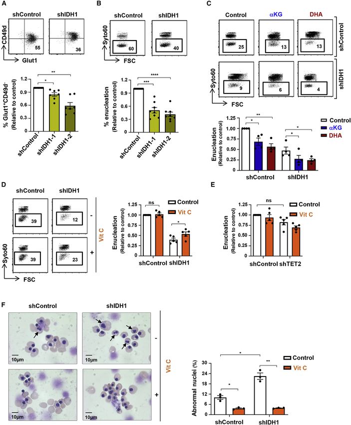
Figure 7. Vitamin C rescues dyserythropoiesis due to IDH1 downregulation
(A) Late-stage erythroblast differentiation was monitored as a GLUT1+CD49d phenotype, and representative dot plots are presented (top). The percentages of
GLUT1+CD49d erythroblasts were evaluated following transduction with shIDH1-1 or shIDH1-2 vectors, and mean levels ± SEM relative to control cells are
presented (n = 7, bottom).
(legend continued on next page)
Cell Reports 34, 108723, February 2, 2021 15ll
OPEN ACCESS Article
(Crookston et al., 1969; Goasguen et al., 2018; Iolascon et al., gram at Northwell Health for providing access to BM samples. We thank the
1996). IDH1 knockdown resulted in a significant augmentation Unité of Thérapie Cellulaire and the hospital clinics of Clémentville and
Saint-Roch (Montpellier, France) for their generous efforts in providing access
in the percentage of abnormal multinucleated orthochromatic
to CB samples. We also thank Catherine Winchester for critical reading of the
erythroblasts, and even more importantly, ascorbate reduced manuscript. P.G.-M. was supported by a fellowship from the Clarin-COFUND
these levels back to baseline. Thus, these data provide the first EU Program (Principado de Asturias, Spain), M.R. was supported by a fellow-
evidence that the abnormal erythroblast morphology observed ship from GR-Ex, and L.O. was supported by Ligue and ARC fellowships. S.T.
in diverse dyserythropoietic diseases may be coupled to oxida- is supported by funding from Cancer Research UK (C596/A17196 and
tive stress. Furthermore, our data suggest that antioxidant A23982). This work was supported by generous funding from the NIH (grant
DK32094 to P.G.G., N.M., S.K., and N.T. and grants HL144436 and
treatment of these disorders in humans, a species with subopti-
HL152099 to L.B.); FRM, ARC, and French national (ANR) research grants
mal redox capacity (Johnson et al., 2008), with stable ascorbate
(NutriDiff); and the French laboratory consortiums (Labex) EpiGenMed and
analogs could potentially restore redox homeostasis, quenching GR-Ex. N.T. is presently supported by the NCI Intramural Program.
ROS and subsequently rescuing defective late-stage
erythropoiesis. AUTHOR CONTRIBUTIONS
P.G.-M., M.R., S.K., and N.T. conceived the study; P.G.-M., M.R., H.Y., R.D.,
STAR+METHODS
J.P., L.O., S.T., V.D., C.M., M.S., V.S.Z., L.B., N.M., S.K., and N.T. were
involved in study design; and P.G.-M., M.R., H.Y., R.D., J.P., L.O., S.T.,
Detailed methods are provided in the online version of this paper C.M., M.D., A.-S.D., I.P., X.Q., P.-N.B., and S.K. performed experiments. All
and include the following: authors participated in data analysis. P.-G.M., S.K., and N.T. wrote the manu-
script with important critical input from M.R., H.Y., L.O., S.T., X.A., V.D., M.S.,
d KEY RESOURCES TABLE V.S.Z., P.G.G., L.B., and N.M.
d RESOURCE AVAILABILITY
B Lead contact DECLARATION OF INTERESTS
B Materials availability
M.S. and S.K. are inventors on a patent describing the use of a ligand for eval-
B Data and code availability
uation of GLUT1 expression (N.T. gave up her rights). M.S. is a co-founder of
d EXPERIMENTAL MODELS AND SUBJECT DETAILS METAFORA Biosystems, focusing on metabolite transporters under physio-
+
B CD34 cell isolation and ex vivo differentiation assays. logical and pathological conditions, and is head of the scientific board.
d METHODS DETAIL
B Cytospin preparation Received: June 10, 2020
B Flow cytometry Revised: November 26, 2020
+
B Virus production and transduction of CD34 progenitor Accepted: January 12, 2021
Published: February 2, 2021
cells
B Quantitative real time PCR REFERENCES
B Mass spectrometry (LC-MS)
B Seahorse analysis Abdel-Wahab, O., and Levine, R.L. (2013). Mutations in epigenetic modifiers in
B Glutamine uptake assays the pathogenesis and therapy of acute myeloid leukemia. Blood 121, 3563–
3572.
d QUANTIFICATION AND STATISTICAL ANALYSIS
Adam-Vizi, V., and Tretter, L. (2013). The role of mitochondrial dehydroge-
nases in the generation of oxidative stress. Neurochem. Int. 62, 757–763.
SUPPLEMENTAL INFORMATION
Agathocleous, M., Meacham, C.E., Burgess, R.J., Piskounova, E., Zhao, Z.,
Supplemental Information can be found online at https://doi.org/10.1016/j. Crane, G.M., Cowin, B.L., Bruner, E., Murphy, M.M., Chen, W., et al. (2017).
celrep.2021.108723. Ascorbate regulates haematopoietic stem cell function and leukaemogenesis.
Nature 549, 476–481.
ACKNOWLEDGMENTS An, X., Schulz, V.P., Li, J., Wu, K., Liu, J., Xue, F., Hu, J., Mohandas, N., and
Gallagher, P.G. (2014). Global transcriptome analyses of human and murine
We thank all members of our laboratories for discussions and scientific terminal erythroid differentiation. Blood 123, 3466–3477.
critique. We are grateful to Myriam Boyer of Montpellier Rio Imaging for sup- Ashley, R.J., Yan, H., Wang, N., Hale, J., Dulmovits, B.M., Papoin, J., Olive,
port in cytometry experiments. We are indebted to the Tissue Donation Pro- M.E., Udeshi, N.D., Carr, S.A., Vlachos, A., et al. (2020). Steroid resistance in
(B) Enucleation of shControl- and shIDH1-transduced erythroblasts was assessed on GFP+ cells at day 14, and representative histograms are presented (top).
Enucleation levels, relative to control cells, were quantified (n = 8, bottom).
(C) The impact of aKG and DHA on erythroblast enucleation following IDH1 knockdown was evaluated between days 7 and 10, and representative histograms are
presented (top). Enucleation relative to control conditions was quantified (n = 4, bottom).
(D) The impact of vitamin C on enucleation of control and IDH1-downregulated erythroblasts was evaluated between days 7 and 10. Representative histograms
(top) and quantifications (bottom) are presented (n = 5). Changes in mitochondrial superoxide were evaluated by MitoSOX staining, and MFIs are presented (n =
4).
(E) TET2 was downregulated in erythroid progenitors by shTET2 lentivirus (day 4), and enucleation was monitored at day 10 in the presence or absence of vitamin
C. Means ± SEM are presented (n = 5, one-way ANOVA with Tukey’s post hoc test).
(F) Nuclear morphology of orthochromatic erythroblasts differentiated from shControl- and shIDH1-transduced progenitors in the absence or presence of vitamin
C was monitored. Representative cytospins are shown (left), and the quantification of orthochromatic erythroblasts with abnormal nuclei was determined by
counting 500–1,000 cells (n = 3, right).
ns, not significant; *p < 0.05; **p < 0.01; ***p < 0.001; ****p < 0.0001.
16 Cell Reports 34, 108723, February 2, 2021You can also read