ATR is essential for preservation of cell mechanics and nuclear integrity during interstitial migration - Nature
←
→
Page content transcription
If your browser does not render page correctly, please read the page content below
ARTICLE
                  https://doi.org/10.1038/s41467-020-18580-9                 OPEN

                  ATR is essential for preservation of cell mechanics
                  and nuclear integrity during interstitial migration
                  Gururaj Rao Kidiyoor 1, Qingsen Li1, Giulia Bastianello1, Christopher Bruhn 1, Irene Giovannetti1,
                  Adhil Mohamood1, Galina V. Beznoussenko1, Alexandre Mironov1, Matthew Raab2, Matthieu Piel 2,
                  Umberto Restuccia1, Vittoria Matafora1, Angela Bachi 1, Sara Barozzi1, Dario Parazzoli 1, Emanuela Frittoli1,
                  Andrea Palamidessi 1, Tito Panciera3, Stefano Piccolo1,3, Giorgio Scita 1,4, Paolo Maiuri 1,
                  Kristina M. Havas1, Zhong-Wei Zhou5,6, Amit Kumar 7, Jiri Bartek8,9, Zhao-Qi Wang5,10 & Marco Foiani 1,4 ✉
1234567890():,;

                  ATR responds to mechanical stress at the nuclear envelope and mediates envelope-
                  associated repair of aberrant topological DNA states. By combining microscopy, electron
                  microscopic analysis, biophysical and in vivo models, we report that ATR-defective cells
                  exhibit altered nuclear plasticity and YAP delocalization. When subjected to mechanical
                  stress or undergoing interstitial migration, ATR-defective nuclei collapse accumulating
                  nuclear envelope ruptures and perinuclear cGAS, which indicate loss of nuclear envelope
                  integrity, and aberrant perinuclear chromatin status. ATR-defective cells also are defective in
                  neuronal migration during development and in metastatic dissemination from circulating
                  tumor cells. Our findings indicate that ATR ensures mechanical coupling of the cytoskeleton
                  to the nuclear envelope and accompanying regulation of envelope-chromosome association.
                  Thus the repertoire of ATR-regulated biological processes extends well beyond its canonical
                  role in triggering biochemical implementation of the DNA damage response.

                  1 IFOM- FIRC Institute of Molecular Oncology, Milan, Italy. 2 Institut Curie/CNRS, Paris, France. 3 University of Padova, Padova, Italy. 4 University of Milan,

                  Milan, Italy. 5 Leibniz Institute on Aging, Fritz Lipmann Institute, Jena, Germany. 6 School of Medicine, Sun Yat-Sen University, Shenzhen, China. 7 Genome
                  and Cell Integrity Lab, CSIR-Indian Institute of Toxicology Research, Lucknow, India. 8 Danish Cancer Society Research Center, Copenhagen, Denmark.
                  9 Karolinska Institute, Stockholm, Sweden. 10 Friedrich-Schiller University, Jena, Germany. ✉email: marco.foiani@ifom.eu

                  NATURE COMMUNICATIONS | (2020)11:4828 | https://doi.org/10.1038/s41467-020-18580-9 | www.nature.com/naturecommunications                                     1
ARTICLE                                                                 NATURE COMMUNICATIONS | https://doi.org/10.1038/s41467-020-18580-9

M
           echanical properties of the nucleus and nuclear                   invaginations)13, associated with condensed chromatin and/or
           mechanosensing affects genome integrity, nuclear archi-           nucleoli (Fig. 1f and Supplementary Fig. 1n–r). NE invaginations
           tecture, gene expression, cell migration, and differenti-         also associated with nucleoli forming nucleolar canals that
ation1,2. The physical properties of the nucleus are conveniently            represent intermediates in rRNA export through the NE14,15
modulated following the inputs from the cell microenvironment                (Fig. 1f and Supplementary Fig. 1r). We also found, within the
or from chromatin dynamics. The nuclear envelope (NE) plays a                nucleus, inner membrane invaginations/fragments attached to
critical role in this process by connecting the cytoskeleton and the         chromatin and micronuclei (Supplementary Fig. 1r, s).
chromatin1,2.
   Ataxia Telangiectasia and Rad3-related protein (ATR) regulates            ATR depletion alters nuclear mechanical properties. NE
the DNA damage response (DDR)3 and protects genome integrity by              abnormalities can affect the mechanical properties of the nucleus1,16.
regulating multiple pathways4. ATR mutations cause the Seckel                When we measured the elastic modulus of ATR-depleted cells by
syndrome, an autosomal recessive disorder characterized by growth            atomic force microscopy (AFM)17, we found a reduced elasticity
retardation, dwarfism, and microcephaly with mental retardation5.             compared to controls (Fig. 2a). As the nucleus is the stiffest orga-
We previously reported that ATR directly senses mechanical stress at         nelle in the cell18, we performed the same analysis on isolated ATR-
the NE/chromatin interface and facilitates release of chromatin from         defective nuclei and found, again, a reduced elasticity, compared to
the NE6. This possibility is supported by the fact that ATR comprises        controls (Fig. 2b). Acute treatment with ATR inhibitors for 4 h did
HEAT (huntingtin, elongation factor 3, A subunit of protein phos-            not alter nuclear stiffness (Supplementary Fig. 2a). Hence, the
phatase 2A, and TOR1) repeats, which are elastic connectors, ideal to        reduced nuclear elasticity results from chronic ATR depletion.
sense mechanical stimuli7,8.
   Here we explore the possibility that ATR-mediated mechanical
                                                                             Lipid composition of the NE is altered in ATR-defective cells.
communication are also important for the state of the NE itself
                                                                             Nuclear stiffness is influenced by lamins and nuclear membrane
and, having obtained evidence to this effect, explore its functional
                                                                             fluidity. We did not find significant alterations in Lamins protein
implications.
                                                                             levels or in their relative cellular localization in ATR-depleted cells
                                                                             (Supplementary Fig. 2b). However, when we measured the lipid
Results                                                                      composition of isolated nuclear membranes from ATR-depleted
ATR is enriched at membranes and actin filaments around the                   and control cells, out of 855 lipid species analyzed, we observed
nucleus. We visualized ATR distribution in exponentially grow-               significant differences in the phosphatidylcholine (PC)/phosphati-
ing HeLa cells by electron microscope (EM) and found ATR in                  dylethanolamine (PE) ratio (Fig. 2c). In particular, we observed a
the nucleus, cytosol, and other organelles, including endoplasmic            specific altered ratio in the 18-carbon and 17-carbon lipid species
reticulum (ER), Golgi, and mitochondria (Supplementary                       that represent the most common lipids in membranes (Fig. 2c). Of
Fig. 1a). ATR (18.8%) was bound to actin filaments, particularly              note, when we performed a whole cell metabolic analysis, we did
in the proximity of the NE and more than 20% was bound to                    not observe specific alterations at the levels of PC/PE ratios.
cellular membranes (Fig. 1a). Membrane fractionation analysis
confirmed that 17% of ATR co-fractionated with membranes,                     ATR depletion alters chromatin organization. Chromatin con-
also when nucleic acids were degraded by Benzonase treatment                 formation and distribution can also influence mechanical properties
(Supplementary Fig. 1b). We used TopBp1, a chromatin-bound                   of the nucleus19. We first performed the DNAse I sensitivity assay20
protein, tubulin, a cytoplasmic protein, and Nup133, a NE pro-               to analyze the chromatin state of control and ATR-depleted cells
tein, as controls in our fractionation experiments (Supplementary            (Supplementary Fig. 2c). DNAse preferentially cleaves euchromatin,
Fig. 1b). The Kyte and Doolittle9, and the SOSUI and WoLF                    which is more accessible than heterochromatin to its enzymatic
PSORT analyses10, which recognize hydrophobic and membrane-                  activity21. We found that at early time points, ATR-defective cells
associated domains, respectively, identified seven putative mem-              exhibited a higher level of undigested DNA that failed to migrate in
brane binding and hydrophobic regions in ATR (Supplementary                  the gel and likely resulted from heterochromatin accumulation
Fig. 1c).                                                                    (Supplementary Fig. 2c). Perinuclear chromatin is generally in the
                                                                             heterochromatic state and is influenced by the levels of H3K9 tri-
ATR depletion results in multiple nuclear membrane defects.                  methylation22. ATR-defective cells exhibited a 20% increase in K9
Short hairpin RNA (shRNA)-mediated ATR depletion in HeLa                     trimethylated histone H3 compared to control (Supplementary
cells caused 80% reduction of ATR (Supplementary Fig. 1d) and                Fig. 2d), implying that ATR depletion promotes increased hetero-
no obvious cell cycle anomalies (Supplementary Fig. 1e). Immu-               chromatization. This conclusion is confirmed by the fluorescence
nofluorescence (IF) analysis showed that shATR cells have com-                energy transfer (FRET)-based fluorescent lifetime imaging micro-
promised nuclear morphology, characterized by altered nuclear                scopy (FLIM) assay utilizing GFP- and mCherry-tagged histone
circularity index, invaginations, and micronuclei (Fig. 1b–e).               H2B to measure chromatin compaction23. We found that, under
Similar defects were observed in ATR-depleted U2OS cells (Sup-               unperturbed conditions, loss of ATR causes a reduction in the
plementary Fig. 1f), human ATR Seckel fibroblasts, and non-                   fluorescent lifetime signal, indicating that H2B histones are more
cycling primary neurons isolated from humanized Seckel mice11                compacted (Fig. 2d). However, we did not observe changes at the
(Supplementary Fig. 1g, h). ATRflox/− HCT116 cells, which have                level of FRET signal or H3K9 trimethylation upon short-term
reduced ATR levels12 (Supplementary Fig. 1i), also displayed                 inhibition of ATR using kinase inhibitors (Fig. 2d and Supple-
compromised nuclear morphology (Supplementary Fig. 1j, k). We                mentary Fig. 2e), suggesting that the aberrant chromatin state
transfected ATRflox/− cells with wild-type green fluorescent pro-              owing to ATR depletion results from long-term effects.
tein (GFP) tagged ATR (GFP-ATR) or with a kinase inactive
version of GFP-ATR. Although wild-type GFP-ATR rescued the                   ATR depletion affects LINC-mediated nuclear-cytoskeleton
nuclear defects, the mutant form did not (Supplementary Fig. 1l,             connections. Another parameter affecting the mechanical proper-
m). We then performed EM analysis of shATR nuclei (Fig. 1f,                  ties of the nucleus is the connection between the NE and the
Supplementary Fig. 1n–s, and Supplementary Video. 1). ATR-                   cytoskeleton, which is mediated by the Linker of Nucleoskeleton
depleted cells exhibited NE invaginations of type II (outer and              and Cytoskeleton (LINC) complex24. We measured this parameter
inner membranes invaginations) and type I (inner membranes                   in cells with or without ATR, using a FRET sensor of Nesprin 2G25,

2                NATURE COMMUNICATIONS | (2020)11:4828 | https://doi.org/10.1038/s41467-020-18580-9 | www.nature.com/naturecommunications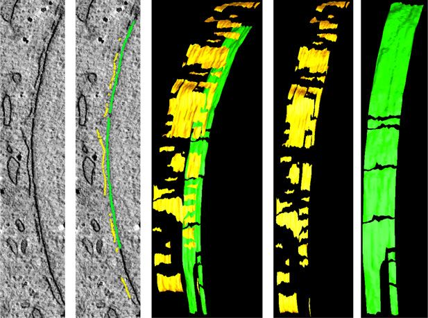
NATURE COMMUNICATIONS | https://doi.org/10.1038/s41467-020-18580-9                                                                                                                                                                   ARTICLE

                       a                                                                                                                Cytoplasm
                                                                                                                                                                 b
                                              Actin                              Cytoplasm

                                                                                                                                                                 sh Ctrl
                                                                                                                Actin

                                                                                                                                                                                                    DAPI                  LaminB1

                                                   Ch
                                                      ro

                                                                                                                                                                 shATR1
                                                        m                      NE
                                                           at
                                                             in
                                                                                                                                             NE
                                             Nucleus                                                            Nucleus

                       c                                                 Nuclear deformations                     d                               Micro-nuclei                  e                          Nuclear circularity
                                             100               ****                                    shCtrl                           30                   ****                                                         ****
                                                              p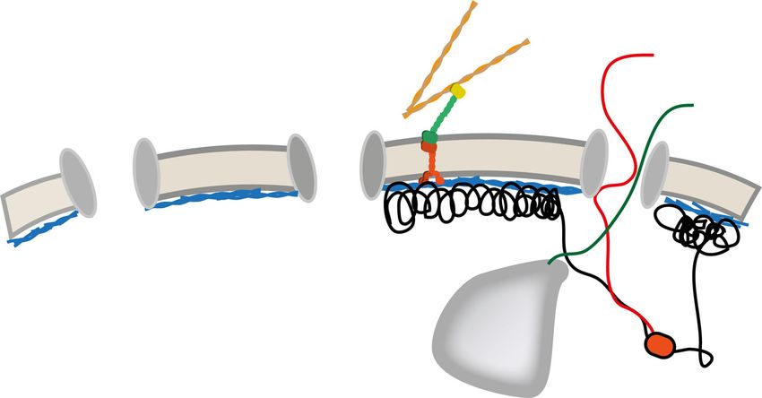
ARTICLE                                                                                                                                                                              NATURE COMMUNICATIONS | https://doi.org/10.1038/s41467-020-18580-9

           a                                                                              b                                                                  ****                c                                                                                                                                               shCtrl
                                                                   ****                                                                                     p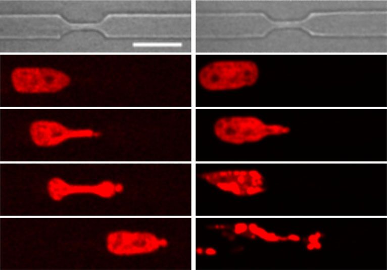
NATURE COMMUNICATIONS | https://doi.org/10.1038/s41467-020-18580-9                                                                                                                                    ARTICLE

                             a              Microfluidic cell compressor      b

                                                                                                              Pressure (mBar)
                                         Microfluidic pump                                            2.5                       40

                                                                                                                                                                                          sh Ctrl
                                                                   Silicone                                                     20
                                                                                                                                                                                          shATR1

                                                                              Relative nuclear area
                                                                   membrane
                                                                                                                                                                                          shATR2
                                                                                                      2.0                            10      20
                                                                                                                                     Time (min)
                                                                                                                                                  30

                                      PDMS
                                      pillars                                                         1.5

                                                                                                      1.0

                                                                                                                                     10       20     30 40 50 release
                                                                                                                                                   Pressure (mBar)

                             c                                                                                                                          d                                   ****
                                                                                                                                                                                          p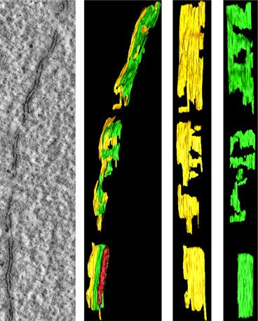
ARTICLE NATURE COMMUNICATIONS | https://doi.org/10.1038/s41467-020-18580-9
a shCtrl shATR
b c n.s
40
***
p=0.0009
30 p=0.5465
Min
Percentage cell death
0
53BP1 foci change
30 20
30 10
20
60 10 0
0
90 DMSO ATRinh
rl R
Ct AT
sh sh
d e n.s
Nuclei 1.5 p=0.0298
*
p=0.8594
Channel
1.5 *
p=0.0209
Ratio front/back
3
Ratio front/back
1.0
1.0
FRET index
0.5
0.5
1
Without Before Inside After
Constriction Nuclear position Outside Inside Outside Inside
DMSO ATRinh
f ** shCtrl
100 p=0.0016
shATR
Percentage of cells
***
80 ***
p=0.0006 ATRinh h shCtrl ONM INM
p=0.0003
60 n.s
p=0.903 ****
p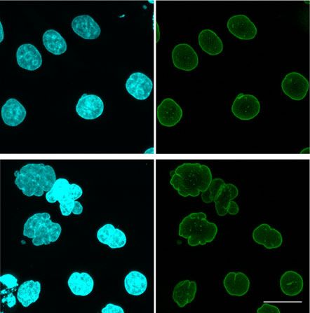
NATURE COMMUNICATIONS | https://doi.org/10.1038/s41467-020-18580-9                                                                             ARTICLE

Fig. 4 ATR-defective nuclei are inefficient in migrating through narrow pores. a Snapshots of H2B-mCherry labeled control and shATR nuclei passing
through constriction. b Cell death measured as the percentage of engaged cells that burst at the constriction (n = 122 and 88 cells for shCtrl and shATR1;
data pooled from three independent experiments). c Quantification of 53BP1-GFP foci generated due to constriction in HeLa cells expressing 53BP1-GFP in
the presence of DMSO or ATR inhibitor, VE-821 (n = 24 and 17 for DMSO and ATRinh; pooled from two independent experiments). Cells that undergo cell
death in the constriction are highlighted in red (for ATRinh). d, e FRET signal measurements of cells engaged in constrictions. d Images of FRET signal at
various stages of migration through the constriction and measurement of signal ratio between front (leading half of the nucleus) and back (lagging half of
the nucleus) of a nuclei at various stages of migration (n = 11, 10, 10, and 6, respectively). e Ratio of front to back FRET signal in migrating cells (inside or
outside the constriction) in the presence DMSO or ATRinh (n = 11, 10, 9, and 10; data from 2 to 3 experiments). f Quantification of nuclear position in the
constriction during the first cGAS foci formation (n = 47, 43, and 28; numbers pooled from 3 experiments). g EM images of control and shATR nuclei in
constriction (routine 200 nm EM sections). Arrowheads indicate invaginations and NE attached chromatin or nucleoli. h 3D reconstruction of NE at the
leading edge from control nucleus in constriction. Green color indicates inner nuclear membrane (INM) and yellow indicates outer nuclear membrane
(ONM). i 3D reconstruction of NE section from leading edge of shATR nucleus in constriction. j Quantification of ratio between number of inner nuclear
membrane breaks to that of the outer membrane (n = 15 and 13). Scale bar for a, d is 20 μm, for g is 9 μm, and for i, h is 200 ηm). Bar graphs presented as
mean ± SEM and dot-plot as mean ± SD. P-value calculated using two-tailed Student’s t-test for b, c, j. One-way ANOVA for d, e with Tukey’s or Sidak’s
multiple comparisons test, and two-way ANOVA for f (****P < 0.0001, ***P < 0.001, **P < 0.01, and *P < 0.05; n.s., not significant).

part of the nucleus, often associated with semicondensed                             We then examined the contribution of ATR in the migration of
chromatin or nucleoli (Fig. 4g and Supplementary Fig. 3f, h).                     cancer cells. We injected equal number of shRNA control and
Both control and ATR-depleted cells exhibited sporadic NE                         ATR-depleted HeLa cells labeled with a vital dye into the tail vein of
ruptures (Fig. 4h). Our EM analysis allowed us to establish that                  immunocompromised mice and recovered lung disseminated cells
control cells accumulated extensive outer membrane ruptures and                   at 2 and 48 h after injection (see scheme in Fig. 5d). We found a
fewer inner membrane ruptures at the leading edge of the nucleus                  significant reduction of fluorescent-positive shATR cells in the lung
(Fig. 4h), whereas ATR-depleted cells accumulated extensive NE                    48 h after injection compared to controls (Fig. 5e, f), indicating that
ruptures involving both the outer and inner membranes, with a                     ATR is essential to allow cells to sustain the harsh mechanical
higher frequency of inner membrane damage (Fig. 4i, j). Moreover,                 environment imposed by blood flow and extravasation.
we noticed that the leading edge of ATR-defective nuclei exhibited
NE portions with a disorganized distribution of outer and inner                   ATR interactors known to influence nuclear mechano-
membranes, likely due to aberrant nuclear membrane remodeling.                    response. To identify potential ATR interactors and targets
The NE at the rear part of the nucleus was normal and comparable                  contributing to nuclear mechanics and dynamics, we performed
in control and in ATR-depleted cells (Supplementary Fig. 3i).                     high-resolution mass spectrometry screens (IP-liquid chromato-
Hence, as soon as ATR-defective cells engage narrow constrictions,                graphy (LC)-MS/MS) in exponentially growing U2OS cells
they fail to adequately respond to the mechanical stress arising at               expressing GFP-ATR. Combining data from three SILAC (stable
the leading edge of the nucleus and undergo NE deformation and                    isotopes labeling with amino acids in cell culture) quantitative
extensive NE damage, which, in turn, cause nuclear collapse and                   approaches (Supplementary Fig. 4a), we obtained 479 unique
cell death. We failed to observe NE ruptures in ATR-defective cells               interactors of ATR (analysis details in “Methods” and Supple-
grown under normal conditions, suggesting that they represent a                   mentary Data 1). Our ATR interactome exhibited a 55%
consequence of mechanical compression. Although the intrinsic                     enrichment (265/479) for proteins with (S/T-Q) motif, a potential
alterations of the mechanical properties of ATR-defective nuclei                  targets of ATR phosphorylation, while such proteins represent
may not affect cell viability under normal conditions, the con-                   only 22% (5137/17522) in the total phospho-proteins of the
sequences of nuclear collapse following mechanical stress certainly               PhosphoSitePlus database34 (Supplementary Fig. 4b). Several of
contribute to cell lethality when cells are forced through narrow                 these interactors were reported to be phosphorylated during S
passages.                                                                         phase, mitosis35 (71/479) and in response to DNA damage when
                                                                                  ATR is hyperactive4 (48/479) (Supplementary Fig. 4c).
                                                                                     We found several ATR interactors for which previous studies
ATR influences neurogenesis and metastatic dissemination.                          have identified roles in the mechanical properties of the nucleus and
During neurogenesis and metastasis, cells migrate through nar-                    whose depletion mimics, at least in part, some of the phenotypes
row places. The in vitro observations described above suggest that                observed in ATR-defective cells (Fig. 6a and Supplementary
ATR may play a relevant role in these processes. We analyzed the                  Fig. 4d–f). TOPII, an ATR/ATM phosphorylation target4, is
contribution of ATR in neurogenesis by performing a transwell                     involved in modulating DNA topology in S phase and in prophase
migration assay of neuroprogenitors isolated from ATR-                            to deal with the mechanical stress caused by chromosome
conditional knockout mouse brain (E13.5 days) (Fig. 5a). ATR                      dynamics. Moreover, genetic evidence36 suggests that Top2 activity
depletion impaired migration of neurosphere-derived cells                         is restrained by Mec1ATR. We also found HDAC2 and CHD4,
through 3 or 8 μm pore size membranes and, as expected, the                       which are members of the Nuclear Remodeling and Deacetylating
defect was more pronounced in the smaller pore size. We next                      (NRD) complex, previously identified as ATR interactors37. An
depleted ATR in vivo, in a developing mouse brain. GFP-tagged                     ATR-mediated regulation of the TOP2 and NRD complex could
shRNAs against ATR were electroporated into a developing brain                    account for the phenotypes associated with the heterochromatic
(at day 14.5), to selectively deplete ATR in a subpopulation of                   and condensed chromatin observed in ATR-defective cells. The
migrating neurons (Fig. 5b). Cortical plates from these brains                    screen identified four proteins of the nuclear pore complex (Nup50,
were analyzed in later stages (E18.5) of embryonic development.                   107, 133, 160), regulating nuclear transport, centrosome attachment
By using two independent shRNAs against ATR, we observed a                        to the NE during mitosis, as well as YAP mechanotransduction26.
compromised neuronal migration in the cortical plate: although                    In addition, we identified several transport proteins including
many of the neurons transfected with Luciferase (control)                         Exportin1 (XPO1/CRM1), an ATR/ATM phosphorylation target4,
reached the top layers of cortical plate, ATR-depleted neurons                    involved in rRNA transport38, which might be connected to the
were stuck in lower layers (Fig. 5b, c).                                          accumulation of nucleolar canals described in this study. We also

NATURE COMMUNICATIONS | (2020)11:4828 | https://doi.org/10.1038/s41467-020-18580-9 | www.nature.com/naturecommunications                                       7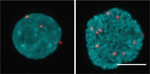
ARTICLE                                                                                                                                    NATURE COMMUNICATIONS | https://doi.org/10.1038/s41467-020-18580-9

                            a                                                     8 mm                                     3 mm                         b
                                                                                       ****                                   ***                                      shLuc    shAtr-4     shAtr-6
                                                                                                                            p=0.0008
                                                                                      p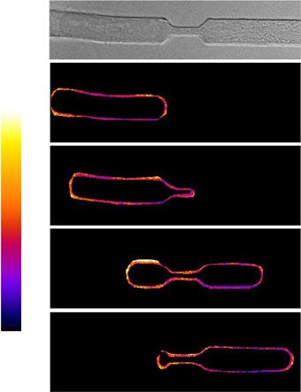
NATURE COMMUNICATIONS | https://doi.org/10.1038/s41467-020-18580-9                                                                                                      ARTICLE

                      a

                                                                                                F-actin                                                mRNA

                                                                            YAP                                                                           rRNA
                                                                                                Nesprin-2
                                                             NUP160
                                                              NUP107                       SUN1/2
                                                              NUP133
                                                               NUP50
                                                       Lamins     TPR
                                                                            XPO1          TOP2B TOP2A
                                                                                                                               CHD4
                                                                                                                                                             HDAC2
                                                tRNA

                                                                                                  Nucleolus

                                                                                                                     Transcription

                      b              Total             IP                     c    Interphase             Prophase
                                                                                                                       d                   6              ****
                                                                                                                                                         p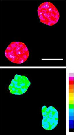
ARTICLE                                                                 NATURE COMMUNICATIONS | https://doi.org/10.1038/s41467-020-18580-9

might contribute to explain the low nuclear stiffness of ATR-                invaginations and extensive ruptures at both nuclear membranes
defective cells.                                                             at the leading edge of the nucleus. Moreover, the extensive NE
                                                                             invaginations tethered with semicondensed chromatin may hin-
Nuclear membrane defects owing to ATR depletion. The lack of                 der efficient nuclear squeezing and prevent an efficient repair of
coordination between chromatin condensation and NEBD during                  the nuclear membranes.
cell division in ATR-defective cells causes accumulation of semi-               Cancer cell migration through the ECM requires nuclear
condensed chromatin attached to NE fragments6. Moreover, our                 deformability, particularly when cells must meander through
EM analysis showed that ATR-depleted cells accumulate mem-                   dense and highly crosslinked collagen type I-rich stroma,
brane ruptures already under unperturbed conditions and, fol-                extravagate, and sustain the harsh conditions of the blood
lowing nuclear deformation during interstitial migration, they               circulation before extravasating, passing through pores as small as
exhibit massive breakage of the outer nuclear membrane and                   2 μm in diameter50. In fact, altered NE morphology is typical of
aberrant membrane remodeling. The ESCRTIII complex plays a                   cancer cells and crucial in the tumor grade assessment, and
key role in sealing membrane holes in the reforming NE during                correlates with prognosis. Cancer cells can adapt to metastatic
mitotic exit48 and in repairing the NE upon migration-induced                migration by deregulating the expression of Lamins, but a certain
rupture28,29. It is possible that the activity of the ESCRTIII com-          degree of NE stiffness is required to allow a productive migration
plex becomes limiting in ATR-defective cells, due to the massive             and to prevent massive NE ruptures50. Our observations suggest
damage of nuclear membranes. The extensive NE damage and                     that ATR activity might be therefore beneficial for cancer cell
remodeling in ATR-defective cells may also represent the primary             migration, thus implying that ATR might play opposite roles in
cause of the aberrant phospholipid composition of their nuclear              cancer progression, by preventing genome instability and by
membranes. In agreement with this hypothesis, the aberrant                   promoting metastasis. Along this idea, it is interesting to note that
phospholipid composition of the NE in ATR-defective cells does               our experiments indicate that ATR depletion impairs three-
not reflect a direct metabolic problem and we failed to identify              dimensional (3D) invasion and lung homing of cancer cells.
ATR interactors involved in lipid metabolism.                                   Our findings describe a variety of nuclear defects and their
                                                                             pathological consequences (Fig. 7). However some of this defects such
                                                                             as the altered mechanical coupling between cytoskeleton and NE, and
Altered NE-cytoskeleton coupling upon ATR inactivation. A                    the YAP cytoplasmic retention occur soon after ATR catalytic
key finding of this work is that ATR is a component of the cell               inactivation, suggesting that these two phenotypes represent a direct
mechanotransduction machinery by ensuring appropriate                        consequence of ATR deregulation. Considering that during develop-
mechanical coupling of the cytoskeleton to the NE. The NE is                 ment, organogenesis requires that stem/progenitor cells migrate
exposed to forces acting in opposite directions: forces deriving             towards destination tissues, our observations may contribute to
from chromatin dynamics (as outlined above) and opposite forces              explain some of the developmental defects of Seckel patients bearing
generated by extracellular matrix (ECM) attachment and con-                  genetic defects in ATR. Our results might also explain the increased
veyed to the NE through the LINC complex and the NE-                         cell death in non-proliferating neuroprogenitors and neurons of ATR-
associated cytoskeleton. ATR orchestrates the integration of all             knockout mice51,52, which cannot be directly ascribed to the role of
these mechanical inputs, by regulating at once NE dynamics,                  ATR in replication stress. Moreover, the delocalization of YAP might
chromatin condensation, and the LINC function as the                         also contribute to a variety of pathological outcomes such as loss of
mechanosensory properties of the entire cell; this is visualized by          stem cells and cardiomyopathies27,53. Intriguingly, ATR-conditional
the here discovered ATR-YAP mechano-signaling axis.                          knockout mice exhibit a progeroid phenotype that has been related to
   Although abnormalities in nuclear shape and mechanics can                 stem cell loss54.
impact on genome integrity by generating chromatin fragmenta-
tion36 and fork collapse42, under normal conditions, the nuclei
                                                                             Methods
remain relatively stable as well as the NE. However, at raising              Plasmids. ATR shRNA1 and control (pLKO1) plasmids were from Dr. O.F. Capetillo
levels of mechanical strain, cells must promptly respond to                  (CNIO, Spain); ATR shRNA2 was purchased from Sigma (TRCN0000219647); the
mechanical stress. Here we show that ATR is critical also for the            GFP-ATR plasmid was from Dr. R.Tibbetts (Wisconsin, USA)55. GFP-AU1-ATR
nuclear response to more severe mechanical challenges. ATR-                  plasmid was digested with BamHI to excise out GFP cDNA. The BamHI-digested
                                                                             GFP cDNA insert was cloned into FLAG-ATR-KD plasmid56 also linearized with
defective cells fail to properly respond to sub-lethal compression           BamHI, followed by transformation in Escherichia coli. Positive clones after trans-
forces and undergo extensive nuclear collapse characterized by               formations were screened by PCR and BamHI restriction digestion, finally sequenced
NE ruptures. In turn, this imposes an additional stress at the level         (list of primers used are provided in Supplementary Table 1). pTRIP-CMV-GFP-
of nuclear membrane remodeling, as revealed by the presence of               FLAG-cGAS (Plasmid #86675)28, Nesprin tension sensor (pcDNA nesprin TS; plas-
                                                                             mid #68127), and nesprin headless control (pcDNA nesprin HL; plasmid #68128)25
mixed portions of the outer and inner membranes at the NE                    were acquired from Addgene plasmid repository.
engaged in the constrictions. NE fragmentation under high level
of mechanical stress exposes nuclear DNA into the cytoplasm,
leading to activation of the cGAS-STING pathway49. The                       Antibody                  Source         Catalog number Dilution
functional consequences of the ATR and cGAS connection                       1.   ATR                  Cell Signal    2790             1 : 1000 (WB); 1 : 100 (IF)
remain unexplored but may hold relevant pathological implica-                2.   TopBP1               Abcam          ab2402           1 : 1000 (WB)
                                                                             3.   Nup133               SantaCruz      sc-27392         1 : 500 (WB)
tions, particularly in tissues undergoing mechanical stress.                 4.   Tubulin              Sigma          T5168            1 : 5000 (WB)
                                                                             5.   Lamin B1             Abcam          ab16048          1 : 10,000 (WB); 1 : 1000 (IF)
                                                                             6.   Lamin A/C            SantaCruz      sc-7292          1 : 500 (WB); 1 : 200 (IF)
Pathological consequences owing to ATR defects. Our obser-                   7.   Nesprin 2            Thermo         MA5-18075        1 : 500 (WB); 1 : 500 (IF)
                                                                                                       Scientific
vations indicate that, when the nucleus engages the narrow                   8. Histone H3(tri-met K9) Abcam          ab8898           1 : 1000 (WB)
constrictions, the LINC complexes at the leading edge of the                 9. Total Histone H3       Abcam          ab1791           1 : 5000 (WB)
                                                                             10. Total YAP(63.7)       SantaCruz      sc-101199        1 : 500 (WB); 1 : 200 (IF)
nucleus are tightly bound to the cytoskeleton and the mechanical             11. Phospho YAP (Ser127) Cell Signal     4911S            1 : 1000 (WB)
strain generates extensive ruptures at the outer nuclear mem-
branes. Hence, the nucleus during interstitial migration is
polarized at the level of NE. ATR-defective cells fail to maintain             Secondary antibodies were obtained from IFOM imaging facility:
the coupling between Nesprin-2 and the cytoskeleton, and to                    Polyclonal Donkey anti-mouse AlexaFluor-488 AB_2340846 (Jackson
polarize the NE under these conditions and accumulate NE                     ImmunoResearch).

10               NATURE COMMUNICATIONS | (2020)11:4828 | https://doi.org/10.1038/s41467-020-18580-9 | www.nature.com/naturecommunications
NATURE COMMUNICATIONS | https://doi.org/10.1038/s41467-020-18580-9 ARTICLE
Cell lines. U2OS cells stably expressing GFP-ATR and HeLa cells stably expressing
mCherry-H2B were reported previously6. U2OS cells expressing the FUCCI
UNPERTURBED CONDITIONS PATHOLOGICAL reporter was a kind gift from Libor Macůrek33. Human primary Seckel fibroblasts
CONSEQUENCES (GM18366) and IMR90 were from Coriell Cell Repository. HCT116 and ATRflox/−
cells were from The American Type Culture Collection.
RNA EXPORT EPIGENETIC AND MITOTIC
DEFECTS TOPOLOGICAL DEFECTS DEFECTS
MICRONUCLEI
Cell culture, transfection, and inhibitor treatments. HeLa and U2OS cells were
maintained in Dulbecco’s modified Eagle’s medium (DMEM) with GlutaMAX
(Life Technologies) supplemented with 10% (vol/vol) fetal bovine serum (FBS,
Biowest) and penicillin–streptomycin (Microtech). Human primary fibroblasts
CHROMATIN derived from Seckel patient were maintained in DMEM supplemented with 15%
NUCLEAR FRAGMENTATION FBS (not activated, Sigma-Aldrich) and IMR90 were grown in 10% FBS (not
DEFORMATION activated). HCT116 and ATRflox/− cells were grown in McCOY’s 5A media. All
cells were grown in a humidified incubator atmosphere at 37° and 5% CO2.
We used Lipofectamine 2000 (Invitrogen) for transfecting plasmids into cells,
using the protocol recommended by the manufacturer.
NUCLEAR MEMBRANE STEM CELL LOSS
HEK293T cells were transfected with shRNA plasmids and viral packaging
REMODELLING
plasmids to generate lentiviral particles. Desired cell lines were then infected for
16 h followed by 2 μg/ml puromycin selection for 24 h. Infected cells were cultured
in 1 μg/ml puromycin containing media and were utilized for experiments up to
ALTERED NUCLEAR YAP 10 days after infection.
MECHANICS DELOCALIZATION Cells were treated with ATR inhibitors (2 μM ETP46464, 10 μM VE-821, or
1 μM AZ-20) 1 h before (unless mentioned otherwise) starting the experiment and
were maintained in the media throughout the course of the experiment.
For cell cycle analysis, cells were fixed with ice-cold ethanol, DNA was labeled
MECHANICAL STRESS PATHOLOGICAL with propidium iodide, and quantified using FACS calibur (BD bioscience) system.
CONSEQUENCES
Membrane fractionation using Mem-Per Plus kit. Membrane fractionations
CELL
MIGRATION DEFECTS were performed following protocol provided by the vendor. Briefly, cells were
NUCLEAR trypsinized, washed with cell wash solution, resuspended in permeabilization buffer
COLLAPSE (with or without Benzonase), and incubated for 30 min at 4 °C with constant
DEFECTIVE TUMOR CELL
MIGRATION mixing. Permeabilized cells were then centrifuged for 15 min at 16,000 × g, soluble
fraction was collected, and pellet resuspended and incubated in solubilization
buffer for 30 min at 4 °C. Samples were then centrifuged for 15 min at 16,000 × g
NEUROGENESIS
NE RUPTURES DEFECTS and supernatant was collected as a membrane fraction.
Cell lysis and immunoblotting. Total cell lysates were prepared in lysis buffer
PREMATURE AGEING
Perinuclear cGAS RECRUITMENT (50 mM Tris-HCl pH 8.0, 1 mM MgCl2, 200 mM NaCl, 10% Glycerol, 1% NP-40)
Protease (Roche) and Phosphatase inhibitors (Sigma) were added at the time of
experiment, and Benzonase (50 U/ml) was added if degradation of nucleic acid was
Fig. 7 Graphical summary of ATR defects affecting nuclear morphology needed. Cell lysates boiled with Laemmli buffer were (20–50 μg) resolved using
and mechanics and relative pathological consequences. In the absence NuPAGE® (Invitrogen) or Mini-PROTEAN® (Biorad) precast gels, transferred to
of external stimuli ATR coordinates chromatin processes (such as RNA nitrocellulose membrane, and probed as with primary (2 h at roomtemperature
(RT) or overnight at 4 °C) and secondary antibodies (1 h at RT), and acquired
export, epigenetic and topological transitions, and chromatin condensation in using ChemiDoc imaging system (Image Lab v5.0). Image intensity measurements
prophase) with NE dynamics, influencing nuclear morphology. ATR defects were performed using ImageJ.
lead to nuclear deformation, NE remodeling, altered nuclear mechanics, and
YAP delocalization. In response to mechanical stress, ATR-defective nuclei IF assays and quantifications. Briefly, cells were fixed with 4% formaldehyde
collapse leading to NE ruptures and cGAS recruitment at the nuclear (15 min), permeabilized with 0.2% Triton X-100 in phosphate-buffered saline
periphery. Pathological consequences are also described. See text for details. (PBS) (15 min), blocked with 1% bovine serum albumin in PBS for 1 h (blocking
buffer), incubated with primary antibodies (diluted in blocking buffer) for 1 h
in RT, followed by three PBS washes and then incubated in secondary antibodies
Polyclonal Donkey anti-mouse AlexaFluor-594 AB_2340854 (Jackson (1 : 400 in blocking solution) for 1 h in the dark at RT followed by three PBS
ImmunoResearch). washes. Samples were mounted with VectaShield mounting medium containing
Polyclonal Donkey anti-mouse AlexaFluor-Cy3 AB_2340813 (Jackson 4′,6-diamidino-2-phenylindole (DAPI). Image acquisition was performed using
ImmunoResearch). Leica TCS SP2 confocal scanning microscope, equipped with a ×63/1.4 numerical
Polyclonal Donkey anti-rabbit AlexaFluor-488 AB_2313584 (Jackson aperture (NA) objective. Single optical sections of the images or maximum pro-
ImmunoResearch). jections (step size 0.5 μm) were processed using ImageJ and smoothed to reduce the
Polyclonal Donkey anti-rabbit AlexaFluor-594 AB_2340621 (Jackson background noise.
ImmunoResearch). Quantification of nuclear morphology, YAP localization: images from random
Polyclonal Donkey anti-rabbit AlexaFluor-Cy3 AB_2307443 (Jackson fields (upto 50) were acquired from coverslips stained with DAPI and Lamin or
ImmunoResearch). YAP on a UltraVIEW VoX spinning-disc confocal unit with Velocity software
(PerkinElmer). Nuclei from each field were manually binned into normal, mild
(blebs, invaginations, wrinkles, micronuclei, and multi-nuclei), and severely
Other reagents Source Catalog number
deformed (with multiple defects), as well as with or without micronuclei alone (in
1. ATR inhibitors case if field hasARTICLE NATURE COMMUNICATIONS | https://doi.org/10.1038/s41467-020-18580-9
performed exactly as it has been reported previously6,57,58. A brief description of using the Kolmogorov–Smirnov test. Estimation of the minimal set of samples was
each process is described below. performed according to ref. 62. Correlation between two variables was calculated
Embedding: cells grown on MatTek dishes (MatTek Corporation, USA) were using Pearson’s product moment correlation.
fixed with of 4% paraformaldehyde and 2.5% glutaraldehyde (EMS, USA) mixture Analysis of cells in channels: we embedded Poly-di-methyl-siloxane (PDMS)
in 0.2 M sodium cacodylate buffer (pH 7.2) for 2 h at RT, followed by six washes in molds on MatTek dishes, loaded cell, and incubated for 24 h to facilitate cell
0.2 M sodium cacodylate buffer at RT. Then cells were incubated in 1 : 1 mixture of migration into the channels. Cells migrating within the channels were examined
2% osmium tetraoxide and 3% potassium ferrocyanide for 1 h at RT followed by six under the UltraVIEW VoX spinning-disc confocal unit (PerkinElmer) and
times rinsing in 0.2 M sodium cacodylate buffer (pH 7.2). Then the samples were acquired images of cells suitable for the future CLEM analysis. The remaining cells
sequentially treated with 0.3% Thiocarbohydrazide (in 0.2 M sodium cacodylate from the loading wells were eliminated and 0.05% glutaraldehyde + 4%
buffer) for 10 min and 1% OsO4 (in 0.2 M cacodylate buffer, pH 6.9) for 30 min. formaldehyde solution (in 0.1 M cacodylate buffer, pH 7.2) was added to the dish
Samples were then rinsed with 0.1 M sodium cacodylate (pH 6.9) buffer until all for 5 min. Cells were then fixed with 2.5% glutaraldehyde + 4% formaldehyde (in
traces of the yellow osmium fixative have been removed. Then samples were 0.2 M cacodylate buffer, pH 7.2) for 10 days, to make cell bodies resistant to the
washed in de-ionized water, treated with 1% uranyl acetate (in distilled water) for 1 process of the mechanical detachment of PDMS from the MatTek. Then PDMS
h and washed in water again57,59. The samples were subsequently embedded in mold was detached from the MatTek dish and the cells attached to the dishes were
Epoxy resin at RT and polymerized for at least 72 h in a 60 °C oven. Embedded processed for EM analysis as described above. After mold detachment, cells were
samples were then sectioned with diamond knife (Diatome, Switzerland) using the additionally stained with 1% methylene blue in PBS for 3 min at RT and again
ultramicrotome (LeicaEM UC7, Leica Microsystem, Vienna). Sections were examined under a light microscope to confirm the presence of selected cells on the
analyzed with a Tecnai20 EM (FEI, Thermo Fisher Scientific, Eindhoven, The MatTek glass.
Netherlands) operating at 200 kV58.
Nano-gold labeling: cells grown on MatTeks were fixed with a mixture of 4%
DNAse I sensitivity assay. Cells were trypsinized, washed in ice-cold PBS, and
paraformaldehyde and 0.05% glutaraldehyde (0.15 M Hepes buffer, pH 7.2) for 5
resuspended in 2 ml of ice-cold cell lysis buffer (300 mM sucrose, 10 mM Tris pH
min at RT and then replaced with 4% paraformaldehyde (in 0.15 M Hepes buffer,
7.4, 15 mM NaCl, 5 mM MgCl, 0.5% NP-40, 0.5 mM dithiothreitol, protease
pH 7.2) for 30 min. Cells were washed six times in PBS and incubated with
inhibitor (Complete, Roche)). After 30 min, the lysed cells were centrifuged at
blocking solution for 30 min at RT. Then cells were incubated with primary
500 × g for 5 min at 4 °C and supernatant was discarded. The nuclei were gently
antibody diluted in blocking solution overnight at 4 °C. On the following day, the
resuspended in appropriate amount of reaction buffer (30 μl per DNAse I condi-
cells were washed six times with PBS and incubated with goat anti-rabbit Fab’
tion). Separate 30 μl aliquots were then taken and gently mixed with 70 μl of DNase
fragments coupled to 1.4 nm gold particles (diluted in blocking solution 1 : 100) for
I mix (of varying units) on ice. It was incubated for 15 min at 25 °C and then 700 μl
2 h and washed six times with PBS. Meanwhile, the activated GoldEnhanceTM-EM
of nuclei lysis buffer (100 mM Tris-HCL pH 8, 5 mM EDTA pH 8, 200 mM NaCl,
was prepared according to the manufacturer’s instructions and 100 μl of it was
0.2% SDS) was added to each sample with 50 mg proteinase K. Samples were
added into each sample well. The reaction was monitored by a conventional light
incubated at 55 °C for 1 h; RNaseA (10 mg) was added and again incubated at 37 °C
microscope and was stopped after 5–10 min when the cells had turned “dark
for 30 min. DNA was then extracted using standard phenol–chloroform technique
enough” by washing several times with PBS. Osmification followed: the cells were
and was resuspended in 200 μl of 0.1 TE, quantified, and were run on 1%
incubated for 1 h at RT with a 1 : 1 mixture of 2% osmium tetraoxide (in water) and
agarose gels.
3% potassium ferrocyanide (in 0.2 M sodium cacodylate pH 7.4) and then rinsed
six times with PBS and then with distilled water. The samples were then
dehydrated: 3 × 10 min in 50% ethanol; 3 × 10 min in 70% ethanol; 3 × 10 min in AFM measurements. The AFM measurements were performed using Nanowizard
90% ethanol; 3 × 10 min in 100% ethanol. The samples were subsequently 3 (JPK Instruments, Germany) and a modified silicon nitride cantilever (NovaScan,
incubated for 2 h in 1 : 1 mixture of 100% ethanol and Epoxy resin (Epon.EMS) at USA) with a spring constant of 0.03 N/m and a 5 μm diameter polystyrene bead
RT; the mixture was then removed with a pipette and finally samples were adhered at the tip. Central region of the cell was indented with a loading rate of
embedded for 2 h in Epoxy resin at RT. The resin was polymerized for at least 10 h 1.5 μm/s. The ramp size was 3 μm was used. All the measurements were performed
at 60 °C in an oven. as previously described in ref. 17. Nuclei were isolated by treating cells with 1 ml of
Tomography: two-step CLEM based on the analysis of tomographic a 0.01% Igepal CA-630 (a non-ionic detergent, Sigma) and 1% citric acid solution
reconstructions acquired under low magnification with consecutive in water for 5 min. Expelled nuclei from the adherent cells were collected, washed
reacquisition of EM tomo box under high magnification (×60,000) and its re- with 5 ml PBS, centrifuged at 800 × g for 5 min, resuspended in PBS, and dropped
examination was used exactly as described previously 60. Briefly, an ultratome onto coverslip for AFM experiments.
(LeicaEM UC7; Leica Microsystems, Vienna) was used to cut 60 nm serial thin
sections and 200 nm serial semi-thick sections. Sections were collected onto 1% FRET image acquisition and analysis. Cells grown on coverslips were injected
Formvar films adhered to slot grids. Both sides of the grids were labeled with with the Nesprin 2G-TS construct (50 ng/cell) and the following day, imaged using
fiduciary 10 nm gold (PAG10, CMC, Utrecht, The Netherlands). Tilt series a DeltaVision Elite imaging system using an Olympus ×60/1.42 Plan Apo N oil-
were collected from the samples from ±65° with 1° increments at 200 kV in immersion objective. Three images were collected in sequence at each point: Cyan
Tecnai20 EMs (FEI, Thermo Fisher Scientific, Eindhoven, Tthe Netherlands). Fluorescent Protein (CFP) (for mTFP1) (ex: 438/24 nm, em: 470/24 nm), FRET (ex:
Tilt series were recorded at a magnification of ×20,000 or ×60,000 using 438/24 nm, em: 559/38 nm), and Yellow Fluorescent Protein (for mVenus) (ex:
software supplied with the instrument. The nominal resolution in our 513/17 nm, em: 528/38 nm). A single-plane image was background corrected,
tomograms was 4 nm, based upon section thickness, the number of tilts, tilt realigned, converted into 32 bits, and analyzed using an in-house macro in ImageJ.
increments, and tilt angle range. The IMOD package and its newest viewer, The nuclear membrane of each cell was manually selected as a region of interest
3DMOD 4.0.11, were used to construct individual tomograms and for the and average FRET/CFP ratios calculated for the nuclear membrane region.
assignment of the outer leaflet of organelle membrane contours, and best-fit Approximately 30 cells/conditions were analyzed for n = 2 experiments. ATR
sphere models of the outer leaflet were used for vesicle measurements. Videos inhibitor was added (ETP46464 2 μM) 3 h before image acquisition. ATR inhibitor
were made in 3DMOD and assembled in QuickTime Pro 7.5 (Apple) and the VE-821 could not be used for this measurements, as they exhibited auto fluores-
video size was reduced by saving videos at 480p in QuickTime. CLEM was cence. HeLa cells stably expressing Nesprin 2G sensor or the Headless control
performed exactly as described previously 59. sensor were generated by Lipofectamine 2000 transfection, Neomycin (G418)
FIBSEM: FIBSEM analysis was performed using a FEI Helios NanoLab 660 selection, and single-cell fluorescence-activated cells sorting of the mVenus/
FEGSEM or G3 equipped with SEM Multi-Detector and ICD detector at mTFP1-positive population. These cells were loaded onto the channels in the
accelerating voltage 2.0 kV. Access to both of which was kindly provided by FEI, presence of ATR inhibitor or DMSO. Single stack image acquired for each field of
Co. (FEI, Thermo Fisher Scientific, Eindhoven, The Netherlands). For all high- view every 2 h for 10 h duration. Image acquisition parameters and analysis were
resolution EFSEM images, a primary beam energy of 2.0 kV was used with a similar to the above-mentioned experiment.
working distance of 1 mm, 3 ms dwell time, and tube bias of 140 V. An Auriga 60
FIB-SEM (Zeiss) microscope with Atlas3D software (FIBICS) was additionally used
to collect the 3D data of two cells. Acquisitions were performed according to FLIM-FRET analysis. For the acquisition, we used the Leica TCS SP8 confocal
instructions of the manufacturers. microscope with White light laser as excitation source tuned at 488 nm and HC PL
Quantification and statistics: all acquired images were aligned using the APO CS2 ×63/1.40 oil-immersion objective, everything managed by Leica Appli-
TrakEM2 plugin of FIJI. Images were segmented by thresholding with Amira ((FEI, cation Suite X software, ver. 3.5.2.18963. For the lifetime measurements, the above
Thermo Fisher Scientific, Eindhoven, The Netherlands). The number of ATR- system was implemented with PicoQuant Pico Harp 300 TCSPC module and
tagged gold particles in different compartments of the cell was counted and picosecond event timer, managed by PicoQuant software (SymPho Time 64, ver.
percentages were calculated. The labeling density of ATR on different cellular 2.4). Data were imported and analyzed using in-house ImageJ macro.
structures was assessed and calculated as described in ref. 61. For this we used the
following criteria: gold particles were considered to label the NE, ER, or Micro-fabricated cell compression chamber. A custom-made cell compression
mitochondria when these particles were observed over lumens or membranes of device has been invented based on movement of thin membrane attached with a
these compartments; gold particles were considered as a label of the PM when these piston, which is precisely controlled by air pressure regulator. The cell compression
particles were observed over the PM. Normality of variant distribution was assessed device was designed using Solidworks and device components were 3D-printed
with Shapiro–Wilk tests. Cumulative probability distributions were compared using Dental SG resin (Formlabs) for its biocompatibility. All the components were
12 NATURE COMMUNICATIONS | (2020)11:4828 | https://doi.org/10.1038/s41467-020-18580-9 | www.nature.com/naturecommunicationsNATURE COMMUNICATIONS | https://doi.org/10.1038/s41467-020-18580-9 ARTICLE
printed and then washed with IPA for 20 min, followed by post processing in processes (GOTERM_BP) (p-value with Benjamini correction < 0.05). Revigo tool
ultraviolet chamber as suggested by Formlabs. A 20 mm diameter coverslip was was utilized to simplify GO terms (resulting list size: 0.7, database: Homo sapiens,
stick on the top center of the cell compression device. Silicon membrane was semantic similarity measure: SimRel) and R Studio for plotting of Revigo output
sticked with a piston and then clamped to the bottom of the cell compression (size = log size, color = log10 p-value). Candidates where then manually curated to
device by clamping tools. The assembled cell compression device was then con- generate non-overlapping sub-categories of interest (for this study). A network was
nected to the air pressure regulator. Cells were plated on glass-bottom petridish generated for each sub-category using STRING interaction analysis and the output
and maintained in cell incubator. Before the experiment, a cell compression device was plotted using cytoscape.
was capped and locked on the cell culture dish. Images were acquired using ×40 oil The MS proteomics data have been deposited to the ProteomeXchange
lens (NA = 1.3) in PerkinElmer spinning disk microscope. Consortium via the PRIDE65 partner repository with the dataset identifier
PXD020622 (Project Name: ATR interactome in H. sapiens bone osteosarcoma
U2OS cells). Files are named SILAC1, SILAC2, and SILAC3.
Analysis of cell migration in micro-fabricated channels. We followed the pro-
tocol established previously28 for PDMS channels preparation. Briefly, Polymer
and crosslinking agent (RTV615 kit) (mixed in 1 : 10 ratio) was used to prepare
Lipidomic analysis. (I) Lipid extraction: Nuclei were isolated according to the
PDMS channels. These are then plasma treated and embedded onto a glass-bottom
protocol66. Nucleus and total cell samples were resuspended in 150 mM ammo-
dish or a two-chamber LAB-TEK II dish (Thermo Fisher). Then channels were
nium bicarbonate and passed through a 26 G syringe needle to fragment nucleic
fibronectin-coated and cells were loaded the day before the beginning of time lapse.
acids. Samples were centrifuged at 10,000 × g for 10 min at 4 °C to eliminate cell
We chose 15 μm-long, 4 μm-wide constriction for experiments involving HeLa and
debris. Lipids were extracted starting from an sample size equivalent of 50 µg of
U2OS cell lines. Time-lapse images were acquired (every 10 or 15 min, with z-
proteins, using a two-step extraction protocol (Folch method) with methanol and
stacks) on a UltraVIEW VoX spinning-disc confocal unit with Velocity software
chloroform in different proportions67. Organic phase fractions were then dried out
(PerkinElmer), equipped with an Eclipse Ti inverted microscope (Nikon) and a
and resuspended in 50 µL of 95% phase A (CH3CN : H2O 40 : 60; 5 mM
C9100-50 electron-multiplying CCD (charge-coupled device) camera (Hama-
NH4COOCH3; 0.1% FA) plus 5% phase B (IPA : H2O 90 : 10; 5 mM NH4COOCH3;
matsu) or Confocal Spinning Disk microscope (Olympus) equipped with IX83
0.1% FA) for subsequent analysis. Before extraction, samples were spiked in with
inverted microscope provided with an IXON 897 Ultra camera (Andor) with
16 internal standards: PC (12 : 0/13 : 0) 40 pmol, PE (12 : 0/13 : 0) 52 pmol, phos-
OLYMPUS cellSens Dimension software, or on a DeltaVision Elite imaging system
phatidylglycerol (PG) (12 : 0/13 : 0) 7.5 pmol, phosphatidylserine (PS) (12 : 0/13 : 0)
using ×40 oil-immersion (for 53BP1 foci counting) or ×20 dry objective for a
43 pmol, phosphatidylinositol (PI) (12 : 0/13 : 0) 54 pmol, Cer (d18 : 1/25 : 0)
duration of 18–24 h. The images were processed using ImageJ and smoothed to
100 pmol, cholesterol ester (CE) (19 : 0) 100 pmol, GlcCer (d18 : 1/12 : 0) 50 pmol,
reduce the background noise. All the quantifications were performed manually.
LacCer (d18 : 1/12 : 0) 50 pmol, sphinganine (d17 : 0) 50 pmol, sphingosine-1-P
Number of cells reaching the constriction within the experimental period was
(d17 : 1) 100 pmol, sphingosine (d17 : 1) 50 pmol, Galactosyl(β) Sphingosine-d5
considered as a total cell number. Number of cell death and cell passing were
20 pmol, d5-TG ISTD Mix I 20 pmol, d5-DG ISTD Mix I 20 pmol, and cholesterol
counted per field to calculate the percentages. Fields with no cell migration or death
(d7) 800 pmol. (II) Protein quantification: proteins were extracted form 20 µL of
were discarded from analysis. 53BP1 foci were counted manually using ImageJ.
ammonium bicarbonate resuspended fractions by adding 5 µL of lysis buffer (10%
Difference was calculated by subtracting number of Foci before engaging the
NP-40, 2% SDS in PBS) and quantified by BCA protein assay kit (Thermo Fisher
constriction from the number of foci present in the constriction.
Scientific). (III) Lipid profiling data acquisition: lipid extracts were diluted 1 : 5 and
1 µL injected on a LC system nLC Ekspert nanoLC400 (Eksigent, 5033460C; Sin-
IP, MS, and data analysis. U2OS cells expressing GFP-ATR or GFP alone are gapore) coupled with a Triple TOF 6600 (AB Sciex, Singapore). Chromatography
cultured in SILAC medium containing light or heavy-labeled L-lysine and L-argi- was performed using an in-house packed nanocolumn Kinetex EVO C18, 1.7 µm,
nine for 5 days ensuring adequate incorporation of isotopes. These cells were 100 A, 0.75 × 100 mm. The mobile phases A (CH3CN : H2O 40 : 60; 5 mM
collected with Lysis buffer (50 mM Tris-HCl pH 8.0, 1 mM MgCl2, 200 mM NaCl, NH4COOCH3; 0.1% FA) and B (IPA : H2O 90 : 10; 5 mM NH4COOCH3; 0.1% FA)
10% Glycerol, 1% NP-40 + Protease, and Phosphatase inhibitors) containing were used in positive mode. The gradient elution was initially started from 5% B,
Benzonase (50 U/ml) and incubated for 1 h on ice. Lysates were pre-cleaned by linearly increased to 100% B in 5 min, maintained for 45 min, then returned to the
incubating with Protein-A beads for 1 h at 4 °C. GFP and GFP-ATR (4 mg of initial ratio in 2 min, and maintained for 8 min. Acquisition in MS was performed
protein lysate per sample) were immunoprecipitated by incubating the lysates with in positive with the following parameters: mass over charge (m/z) range 100–1700,
200 μl of anti-GFP-conjugated magnetic beads overnight on a rotor at 4°. Beads T source 80 °C, Ion Spray Voltage 2000, declustering potential 80, fixed collision
were washed (with Lysis buffer) and eventually pooled before elution with sample energy 40 V (+). For Information Dependent Acquisition analysis (Top 8), range
buffer. Proteins were then resolved onto a 4–12% NuPAGE® precast gel (Invi- of m/z was set as 200–1800 in positive ion mode; target ions were excluded for 20 s
trogen) and stained by Coomassie colloidal blue. The gel lane was cut into eight or after two occurrences (Analyst TF 1.7.1). (IV) Data processing: Lipidview work-
ten slices each of which has been reduced, alkylated, and digested with trypsin as station (version 1.3 beta, AB SCIEX, USA) was for lipids identification and
reported elsewhere63. Peptide mixtures were desalted and concentrated on a home- quantification. Lipid identification was based on exact mass, retention time, and
made C18 desalting tip, then peptides were injected in a nanoHPLC (EasyLC MS/MS pattern. Lipid species based on precursor fragment ion pairs were deter-
Proxeon, Denmark). Peptides separation occurred onto a 25 cm-long column, mined using a comprehensive target list in LipidView (Sciex). Lipid species iden-
reverse-phase spraying fused silica capillary column (75 μm i.d.) packed with 3 μm tification was performed using the mass tolerance of 0.05 in MS and 0.02 in MS/
ReproSil AQ C18 (Dr. Maisch GmbH, Germany). A gradient of eluents A (high- MS, s/n of 3, and % peak intensity >0 for positive ion mode. Lipid classes included
performance liquid chromatography-grade water with 0.1% v/v formic acid) and B for statistics and downstream analysis were cholesterol ester (CE), sphingomyelin,
(Acetonitrile) with 20% v/v water with 0.1% v/v formic acid) were used to achieve diacylglycerol, triacylglycerol, ceramide (Cer), PC, PE, PG, PI, PS and lysopho-
separation, from 7 to 60% of B in 30 min, at a constant flow rate of 250 nl/min. The sphatidylcholine, lysophosphatidylethanolamine, lysophosphatidylglycerol, lyso-
LC system was connected to a QExactiveHF mass spectrometer (Thermo Scientific, phosphatidylinositol, lysophosphatidylserine, hexosylceramide, dihexosylceramide,
Bremen, Germany) equipped with a nanoelectrospray ion source (Proxeon Bio- trihexosylceramide, sulphatides (SGalCer), and Cer-phosphate, in positive mode.
systems, Odense, Denmark). Full scan mass spectra were acquired in the LTQ All statistical analysis was performed using Metaboanalyst 4.0 web tool68 (https://
Orbitrap mass spectrometer with the resolution set to 60,000 (@200 m/z) accu- www.metaboanalyst.ca/MetaboAnalyst/faces/home.xhtml). Three experimental
mulating ions to a target value of 6,000,000. The acquisition mass range for each and two technical replicates were measured for each condition and 854 lipid
sample was from m/z 300–1650 Da and the analyses were made in duplicates. The metabolites were detected in total. Missing metabolite intensities were imputed
15 most intense doubly and triply charged ions were automatically selected and with half of the lowest detectable value. Intensities were then normalized by the
fragmented in the ion trap after accumulation to a “target value” of 15,000. Target median of the 322 technically most reliable metabolites (detectable in ~95% of all
ions already selected for the MS/MS were dynamically excluded for 20 s. Identifi- samples) and averaged over two technical replicates. The total intensities of PC and
cation and quantification of peptides and proteins were performed with MaxQuant PE were calculated as sum of all individual phospholipids with choline and etha-
1.5.2.8 against the human Uniprot complete proteome set, having identified a nolamine head groups.
protein with at least two peptides (one unique), six amino acids of minimal length,
false discovery rateARTICLE NATURE COMMUNICATIONS | https://doi.org/10.1038/s41467-020-18580-9
dark/light cycle. Animal experiment protocol was approved by Thüringen Land- Received: 6 June 2019; Accepted: 25 August 2020;
esamt für Verbraucherschutz, Germany.
Primers used for genotyping are as follows:
ATR10 (5′-CTATTTTTTGTTGCTGGTTTTG-3′)
ATR15 (5′-CTTCTAATCTTC-CTCCAGAATTGTAAAAGG-3′)
Cre1 (5′-CGGTCGATGCAACGAGTGATG-3′)
Cre2 (5′-CCAGA-GACGGAAATCCATCGC-3′).
References
1. Cho, S., Irianto, J. & Discher, D. E. Mechanosensing by the nucleus: from
Transwell membrane migration assay. Neuroprogenitors were isolated from
pathways to scaling relationships. J. Cell Biol. 216, 305–315 (2017).
E13.5 embryonic brain of Atr-inducible deletion (ATR-CER) mouse line brain and
2. Kirby, T. J. & Lammerding, J. Emerging views of the nucleus as a cellular
cultured in neurosphere medium (DMEM/F12 supplemented with B-27, penicillin/
streptomycin, 10 ng/ml epidermal growth factor (EGF), and 20 ng/ml basic fibro- mechanosensor. Nat. Cell Biol. 20, 373–381 (2018).
blast growth factro (bFGF)) for 1~2 days. Then 4-hydroxytamoxifen (4-OHT) was 3. Branzei, D. & Foiani, M. Regulation of DNA repair throughout the cell cycle.
added to induce Atr deletion. Four days after tamoxifen or 4-OHT treatment, Nat. Rev. Mol. Cell Biol. 9, 297–308 (2008).
neurosphere were trypsinized and resuspended in EGF- and bFGF-free medium. 4. Matsuoka, S. et al. ATM and ATR substrate analysis reveals extensive protein
Cells were plated into polycarbonate membrane insert (ThinCert™, Greiner-Bio- networks responsive to DNA damage. Science 316, 1160–1166 (2007).
One GmbH, Frickenhausen Germany) coated with poly-L-lysine. The cells were 5. O’Driscoll, M., Ruiz-Perez, V. L., Woods, C. G., Jeggo, P. A. & Goodship, J. A.
allowed to migrate for 20 h at 37 °C in 5% CO2 before fixation with paraf- A splicing mutation affecting expression of ataxia-telangiectasia and Rad3-
ormaldehyde and staining with DAPI. All cells in the upper side of membrane were related protein (ATR) results in Seckel syndrome. Nat. Genet. 33, 497–501
carefully removed with a cotton ball before mounting the membrane to glass slice (2003).
with coverslip on the top. Cells on the underside of filter membrane were counted 6. Kumar, A. et al. ATR mediates a checkpoint at the nuclear envelope in
under a fluorescence microscope and migration activity was calculated by average response to mechanical stress. Cell 158, 633–646 (2014).
number of cells per object (×20) field. 7. Perry, J. & Kleckner, N. The ATRs, ATMs, and TORs are giant HEAT repeat
proteins. Cell 112, 151–155 (2003).
8. Grinthal, A., Adamovic, I., Weiner, B., Karplus, M. & Kleckner, N. PR65, the
In vivo neuronal migration. The construction of shRNA expression vectors was
HEAT-repeat scaffold of phosphatase PP2A, is an elastic connector that links
reported in ref. 69. All oligonucleotides contained the hairpin loop sequence 5′-
force and catalysis. Proc. Natl Acad. Sci. USA 107, 2467–2472 (2010).
TTCAAGAGA-3′. The targeting sequences are as follows:
shLuc: 5′-GGCTTGCCAGCAACTTACA-3′; 9. Kyte, J. & Doolittle, R. F. A simple method for displaying the hydropathic
shATR-4: 5′-GGACCTAAACATGTCAGTTCT-3′; character of a protein. J. Mol. Biol. 157, 105–132 (1982).
shATR-6: 5′-GCATGCCATCAGTACCCAAGA-3′. 10. Mitaku, S., Hirokawa, T. & Tsuji, T. Amphiphilicity index of polar amino
The efficiency of these shRNA was screened with mouse embryonic acids as an aid in the characterization of amino acid preference at membrane-
fibroblasts cells and Neuro 2A cells. In utero electroporation was performed as water interfaces. Bioinformatics 18, 608–616 (2002).
described in ref. 70. One microgram of plasmid DNA in PBS was injected into 11. Murga, M. et al. A mouse model of ATR-Seckel shows embryonic replicative
the lateral ventricle of E14.5 embryos followed by electroporation. The embryos stress and accelerated aging. Nat. Genet. 41, 891–898 (2009).
were isolated 4 days after electroporation (at E18.5) and processed for cryo- 12. Cortez, D., Guntuku, S., Qin, J. & Elledge, S. J. ATR and ATRIP: partners in
section. Images were acquired with Zeiss ApoTome (Carl Zeiss, Germany) after checkpoint signaling. Science 294, 1713–1716 (2001).
immunostaining with DAPI. GFP-tagged shRNA vectors were electroporated 13. Malhas, A., Goulbourne, C. & Vaux, D. J. The nucleoplasmic reticulum: form
into wild-type embryonic brain ventricles at E14.5. The embryonic brain is and function. Trends Cell Biol. 21, 362–373 (2011).
analyzed by imaging at E18.5. Brain cortex was equally divided into ten 14. Bourgeois, C. A., Hemon, D. & Bouteille, M. Structural relationship between
segmentations and percentage of GFP-positive (GFP+) cells from each the nucleolus and the nuclear envelope. J. Ultrastruct. Res. 68, 328–340 (1979).
segmentation were quantified based on 1~2 sections from indicated number of 15. Duheron, V., Chatel, G., Sauder, U., Oliveri, V. & Fahrenkrog, B. Structural
animals for each plasmid. characterization of altered nucleoporin Nup153 expression in human cells by
thin-section electron microscopy. Nucleus 5, 601–612 (2014).
Short-term lung colonization assay. Control HeLa cells (5 × 10⁵) and shATR HeLa 16. Dahl, K. N. et al. Distinct structural and mechanical properties of the nuclear
cells (5 × 10⁵) were labeled with E-Fluor 670 (Molecular Probes), mixed in 200 μl PBS, lamina in Hutchinson-Gilford progeria syndrome. Proc. Natl Acad. Sci. USA
and injected intravenously. Mice were then sacrificed after 2 and 48 h. The lungs were 103, 10271–10276 (2006).
isolated and fixed in 4% phosphate-buffered formalin. Micrometastases were visua- 17. Li, Q. S., Lee, G. Y., Ong, C. N. & Lim, C. T. AFM indentation study of breast
lized using a confocal microscope and counted. All animal experiments were cancer cells. Biochem. Biophys. Res. Commun. 374, 609–613 (2008).
approved by the OPBA (Organisms for the well-being of the animal) of IFOM and 18. Guilak, F., Tedrow, J. R. & Burgkart, R. Viscoelastic properties of the cell
Cogentech. All experiments complied with national guidelines and legislation for nucleus. Biochem. Biophys. Res. Commun. 269, 781–786 (2000).
animal experimentation. All animal experiments were performed in accordance with 19. Mazumder, A. & Shivashankar, G. V. Emergence of a prestressed eukaryotic
national and international laws and policies. Mice were bred and housed under nucleus during cellular differentiation and development. J. R. Soc. Interface 7,
pathogen-free conditions in our animal facilities at Cogentech Consortium at the S321–S330 (2010).
FIRC Institute of Molecular Oncology Foundation under the authorization from the 20. Krajewski, W. A. & Becker, P. B. Reconstitution of hyperacetylated, DNase I-
Italian Ministry of Health (Autorizzazione Number 604-2016). sensitive chromatin characterized by high conformational flexibility of
nucleosomal DNA. Proc. Natl Acad. Sci. USA 95, 1540–1545 (1998).
21. Weintraub, H. & Groudine, M. Chromosomal subunits in active genes have an
Statistics and reproducibility. Statistical calculations and graphs were generated
with GraphPad Prism5 and Prism7 or using Microsoft Excel (2011) software. All altered conformation. Science 193, 848–856 (1976).
bar graphs are represented as mean ± s.e.m. Box plot, whiskers, and outliers are 22. Cavalli, G. & Misteli, T. Functional implications of genome topology. Nat.
plotted in Graphpad Prism7 using Tukey’s method. Each dot on the box plots Struct. Mol. Biol. 20, 290–299 (2013).
represents a measurement from a single cell. P-values were calculated by Student’s 23. Lleres, D., James, J., Swift, S., Norman, D. G. & Lamond, A. I. Quantitative
t-test, one-way or two-way analysis of variance with Sidek’s, Tukey’s, Dunnett’s, or analysis of chromatin compaction in living cells using FLIM-FRET. J. Cell Biol.
Bonferroni multiple comparisons as indicated in the figure legends of the respective 187, 481–496 (2009).
figure. All detailed report of statistical analysis for each graph is included in the 24. Meinke, P. & Schirmer, E. C. LINC’ing form and function at the nuclear
source data file. All the experiments presented in this manuscript are successfully envelope. FEBS Lett. 589, 2514–2521 (2015).
reproduced at least in two independent experiments. Exact numbers of replicates 25. Arsenovic, P. T. et al. Nesprin-2G, a component of the nuclear LINC complex,
are included in the figure legend. is subject to myosin-dependent tension. Biophys. J. 110, 34–43 (2016).
26. Elosegui-Artola, A. et al. Force triggers YAP nuclear entry by regulating
transport across nuclear pores. Cell 171, 1397–1410 e1314 (2017).
Reporting summary. Further information on research design is available in the Nature
27. Panciera, T., Azzolin, L., Cordenonsi, M. & Piccolo, S. Mechanobiology of
Research Reporting Summary linked to this article.
YAP and TAZ in physiology and disease. Nat. Rev. Mol. Cell Biol. 18, 758–770
(2017).
Data availability 28. Raab, M. et al. ESCRT III repairs nuclear envelope ruptures during cell
The mass spectrometry proteomics data have been deposited to the ProteomeXchange migration to limit DNA damage and cell death. Science 352, 359–362
Consortium via the PRIDE65 partner repository with the dataset identifier PXD020622. (2016).
All other data supporting the findings of this study are available in main text, the 29. Denais, C. M. et al. Nuclear envelope rupture and repair during cancer cell
Supplementary Material or in Source Data file. Any additional information can be made migration. Science 352, 353–358 (2016).
available personally upon reasonable request to the corresponding author. Source Data 30. Lammerding, J. & Wolf, K. Nuclear envelope rupture: actin fibers are putting
are provided with this paper. the squeeze on the nucleus. J. Cell Biol. 215, 5–8 (2016).
14 NATURE COMMUNICATIONS | (2020)11:4828 | https://doi.org/10.1038/s41467-020-18580-9 | www.nature.com/naturecommunicationsYou can also read