Telomerase and telomeres in aging theory and chronographic aging theory (Review) - Spandidos Publications
←
→
Page content transcription
If your browser does not render page correctly, please read the page content below
Molecular Medicine REPORTS 22: 1679-1694, 2020

                    Telomerase and telomeres in aging theory and
                        chronographic aging theory (Review)
                       MAYYA P. RAZGONOVA1,2, ALEXANDER M. ZAKHARENKO1,2,
                KIRILL S. GOLOKHVAST1‑3, MARIA THANASOULA4, EVANGELIA SARANDI4,
           KONSTANTINOS NIKOLOUZAKIS5, PERSEFONI FRAGKIADAKI5,6, DIMITRIS TSOUKALAS4,
                         DEMETRIOS A. SPANDIDOS7 and ARISTIDIS TSATSAKIS5,6

                    1
                  N.I. Vavilov All‑Russian Institute of Plant Genetic Resources, 190000 Saint‑Petersburg;
                   2
                     Far Eastern Federal University, 690950 Vladivostok; 3Pacific Geographical Institute,
                  Far Eastern Branch of The Russian Academy of Sciences, 690041 Vladivostok, Russia;
              4
                Metabolomic Μedicine, Health Clinics for Autoimmune and Chronic Diseases, 10674 Athens;
        5
         Laboratory of Toxicology, Medical School, University of Crete, 71003 Heraklion; 6Spin‑Off Toxplus S.A.,
    71601 Heraklion; 7Laboratory of Clinical Virology, School of Medicine, University of Crete, Heraklion 71003, Greece

                                         Received April 17, 2020; Accepted June 24, 2020

                                                   DOI: 10.3892/mmr.2020.11274

Abstract. The current review focuses on the connection               cells, as well as the consequences of the forced expression of
of telomerase and telomeres with aging. In this review, we           telomerase (telomerization) in human cells. Additionally, it
describe the changes in telomerase and telomere length (TEL)         presents the effects of chronic stress exposure on telomerase
during development, their role in carcinogenesis processes,          activity, the effect of TEL on fertility, and the effect of nutra-
and the consequences of reduced telomerase activity. More            ceutical supplements on TEL. Finally, a comparative review
specifically, the connection of TEL in peripheral blood              of the chronographic theory of aging, presented by Olovnikov
cells with the development of aging‑associated diseases is           is provided based on currently available scientific research
discussed. The review provides systematic data on the role of        on telomere, telomerase activity, and the nature of aging by
telomerase in mitochondria, the biology of telomeres in stem         multicellular organisms.

                                                                     Contents

Correspondence to: Professor Aristidis Tsatsakis, Laboratory of      1.	Introduction
Toxicology, Medical School, University of Crete, P.O. Box 1393,
                                                                     2. Telomeres and telomerase: The generation of aging theory
71003 Heraklion, Greece
                                                                     3. Telomerase activity and aging
E‑mail: tsatsaka@uoc.gr
                                                                     4.	Chronographic theory of aging: Neural chronograph
Abbreviations: hTERT, human telomerase reverse transcriptase;        5.	Conclusions and future research directions
TRF1, telomeric repeat binding factor 1; TRF2, telomeric repeat
binding factor 2; PB, peripheral blood; HPMCs, human peritoneal
mesothelial cells; ALT, alternative lengthening of telomeres;        1. Introduction
Q‑FISH, quantitative fluorescence in situ hybridization; SDH,
succinate dehydrogenase; FH, fumarate hydratase; IDH1, isocitrate    Genetic information is stored in linear DNA molecules - chro-
dehydrogenase 1; lncRNAs, long non‑coding RNA; tRNA, transfer        mosomes in eukaryotic cells (1). McClintock and Möller in
RNA; rRNA, ribosomal RNA; mRNA, messenger RNA; TERC,                 1930 found that complete chromosomes and their fragments
telomerase RNA component; PNPase, polynucleotide phosphorylase;
                                                                     behave differently in the cells (2,3). The different proper-
PDL, population doublings; TEL, telomere length; PBLs, peripheral
                                                                     ties of chromosomes could be due to the presence of special
blood leukocytes; Flow‑FISH, fluorescence in situ hybridisation
and flow cytometry; WB, whole blood; HSC, hematopoietic stem         nucleotide sequences at the chromosomal ends, which are
cells; MSC, mesenchymal stem cells; ATP, adenosine triphosphate;     called telomeres (4). Telomeres consist of repeating nucleotide
NPC, Niemann‑Pick C disease; SMCs, smooth muscle cells; LDL,         sequences and a set of special proteins that interact with DNA
low‑density lipoprotein cholesterol; ROS, reactive oxygen species    to form a nucleoprotein complex (5). When these repetitive
                                                                     sequences were used for in situ hybridization, the following
Key words: chronomere, stem cells, telomerase, telomere, theory of   conclusions were made: Human heterochromatin is composed
aging                                                                of some heterochromatic pieces, (e.g., that of chromosome) that
                                                                     appear to have more heterogeneous composition than others,
                                                                     while the highly repetitive human DNA fractions are located
1680 RAZGONOVA et al: TELOMERASE AND TELOMERES IN AGING
primarily at the centromeric and telomeric regions. Moreover, At first, the telomere functions were unknown, and the
more slowly re‑associating repetitious sequences are distrib- sequence of their nucleotides was not known either. In 1958,
uted along the chromatid, with a slight bias towards telomeric the DNA polymerase enzyme responsible for DNA replication
regions (6). During each cell division, the 5' end of the newly was discovered by Lehman et al (17) and Bessman et al (18).
synthesized strand shortens due to the deletion of terminal DNA polymerase binds to a primer, a short RNA fragment
RNA primer, leading to incomplete replication of linear DNA sitting on a DNA strand, that is synthesized by another enzyme
molecules. Olovnikov (1971) and Watson (1972) independently in order to initiate replication of telomeres and is subsequently
formulated the ‘problem of insufficient replication’ in the removed. During replication, DNA polymerase moves from
1970s (7,8). the 5'‑end to the 3'‑end, and as a result, it cannot replicate the
Aging is caused by degenerative diseases, which generates entire DNA molecule (there should be a non‑copied fragment
an irreversible accumulation of mutations, inability of cell on one of the ends to which it is attached), the so‑called end
division, increased susceptibility to morbidity, and ultimately replication problem. Olovnikov (1971) and Watson (1972) first
death. Human cells can undergo a limited number of cell divi- noticed this in their independent observations (7,8). Later,
sions and eventually reach an indivisible state called replicative Olovnikov (1973) published the ‘theory of marginotomy’ that
senescence, as telomeres get shorter in each cell division. The can be considered a response to the work of Hayflick (1961),
link between aging and cancer is a critical aspect of modern explaining how the division counter works (19). In this work
research. Unlimited replicative potential, or immortality, is it was predicted that in the case of the matrix synthesis of
considered the main factor distinguishing cancer cells from a linear polynucleotide (chromosome replication), the copy
normal cells. Numerous studies have shown that tumour cell should be shorter leading to the limitation of the number
immortality is associated with the activation of the enzyme of cell divisions observed. Also, the existence of a special
telomerase that is responsible for telomere replication and enzyme that extends the ends of chromosomes in immortal
compensates for the telomere shortening that occurs during cells was discussed.
each cell division. Since the development of methods able to The studies of Greider and Blackburn led the discovery
determine telomerase activity, many human tissue samples of telomerase in 1984, suggesting that the sequences at the
have been examined for the presence of telomerase activity. ends of chromosomes may have some special replication
Telomerase activity has been recorded in more than 85% of mechanism (20). Moreover, Szostak and Blackburn studied
malignant tumours, whereas it was absent in normal tissue the molecular mechanisms of the theory of Möller and
cells (9). It is claimed that telomere shortening may be the McClintock, according to which, telomeres are required for
molecular clock that triggers aging. Studies attempted to chromosome stabilization (2,3). Importantly in 1988, Harley
verify this hypothesis, by transfecting two telomerase‑negative combined the idea of Olovnikov with the studies of Greider
normal human cell types, retinal epithelial cells and foreskin and Blackburn, leading to a breakthrough in the field (21)
fibroblasts, with vectors encoding the catalytic subunit of i.e., observing a shortening of telomeres in human cells
human telomerase (hTERT). Unlike the control clones that overtime (22).
were telomerase‑negative and exhibited telomere shortening
and aging, telomerase‑positive clones had elongated telomeres 3. Telomerase activity and aging
and exhibited reduced staining for β ‑galactosidase, which
is an aging biomarker. Importantly, telomerase‑positive clones Telomerase is an enzyme responsible for the replication of
exceeded their normal life expectancy by at least 20 doublings, the telomeric regions of chromosomal DNA. It is known that
making it possible to establish a link between telomere short- shortening of telomeric regions of DNA to critically short
ening and cellular aging in vitro (10,11). Moreover, apart from length can serve as a signal for the replicative senescence of
the inhibition of telomerase activity, telomere shortening may somatic cells and destabilization of their chromosomes (23).
be a result of loss of telomere protection and stability. This is Telomerase activity combined with the telomere length (TEL)
caused by the accumulation of unrepaired DNA damage during reflects the cell's proliferative potential (24). Telomerase
the cell cycle via destabilization of telomeric protein complexes activity is mainly present in stem, sex, and tumor cells but
such as Shelterin complex components i.e. telomeric repeat there is also evidence of detectable activity in intestinal
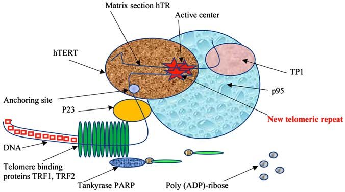
binding factor 1 and 2 (TRF1 and TRF2) (Fig. 1) (12‑15). mucous cells, in lymphocytes of peripheral blood (PB), and
thymus (25). In addition, the catalytic subunit of telomerase
2. Telomeres and telomerase: The generation of aging (hTERT), which is an RNA‑dependent DNA polymerase that
theory synthesizes telomeric repeats, as well as the mRNA of this
subunit, is also expressed in these cells at a relatively low, but
In a historical perspective, Hayflick and Olovnikov greatly detectable level.
contributed to the field of telomeres and telomerase, with the During life, telomeres of PB lymphocytes gradually shorten
work of Hayflick in 1961, triggering many scientists to try to due to cell division, accumulating with age in vivo (25). It has
explain how cells count their divisions (16). He showed that been shown that in many diseases, including immune‑asso-
human cells divide only a limited number of times in culture, ciated diseases (rheumatoid arthritis, atopic dermatitis, and
while at that time the public opinion was dominated by the bronchial asthma), PB lymphocytes are characterized by short-
conviction that T cells are immortal in the hands of a skilled ened telomeres and increased proliferation compared with
researcher. The article received a tremendous public response those in healthy lymphocytes. Numerous studies have shown
and the term ‘Hayflick limit’ was created and included in the that telomere shortening may contribute to osteoarthritis and
dictionaries. osteoporosis as an epigenetic factor and it is further supportedMolecular Medicine REPORTS 22: 1679-1694, 2020 1681
Figure 1. Schematic representation of the interaction of the telomeric complex (comprised of TRF1, TRF2) with a part of telomeric DNA. Protein components
include p95, p23 and ТР1 proteins, as well as the catalytic subunit hTERT, which performs the synthesis of telomeric repeats. Moreover, tankyrase PARP
is depicted to also interact with the telomeric complex. TRF, telomeric repeat binding factor; hTERT, human telomerase reverse transcriptase; PARP, poly
(ADP)-ribose polymerase.
that TEL measurement of chondrocytes and PB cells may be ALT‑alternative lengthening of telomeres. In addition to
an appropriate marker for the evaluation of the progression of telomerase activation, there is a telomere lengthening mecha-
these diseases (26‑29). Moreover, other factors can influence nism, alternative to telomerase (ALT‑alternative lengthening
the rate of telomere shortening in lymphocytes, such as the of telomeres). However, this mechanism in mammalian cells
presence of chronic stress (30) and drug abuse (31). It has been is only present in abnormal situations, such as in cancer cells,
shown that telomerase expression in lymphocytes is strictly immortalized cell lines, and murine cell knockout for the telom-
controlled during their development, differentiation, and acti- erase gene, whereas normal human lymphocytes cannot use
vation (30), and because of stimulation, ‘adult’ lymphocytes ALT to maintain their telomeres. A minority of immortal cell
become capable of expressing telomerase at a rather high lines and tumor cells use ALT to counteract telomere erosion
level. After any re‑stimulation, telomerase expression may due to cell division (35,36). Observations on the dynamics of
also increase but its level does not reach the level of response the TEL of immortalized cells suggest that this mechanism
to the primary stimulus (32). Such dynamics may reflect the is based on recombination, resulting in individual telomeres
regulatory mechanisms of clonal expansion of lymphocytes, that undergo rapid shortening and rapid elongation (37).
which is part of the immune response and control mechanism Besides, ALT cells can lengthen their telomeres using telo-
of their proliferation. meric sequences from other chromosomes as a template
Somatic cells are mostly devoid of telomerase activity. for new DNA synthesis (38,39). Cesare and Reddel (2010)
It is suggested that telomerase activity can be regulated in convincingly proved in their research that tumor cells treated
human cells by intranuclear transfer of key components. It was with telomerase inhibitors can activate the ALT pathway for
shown in HeLa cervical carcinoma cells that hTR and hTERT, telomeric maintenance through recombination (40). This can
during most of the cell cycle, accumulate in intranuclear sites explain the therapeutic failures and/or a resistance against
separately from telomeres, whereas during S phase, they are telomerase inhibition‑based anti‑cancer therapy. Therefore,
specifically recruited at telomeres. Therefore, the recruitment developing new therapeutics targeting proteins known to be
of telomerase at telomeres is dynamic, reaching a peak in involved in the latter pathway will be crucial for the targeting
the middle of S phase. Moreover, both hTR and hTERT are of ALT‑specific cells (41).
associated with nucleoli and Cajal bodies during S phase, Telomerase is the main mechanism for stabilizing telomeres
participating in telomerase transfer (33). Interestingly, human in lymphocytes and they use it to reduce telomere shortening
peritoneal mesothelial cells (HPMCs) appear to have unusu- during cell proliferation (42). Thus, in intact lymphocytes in
ally short telomeres (3.5 kbps) despite the presence of active systemic lupus erythematosus, multiple sclerosis, and atopic
telomerase. These telomeres do not shorten due to aging but dermatitis, telomerase activity is enhanced (43). Moreover,
due to a decrease in telomerase activity. HPMC aging is asso- quantitative fluorescence in situ hybridization (Q‑FISH) on
ciated with mitochondrial dysfunction, leading to increased metaphase chromosomes in the PB lymphocytes showed
production of active oxygen species and reduced mitochondrial that the distribution of TEL for individual chromosomes
membrane potential, indicating that premature aging is highly differs in normal and pathological conditions. Patients with
associated with oxidative damage in non‑telomeric regions of rheumatoid arthritis had significantly shorter telomeres of
the genome (34). chromosome 4p, an observation that could be related to the
Telomerase activity in autoimmune diseases, such as pathology of the disease, while the activity of the disease is
rheumatoid arthritis, lupus erythematosus, atypical derma- inversely correlated with the length of the telomeres of the 10p
titis, psoriasis, multiple sclerosis, and aplastic anaemia is chromosome which carries the genes involved in the differen-
summarized and presented in Table I. tiation and proliferation of T‑cells (44). The dynamic activityTable I. Telomerase activity in autoimmune diseases.
1682
No. Disease Telomerase activity in autoimmune disease (Refs.)
1 Rheumatoid The telomerase activity level in synovial infiltrating lymphocytes was significantly correlated with the intensity of Yudoh et al (26)
arthritis synovial lining hyperplasia, microvessel proliferation, lymphocyte infiltration, and percentage of synovial cells
positive for proliferating cell nuclear antigen in rheumatoid synovium
Activated lymphocytes with high telomerase activity distribute systemically through the circulation and contribute Hiyama and Hiyama (27);
disease activity in connective tissue disease. The stimulatory effect of estrogens on telomerase activity and female Yamanishi et al (187);
predominance in connective tissue disease may contribute to high telomerase activity in these pathologies. Tarhan et al (188)
Telomerase activity in lymphoid cells of lymphoid organs is higher than in peripheral blood. Hiyama and Hiyama (27);
Highest hTERT values were observed in Rheumatoid arthritis group (21.24±28.54). hTERT values were significantly Tarhan et al (188);
higher than the healthy control group (7.62±4.21) (PMolecular Medicine REPORTS 22: 1679-1694, 2020 1683
of telomerase is correlated with the action of an acute stressor
Yamaguchi et al (198); Marrone et al (199)
Yamaguchi et al (198); Marrone et al (199)
and with two types of reactions to stress‑psychological stress
and neuroendocrine (cortisol) responses to the stressor (24).
Several studies indicate that in most of the above situations
(immunopathological diseases, stress, and many others) the
telomeres of lymphocytes shorten with age faster than in
healthy individuals (45,46).
Brümmendorf et al (194);
(Refs.)
The telomere reduction and short cell life can be explained by the presence of mutations associated with this Ly (196); Young (197);
Most patients carrying these mutations are insensitive to immunosuppressive therapy. Ly (196); Young (197);
Yamaguchi et al (198)
Role of telomerase in mitochondria. It is known that mito-
Ogoshi et al (192);
Fogarty et al (195)
chondrial dysfunctions are found in metabolic disorders,
Taylor et al (193)
The level of telomerase activity in skin diseases that are not malignant is much lower than in malignant tumors. Taylor et al (193)
Atopic dermatitis Telomerase activity in the affected areas of the skin is significantly higher than in intact skin and is associated Jang et al (191)
neurodegenerative diseases, and autoimmune diseases (47,48).
Wu et al (46)
Mitochondria are important organelles for the production of
bioenergy, for biosynthesis and signaling, thus an integral
part of cellular adaptation to the environment. Concomitantly,
mitochondria are important mediators of carcinogenesis,
since this process requires flexibility for the cells to adapt to
the environmental changes. Many aspects of mitochondrial
The length of telomeres in blood cells progressively decreases by 216 base pairs in addition to the age loss of
Among patients with polymorphisms in the TERT gene, the telomere length was within 90% of the values of
biology support transformation, including mitochondrial
Lymphocytes that infiltrate the epidermis are not responsible for the level of telomerase, since it does not
biogenesis and metabolism, fusion dynamics, regulation of
oxidative stress, and cell‑signaling. Thus, understanding
the mechanisms of mitochondrial function during cancer
genesis is critical for future cancer treatment (49). Mutations
A high level of telomerase activity in peripheral blood mononuclear cells compared to donors.
in mitochondrial genes are common in cancer cells, they do
not inactivate mitochondrial energy metabolism but rather
alter the mitochondrial bioenergetic and biosynthetic state,
including succinate dehydrogenase, fumarate hydratase, and
isocitrate dehydrogenase 1, which lead to an increase in the
Telomerase activity in autoimmune disease
level of succinate, fumarate or R‑2‑hydroxyglutarate. These
metabolic changes can inhibit α‑ketoglutarate‑dependent
dioxygenases, possibly contributing to oncogenesis (50).
Disruption of mitochondrial activity is directly related to
neurodegenerative diseases, indicative of the importance of
mitochondrial functions in normal cellular physiology. Recent
studies focused on mitochondrial function in reactive oxygen
generation and its participation in the development of neurode-
generative diseases (51).
As is generally known, the mitochondrial genome is a highly
correlate with the number of these lymphocytes.
replicated, specialized genetic system that allows individual
mitochondria to respond to changes in membrane potential
and support oxidative phosphorylation by gene expression (52).
Studying this special role, one should not be surprised that the
disease in the hTR and hTERT genes.
with proliferative activity of the cell.
mitochondrial genome is regulated and expressed in a unique
way. Human mitochondria contain a compact ring genome
of 16,569 bp in length (53). Replication and transcription of
mitochondrial DNA (mtDNA) is initiated from the non‑coding
region, loop D, and is regulated by nuclear‑encoded proteins
36 base pairs per year.
that are post‑translationally imported into mitochondria.
Mitochondrial RNAs are transcribed as long polycistronic
healthy donors.
precursor transcripts from ‘heavy’ and ‘light’ chains, which
are processed according to the ‘tRNA punctuation model’,
whereby 22 disseminated tRNAs are excised to release rRNA
and mRNA (54). The released RNAs then undergo maturation,
which involves the polyadenylation of 30 ends of mRNA and
rRNA (55). Together, this includes a unique genetic system
that is able to translate genes encoded by mitochondria into
Table I. Continued.
and psoriasis
No. Disease
13 protein subunits of the electron transfer chain. The subtle
features of the mitochondrial transcriptome are still poorly
understood: Regulation of the number of transcripts, RNA
processing and modification sites, and the possible presence
5
of non‑coding RNAs. The recent advent of deep sequencing
41684 RAZGONOVA et al: TELOMERASE AND TELOMERES IN AGING
has provided a global nuclear transcriptome profile, revealing The regulation of TEL and telomerase activity is a complex
unanticipated transcription complexity, which includes wide- and dynamic process that is tightly linked to cell cycle regula-
spread post‑transcriptional processing and an abundance of tion. HSCs demonstrate telomeric shortening during replicative
non‑coding RNA (56‑58). Mercer et al (2011) used a similar aging despite expression of low levels of telomerase (66).
approach to study mitochondrial transcriptome (59). Granulocytes and naive T cells showed a parallel two‑phase
Wang et al (60) (2010) investigated the import functions decrease in TEL with age, which most likely reflected the
of some lncRNAs in mitochondria, showing that one of the accumulated cell divisions in the HSCs that are the common
RNAs, the RNA component of the telomerase (TERC), is precursors of both types of cells. Memory T cells also first
processed in mitochondria and then exported out of mitochon- showed a rapid decrease in TEL with age, which continued for
dria. Partial disruption of mitochondrial function caused the several years, resulting in lymphocytes with shorter telomeres
accumulation of mitochondrial‑processed RNA, TERC, in the that granulocytes in older people. These results indicate a sharp
cytosol. These results provide a potential link between mito- decrease in stem cell turnover in early childhood and confirm
chondrial function and telomerase activity. The import of RNA that cell divisions in HSCs and T cells lead to telomere short-
into mitochondria is very important for replication, transcrip- ening (67,68). Telomere shortening is most pronounced during
tion, and translation of the mitochondrial genome. PNPase is bone marrow resection, as it has been observed that there is an
an important regulator of the import of nuclear‑encoded RNA inverse correlation between the number of transplanted mono-
into the mitochondrial matrix. It has been shown that the nuclear cells and the degree of telomere shortening. A bone
decrease in PNPase impairs mitochondrial RNA processing. marrow transplant results in a telomere shortening equivalent
Moreover, the increase in imports of RNase P, 5S rRNA, and to a period of 15 years of life (69).
multidrug resistance proteins RNA is directly dependent on Interestingly, Scheding et al (70) (2003) attempted to assess
the expression of PNPase. These observations led the iden- whether repeated stimulation of the hematopoietic system due
tification of PNPase‑imported RNA interactions, suggesting to regular blood sampling will negatively affect the TEL of PB
a new role of PNPase in providing translocation of RNA to leukocytes (PBLs). It is known that the TEL of PBLs can be
mitochondria (60). In addition, Cheng et al (61) (2018) showed used to evaluate HSCs turnover. A study using fluorescence
that the RNA component of mammalian TERC telomerase is in situ hybridisation and flow cytometry (Flow‑FISH), showed
imported into mitochondria, processed into the shorter form that when compared with the corresponding non‑donor data,
of TERC‑53, and then exported back to the cytosol. These data no differences were observed. In addition, neither age‑adjusted
collectively reveal the existence of a pathway of mitochondrial donor data for TEL for granulocytes nor TEL for lympho-
RNA transfer and provide a potential mechanism for trans- cytes correlated with either the total number of years or the
ferring the functional states of mitochondria to other cellular total number of whole blood (WB) and/or PLT donations.
compartments. Long‑term donation of WB does not affect TEL, as measured
Several recent reports have studied the transport of hTERT by Flow‑FISH, which indicates a significant increase in stem
to mitochondria, which is most likely controlled by a mito- cell turnover (70). Moreover, lymphoid cells are capable of
chondrial sequence at the N‑terminus of hTERT. It has been very intensive clonal reproduction with antigenic stimulation.
shown that the accumulation of endogenous oxidative stress Despite the induction of telomerase, their telomeres are more
leads to the hTERT nuclear export to the cytosol, reducing significantly shortened than in granulocytes. Accordingly,
the nuclear and total telomerase activity. The addition of memory cells have significantly shorter telomeres than
antioxidants slows down this process significantly (62). Aging lymphocytes (71).
is associated with an increase in the number of intracellular There are many unresolved issues in telomeric stem cell
reactive oxygen species (ROS) and a loss of telomerase biology and the various stem cells of an adult organism vary
activity. Haendeler et al (62) (2004) investigated whether greatly regarding the role of telomeres and telomerase in
an age‑related increase in the number of ROS can induce maintaining proliferation. These stem cells include HSCs and
nuclear export of TERT and promote endothelial cell aging. mesenchymal stem cells (MSCs) from human bone marrow,
Haendeler et al found that in parallel with increased ROS that are extensively studied. hMSCs are a type of adult stem
formation, the TERT nuclear protein is exported from the cells from the bone marrow, which can differentiate into bone,
nucleus to the cytoplasm and Src kinase is activated. Incubation cartilage, adipose, and muscle cells (72). hMSCs show a total
with N‑acetylcysteine reduced the formation of intracellular absence of telomerase activity, in contrast to HSC and other
ROS and prevented mitochondrial DNA damage, as well as adult stem cells (73). Ectopic telomerase expression is able
low doses of atorvastatin that had a similar effect. Therefore, to expand their limited replicative ability in tissue culture,
it has been suggested that antioxidants and statins can delay maintaining their functional characteristics (74), a feature that
the onset of replicative aging by counteracting the increase in can be developed for the application of these cells in tissue
ROS associated with aging of endothelial cells (62‑64). engineering (75).
Importantly, stem and cancer cells share several charac-
Telomeric stem cell biology. Most human stem cells are teristics, including factors that regulate self‑renewal in both
characterized by a rather slow proliferation rate, which was normal HSCs and leukemic cells (76), leading to the hypoth-
estimated for the hematopoietic stem cells (HSCs), as one divi- esis of ‘cancer stem cells’ (77). Cancer stem cells resemble
sion in 1‑2 years (65). During the intervals between divisions, normal stem cells in the sense that when they divide, one of the
the cells are in proliferative rest. However, even with such daughter cells differentiates into a certain type of cell, which
proliferation rates, it is clear that stem cells have a proliferative eventually ceases to divide, while the other retains its stem cell
potential that is bigger than the Hayflick limit. properties, being able to divide constantly. Cancer stem cells,Molecular Medicine REPORTS 22: 1679-1694, 2020 1685
which constitute only a small fraction of the total population studies have shown that histone modification is an important
of tumor cells, are the only tumor cells capable of supporting means of regulating telomerase gene expression (83,84). The
tumor growth. Therefore, a scientific assumption was made loss of telomerase activity is accompanied by a shortening of
that a complete cure for cancer may require the development telomeres, which is especially fast in the early stages of differ-
of treatments targeting cancer stem cells, assuming that this entiation. Telomere shortening can be accelerated due to high
can be accomplished without destroying the stem cells needed ROS levels since telomeres are at least partially deficient in
to maintain tissue regeneration, such as bone marrow and repairing oxidative DNA damage (85). It was also shown that
intestines (77). In addition, stem cells may be the source of the self‑renewing potential and longevity of HSCs in vivo are
some types of cancer, increasing the risk when used to repair largely determined by the ROS levels (86).
organs, while they can also be of prognostic value as the Lonergan et al (87) (2006) suggested that mitochondrial
proportion of stem cells in a tumor can determine how deadly characteristics may be an additional and reliable way to test
it is. stem cell competency. The results indicated that, the peri-
Some therapeutic strategies for cancer treatment are nuclear arrangement of mitochondria in the primate stem cell
focused on the destruction of cancer stem cells. However, many line, accompanied by a low ATP/cell content and high oxygen
potential uses require stem cells to divide to produce enough consumption, may be a true indicator of competence in stem
cells to replace damaged tissue, a fact that could possibly cell differentiation, and deviations from this profile suggest
contribute to the accumulation of potentially cancer‑causing thaT cells differentiate or possibly age.
mutations (77). The origin of stem cells for some types of Passos et al (88) (2007) found in their studies that an
cancer means that these stem cells have telomerase that does increase in mitochondrial density in combination with mito-
not need to be reactivated, although the enzyme activity may chondrial dysfunction can accelerate aging. This is confirmed
be further increased in the later stages of carcinogenesis (78). by a negative correlation between the density of mitochondria
On the contrary, the limited activity of telomerase, associated and the maximum life expectancy in interspecific comparisons
with the final lifespan of adult stem cells, can both contribute in mammals. Strengthening mitochondrial biogenesis and,
to aging and cancer prevention (79,80). possibly, impaired mitochondrial degradation and segregation,
if this occurs as an adaptation to pre‑existing mitochondrial
Forced expression of telomerase (telomerization of cells). In dysfunction, it can significantly increase ROS production and,
many cell types, telomerase activity appears only after the thus, actively contribute to aging. In other words, both ATP and
forced expression of the hTERT gene, a process called telom- ROS production increase in parallel with the bulk density of
erization. The RNA component of telomerase is expressed in mitochondria. In an aging organism, the mitochondrial mass
most human cells regardless of telomerase activity; therefore, and ROS production increase, while the membrane potential
after the transfection of human cells with the hTERT gene, decreases due to the activation of the uncoupling protein (89).
telomerase activity appears in them, and cells gain the ability A rather interesting result was obtained after the introduc-
to maintain TEL. Such cells are called telomerized. The pres- tion of the hTERT gene into the cells of a patient suffering
ence of telomerase activity does not violate the mechanisms of from Niemann‑Pick C disease (NPC). NPC is a recessive lipi-
cell growth regulation but it contributes to the stability of the dosis that is characterized by excessive accumulation of free
genome, preventing chromosomal rearrangements instead. The cholesterol and glycosphingolipids in the endosomal lysosomal
sensitivity of telomerized and initial fibroblasts of adult skin system. Walter et al (90) (2003) confirmed that ectopic expres-
to common cytostatics i.e., doxorubicin, cisplatin, etoposide, sion of hTERT in human cells leads to increased regulation of
and dacarbazine was studied. No significant differences in small GTPase Rab9 and its p40 effector. Telomerase immor-
sensitivity were found. These observations directly support the talization of human cells affects the function of the endosomal
safekeeping of normal mechanisms of homeostasis in telomer- lysosomal system and in particular the efflux of cholesterol
ized cells and indirectly support the assumption that they can from this system, independently of the NPC1 protein. Such
be used in cell therapy (81). changes may be necessary to maintain cells in a continuous
Saretzki et al (82) (2004) found very similar molecular proliferative state (90,91). In fact, a recent study has provided
events in two unrelated human embryonic stem cell (hESC) evidence for a correlation between cholesterol content and cell
lines that regulate the suppression of telomerase activity, cycle progression (92). Inhibition of cholesterol biosynthesis
increase oxidative stress, and decrease DNA repair activity with low concentrations of lovastatin blocked cell prolifera-
during spontaneous differentiation. This study characterized tion. When LDL‑cholesterol was added to these cells, cell cycle
resistance to oxidative stress and restoration of DNA breaks progression resumed (92). In NPC1 disease, cells are unable to
in parallel with the regulation of telomerase activity and TEL metabolize LDL‑derived cholesterol due to its accumulation
during differentiation of hESC. Previously, it was reported in the late endosomal lysosomal system. The expression of
that murine ESC has a high level of antioxidant protection hTERT in NPC cells leads to the correction of the cell pheno-
and effectively repair DNA chain breakage but the level of type, including the elimination of the accumulated cholesterol.
antioxidant protection and restoration of chain breakdown In particular, in NPC‑TERT cells, cholesterol transport from
noticeably decrease during differentiation (82). The data the endosomal lysosomal system to the plasma membrane is
obtained indicated the deacetylation of histones H3 and H4 in restored with a concomitant increase in cholesterol esterifica-
the hTERT promoter region. The deacetylation of histone H3 tion (90).
in the hTR promoter was suggested as a candidate mechanism Transfection of cells with expression vectors containing
for explaining the suppression of expression of both genes and hTERT supports TEL and provides normal cells with an
the loss of telomerase activity during differentiation. Other unlimited life span in culture. It is important that hTERT1686 RAZGONOVA et al: TELOMERASE AND TELOMERES IN AGING
prolongs the life span of cultured cells well beyond normal telomeres and may be a marker of aging (110). Moreover,
aging without causing a neoplastic transformation (93). chronic stress and depression are directly related to high levels
Condon et al (93) developed a cell line from hTERT‑infected of hydroxy‑deoxyguanosine and reduced levels of antioxidant
human myometrial cells (hTERT‑HM). Cells expressing enzymes (111,112). In addition, oxidative stress affects telo-
telomerase contained mRNA for α‑smooth muscle actin, meres more frequently compared to other areas of genomic
smoothelin, an oxytocin receptor, and an estrogen receptor α. DNA and inhibits telomerase activity in vitro in various cell
Myometrial cells immortalized with hTERT retained the types (113,114).
differentiation markers that are observed in primary smooth An antioxidant function is associated with various micro-
muscle cell (SMC) cultures. nutrients, including vitamin C, E, folic acid, and ω‑3 fatty acids
As known, the ectopic expression of hTERT prolongs and, therefore, can positively affect TEL (115). Stress leads to
the lifespan of certain human cells. McKee et al (94) (2003) having high levels of pro‑inflammatory cytokines, including
introduced hTERT into human SMCs and found that the IL‑6 and TNF‑α (116,117).
resulting cells proliferated well beyond their normal lifespan Insulin‑like growth factor 1 (IGF‑1) exerts a mitogenic
and, remarkably, retained the characteristics of normal control effect implicated in cell proliferation. It was first reported that
SMCs. Using these neonatal SMCs, they were able to create IGF‑1 is involved in the modulation of telomerase activity by
reliable human vessels, and then it becomes possible to enhancing the stimulation of telomerase activity caused by
create coronary arteries for coronary artery bypass grafting. phytohemagglutinin (PHA) in cord blood mononuclear cells,
Klinger et al (95) (2006) concluded that this tissue engineering which have characteristically low levels of telomerase activity
model emphasizes the effect of donor age on cellular synthetic and hTERT expression (118,119).
function, which apparently does not depend on the increase in Fibroblast growth factor 2 (FGF2) has been shown to
hTERT life span. induce a dose‑dependent increase in both neural precursor
cell proliferation and telomerase activity in primary cortical
Chronic stress exposure and telomerase activity. PB mono- cultures (118,120). In contrast, in both neurons and glial cells
nuclear cells (PBMCs), are most suitable for the study of in primary cultures of E15 mouse embryo cortex, down‑regu-
telomerase activity. Maximow (96) (1909), the founder of the lation of telomerase activity and mTERT gene expression
stem cell theory, revealed that PBMCs are stem cells that are occurred in parallel with cell differentiation (120). In primary
common to various blood elements in embryonic development endothelial cells, freshly isolated from intact endothelium,
and during the fetal life in mammals. It has been shown that telomerase activity is observable during logarithmic growth but
there is an accelerated telomere shortening and reduced telom- not when cells enter quiescence (121). Treatment of quiescent
erase activity in PBMC in various situations. Recent studies human umbilical vein endothelial cells (HUVECs) with FGF2
have shown changes in TEL and telomerase activity due to reactivates telomerase activity in a time‑ and dose‑dependent
psychological and social factors (64). An earlier study showed manner, in contrast to vascular endothelial growth factor‑A
that telomere shortening and the decrease in the telomerase (VEGF‑A), which has no effect (121). Consistently, FGF2 but
activity is higher in PBMC of women with high stress levels not VEGF‑A, up‑regulates hTERT expression in parallel with
compared with the control group (24). This study initiated a transcription factor Sp1 (121).
large number of similar studies, showing that telomeres are VEGF has angiogenic activity that can contribute to tumor
shorter in patients with mood disorders (97), in hemodial- development by inducing vessel permeability that facilitates
ysis‑depended patients (98) and in patients with depressive tumor cell proliferation and metastasis (122). Although
disorder (99). VEGF‑A appears not to regulate telomerase activity in human
To date, the effect of chronic stress on telomerase activity umbilical vein endothelial cells (123), VEGF and lysophos-
in humans has been described in many studies (68,100‑102). phatidic acid (LPA), both secreted from ovarian cancer cells
This effect includes an increase in the oxidative level, which and known to promote cancer cell growth, have been shown to
stimulates hTERT nuclear export to the cytosol and decreases regulate telomerase activity in non‑endothelial cells (118,124).
the nuclear and total telomerase activity (62,103,104). It has Up‑regulation of telomerase activity by VEGF appears to be
been observed that long periods of psychological stress can receptor‑dependent, as unsaturated vitamin E, tocotrienol,
increase vulnerability to the effects of the virus that induces has been shown to down‑regulate telomerase activity through
aging of specific T cells (32). down‑regulating the VEGF receptor (125). Vitamin E has also
According to Lansdorp (105) (2006), telomere shortening been reported to down‑regulate telomerase activity in ovarian
can be due to the influence of psychological stress and social cancer cells (118,126).
rank, whereas Adams et al (106) (2007) contradict the Genetic factors are also related to telomere shortening.
theory that socio‑economically deprived people age faster Monoamine oxidase‑A and apolipoprotein E (APOE) poly-
and, therefore, have shorter telomeres than the more affluent morphisms were statistically significant for TEL only in
part of society. Chronic stress can affect human health people with mental and depressive disorders but not in healthy
through a variety of behavioural aspects and biochemical respondents. In addition, sex, the APO‑ε2 allele, and the poly-
pathways (107). The shortening of telomeres in cells of the morphism of the monoamine oxidase‑A have an indirect effect
immune system occurs through biochemical reactions (108), on TEL. Thus, mental and depressive disorder is an additional
while stress can also affect the occurrence of age‑related mediator between monoamine oxidase‑A and APOE poly-
diseases (109). It has been shown that metabolic stress plays morphisms and TEL (99). Further, studies revealed similar
an important role in telomere shortening and that the decrease patterns in the European population for the APOE‑ε4 gene
in the level of ω ‑3 fatty acids in the blood correlates with polymorphism, which is associated with a risk of Alzheimer'sMolecular Medicine REPORTS 22: 1679-1694, 2020 1687
disease. Although telomeres are shortened significantly faster adaptation. Accordingly, changes in TEL and telomerase
in female carriers of the APOE‑ε4 mutant allele, hormone activity may reflect the effectiveness of the treatment
replacement therapy in middle‑aged women can change the of age‑related diseases. Most of the recent studies on telomeres
effect of allele transfer on aging (127). and age‑related diseases are based on the fact that increase
In order to explain the effect of chronic stress on telomere in telomerase activity corresponds to either a decrease in
shortening, Damjanovic et al (128) (2007) studied immuno- telomere shortening (30), or telomere elongation (142).
logical changes in caregivers for relatives with Alzheimer's
disease. TEL was significantly shorter in caregivers than in Effect of TEL on fertility and pregnancy. Vasilopoulos et al (143)
control subjects and their immune response was weaker. (2019) investigated the potential relationship between TEL
Moreover, caregivers had significantly lower T cell prolif- and various factors of female and male infertility. Most female
eration but higher production of immunoregulatory cytokines infertility factors have been associated with shorter TEL, with
(TNF‑α and IL‑10) than control subjects in response to in vitro the exception of endometriosis, premature ovarian failure,
stimulation. These results demonstrate that chronic stress transparent cell carcinoma, and polycystic ovary syndrome
leads to a reduction in TEL and is associated with accelerated (PCOS). These types of diseases have been associated with
aging of T‑cells. a longer TEL, which has shown conflicting results in several
However, there is some controversy among different studies studies. On the other hand, male infertility factors were associ-
on telomere shortening under stress conditions. Effros (129) ated with critically shorter TEL (143).
(2001) proposed an interesting model for the study of PB cells Fragkiadaki et al (144) (2016) summarized and critically
in vitro, where lymphocytes from healthy donors, namely discussed the evidence linking telomerase activity with preg-
men and women aged 25‑55 years, were exposed to various nancy complications. Maternal age is a determining factor
concentrations of cortisol, which is a stress‑related hormone in fertilization success and numerous studies have focused
or dimethyl sulfoxide. He showed that the level of telomerase on telomerase activity and its correlation with mammalian
activity decreases under stress, which explains the decrease fertilization, as well as subsequent splitting processes and
in both the number of immune‑competent T cells and the preimplantation development. It has been also shown that
length of telomeres in these cells under chronic stress, further there is a relationship between telomerase activity and compli-
accelerating cell aging and deterioration of the immune cations during pregnancy.
system (130,131). Additionally, the effect of psychosocial stress on pregnancy
Ornish et al (132) (2008) investigated for three months is an important risk factor for the early onset of common
the comprehensive lifestyle changes for 30 men with age‑related diseases (145). Human studies have demonstrated
biopsy‑diagnosed low‑risk prostate cancer, which led to a a link between chronic or excessive psychosocial stress
significant increase in telomerase activity by 10%. Increased and telomere biology (146). It was shown that stress‑related
telomerase activity was associated with a decrease in the level changes in telomeres might be a possible mechanism linking
of low‑density lipoprotein cholesterol (LDL) and a decrease in psychosocial stress with age‑related diseases (147). Indeed,
psychological stress. Physical activity appears to protect against the accelerated shortening of telomeres reflects stress‑induced
metabolic and psychological stress and maintain TEL (133). oxidative cell damage and accelerated aging (97). Therefore,
Moreover, a study on various diets showed that chronic food Lazarides et al (148) (2019) examined the hypothesis that the
restriction (regardless of age), body mass index, or smoking maternal pro‑inflammatory state during pregnancy, which is a
might be a risk factor for telomere premature shortening (134). balance between TNF‑α, the main pro‑inflammatory cytokine,
Furthermore, regular sleep, exercise, and a healthy lifestyle and IL‑10, the main anti‑inflammatory cytokine, are directly
contribute to normal TEL and telomerase activity, regard- related to the TEL of the leukocytes (LTL) of the newborn
less of the level of the depressive disorder on the Hamilton at birth. The higher average ratio of TNF‑ α /IL‑10 during
Depression Rating Scale (135). These data are consistent with pregnancy was significantly associated with shorter TEL of
the results of another study, which showed that the effectiveness the newborn after adjusting for the mother's age, body mass
of an antidepressant depends on the initial level of telomerase index before pregnancy and sex of the child.
activity (136). In addition, De Punder et al (137) (2018) recently
reported that high levels of chronic stress reduce the ability of Ef fect of nutraceutical supplements on TEL. The
immune cells to induce telomerase activity by 25% compared Dietary Inf lammatory Index (DII) was developed by
to moderate or low levels of chronic stress. Chronic stress is Shivappa et al (2014) to measure the inflammatory potential
associated with a decrease in mitogen‑induced lymphocyte of diet and it can be used in diverse populations to predict
proliferation and mitogen‑induced IL‑2 production (138), levels of inflammatory markers, including C‑reactive protein
which are activators of signalling pathways that stimulate (CRP) and interleukin‑6 (149‑151). Shivappa et al (152) (2017)
telomerase activity (139,140). Chronic stress exposure is examined whether inflammatory potential of diet, as measured
also associated with a higher level of oxidative stress, which by the Dietary Inflammatory Index (DII) has an impact on
ultimately leads to the accumulation of senescent (CD28 ‑) telomere shortening in the National Health and Nutrition
T‑cells (103). This suggests stringent telomerase regulation in Examination Survey (NHANES). Validation of the DII with
human T cells, which may contribute to telomere shortening C‑reactive protein (CRP) was also carried out, showing that
and ultimate replicative potential and loss of control over there was an association between DII and leukocyte TEL
certain pathogens (141). in these NHANES data. The impact of specific fatty acids
In conclusion, the length of telomeres in PB cells can on inflammation can be crucial to the effect of dietary fats
be an indicator of human health, stress resistance, and cell on human health, for example polyunsaturated fatty acids,1688 RAZGONOVA et al: TELOMERASE AND TELOMERES IN AGING mainly ω ‑3 fatty acids, have anti‑inflammatory and immu- related to temperature and metabolic rate. Studies of relative nomodulating properties. Research studies have confirmed temporary scaling at long‑living mutants of C. elegans showed that excessive amounts of ω ‑6 fatty acids leading to a high that the life expectancy is controlled by some physiological ω ‑6: ω ‑3 fatty acid ratio can promote the pathogenesis of hours different from chronological time (170). chronic diseases (153). Interestingly, a Mediterranean diet Olovnikov (171) (2007) claims that the lunar cycle plays a supplemented with two fatty fish meals per week, rich in ω‑3 role in aging of living organisms. He suggested that pineal cells fatty acids, has been shown to have an anti‑inflammatory called pinealotcytes regularly change the endocrine secretory effect reducing airway inflammation in childhood asthma that activity, responding to periodic changes of the gravitational field is a chronic disease (154). Therefore, it is likely that blood of the moon. The theory is based on the idea of a controlled loss polyunsaturated fatty acid levels are involved in preventing by a neuron in the brain of chronomeres. The regular process of telomere shortening over time (155). In agreement with this, loss of chronomeres in neurons is controlled by the pineal gland recent studies revealed that the administration of nutraceu- and activated at least once a lunar month. Hormonal signals can tical supplements to healthy individuals is implicated in TEL be generated by the pineal gland and depend on the gravity maintenance (156). Participants were selected from healthy of the periodic shift of the mineralized deposits located at the outpatients and divided into an intervention group that intra‑pinealotcyte of neighbour cellular structures. The calcific received nutraceutical supplements and a control group. The large particles that are in extracellular spaces of a pineal gland length of individual telomeres was estimated in metaphase can also contribute to the hormonal secretion. leukocytes isolated from PB using Q‑FISH analysis. The This pineal secretion activates the hypothalamus, enhancing median length of the telomeres was significantly increased the hormonal response, which allows a sharp increase in the (P
Molecular Medicine REPORTS 22: 1679-1694, 2020                                            1689

during normal development. Dedifferentiation or decreased            Acknowledgements
treatment specificity is a robust characteristic of cognitive
aging. Voss et al proved that neural dedifferentiation was not       The authors would like to thank Dr Muhammad Amjad
ubiquitous in different categories of stimuli, while nervous         Nawaz from SEC of Nanotechnology, Far Eastern Federal
dedifferentiation was relatively stable according to the age         University (Vladivostok, Russia) for his assistance with the
of the elderly (178,179). Moreover, an age‑related decline in        copy‑editing of the manuscript and for correcting the final
specialization has also been shown for the cognitive function        proofs.
of the frontal lobes of the hemispheres (180). Dreher et al
examined the relationship between dopamine synthesis in the          Funding
midbrain and the prefrontal ligament with reward. It was found
that healthy aging causes functional changes in the reward           This study was supported by Spin‑Off Toxplus S.A. and
system, and reveals age‑related changes in the relationship          by the Special Research Account of University of Crete
between dopamine synthesis in the midbrain and prefrontal            (ELKE nos. 4602, 4920 and 3963).
activity. These results gave insight into the interaction between
dopamine function of the midbrain and the reward system in           Availability of data and materials
young people and the elderly, and also determined the changes
that accompany aging (181). Furthermore, it has been shown           Not applicable.
that the contraction of the hippocampus, inferior temporal
cortex and prefrontal white matter are increased with age,           Authors' contributions
while shrinkage in the hippocampus and cerebellum acceler-
ates (182,183). The weakening of the functional correlations         All the authors contributed in conceiving and designing the
of individual parts of the brain spreads with age from the           study. MPR, AMZ and KSG searched the literature for inclu-
forehead to the back of the head (184).                              sion in the study and wrote the manuscript. MT, ES, and PF
    The decrease in the volume of a brain becomes noticeable         checked and reviewed the manuscript. KN, DT, DAS, and AT
approximately from 30 years of age (185). Total brain volume         gave advice and critically revised the manuscript. All authors
for mentally healthy people is steadily declining by 0.22% per       have read and approved the final version of the manuscript.
year between 20 and 80 years, and this decrease is acceler-
ated in older age (186). The brain in some patients has vital        Ethics approval and consent to participate
deposits of amyloid protein indicative of the preclinical phase
of Alzheimer's disease: Their brain volume is reduced by 2.5%        Not applicable.
compared with the brain of healthy volunteers. All of the listed
age‑related changes, including a decrease in brain volume, are       Patient consent for publication
considered a result of aging caused by various factors, among
which is the accumulation of damage caused by free radicals.         Not applicable.
However, qualitative changes in the brain can be interpreted in
a completely different way, meaning that all these changes in        Competing interests
the brain are not the result of errors, but the product of genetic
programming.                                                         DAS is the Editor‑in‑Chief for the journal, but had no personal
                                                                     involvement in the reviewing process, or any influence in
5. Conclusions and future research directions                        terms of adjudicating on the final decision, for this article. The
                                                                     other authors declare that they have no competing interests.
As discussed in this review, telomerase activity is regulated in
response to various biological factors and systems, including        References
stress hormones, oxidative stress, inflammatory mediators,
and circadian rhythm. Therefore, future studies of telomerase         1. Morgan TH: Random segregation versus coupling in Mendelian
activity in PB mononuclear cells should include measurements             inheritance. Science 34: 636‑638, 1911.
                                                                      2. McClintock B: Cytological observations of deficiencies involving
of biomarkers that participate in these processes, which will            known genes, translocations and an inversion in Zea mays.
significantly contribute to enhancing our knowledge in the               Mo Agric Exp Res Stn Res Bull 163: 1‑30, 1931.
field. Moreover, it is necessary to further study the role of         3. Möller HJ: The remaking of chromosomes. Collecting Net 8:
                                                                         182‑198, 1938.
telomerase in the defence mechanism of mitochondria against           4. Blackburn EH and Gall JG: A tandemly repeated sequence at
oxidative stress to gain a broader understanding of the role             the termini of the extrachromosomal ribosomal RNA genes in
of telomerase in human health and the risk of stress‑related             Tetrahymena. J Mol Biol 120: 33‑53, 1978.
                                                                      5. De Lange T, Lundblad V and Blackburn EH (eds): Telomeres.
diseases.                                                                Cold Spring Harbor Laboratory Press, New York, NY, pp21‑48,
    A decrease in telomerase activity may explain why telo-              2006.
meres shorten faster under pathological conditions. The study         6. Hsu TC, Arrighi FE and Saunders GF: Compositional heteroge-
                                                                         neity of human heterochromatin. Proc Natl Acad Sci USA 69:
and characterization of individual differences in the ability            1464‑1466, 1972.
of the telomere biology system to respond to a challenge, in          7. Watson JD: Origin of concatemeric T7 DNA. Nat N Biol 239:
addition to indicators of basal telomerase activity and TEL,             197‑201, 1972.
                                                                      8. Olovnikov AM: The principle of marginotomy in the matrix
is an interesting non‑standard approach for future studies of            synthesis of polynucleotides. Dokl Akad Nauk SSSR 201:
telomere biology and aging.                                              1496‑1499, 1971.
You can also read