VACCINATION AGAINST CHOLERA AND ETEC DIARRHEA AND INTERVENTIONS TO IMPROVE VACCINE IMMUNE RESPONSES - TANVIR AHMED
←
→
Page content transcription
If your browser does not render page correctly, please read the page content below

VACCINATION AGAINST CHOLERA AND ETEC
DIARRHEA AND INTERVENTIONS TO IMPROVE
      VACCINE IMMUNE RESPONSES

                    TANVIR AHMED

         Department of Microbiology and Immunology
                   Institute of Biomedicine
     The Sahlgrenska Academy at University of Gothenburg
                         Sweden 2009
ISBN 978-91-628-7789-7
http://hdl.handle.net/2077/19796
2009 Tanvir Ahmed
The pictures on the cover page show a hospitalized child with
diarrhea, a child receiving oral cholera vaccine at the field clinic and a
child receiving zinc supplementation.
Printed by Geson Hylte Tryck
Gothenburg, Sweden, 2009
‐2‐
Dedication

      This thesis is dedicated
      -to my late father Amjad and to my wonderful mother, Fahmida, who
      have raised me to be the person I am today and always, supported my
      endeavors
      -to my beloved wife, Chuty, who inspires me to be all that I can be
      -and my inspiration, of course, to my two kids, Ariana and Tanisha, who
      are my constant companions, delights, and irritants

“The world is my country, all mankind are my brethren,

and to do good is my religion”

-Thomas Paine

                                    ‐3‐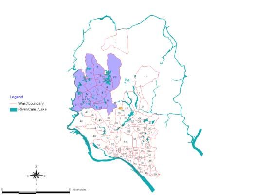
Vaccination against cholera and ETEC diarrhea and interventions to improve
vaccine immune responses
Tanvir Ahmed
Department of Microbiology and Immunology, Institute of Biomedicine at the Sahlgrenska
Academy, University of Gothenburg
Abstract
Vibrio cholerae O1 and enterotoxigenic Escherichia coli (ETEC) together account for the
majority of bacterial causes of acute dehydrating diarrhea in children in Bangladesh. Vaccines
should be considered as an important public health tool for prevention of these diarrheal diseases.
However, a limitation for the use of vaccines in developing countries is that the efficacy and
immunogenicity of vaccines, especially oral enteric vaccines, are lower in these countries than in
the industrialized world. The main objectives of the thesis were to study the safety and
immunogenicity of oral cholera toxin B subunit (CTB) containing inactivated whole cell ETEC
and cholera vaccines in young children in a developing country and to identify possible immune
modulating factors, e.g. vaccine dose, different buffer formulations, effects of breast milk
withholding and zinc supplementation.
For determining optimal doses of the ETEC vaccine, we immunized 6 months to 12 year old
children with full, half and quarter doses of the ETEC vaccine. Safety and immunogenicity of
different vaccine doses were compared. All doses of the ETEC vaccine were found to be equally
immunogenic in the older children. However, a quarter dose, although giving somewhat lower
antibacterial responses than a full dose, was required for children 6-18 months to avoid
reactogenicity.
For determining the safety and immunogenicity of the cholera vaccine in young children and the
effect of different interventions to try to enhance immune responses, children 6-18 months of age
were given two doses of the vaccine according to the standard protocol or with different
modifications. In addition to analyzing antibacterial and antitoxic B-cell responses, T-cell
responses were determined using a new flowcytometric technique, FASCIA. The vaccine was
found to be safe and to induce both antibody and Th1 type T-cell responses. Vibriocidal antibody
responses were improved by temporarily withholding breast-feeding for three hours before
immunization as well as by giving 20 mg of zinc from 3 weeks prior to and one week after the
second dose of vaccine. Zinc supplementation also enhanced IFN- responses to CTB.
Further objectives of this thesis were to analyze the immune responses to one of the most
prevalent ETEC colonization factors (CFs), i.e. CS6, in patients infected with CS6-positive ETEC
and to evaluate if there is an association between expression of certain Lewis blood group
antigens of the host and infection by ETEC expressing different CFs. Natural infection with CS6
ETEC was found to induce robust systemic and mucosal immune responses in 70-90% of adults
and children with diarrhea caused by CS6 positive ETEC strains, suggesting that CS6 could be
an important immunogenic component of a new ETEC vaccine. We could also show that
individuals with Le (a+b-) blood group had increased susceptibility to infection with ETEC
expressing CFA/I group fimbriae.
The results of these studies give important background information regarding the possibility of
inducing effective immune responses to oral inactivated enteric vaccines in young children in
developing countries.
Keywords: Vibrio cholerae, ETEC, oral vaccine, CS6, CFA/I, Lewis blood group, zinc, breast
feeding, T cell, B cell
ISBN 978-91-628-7789-7
‐4‐Original Papers
This thesis is based on the following papers referred to in the text by the given Roman
numerals:
I Qadri F, Ahmed T, Ahmed F, Begum YA, Sack DA and Svennerholm AM:
Reduced doses of oral killed enterotoxigenic Escherichia coli plus cholera toxin B
subunit vaccine is safe and immunogenic in Bangladeshi infants 6–17 months of
age: Dosing studies in different age groups.
Vaccine 24, 1726-33, 2006.
II Qadri F, Ahmed T, Ahmed F, Bhuiyan MS, Mostofa MG, Cassels FJ, Helander A
and Svennerholm AM: Mucosal and systemic immune responses in patients with
diarrhea due to CS6-expressing enterotoxigenic Escherichia coli.
Infect Immun 75, 2269-74, 2007.
III Ahmed T, Lundgren A, Arifuzzaman M, Qadri F, Teneberg S, Svennerholm AM:
Children with Lewis (a+b-) blood group have increased susceptibility to diarrhea
caused by enterotoxigenic Escherichia coli expressing colonization factor I-group
fimbriae.
Infect Immun 77, 2059-2064, 2009.
IV Ahmed T, Svennerholm AM, Tarique AA, Sultana GN and Qadri F: Enhanced
immunogenicity of an oral inactivated cholera vaccine in infants in Bangladesh
obtained by zinc supplementation and by temporary withholding breast feeding.
Vaccine 27, 1433-1439, 2009.
V Ahmed T, Arifuzzaman M, Lebens M, Qadri F, Lundgren A: CD4+ T-cell
responses to an oral inactivated cholera vaccine in young children in a cholera
endemic country and the enhancing effect of zinc supplementation.
Submitted for publication.
Reprints were made with permission from the publishers.
‐5‐Table of Contents
ABBREVIATIONS 8
INTRODUCTION 9
CHOLERA 11
Cholera epidemiology 11
Natural protection against cholera 11
Cholera vaccines 12
ETEC 15
ETEC epidemiology 15
Pathogenesis and mechanisms of immunity against ETEC diarrhea 15
ETEC vaccines 16
FACTORS INFLUENCING THE IMMUNE RESPONSES TO ORAL VACCINES 18
Hyporesponsiveness of vaccines in children in developing countries 18
Interventions to overcome hyporesponsiveness 19
Influence of the genetic diversity of the host to natural infection 20
AIMS 23
MATERIALS AND METHODS 24
Study sites 24
Study participants 26
Vaccination studies (Paper I, IV, V) 26
Lewis blood group study (Paper III) 27
CS6 study (Paper II) 27
ETEC and V. cholerae antigens and strains used for the studies 27
Standard vaccination protocols (Paper I, IV & V) 29
Dose finding study for ETEC vaccine (Paper I) 30
Enhancement of cholera vaccine specific immune responses (Paper IV and V) 30
Collection of clinical samples (Paper I-V) 31
Identification of ETEC and other enteric pathogens in stool (Paper I-V) 31
Determination of antibody responses in serum or plasma (Paper I, II, III & V) 32
Determination of T-cell responses (Paper V) 32
Determination of mucosal antibody responses (Paper I, II & IV) 34
ASC responses 34
ALS responses 34
Fecal IgA antibody responses 34
Determination of Lewis blood group phenotypes (Paper III) 35
Determination of zinc levels (Paper IV and V) 36
Statistical analysis 36
‐6‐RESULTS AND COMMENTS 37
Safety and immunogenicity of reduced doses of ETEC vaccine
in Bangladeshi infants (Paper I) 37
Mucosal and systemic immune responses to CS6-expressing ETEC in hospitalized
diarrhoea patients (Paper II) 39
Identification of CS6-ETEC patients 39
Immune responses to CS6 39
Children with Lewis (a+b-) blood group are more susceptible to diarrhea caused
by ETEC expressing CFA/I group fimbriae (Paper III) 41
Determination of ETEC infection in birth cohort children 41
Lewis blood group phenotypic distributions 42
Lewis blood group phenotypes and association with ETEC expressing
major CFs and different toxin profiles 43
Combined association of ABO and Lewis blood groups with ETEC infection 44
Studies of immune responses to cholera vaccine in young Bangladeshi children and
the effect of different interventions (Paper IV & V) 44
Cholera vaccination and evaluation of reactogenicity 45
Systemic and mucosal antibody responses 45
Cellular immune responses 46
Interventions to improve vaccine specific antibody responses 47
Influence of zinc on vaccine specific cellular responses 50
GENERAL DISCUSSION 52
ACKNOWLEDGEMENTS 60
REFERENCES 62
‐7‐ABBREVIATIONS
Ag Antigen MSHA Mannose-sensitive haemagglutinin
ALS Antibody in lymphocyte supernatants NCHS National center for health statistics
ASC Antibody secreting cell n.t. Not tested
BC Birth cohort PBMC Peripheral blood mononuclear cell
CT Cholera toxin PHA Phytohaemgglutinin
CTB Cholera toxin B subunit RBC Red blood cell
CF Colonization factor rCTB Recombinant CTB
CFA Colonization factor antigen RF Responder frequency
CFU Colony forming unit SD Standard deviation
chMP Vibrio cholerae O1 membrane protein SEM Standard error of mean
cAMP Cyclic adenosine monophosphate sIgA Secretory IgA
cGMP Cyclic guanosine monophosphate ST Heat stable toxin
CS Coli surface TCP Toxin-coregulated pilus
ELISA Enzyme linked immunosorbent assay Th T helper
ELISPOT Enzyme linked immunospot TNF Tumor necrosis factor
ETEC Enterotoxigenic Escherichia coli Vacc Vaccine
FACS Fluorescent activated cell sorter VCO1 Vibrio cholerae O1
FASCIA Flow cytometric assay of specific WC Whole cell
cell-mediated immune response in Zn Zinc
activated whole blood ZnDef Zinc deficient
Fuc L-Fucose ZnSuf Zinc sufficient
FUT Fucosyl transferase ZnVacc Zinc plus vaccine
Gal D-Galactose
GlcNAc N-acetylglucosamine
GM1 Ganglioside monosialic acid 1
GMT Geometric mean titer
HIV Human immunodeficiency virus
ICDDR,B International Centre for Diarrhoeal
Disease Research, Bangladesh
IFN Interferon
Ig Immunoglobulin
IL Interleukin
LPS Lipopolysaccharide
LT Heat labile toxin
Le Lewis
mCTB Mutant/modified CTB
‐8‐INTRODUCTION
The noninvasive diarrheal pathogens Vibrio cholerae O1 and enterotoxigenic Escherichia
coli (ETEC) together account for the majority of bacterial causes of acute diarrhea in
hospitalized and community based settings in children in Bangladesh. Overall, these two
pathogens cause about 35% of the hospitalization due to diarrhea in children up to 5 years
of age. The two pathogens share many clinical and epidemiological features. Peak rises
in rates are seen twice a year, once in the spring and then again in the post-monsoon
season with additional peaks during natural disasters (Figure 1).
25
ETEC
VCO1
20
% of pathogens isolated
15
10
5
0
Jan Feb Mar Apr May Jun Jul Aug Sep Oct Nov Dec
Month of isolation
Figure 1. Isolation of enterotoxigenic E. coli (ETEC) and V. cholerae O1 (VCO1) from
diarrheal stools of under-5 children obtained from the 2% systematic sampling at
International Centre for Diarrhoeal Disease Research, Bangladesh (ICDDR,B) Dhaka
Hospital during the period of 2002-2007.
‐9‐Both ETEC and V. cholerae O1 cause dehydrating diseases in adults and children.
Cholera can cause severe disease in both children and adults while ETEC diarrhea is
often more severe in adults (128). Both pathogens induce mucosal and systemic antitoxic
as well as antibacterial immune responses in patients (124, 181) and effective vaccines
should stimulate such responses. Immunity in these diseases is dependent on the
stimulation of the mucosal immune system and generation of secretory IgA (sIgA)
antibodies in the gut associated lymphoid tissue (72, 96), and antibodies present on the
mucosal surfaces of the gut as well as memory B cells can protect against subsequent
disease.
The control of diarrheal diseases has made progress over the past decade. However, even
now about 2.0 million children die each year from diarrheal diseases that are potentially
vaccine preventable. If effective vaccines could be made available against V. cholerae
and ETEC, a large proportion of the diarrheal disease burden would be decreased.
Additionally, the prevention of disease in children during the first 5 years of life could
also reduce mortality. The World Health Organization and other international agencies
have given high priority to the control of cholera and ETEC diarrhea through vaccination,
since effective vaccines appear to be the most appropriate preventive interventions for the
developing world.
The development of candidate vaccines for children in developing countries is however
associated with substantial problems, since these children often fail to mount strong
immune responses to different vaccines. Effective vaccination strategies require to be
optimized to overcome the hyporesponsiveness and studies to determine the role of
undernutrition, including micronutrient deficiency, environmental factors, breast feeding
patterns and the influence of genetic factors would be important to improve
immunogenicity as well as the effect of different doses of vaccine and the role of
adjuvants.
A whole cell killed cholera vaccine containing B subunit of cholera toxin (CTB) is
licensed in many countries of the world, while an oral inactivated ETEC vaccine with a
similar formulation as that of the cholera vaccine has been tested in Phase III studies in
‐ 10 ‐large groups of both adults and children (139, 145). Both of these vaccines have proved
efficacious when tested in adults but particularly the ETEC vaccine has been found to be
less effective in children in resource poor settings, e.g. in Egypt and Bangladesh (116,
139, 145). To make vaccines effective for infants and young children in such settings,
there is a need for improved composition of the candidate vaccines and/or modified
immunization regimens. The issues relevant to the composition of the candidate vaccines
need attention, but equally important are other factors that may affect vaccine responses,
e.g. the nutritional status of the vaccinees, environmental factors and genetic diversity.
CHOLERA
Cholera Epidemiology
V. cholerae O1 is a major diarrheal pathogen (35) causing millions of cases and at least
200,000 deaths in adults and children each year (35, 91, 93). It is assumed that there are
at least 300,000 severe cases and 1.2 million infections in people in Bangladesh alone.
The rate of cholera varies from around 1 to 8 per 1000 people and the highest attack rate
is in children 2 to 9-year of age (124). Cholera is now also being documented in very
young children (35, 148). After colonizing the proximal small intestine, the bacteria
produce cholera toxin (CT), the major virulence factor for all toxigenic strains of V.
cholerae. CT is a heterodimeric exotoxin which consists of a single, enzymatically active
A subunit non-covalently associated with five identically-sized B subunits responsible for
binding to ganglioside monosialic acid 1 (GM1) receptors on epithelial cells (50). CT
activates adenylate cyclase in the mucosal epithelium causing a profuse secretory
diarrhea, which is a characteristic feature of cholera disease.
Natural protection against cholera
Studies to-date in patients with cholera suggest that different components of the immune
system, both humoral and cell mediated, innate as well as adaptive, are activated in
response to natural infection (8, 119, 125). The best studied responses are the humoral
immune responses and both mucosal and systemic antibody responses have been found to
be related to protection (70, 155, 158). The serological responses such as the complement
mediated vibriocidal antibody response, antibody responses to lipopolysaccharide (LPS)
‐ 11 ‐and CT as well as to protein antigens have been found to be significantly increased in
response to clinical cholera (26, 70, 158). The antibacterial responses include, in addition
to LPS, responses to the toxin-coregulated pilus (TCP), which is a colonization factor and
potentially protective antigen (9, 165, 177), as well as to the mannose sensitive
haemagglutinin (MSHA), a type IV pilus antigen (76) which is also immunogenic and
gives rise to antibody secreting cell (ASC) responses and fecal as well as plasma
antibodies in patients (123) (Table 1). SIgA antibodies to the major protective antigens
have been detected in mucosal secretions of patients, e.g. in intestinal lavages, feces as
well as in breast milk and saliva specimens. Of these, fecal extracts have been found
useful due to the ease of collection, and relatively satisfactory mucosal responses have
been estimated in patients and vaccinees using these samples (70, 72, 147, 155). There is
however a need for more sensitive analytical methods and appropriate clinical specimens
to better gauge the mucosal response.
Table 1. Immune responses to specific protective antigens of Vibrio cholerae O1 in
response to natural infection.
Antibody responses in
Serum Stool Saliva
CTB +++ ++ +
LPS + + +
TCP + + n.t.1
MSHA ++ + n.t.
Vibriocidal +++ - -
1
n.t. stands for ‘not tested’
Cholera vaccines
Vaccines which reduce the rates of cholera will provide an overall health benefit for
children and adults who are at risk of disease. There are currently three oral cholera
vaccines that are licensed in different parts of the world. The first, Dukoral, has been
developed at the University of Gothenburg and is commercially produced by SBL
Vaccin, Stockholm, Sweden. This vaccine contains recombinant CTB plus heat and
‐ 12 ‐formalin killed V. cholerae organisms thus stimulating both anti-bacterial and anti-toxic
immunity (Box 1).
Box 1. Composition of the cholera vaccine used in the studies.
WC-CTB-Cholera Vaccine (Dukoral)1
Consists of the following V. cholerae O1
components (1x10¹¹ bacteria/dose):
Formalin-killed El Tor Inaba (strain Phil 6973)
Heat-killed Classical Inaba (strain Cairo 48)
Heat-killed Classical Ogawa (strain Cairo 50)
Formalin-killed Classical Ogawa (strain Cairo 50)
plus 1 mg of rCTB
1
WC stands for whole cell
This cholera vaccine should be given as two doses to individuals 6 years, and as three
doses to children aged 2-6 year, at 1–6 week intervals between doses, with a buffer to
protect CTB against stomach acidity. Before being licensed, this vaccine was extensively
tested in both adults and children in large field trials in cholera endemic areas (24, 28, 85,
94) and it is now licensed in over 50 countries of the world, including Sweden and
Bangladesh. The vaccine provided a very high degree of short term protection in all age-
groups, 85-90% (26), but a more lasting protection in adults (~60% during 3 years) than
in children in a field trial carried out in Matlab in Bangladesh (26). Subsequent analyses
of data from the field trial in Bangladesh showed that a greater than 90% reduction in
cholera disease burden could be achieved by this vaccine through herd protection, even
when the level of coverage was only moderate (~50% - 60%) (5, 6, 91). The vaccine
gives rise to intestinal sIgA responses directed against CTB as well as against V. cholerae
LPS, which are thought to synergistically contribute to the protection afforded by the
vaccine (118, 125, 147, 155, 156) (Table 1). The vaccine enhances serum vibriocidal
antibody responses, which is known to be the best available indirect correlate of
‐ 13 ‐protection after oral immunization or infection (105, 106); it also induces systemic
antibody responses against CTB and LPS (71, 125, 155). However, less is known about
the T-cell responses induced after immunization with this cholera vaccine. In mice, T-cell
responses to CT are strictly dependent on the presence of CD4+ T cells (39, 97, 98).
Studies also suggest that humans mount CTB-specific T-cell responses to the oral cholera
vaccine (21, 87).
The cholera vaccine has mostly been tested in adults and children >2 years, but the
disease is also seen in infants and under 2 year old children (148, 153). Therefore, it is
important to test the vaccine in younger children down to 6 months of age where the
disease is prevalent, especially when maternal antibody protection wanes and weaning
from breast feeding is generally initiated (53, 54, 104).
The second licensed oral cholera vaccine, CVD 103HgR or Orochol that was
previously produced by Berna/Crucell, is a single-dose, live attenuated vaccine. It was
derived from the classical Inaba 569B strain with 94% deletion of the enzymatically
active A-subunit of the cholera toxin leaving only the immunologically active B-subunit
(29). This vaccine was shown to be safe and immunogenic in various trials in North
America (81), Switzerland (30), Peru (55), Indonesia (149, 152) and in HIV seropositive
individuals in Mali (110) and was also protective in challenge studies in the US (164).
However, a large field trial with more than 67,000 subjects in Indonesia failed to show
protective efficacy (133). Production of this vaccine was stopped several years ago (93).
Another killed oral whole cell cholera vaccine is available which is produced in Vietnam
by the local manufacturer Vabiotech following technology transfer from Sweden. This
vaccine consists of killed V. cholerae O1/O139 whole cells (WC) and has been shown to
be safe and immunogenic in subjects aged 1 year and older (171) and to have 50% long
term effectiveness in Vietnam (168). This vaccine was initially only licensed in Vietnam
but has very recently also been licensed in India. In order to expand the use of this
vaccine globally, the vaccine has been reformulated, and is currently under trial in
Kolkata, India (99); production is now being conducted by a WHO-prequalified vaccine
manufacturer in India (Shanta Biotech, India).
‐ 14 ‐Several other live and killed candidate vaccines have been developed or are currently in
development. Among them, Peru-15 (80, 120, 121, 166), V. cholerae 638 (48), CVD 111
(163, 167) and a combined B-subunit bivalent O1/O139 vaccine (70) should be
mentioned.
ETEC
ETEC epidemiology
It has been estimated that diarrhea due to ETEC alone causes 650 million episodes of
diarrhea and over 380,000 deaths annually in children less than five years of age (13, 15),
but ETEC diarrhea are also frequent in adults in endemic countries (184) as well as in
travelers to these regions (14, 73). The clinical symptoms of the disease include watery
diarrhea often accompanied with abdominal cramps, malaise, and low grade fever. The
disease may last from 3-7 days and symptoms range from mild diarrhea to dehydrating
cholera like disease, which is seen in about 5% of cases and mostly in adults (128).
Pathogenesis and mechanisms of immunity against ETEC diarrhea
The pathogenicity of ETEC is due to the ability of the bacteria to colonize the small
intestine and produce one or both of two types of toxins, the heat-stable (ST) and/or
heat-labile (LT) enterotoxin (6, 13, 128, 141, 160). The bacteria also possess a variety of
surface located adhesins, termed colonization factors (CFs) that attach them to intestinal
mucosal receptors (41, 45, 172). The LT toxin has a similar structure as CT, whereas ST
is a small non-immunogenic protein. After colonization, toxin secretion increases
intracellular cAMP or cGMP which leads to hypersecretion of water and electrolytes into
the bowel lumen in a similar way as CT.
Natural ETEC infections are protective with an age related decrease in infection starting
from 5 years of age (10, 92). Antibodies that can be induced locally in the gut are
believed to be protective and antibodies directed against the CFs have been shown to
cooperate synergistically with antibodies to LT in providing protection (3, 160). Studies
in animal models and human volunteer studies also suggest that ETEC infections can
protect against reinfections (86, 127, 131, 162).
‐ 15 ‐ETEC express a large number of CFs, of which the most common and best characterized
ones are CFA/I, and the coli surface (CS) antigens CS1, CS2, and CS3 (collectively
designated as CFA/II), CS4, CS5, and CS6 (previously collectively designated as
CFA/IV) (46). There are also different related fimbriae, e.g. within the CFA/I and CS5
families (7); within each of these families there are cross-reactive epitopes that have been
considered as protective antigens for candidate vaccine development (7, 114, 136).
The CS6 colonization factor of ETEC is seen increasingly in clinical ETEC isolates (138,
146, 187). Most CS6-expressing ETEC strains express ST (LT/ST or only ST). CS6 is a
non-fimbrial polymeric protein (3, 128, 131, 135, 189) and has been shown to promote
binding of ETEC to rabbit and human enterocytes but not to cultured intestinal cells and
other human-derived tissue (61, 62). Very recently, CS6 was shown to bind strongly to
sulfatide or sulfatide structures that are present in high concentration in rabbit or human
enterocytes (66). The CS6 antigen is present either alone or in association with CS4 or
CS5 on ETEC strains producing either ST or both enterotoxin types (46, 128, 187). Little
is known about the capacity of CS6 to induce immune responses in humans compared to
the other ETEC CFs (63) and it is not clear if anti-CS6 responses may protect against
reinfection, since detailed studies of immune response to CS6 have not been carried out
in ETEC patients (63). Such information is important for understanding the requirements
for and the design of an effective vaccine to protect against CS6-expressing ETEC.
ETEC vaccines
Efforts have recently been intensified to develop vaccines for protection against ETEC
diarrhea (161, 180). Since both anti-CF and anti-toxic immunity are essential for
protection, both types of antigens have been targeted for inclusion in candidate vaccines.
Based on the epidemiological and clinical data on ETEC, it is believed that a vaccine
suitable for all settings and regions will be one with a multivalent composition containing
the major CF antigens as well as an LT toxoid. The ST toxin, although being a potent
virulence factor, has not yet been included in vaccine formulations since it is not
immunogenic in its native form and efforts to prepare immunogenic conjugates have
failed so far (161). A vaccine containing the most prevalent CFs and an LT toxoid has
‐ 16 ‐the potential to provide protection against over 80% ETEC strains all over the world
(157, 160).
Box 2. Composition of the ETEC vaccine used in the studies.
CF-CTB-ETEC Vaccine
Consists of 5 formalin-inactivated strains of
ETEC (1x10¹¹ bacteria /dose) expressing:
CFA/I
CS1
CS2
CS3
CS4
CS5
plus 1 mg of rCTB
Several groups have conducted work to construct inactivated and live vaccine candidates
to prevent ETEC diarrhea (161, 184). For one vaccine, the oral CF-CTB-ETEC vaccine,
the same concept as used for development of Dukoral has been applied. This ETEC
vaccine is composed of inactivated ETEC strains expressing CFA/I and five of the most
prevalent CFs (CS1, CS2, CS3, CS4, and CS5) as well as recombinantly produced CTB
(rCTB), which is antigenically related to LT (Box 2). This vaccine has been tested
extensively in ETEC endemic countries like Egypt and Bangladesh as well as in Swedish
volunteers and travelers from the US to Guatemala and Mexico over the last 15 years (2,
57, 69, 117, 129, 139, 144, 145, 161, 179). The vaccine has protected travelers from more
severe ETEC disease, whereas it did not afford any significant protection in children in
Egypt (161, 180, 184). In Bangladesh, phase I/II studies showed that the vaccine was
safe and immunogenic in adults as well as in children down to 18 months of age (117,
129). Since ETEC is most prevalent in infants and young children in developing
‐ 17 ‐countries, causing not only mortality and morbidity but also growth retardation and
growth faltering, the vaccine has been tested in children with decreasing age, who are at
risk developing of ETEC diarrhea (15, 16, 59).
Based on the high prevalence of CS6-positive ETEC, this CF is now considered an
important antigen to incorporate in an ETEC vaccine. Efforts have been made to
administer CS6 by different immunization routes, including the oral (42, 79, 180),
transcutaneous (56, 189), and intranasal routes in mice (19, 34). Strategies for designing
CS6 containing ETEC vaccines for use in humans has included the development of an
oral inactivated vaccine (161), oral live attenuated strains expressing CS6 (172, 173) or
recombinant CS6 antigen. Efforts to express CS6 in high amounts on ETEC strains (170)
is one strategy to optimally deliver the antigen in oral or live vaccine preparations.
Another CF antigen, CS7, may also be considered for incorporation in an effective ETEC
vaccine, since recent data suggest that it is becoming the most prevalent ETEC in some
regions (59) and particularly in children (122).
FACTORS INFLUENCING THE IMMUNE RESPONSES TO ORAL
VACCINES
Hyporesponsiveness of vaccines in children in developing countries
The efficacy and immunogenicity of oral mucosal vaccines in children are generally
lower in children in developing than in developed countries (138). This has been found to
be the case for cholera (52, 133), rotavirus (89, 90, 132), ETEC (160, 161, 180), typhoid
vaccines (150) and also for oral polio vaccine (75). There are a number of factors that
may contribute to such decreased vaccine “take rates” in children in these settings. These
factors may include frequent breast feeding behavior, poor nutritional status, maternal
malnutrition and low birth weight of the child. It is believed that maternal trans-placental
antibodies and breast milk antibodies as well as non-immunoglobulin factors in breast
milk might limit stimulation by the vaccine antigens in the gut and adversely influence
the immune responses (138). These effects may be more pronounced in developing
countries where breast feeding is more frequent during the first 24 months of life and
breast milk may contain higher levels of antibodies against specific pathogens compared
‐ 18 ‐to in developed countries. E.g. breast feeding has been shown to interfere with the serum
immune responses to oral rotavirus vaccine, although this effect could be overcome by
administering three rather than one dose of the vaccine (132).
The number of doses of vaccine required for a subject in a developed versus in
developing countries may be different as has been shown e.g. for the dosage required for
oral polio vaccine. The need for higher doses of the live oral cholera vaccine to be
immunogenic was seen for children in Indonesia (81, 133) and Bangladesh compared to
e.g. in the USA (120). In addition, general malnutrition and specific micronutrient
deficiencies can also lead to immune suppression e.g. by inducing villous atrophy which
leads to poor absorption of the vaccine components through the intestinal mucosa.
Interventions to overcome hyporesponsiveness
There have been several potential suggestions to overcome the problems of
hyporesponsiveness such as delaying the vaccine schedule, to lessen the impact of
maternal antibodies by separating vaccination from breast feeding to avoid the
neutralization of antigen and inhibition by factors in breast milk, and by providing
micronutrients e.g. zinc to boost immune responses (4). However, factors which may
contribute to lowered immunogenicity of vaccines have not been well studied. Thus,
although it is well established that zinc has an influence on multiple aspects of the
immune system, including the normal development, differentiation, and function of cells
belonging to both innate and acquired immunity (101, 134, 183), the mechanisms
responsible for the positive effects of zinc treatment observed after vaccination as well as
in diseases such as diarrhea, pneumonia and shigellosis have not been elucidated. Studies
have also shown that zinc supplementation may increase the immunogenicity of Dukoral
in older children in Bangladesh (4) as well as in Norwegian adults (77), and Bangladeshi
infants showed a serotype specific increase in response to a pneumococcal conjugate
vaccine when given zinc (107). However, it is still unclear if zinc only promotes immune
responses in zinc deficient individuals. Since zinc supplementation is now recommended
for all the children with diarrhea in developing countries, it is particularly important to
analyze the effects of zinc in children in relation to their individual zinc status.
‐ 19 ‐Influence of the genetic diversity of the host on natural infection
Expression of different ABO histo-blood group types has been shown to be associated
with different risks of enteric infections (17, 18, 51, 58, 60, 65, 127, 137), presumably
through differential expression of cell surface glycoconjugates that are used as receptors
for pathogens infecting the intestinal mucosa. Blood group antigens are also expressed in
the intestinal mucosa and in the meconium (78). Our recent study showed that ETEC
diarrheal episodes were more common in children with blood group AB and A than in
blood group O individuals (127). A predisposition for dehydrating cholera has been seen
in blood group O individuals (25, 51, 60, 175).
In addition to the interaction with the ABO blood groups, interest has also been focused
on the Lewis blood group antigens which are present in mucosal secretions, on mucosal
epithelial cells and naturally adsorbed on erythrocyte membranes (64, 82, 83, 88, 103). In
the intestinal mucosa, the Lewis antigens are synthesized through a group of
glycosyltransferases, which insert fucose residues in type 1 and type 2 oligosaccharide
precursors (102, 182). The synthesis of Lewis antigens is dependent on the fucosyl
transferase 2 and 3 genes (FUT2 and FUT 3) (Figure 2). If both genes are functional, the
phenotype of the Lewis antigen is Le (a-b+), whereas individuals in whom the FUT2
gene is not expressed are Le (a+b-). Failure to express both FUT2 and FUT3 will result in
the less prevalent Le (a-b-) variant. The Lewis a-b+ phenotype is termed as secretor
positive, while the Lewis (a+b-) is termed as the non-secretor status (33).
Recent studies have shown that CFA/I expressed by ETEC binds to glycosphingolipids
that are associated with Lewis a antigen (67). The glycosphingolipid binding capacity of
CFA/I fimbriae resides in the major CfaB subunit protein and similar binding to
glycosphingolipids has been demonstrated for CS1 and CS4 (12, 25, 67). However,
whether children having specific Lewis blood group antigen phenotypes have different
susceptibilities to diarrhea caused by ETEC expressing major colonization factors has not
previously been investigated.
‐ 20 ‐Gal GlcNAc
Type 1/2 precursor
FUT3 FUT2
Gal GlcNAc Gal GlcNAc
α1,2
α1,4/3
Fuc
Fuc
Lea/x H-type 1/2
FUT3
Gal GlcNAc
α1,2 α1,4/3
Fuc Fuc
Leb/y
Figure 2. Biosynthesis pathways of the human Lewis histo-blood group antigens based
on the type 1 and 2 precursors (Fuc, L-fucose; Gal, D-galactose; GlcNAc, N-
acetylglucosamine).
A holistic approach to increase the understanding of vaccine related interventions to
decrease disease burden from the two major bacterial pathogens causing acute diarrhea in
‐ 21 ‐children is needed. The major aims of this thesis were therefore to determine the immune
responses against natural ETEC disease, to examine the influence of host genetic factors
on susceptibility to ETEC infections and to identify immune modulating factors on ETEC
and cholera vaccine specific humoral and cellular immune responses, including dosing
regimens, zinc supplementation and brief breast milk withdrawal.
‐ 22 ‐AIMS
The overall objective of this thesis was to identify different factors and vaccine
administration regimens that may influence the immunogenicity of oral inactivated ETEC
and cholera vaccines in young children and infants in developing countries.
This includes:
1. To examine the safety and immunogenicity of different doses of a prototype
ETEC vaccine in Bangladeshi infants less than 2 years.
2. To investigate the mucosal and systemic immune responses to one of the most
common colonization factors, CS6, in patients with ETEC diarrhea.
3. To determine the influence of Lewis blood group phenotypes of the host on the
susceptibility to diarrhea with ETEC expressing different colonization factors.
4. To study the safety and immunogenicity of, and different interventions that may
improve antibody responses to, the oral inactivated cholera vaccine Dukoral in
Bangladeshi children less than 2 years of age.
5. To analyze cholera vaccine specific T-cell responses in Bangladeshi infants and
the influence of zinc supplementation on these responses.
‐ 23 ‐MATERIALS AND METHODS
Study sites
Studies were either performed with participants from the ICDDR,B hospital in Dhaka, or in
the Mirpur field area. The ICDDR,B is the only international research centre for enteric
diseases located in a developing country. Mirpur is located in the urban metropolitan area
of Dhaka city around 6-7 km from the ICDDR,B (Figure 3). The area of Mirpur is around
90 sq km and is a densely populated area with 2.5 million inhabitants, corresponding to
about 20% of the population in Dhaka City. We chose the Mirpur site for our studies
since it is representative of a middle to low-income community, where we had experience
in carrying out a large number of field and laboratory based studies over the last 15 years.
Our field clinic is located at the centre of sections 10-12 of the Mirpur area. These
sections cover about 10 sq km and have a population of around 0.3 million. The safety
and immunogenicity studies of vaccines, as well as studies to determine the impact of
interventions to improve the immune responses to cholera and ETEC vaccines in young
children, were conducted in this study area (Paper I, IV & V). A birth cohort study has
previously been performed in Mirpur (127), and was followed up in the present study to
determine the relationship between infections with CFA/I-ETEC and Lewis blood group
antigen expression by the host (Paper III).
In addition, we also enrolled patients with ETEC diarrhea from the Dhaka Hospital at
ICDDR,B to study immune responses against natural ETEC infection (Paper II). The
majority of the immunological work was carried out at the immunology unit of the
ICDDR,B, e.g. studies utilizing ELISA, enzyme linked immunospot (ELISPOT) and
flow cytometric assays (FACS). Additional laboratory work, e.g. FACS and radioactive
thymidine uptake assays for measuring T-cell proliferation, was also carried out at the
Department of Microbiology and Immunology, the Sahlgrenska Academy at the
University of Gothenburg, Sweden.
‐ 24 ‐Bangladesh
Mirpur Area
Dhaka City
Field Clinic
ICDDR,B
Field Site
Figure 3. Study sites
‐ 25 ‐Study participants Vaccination studies (Paper I, IV, V): For the ETEC and cholera vaccination studies, healthy male and female children aged from 6 months to 12 years were enrolled (Table 2). Around 1200 subjects were screened and those with a history of gastrointestinal disorder, diarrheal illness in the past 2 weeks, febrile illness in the preceding week or antibiotic treatment at least 7 days prior to enrollment as well as children, weight-for- length
Lewis blood group study (Paper III): One hundred and seventy nine children, who had
previously participated in a prospective community based birth cohort study (BC) on
ETEC diarrhea (127), were enrolled again about 2 years later for determining their Lewis
blood groups. To evaluate if children below two years of age had similar distribution of
Lewis blood group phenotypes as the older children over four years of age, we also
analyzed the distribution of Lewis antigens in a new group of 112 children less than two
years of age from the same study area. To compare the distribution of Lewis blood group
phenotypes in children and adults, we also studied specimens available from 171 mothers
of the BC children.
CS6 study (Paper II): To determine the mucosal and systemic immune responses to CS6
expressing ETEC diarrhea, patients with acute watery diarrhea caused by ETEC as the
only enteric pathogen were identified at the Dhaka hospital of the ICDDR,B. From 324
ETEC positive patients, 46 patients with diarrhea caused by ETEC expressing CS6 or
CS5 plus CS6 were recruited. In addition, apparently healthy age-matched adults and
children, living in similar socioeconomic background were included as endemic controls.
Written informed consent was obtained from the adult participants as well as from the
parent or guardian of each child before screening and/or enrollment into the study. Assent
was also taken from the children who were more 8 years of age. The studies were
approved by the Research Review Committee (RRC) and Ethical Review Committee
(ERC) of ICDDR,B. Ethical permission was also obtained from the Ethical Committee
for Human Research at the University of Gothenburg.
ETEC and V. cholerae antigens and strains used for the studies
Purified CFs were prepared from disintegrated CFA-positive bacteria using standard
ETEC reference strains (40) (Table 3). The purity and concentration of the preparations
were determined by spectrophotometry and inhibition ELISA (136). In addition, sodium
dodecyl sulfate-polyacrylamide gel electrophoresis and immunoblotting were carried out
(136). Recombinant CS6 was obtained from Dr. Fredrick Cassels at the Walter Reed
Army Research. It was prepared from a bacterial strain Escherichia coli (HB101) and a
plasmid containing the four-gene operon necessary for CS6 expression was inserted by
‐ 27 ‐recombinant techniques. The CS6 genes were cloned from ETEC strain E8875 (188).
Purified CTB was obtained from SBL Vaccin, Stockholm, Sweden; it was highly pure
and free of other antigens and bacterial products. A modified CTB molecule with a single
amino acid substitution causing reduced binding to GM1 was also produced by
recombinant techniques at the University of Gothenburg (74, 84, 140). The ETEC and V.
cholerae strains used for purification of the antigens used in the studies are showed below
(Table 3).
Table 3. ETEC and V. cholerae strains used for antigen preparation and/or
immunological analyses in studies
Strains Antigens Toxin types
ETEC 325542-1 CFA/I ST
258909-3 CFA/I ST/LT
H10407 CFA/I ST/LT
E11881A CS4+CS6 ST/LT
E1392-79 CS1+CS3 ST/LT
278485-2 CS2+CS3 ST/LT
E17018A CS5+CS6 ST/LT
VM75688 CS5+CS6 ST/LT
334A/E29101A CS7 ST/LT
E8875/HB101 rCS6 ST
E7476A CS14 ST
E20738 A CS17 LT
286C2 LT
V. cholerae O1 Ogawa/X25049 LPS CTB
Ogawa/X25049 MP CTB
569B rCTB CTB
569B mCTB CTB
‐ 28 ‐Standard vaccination protocols (Paper I, IV & V)
Both ETEC and cholera (Dukoral) vaccines were obtained from SBL Vaccin, Stockholm,
Sweden. The ETEC vaccine (CF-CTB-ETEC) was composed of a total ~1×10¹¹ CFU of
five strains of ETEC. A full 6-ml dose contained 1 mg of rCTB plus ~1011 formalin-
inactivated bacteria of altogether five different ETEC strains producing CFA/I, CS1,
CS2, CS3, CS4, CS5 (Box 2). The placebo used in the ETEC vaccination study (Paper I)
consisted of ~1×10¹¹ CFU of heat killed E. coli K-12 bacteria. Different volumes of the
ETEC vaccine or placebo were formulated in buffer to prepare the different doses. A
sachet containing 2.8 g of standard bicarbonate buffer (SBL) was diluted with 150 ml of
water. Children over 6 years of age were administered the ETEC vaccine in 75 ml of
buffer while those 2–5 years were administered vaccine with 50 ml of buffer and infants
6–17 month were administered the vaccine in 15 ml of buffer.
The cholera vaccine (Dukoral) consists of ~11011 inactivated Vibrio cholerae O1
bacteria plus 1 mg of rCTB (Box 1). Immediately before use, each dose of Dukoral was
mixed with 20 ml of standard bicarbonate buffer.
Each dose of two-dose regimens of either ETEC or cholera vaccines was given at
intervals of 2 weeks. Both vaccines were given orally using a teaspoon to children 6-18
month old. The study children were not allowed to eat 1 h before and 1 h after
vaccination and were observed for 1 h in the field clinic after vaccination. Post
vaccination surveillance for reactogenicity was carried out for 3 days after each
vaccination. The guardians were requested to return to the health clinic at the field site
with the children in an event of adverse events, in cases in which they needed clinical
support. Each type of reaction was scored as mild (noticeable), moderate (affecting
normal daily activities) or severe (suspending normal daily activities) as defined in an
earlier study (117). All loose stools were tested for enteric pathogens including bacterial
and common parasites.
‐ 29 ‐Dose finding study for ETEC vaccine (Paper I)
For the dose finding ETEC immunization protocol, we initiated an open pilot study in
children 6 months to 12 years of age. The study was carried out in decreasing age groups,
starting with 6–12-year old children followed by 2–5-year old and finally 6–17 month old
children. The children aged 2 years and above received either a full, half or a quarter
dose. Thereafter, 6–17 month old infants received a half or quarter dose of the vaccine in
two different concentrations of bicarbonate buffer (half and full strength buffer) or a
quarter dose of placebo in full strength buffer.
Enhancement of cholera vaccine specific immune responses (Paper IV and V)
To identify factors that may enhance the immunogenicity of the oral inactivated whole
cell cholera vaccine (Dukoral) in young children and infants in Bangladesh, we studied
the effects of different interventions, i.e. breast milk withholding for 3 h prior to and 1
hour after immunization (Paper IV) and zinc supplementation starting 3 weeks before
administration of the first dose of vaccine until 1 week after the second dose (Figure 4)
(Paper IV and V) on the immune responses induced by the vaccine. We also compared
immune responses induced by the vaccine when given it with (i) the standard bicarbonate
buffer (SBL Vaccin, AB), (ii) the same volume of water or (iii) without any additional
fluid (Paper IV).
1st vaccine dose 2nd vaccine dose
Paper IV+V: Vacc
D0 D7 D 14 D 21
Paper IV+V: ZnVacc D0 D 21 D 28 D 35 D 42
Zn
Paper V: Zn Zn
D0 D 42
Figure 4. Vaccination and zinc intervention schedule. ‘Vacc’ stands for vaccine,
‘ZnVacc’ stands for zinc plus vaccine and ‘Zn’ stands for zinc only groups; in addition
‘D’ stands for day.
‐ 30 ‐Collection of clinical samples (Paper I-V)
For the vaccination studies, both stool (5 g) and venous blood (1.5- 3 ml) were collected
from each subject prior to immunization and then 7 days after the first and 7 days after
the second dose of vaccination (Paper I, IV & V). Baseline samples were also collected
prior to initiation of as well as at the end of the zinc supplementation (Paper IV & V). In
addition, to determine cholera vaccine specific T-cell proliferation, 50 ml of blood was
collected from adult Swedish volunteers before and 7 and 14 days after the second dose
of vaccine for validating the novel flow cytometric technique with traditional radioactive
thymidine incorporation methods (Paper V).
To determine the immune responses to CS6 expressing ETEC diarrhea, stool samples (5
g) as well as venous blood (5-10 ml) were collected from the children and adult patients
at the acute stage (~day 2) as well as at different time points (days 7 and 21) after onset
of infection (Paper II). Blood and stool samples were also collected once from healthy
age matched control subjects.
For determining the relation between Lewis blood groups and ETEC infection, venous
blood (3 ml) and saliva samples (500 l) were collected from children of a previous birth
cohort (BC) study (127), who were 4-6 year of age at the time for the renewed sample
collection, and from newly recruited children from the same area, who were less than 2
years of age, as well as from the mothers of the BC children (Paper III).
Identification of ETEC and other enteric pathogens in stool (Paper I-V)
The monthly as well as diarrheal stool samples collected from the participants in the BC
and CS6 studies were analyzed for ETEC as previously described using GM1-ELISA for
LT and ST expression and dot blot assays for analysis of CFs including CS6 (127, 151)
(Paper II and III). The stool samples were also cultured for other enteric pathogens, e.g.
Vibrio cholerae O1/O139, Salmonella, Shigella and Campylobacter spp., as well as
analyzed for rotavirus by ELISA (186) and tested by direct microscopy to detect cyst and
vegetative forms of parasites and ova of helminthes (186). Stools from healthy children
were similarly screened, and those subjects that were found to be negative for enteric
pathogens were recruited as controls for CS6 studies.
‐ 31 ‐Determination of antibody responses in serum or plasma (Paper I, II, IV & V)
Serum separated from blood, or plasma samples collected from the top of the Ficoll
gradient, were stored in aliquots at -20°C until ELISA was performed. Specific IgA and
IgG antibody response to CFs and rCTB were measured by ELISA (68, 143). To
determine immune response to Dukoral, plasma samples were tested for vibriocidal
antibodies using a V. cholerae O1 El Tor Ogawa strain, X25049 as the target bacteria
(125) (Paper IV & V). Plasma samples were also analyzed for LPS specific antibodies of
both IgA and IgG isotypes (126). Antibody titers were calculated using the computer-
based program MULTI (DataTree Inc., USA).
Determination of T-cell responses (Paper V)
For determining the T-cell responses against cholera vaccine in young children by T-cell
proliferation assays, we adopted a new flow cytometric T-cell response assay, the flow
cytometric assay of specific cell-mediated immune response in activated whole blood
(FASCIA) (154) (Figure 5). This assay allows analysis of T-cell proliferation in response
to stimulation with specific antigens using small volumes of whole blood. Briefly, after
dilution of heparinized blood, cells were cultured at 37°C in the presence or absence of
the following antigens: mCTB (10 µg/ml), cholera membrane proteins (chMP, 10 µg/ml
and 1 µg/ml), and positive control antigen phytohaemagglutinin (PHA, 1 µg/ml, Remel,
USA). After 6 days, cell culture supernatants were collected and the cells were stained
with fluorescent tagged antibodies (anti-CD3-APC, anti-CD4-PerCP and anti-CD8-FITC;
BD, USA). After lysing the red blood cells, samples were washed and fixed in
paraformaldehyde and were analyzed using a FACSCalibur machine (BD, USA) and the
FlowJo analysis software (Tree Star Inc., USA). The numbers of blast forming
CD3+CD4+ T cells acquired in each sample during 120 seconds were determined and the
results were expressed as the numbers of CD4+ T-cell blasts/100 l of sample. In
addition, we compared and validated the FASCIA technique with a standard thymidine
incorporation assay (95) in initial setup experiments on vaccinated Swedish volunteers.
The concentrations of different cytokines, e.g. IFN- and IL-13 were measured in culture
supernatants using ELISA as previously described (95), and the levels of IL-4, IL-5, IL-2,
‐ 32 ‐IL-10 and TNF- by the cytometric bead array (BD Pharmingen) as recommended by the
manufacturer.
Whole blood in lithium heparin tubes
Dilution (1:10) in culture medium
Culture in presence of different antigens
CD4+ T‐cells
4.73
11 27.2
53
Incubation for six days
63.6
CD8
34.2
Centrifuge CD4
CD3+ T‐cells
SSC
Culture supernatant Pellet
CD3
Blasts
0
0.32 31.4
Antibody staining
SSC
41.7 29.6
Cytokine ELISA FSC
Unstimulated Ag‐stimulated
Figure 5. Schematic diagram describing the steps of the FASCIA assay for detection of
T-cell responses.
‐ 33 ‐Determination of mucosal antibody responses (Paper I, II & IV) Peripheral blood mononuclear cells (PBMC) were isolated by gradient centrifugation on Ficoll-Isopaque (Pharmacia, Sweden) from heparinized venous blood for determining the specific antibody responses by antibody-secreting cells (ASC) and antibody in lymphocyte supernatant (ALS) at different time points for patients (Paper II) as well as for vaccinees (Paper I, IV). For determining anti-CS6 fecal IgA responses, fecal extracts were prepared and aliquots were frozen at -70°C until ELISA was conducted (117). To assess ASC responses, PBMCs were assayed for total and ETEC-specific numbers of ASC by the two-color enzyme-linked immunospot technique (ELISPOT) (31, 69, 115). Cells secreting antibodies of the IgA isotype against CFA/I antigen and rCTB (Paper I) as well as CS6 (Paper II) were determined as described (31, 69, 115). Numbers of antibody secreting cells (per 107 PBMC) against the different antigens were determined; a post dosing value of ≥10 ASC/106 was considered as a significant response (144). ALS responses were determined for CFs as well as CTB and LPS (Paper II & IV). PBMC (107 cells per ml) from patients and healthy controls and also from children of the vaccination study were cultured in 24-well tissue culture plates for 48 h in 5% CO2, and supernatants of the cultures were stored at -70°C and tested for antibody responses by ELISA (20, 100, 116, 126). Pooled human sera from previous studies on ETEC vaccinees and cholera patients were used as controls to adjust for inter-assay variations. To assess fecal IgA antibody responses, the total IgA content in fecal samples was determined by ELISA, using pooled human Bangladeshi milk with a known IgA concentration (1 mg/ml) as the standard (2, 185). Specific IgA responses were determined by using the conventional ELISA technique as described (2, 185). The fecal antigen- specific IgA responses were expressed as the interpolated IgA ELISA titer per g total IgA; specimens with total IgA contents of
Determination of Lewis blood group phenotypes (Paper III)
Lewis blood groups were typed using fresh whole blood in an agglutination test assay
according to the manufacturers’ instruction (Figure 5) as well as in saliva samples by a
dot-blot immunoassay as described previously (111) (Figure 6).

                Blood Samples                               Saliva Samples

                Removal of plasma

                                            Anti‐Lea

                Washing of RBC

                                            Anti‐Leb                        Addition of saliva
                                                                            to nitrocellulose
                                     Addition of antibody
                                                                            membrane
                4% RBC suspension

                 Incubation
                                                                  Washing
                                                                                  Addition of
                                                                                  secondary
                Centrifugation
                                                                                  Antibody

                                                          Black spots indicate the
Agglutination                                          presence of Lea and Leb antigen

Figure 6. Schematic diagram showing the steps of Lewis blood group determination
using whole blood and salivary samples. RBC stands for red blood cells.

                                         ‐ 35 ‐
Determination of zinc levels (Paper IV and V)
Serum zinc levels were determined at the baseline for all vaccinated children and at the
end of the study period for children given zinc only or zinc plus vaccine. Serum zinc
levels were measured by atomic absorption spectrophotometry. Zinc deficiency was
defined as values ≤0.7 mg/L (49).
Statistical analysis
Data analyses were carried out using the SigmaStat 3.1 program (SPSS Systat Software,
Inc.). Children with 2 fold rises in serum or mucosal antibody levels to CFs, CT or LPS
in ELISA and 4 fold increase of vibriocidal antibodies in serum after vaccination as
compared to before immunization were considered as responders (27, 70, 71). For
determining the T-cell responses, children with 2 fold increase in T-cell counts or IFN-
levels compared to the responses before vaccination were considered as responders. CF-
specific ASC responses of 10 ASC/106 PBMC on day 7 (post-infection) was considered
positive. Responses were also compared where necessary to healthy controls. Cumulative
responder frequency was defined as the responses after intake of the first and/or second
dose of vaccine in vaccination studies, and at early and/or late convalescent responses
compared to acute stage responses in patient studies. Results are expressed as geometric
mean titer (GMT) and standard error of mean (SEM). Paired samples were assessed by
the Wilcoxon signed rank test, non-paired samples by the Mann–Whitney U-test and
proportion of responses using the χ2 or the Fisher exact test. In addition, the Chi-Square
or Fisher Exact Tests were also used for determining the Lewis phenotypes with CFA/I
group ETEC diarrhea (Paper III). P values ≤0.05 were considered to be statistically
significant.
‐ 36 ‐RESULTS AND COMMENTS
Safety and immunogenicity of reduced doses of ETEC vaccine in
Bangladeshi infants (Paper I)
In previous phase I studies in Bangladesh, the CF-CTB-ETEC vaccine was found to be
safe and immunogenic in adults as well as in children 3–9 years of age (129). It was also
well tolerated in children, 18–36 months of age and gave rise to robust systemic and
mucosal IgA antibody responses (117). Since ETEC diarrhea is most common in younger
children (127), the vaccine was also evaluated in a younger age group 6-17 months of
age. A randomized, double blind placebo-controlled study carried out in this age group
showed that a full dose of the ETEC vaccine gave rise to adverse events in the form of
vomiting and hence the study was terminated before being completed (159).
Therefore, studies were undertaken to evaluate whether a lower dose of the ETEC
vaccine would be safe and immunogenic in Bangladeshi infants. For this purpose, a dose
finding study was first carried out in 2-12 year old children, to determine the
immunogenicity of a full, half and a quarter dose of the ETEC vaccine. These analyses
showed comparable plasma antibody responses to vaccine specific antigens against the
different doses of vaccine in these children. Thereafter, half and quarter doses were tested
in children 6-17 months of age showing that the quarter dose was safe and gave almost
comparable immune responses as the higher doses. Based on these results, a randomized
double-blind placebo controlled trial of the reduced quarter dose of the vaccine was
carried out in infants. The latter study showed no differences in symptoms between the
vaccinees and the placebo recipients, confirming that a quarter dose of the vaccine was
safe in children aged 6-17 months. The studies also showed that post-vaccination immune
responses in the 6-17 month old children were comparable after a half and quarter doses
of vaccine.
We also found that response rates and magnitude of responses to CFA/I were somewhat
lower in the infants than in the older children, whereas responses to CTB were
comparable or even slightly higher in the youngest age group given a quarter dose
(Figure 7).
‐ 37 ‐You can also read