Coordination of germ layer lineage choice by TET1 during primed pluripotency - Genes Dev
←
→
Page content transcription
If your browser does not render page correctly, please read the page content below
Downloaded from genesdev.cshlp.org on December 1, 2020 - Published by Cold Spring Harbor Laboratory Press

Coordination of germ layer lineage choice
by TET1 during primed pluripotency
Xinlong Luo,1 Bernard K. van der Veer,1 Lei Sun,1 Michela Bartoccetti,1 Matteo Boretto,2
Hugo Vankelecom,2 Rita Khoueiry,1,3 and Kian Peng Koh1
1
 Laboratory for Stem Cell and Developmental Epigenetics, 2Laboratory of Tissue Plasticity in Health and Disease, Department
of Development and Regeneration, Katholieke Universiteit Leuven, 3000 Leuven, Belgium

Gastrulation in the early postimplantation stage mammalian embryo begins when epiblast cells ingress to form the
primitive streak or develop as the embryonic ectoderm. The DNA dioxygenase Tet1 is highly expressed in the
epiblast and yet continues to regulate lineage specification during gastrulation when its expression is diminished.
Here, we show how Tet1 plays a pivotal role upstream of germ layer lineage bifurcation. During the transition from
naive pluripotency to lineage priming, a global reconfiguration redistributes Tet1 from Oct4-cobound promoters to
distal regulatory elements at lineage differentiation genes, which are distinct from high-affinity sites engaged by
Oct4. An altered chromatin landscape in Tet1-deficient primed epiblast-like cells is associated with enhanced Oct4
expression and binding to Nodal and Wnt target genes, resulting in collaborative signals that enhance mesendo-
dermal and inhibit neuroectodermal gene expression during lineage segregation. A permissive role for Tet1 in neural
fate induction involves Zic2-dependent engagement at neural target genes at lineage priming, is dependent on the
signaling environment during gastrulation, and impacts neural tube closure after gastrulation. Our findings provide
mechanistic information for epigenetic integration of pluripotency and signal-induced differentiation cues.
[Keywords: epigenetics; pluripotency; developmental signaling; cell fate; germ layer segregation; chromatin accessibility]
Supplemental material is available for this article.
Received June 10, 2019; revised version accepted January 27, 2020.

Embryonic development in mammals involves an early                          tence to differentiate in response to inductive signaling
phase of gastrulation, when cell movements result in a                      cues at earlier stages of epiblast transitions (Smith 2017).
massive reorganization of the embryo from a hollow                          The molecular mechanisms acting upstream of germ layer
sphere of cells, or blastocyst, into a multilayered gastrula                lineage decisions remain unclear.
following implantation (Tam and Loebel 2007). The gas-                         The developmental progression of the epiblast through
trula contains three germ layers known as ectoderm, me-                     lineage priming and germ layer fate allocation has been
soderm, and endoderm that contain progenitors of all                        largely recapitulated in vitro using embryonic stem cell
tissues in the adult body. In mice, the onset of gastrulation               (ESC) cultures (Niwa 2010). Murine ESCs derived from
occurs at embryonic day E6.5 with the appearance of a                       the preimplantation E3.5 blastocyst can be maintained
primitive streak in the posterior region of the embryo,                     in a homogenous “ground state” with unrestricted “na-
through which epiblast cells ingress by an epithelial-                      ive” pluripotency in chemically defined medium contain-
mesenchymal transition to form progenitor cells of the                      ing leukemia inhibitory factor (LIF) and inhibitors of two
mesoderm and definitive endoderm, also known as “mes-                       differentiation-inducing kinase pathways, commonly
endoderm” (ME). Descendents of epiblast cells that do not                   known as 2iL (Ying et al. 2008). A 2-d differentiation
pass through the primitive streak form the surface and                      protocol of 2iL-adapted ESCs to transient epiblast-like
neural ectoderm (NE). These early lineages are specified                    cells (EpiLCs) in vitro captures the gastrulation-poised
temporally and spatially through the coordinated activi-                    “primed” pluripotent state of the postimplantation epi-
ties of various signaling pathways downstream from                          blast (Hayashi et al. 2011; Buecker et al. 2014). Earlier dif-
wingless-related integrated site (Wnt), Nodal and bone                      ferentiation models utilizing murine ESCs relied on
morphogenetic protein (BMP) family receptors (Robertson                     cellular aggregation in nonadherent conditions to form
2014; Muñoz-Descalzo et al. 2015; Zinski et al. 2018).                      embryoid bodies (EBs) containing germ layer progenitors
However, recent studies suggest that cells acquire compe-
                                                                            © 2020 Luo et al. This article is distributed exclusively by Cold Spring
3
 Present address: Epigenetics Group, International Agency for Research on   Harbor Laboratory Press for the first six months after the full-issue publi-
Cancer, 69008 Lyon, France.                                                 cation date (see http://genesdev.cshlp.org/site/misc/terms.xhtml). After
Corresponding author: kian.koh@kuleuven.be                                  six months, it is available under a Creative Commons License (Attribu-
Article published online ahead of print. Article and publication date are   tion-NonCommercial 4.0 International), as described at http://creative-
online at http://www.genesdev.org/cgi/doi/10.1101/gad.329474.119.           commons.org/licenses/by-nc/4.0/.

                  GENES & DEVELOPMENT 34:1–21 Published by Cold Spring Harbor Laboratory Press; ISSN 0890-9369/20; www.genesdev.org                   1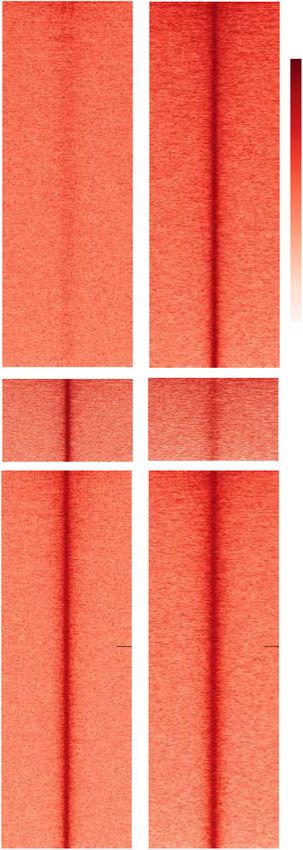
Downloaded from genesdev.cshlp.org on December 1, 2020 - Published by Cold Spring Harbor Laboratory Press

Luo et al.

mimicking the gastrula (ten Berge et al. 2008). Under dif-          sion of all three TET genes is low during gastrulation
ferentiation-permissive conditions, a synergistic interde-          (Khoueiry et al. 2017), suggesting that an epigenetic regu-
pendency between canonical Wnt/β-catenin and Nodal                  latory mechanism allows TET1 to influence lineage segre-
signaling pathways drives ME differentiation (Funa et al.           gation even when expression is down-regulated.
2015; Wang et al. 2017). Further lineage specification de-            Here, we ask how Tet1 controls developmental out-
pends on modulation of collaborative signal inputs; strong          comes by coordinating pluripotency and differentiation
Nodal signals in EBs favor a definitive endoderm fate               signals at entry points in the molecular circuitries of early
while low levels or Wnt favor a mesoderm fate (Gadue                embryonic lineage segregation. Our findings clarify how
et al. 2006), in agreement with how graded Nodal morpho-            Tet1 regulates the primed pluripotency epigenetic land-
gen gradients in the primitive streak drive patterning of           scape at the crossroads of ME and NE fate entry, providing
definitive endoderm anteriorly and mesoderm posteriorly             a cellular memory that regulates engagement of Oct4,
in vivo (Vincent et al. 2003; Schier 2009; Robertson 2014).         Wnt/β-catenin, and Nodal signaling effectors at develop-
   The use of cell culture mimics of early embryonic differ-        mental loci during gastrulation.
entiation has facilitated recent exploration of genomic
regulatory mechanisms underlying early developmental
cell state transitions. For example, the transitions from           Results
naive to primed pluripotency during conversion of 2iL
                                                                    Tet1 is repatterned to developmental fate loci during
ESCs to EpiLCs have revealed global reorganization of
                                                                    pluripotency transition
the master pluripotency transcription factor Oct4 at cell
state-specific enhancer sites (Buecker et al. 2014). In hu-         To examine whether Tet1 genomic occupancy changes
man and murine ESCs, Oct4 co-occupies distal regulatory             during the transition between naive and primed pluripo-
elements with Smad2 and Smad3 (Smad2/3), closely relat-             tency, we first performed Tet1 chromatin immunoprecip-
ed downstream intracellular effectors of TGF-β/Activin/             itation sequencing (ChIP-seq) in ESCs cultured in 2iL
Nodal receptors (Shi and Massagué 2003), to regulate tar-           (naive state) and following in vitro conversion to EpiLCs
get genes including Oct4 (also known as Pou5f1), Nodal,             (primed state). Based on differential binding intensities
and its antagonist, Lefty1 (Mullen et al. 2011), in an auto-        determined from biological replicates (n = 2) of wild-type
regulatory feedback loop. Upon exit from pluripotency,              (WT) cells and normalized to Tet1-deficient (knockout
Oct4 continues to determine germ layer fate selection               [KO]) negative controls (Supplemental Fig. S1A), we
by promoting ME and repressing NE fate (Thomson                     defined Tet1-bound peaks preferentially enriched in
et al. 2011), through new cooperative interactions with             2iL-treated ESCs (ESC-specific, 1256) or EpiLCs (EpiLC-
tissue-specific and signal-dependent factors at lineage-            specific, 5557) and those where Tet1 binding did not
priming genomic sites (Funa et al. 2015; Simandi et al.             show significant differences between the two states (com-
2016). Collectively, these studies suggest that the capacity        mon, 5741) (Fig. 1A; Supplemental Table S1). The sizable
of transcription factors (TFs) to access enhancer sites to          numbers of EpiLC-specific binding peaks suggest the pres-
drive cell fate choice is highly context-dependent. The             ence of de novo Tet1 binding, in line with detection of
complex circuitry that decides which factor(s) dominates            EpiLC-specific and Tet1-dependent differentially hydrox-
in the process of ESC commitment to ME or NE fate,                  ymethylated regions previously observed during ESC-to-
whether stochastic or predetermined by regulators acting            EpiLC differentiation (Khoueiry et al. 2017). Moreover,
upstream of lineage bifurcation, remains to be worked out           even though Tet1 binding profiles were preferentially en-
in further detail.                                                  riched at gene promoter regions in both pluripotency
   Dynamic changes in DNA methylation constitute a                  states, the state-specific binding peaks showed higher pro-
fundamental epigenetic mechanism in cell fate commit-               portions located at gene body and intergenic regions com-
ment in the early embryo (Santos et al. 2002). The Ten–             pared with common peaks (Fig. 1B). To demonstrate that
Eleven-Translocation (TET) DNA dioxygenase (Tet1,                   EpiLC-specific peaks are primarily located at promoter-
Tet2, and Tet3) family proteins mediate DNA demethyla-              distal regulatory sites, we examined the peak regions
tion by iterative oxidation of 5-methylcytosine (5mC)               for enhancer features marked by the presence of p300 his-
(Tahiliani et al. 2009; He et al. 2011; Ito et al. 2011). As im-    tone acetyltransferase occupancy, histone 3 Lys4 mono-
portant regulators of epigenetic reprogramming, they are            methylation (H3K4me1) and histone 3 Lys27 acetylation
intensively studied for their roles in development and dis-         (H3K27ac) using a published data set profiled in EpiLCs
ease (Pastor et al. 2013; Wu and Zhang 2014). In the early          (Buecker et al. 2014). In comparison with strong p300
postimplantation mouse embryo, Tet1 is sustained in ex-             and H3K27ac and weaker H3K4me1 signals detected at
pression without detectable Tet2 and Tet3 (Khoueiry et al.          common and ESC-specific Tet1 peaks, which are consis-
2017). In agreement with a nonredundant biological role             tent with a prevalence at active promoters, EpiLC-specific
of Tet1 in lineage priming, loss of Tet1 in ESCs results            Tet1 peaks displayed stronger p300 and H3K4me1 co-oc-
in hyperactive Nodal signaling in EBs and increased com-            cupancies and much weaker enrichment for the active
petence to differentiate toward ME fate (Koh et al. 2011).          H3K27ac mark (Fig. 1C). These results indicated that de
Furthermore, disruption of Tet1 function can severely               novo Tet1 genomic occupancy in EpiLCs occurs mainly
compromise neurulation in the mouse embryo, manifest-               at primed or poised enhancers.
ed as failed cranial neuropore closure after gastrulation              Gene ontology (GO) analysis showed that EpiLC-specif-
(Fong et al. 2016; Khoueiry et al. 2017). However, expres-          ic binding sites were highly enriched in GO terms for

2       GENES & DEVELOPMENT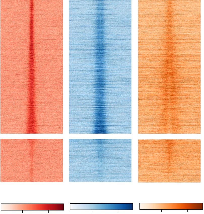
Downloaded from genesdev.cshlp.org on December 1, 2020 - Published by Cold Spring Harbor Laboratory Press
Coordination of lineage choice by TET1
A B
C
D
E F
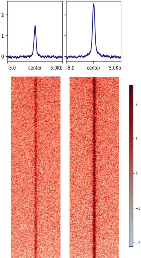
Figure 1. Tet1 is repatterned to developmental fate loci during pluripotency transition . (A) Heat map of Tet1 ChIP-seq signal densities in
2iL-cultured ESCs and EpiLCs at defined EpiLC-specific, ESC-specific, and common peaks (n = 2 biological replicates). See Supplemental
Table S1 for an annotated list of all peak regions. (kb) Kilobases. (B) Pie charts indicating genomic feature distribution of Tet1-bound
EpiLC-specific, ESC-specific, and common peaks. (C) Density aggregation plots of p300, H3K4me1, and H3K27ac ChIP-seq signal densi-
ties (Buecker et al. 2014) in EpiLCs at Tet1-bound EpiLC-specific, ESC-specific, and common peaks. (D) Functional annotation of GO
terms enriched in EpiLC-specific peaks analyzed by GREAT (McLean et al. 2010). The text highlighted in blue indicates terms associated
with lineage development. See Supplemental Table S2 for an annotated list of ranked GO terms. (E,F ) Integrative Genomics Viewer (IGV)
snapshots of Tet1 ChIP-seq tracks in EpiLCs and 2iL-cultured ESCs, and p300, H3K4me1, H3K27ac ChIP-seq tracks in EpiLCs, illustrating
EpiLC-specific loci associated with ME genes (Brachyury and Eomes in E) or NE genes (Six3 and Sox1 in F). Gene structure annotations are
shown below each track set. Blue highlights indicate regions containing statistically significant differential peaks of Tet1 binding between
ESCs and EpiLCs identified by MACS2 from replicates (BED annotations are shown as blue bars below tracks). Gene loci Six3(−133) and
Sox1(+35) are neural lineage-associated enhancers identified from a previous study (Cruz-Molina et al. 2017).
GENES & DEVELOPMENT 3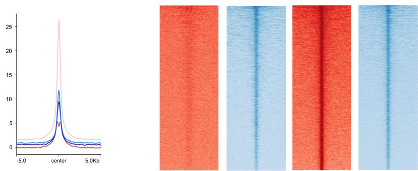
Downloaded from genesdev.cshlp.org on December 1, 2020 - Published by Cold Spring Harbor Laboratory Press

Luo et al.

pattern specification, cell fate commitment, mesoderm              (∼50%) located within introns and intergenic regions
development, and spinal cord-associated neuron differen-           may nonetheless be distal enhancers. Indeed, loss-in-KO
tiation (Fig. 1D; Supplemental Table S2). These regions            loci showed enrichment for markers of active distal regu-
were associated with several genes that are key determi-           latory elements (p300, H3K27ac, and H3K4me1) in WT
nants of lineage fate, including Brachyury, Eomes, Six3,           EpiLCs, which were comparably much weaker in gain-
and Sox1 (Fig. 1E,F; Arnold et al. 2008; Gouti et al. 2014;        in-KO loci (Fig. 2B), suggesting that Tet1 promotes chro-
Cruz-Molina et al. 2017; Koch et al. 2017). Moreover,              matin accessibility predominantly at active enhancers,
pathway analysis from the Molecular Signatures Database            whereas sites that gained accessibility in the absence of
(MSigDB) showed these Tet1-bound EpiLC-specific peaks              Tet1 are normally silent in EpiLCs.
to be highly enriched in Wnt-mediated signal transduc-                When centered at TET1-bound regions in EpiLCs, nor-
tion and regulation of SMAD2/3 signaling (Supplemental             malized ATAC-seq signal intensities were lower in
Fig. S1B). In stark contrast, these developmental-related          Tet1 GT/GT EpiLCs compared with WT (Fig. 2C; Supple-
terms were missing among ESC-specific and common                   mental Fig. S2C). Of all Tet1-bound regions, 84.6% were
peaks, which were instead enriched in general transcrip-           located within accessible genomic regions in EpiLCs,
tional and translational processes (Supplemental Fig.              overlapping considerably with loss-in-KO but not with
S1C,D; Supplemental Table S2). As expected for a func-             gain-in-KO sites (Fig. 2D; Supplemental Fig. S2D). Thus,
tional relevance in blastocyst development, ESC-specific           the latter were not direct targets of Tet1. In our previous
binding sites were also enriched in GO terms related to            study, we had observed that Tet1-bound regions in EpiLCs
placenta development and stem cell maintenance (Sup-               generally lost 5hmC and gained 5mC upon loss of TET1,
plemental Fig. S1C) and can be found at distal sites of            without a corresponding reduction in expression of a ma-
Esrrb and Klf4 (Supplemental Fig. S1E), genes encoding             jority of genes associated with these loci (Khoueiry et al.
naive pluripotency factors known to have pioneer activi-           2017). By overlapping those whole-genome differentially
ties in mouse ESCs (Adachi et al. 2018).                           methylated regions (DMRs) identified by “true 5mC” ox-
                                                                   idative bisulfite sequencing with ATAC-seq loss-in-KO
                                                                   loci, we found 2749 regions where loss of chromatin acces-
Tet1 influences chromatin accessibility at primed
                                                                   sibility were associated with gains in DNA methylation
developmental loci
                                                                   (Fig. 2E). Interestingly, these regions are associated with
We previously detected widespread hypermethylated                  neurodevelopmental lineage genes, including Sox family
genomic regions in Tet1-deficent EpiLCs enriched at                genes Nestin and Sim2.
genes associated with neurological functions (Khoueiry                In addition to developmental processes, GO terms for
et al. 2017). Therefore, we asked whether the presence of          genes associated with loss-in-KO sites include protein lo-
Tet1 in EpiLCs also regulates chromatin accessibility, by          calization to telomeric region and G1 DNA damage
profiling B6.129P2-Tet1 Gt (RRG140) (abbreviated to Tet1 GT)       checkpoint (Supplemental Fig. S2E), consistent with a
WT and Tet1-deficient EpiLCs using the assay for trans-            role for Tet1 in maintaining genomic stability (Cimmino
posase-accessible chromatin sequencing (ATAC-seq).                 et al. 2015; Yang et al. 2016; Khoueiry et al. 2017). On the
Based on analysis of biological replicates (n = 2), we iden-       other hand, gain-in-KO sites enriched for RNA processing,
tified 5661 regulatory elements that were differentially           translation, and protein targeting as top GO terms and
accessible in Tet1 GT/GT (KO) EpiLCs compared with WT              only weakly for a developmental term for mesenchymal
EpiLCs, of which 4280 sites showed decreased chromatin             stem cell differentiation (Supplemental Fig. S2E). The lat-
accessibility (referred to as loss-in-KO sites) and 1381           ter would be consistent with differentiation skewing
sites showed increased accessibility (referred to as gain-         toward ME at the expense of NE (Koh et al. 2011). Howev-
in-KO sites) (Fig. 2A; Supplemental Fig. S2A; Supplemen-           er, gain-in-KO sites within ME-related genes (Pdgfra, Rest,
tal Table S3). The higher incidences and extents of losing         Sox5, and Sox9) included in this GO term were minor
chromatin accessibility compared with gaining in Tet1-             peaks located within broader genomic regions where
deficient EpiLCs are in line with the role of Tet1 in pro-         most peaks exhibited reduced accessibility in KO (data
moting an open chromatin state at regulatory elements              not shown). When we further examined a subset of 359
through DNA demethylation.                                         loss-in-KO sites that overlapped with EpiLC-specific
   Genomic distribution of all loci exhibiting Tet1-depen-         Tet1 ChIP-seq peaks, we observed that these regions, asso-
dent differential chromatin accessibility showed locations         ciated with 505 genes, were significantly enriched for GO
within promoters, intronic and intergenic regions (Supple-         terms for pattern specification and spinal cord develop-
mental Fig. S2B). Promoter regions (0–3 kb upstream of             ment (Fig. 2F). These data suggest that Tet1 occupies
transcription start sites or TSS) were relatively more prev-       new enhancer regions during naive-to-primed pluripo-
alent in loss-in-KO and depleted in gain-in-KO loci,               tency transitions, primarily at neurodevelopmental loci
whereas intergenic and intronic regions were reciprocally          where it promotes an open chromatin state.
more prevalent in gain-in-KO loci (Supplemental Fig.                  To determine whether Tet1-dependent chromatin ac-
S2B). The apparent enrichment of promoter regions in               cessibility in EpiLCs were state-specific or pre-existing
loss-in-KO loci relative to gain in KO may be attributed           in ESCs, we performed further ATAC-seq analysis in
to the presence of common peaks bound by Tet1 in                   WT and KO 2iL-treated ESCs. At loss-in-KO sites defined
both naive and primed states, which mostly occurred                in EpiLCs, ATAC-seq signals in WT and KO ESCs were de-
at promoters (Fig. 1B). We asked whether the fraction              tected at similar levels, as in KO EpiLCs, suggesting that

4       GENES & DEVELOPMENT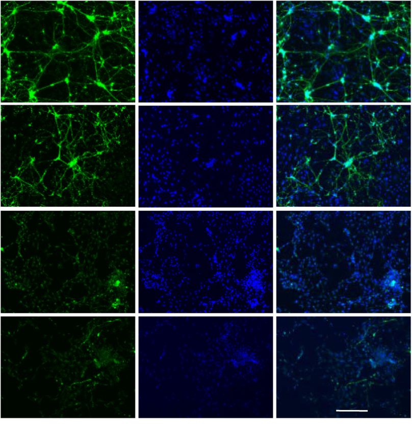
Downloaded from genesdev.cshlp.org on December 1, 2020 - Published by Cold Spring Harbor Laboratory Press

                                                                                                    Coordination of lineage choice by TET1

          A                                 B                                       C

          D                                 E

          F

          G                                                            H

Figure 2. Tet1 influences chromatin accessibility at primed developmental loci. (A) Heat map of ATAC-seq signal densities in EpiLCs
at regions showing differential chromatin accessibility between WT and Tet1 GT/GT (KO) (n = 2 biological replicates) of strain B6.129P2-
Tet1 Gt(RRG140) (Khoueiry et al. 2017). Peak regions with decreased (4280; referred to as “loss in KO”; top) and increased chromatin
accessibility (1381; referred to as “gain in KO”; bottom) in KO EpiLCs are ranked by decreasing and increasing ATAC signal change, re-
spectively. See Supplemental Table S3 for an annotated list of all peak regions. (B) Heat map of p300, H3K27ac, and H3K4me1 ChIP-seq
signal densities in WT EpiLCs at loss-in-KO and gain-in-KO ATAC-seq peaks in EpiLCs as defined in A. (C ) Density aggregation plot of
ATAC-seq signal densities in WT and Tet1 KO EpiLCs at total Tet1-bound peaks in EpiLCs. (D) Venn overlap of total Tet1 ChIP-seq peaks
in EpiLCs with the subsets of loss-in-KO and gain-in-KO ATAC-seq peaks. (E) Venn overlap of loss-in-KO ATAC-seq peaks in EpiLCs with
DMRs previously defined by oxidative whole-genome bisulfite sequencing data (Khoueiry et al. 2017). (F ) Venn overlap of Tet1 EpiLC-spe-
cific ChIP-seq peaks with the subsets of loss-in-KO and gain-in-KO ATAC-seq peaks and GO analysis of the former overlapping subset. GO
terms associated with neural lineage development are highlighted in blue. (G) Density aggregation plots of ATAC-seq signal densities in
WT and Tet1 KO 2iL-cultured ESCs and EpiLCs centered at loss-in-KO (left) and gain-in-KO (right) ATAC-seq peaks in EpiLCs. (H) Density
aggregation plots of ATAC-seq signal densities in WT 2iL-cultured ESCs and EpiLCs centered at Tet1-bound EpiLC-specific (left) and ESC-
specific (right) peaks.

                                                                                                        GENES & DEVELOPMENT             5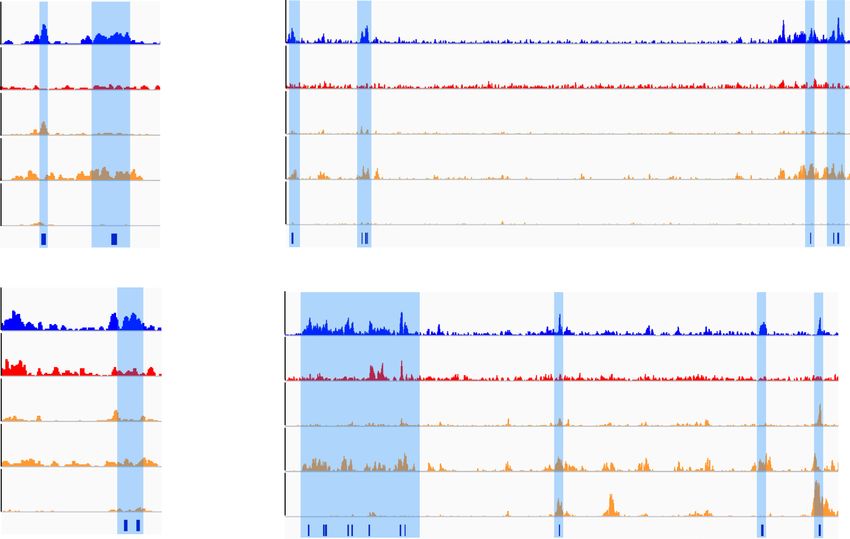
Downloaded from genesdev.cshlp.org on December 1, 2020 - Published by Cold Spring Harbor Laboratory Press

Luo et al.

these sites were already accessible independently of TET1          (KLF) (Klf3, Klf4, Klf5, Klf6, and Klf14), estrogen-related
in ESCs, but that further gain of accessibility in EpiLCs re-      nuclear receptor (NR)-β (Esrrb), and the Wnt/β-catenin
quired Tet1 (Fig. 2G, left panel). On the other hand, gain-        target transcription factors Sp5 and Tcfcp2l1, many of
in-KO EpiLCs sites showed low signal intensities in ESCs           which are naive-specific factors (Fig. 3A). Similar EpiLC-
but were already differentially more accessible in KO              and ESC-specific motifs were found in loss-in-KO- and
ESCs compared with WT; since there were minimal over-              gain-in-KO-accessible sites, respectively (Fig. 3B). Loss-
lap with ESC-specific and common Tet1-bound regions,               in-KO-enriched motifs included Sox3, Sox4, and Zic,
Tet1 may be countering chromatin opening at these loci             whereas gain-in-KO sites were enriched in motifs for the
indirectly (Fig. 2G, right panel). Comparing accessibility         KLF family, Sp2 and Sp5.
of state-specific Tet1 bound regions in the two cell states           Using published RNA sequencing (RNA-seq) gene ex-
showed increased chromatin opening at EpiLC-specific               pression data sets of 2iL-treated ESCs and EpiLCs
sites but only slight reduction at ESC-specific sites during       (Buecker et al. 2014), we verified that cognate transcrip-
ESC-to-EpiLC conversion (Fig. 2H). These observations              tion factors for several EpiLC-specific motifs were indeed
suggest passive engagement of Tet1 at ESC-specific re-             expressed at higher levels in EpiLCs than in ESCs (Supple-
gions where it has minimal impact on chromatin accessi-            mental Fig. S3A,B). Western blot analysis of Zic2, Wt1,
bility. Upon conversion to EpiLCs, Tet1 engages new                and Sox3 showed strikingly an abundance of Zic2 protein
primed loci where it promotes increased chromatin open-            in EpiLCs but undetectable levels in ESCs (Fig. 3C). Using
ing; however, these latter sites already possess an open           a published Zic2 ChIP-seq data set obtained in postim-
chromatin state in ESCs.                                           plantation embryo-derived epiblast stem cells (EpiSCs)
   We then asked whether rescue of Tet1 expression in KO           (Matsuda et al. 2017), we observed that regions where
EpiLCs can restore the chromatin landscape to WT. B6-              EpiLC-specific Tet1 occupancy overlapped with loss-in-
Tet1 GT/GT ESC clonal lines harboring doxycycline-induc-           KO-accessible sites also showed Zic2 occupancy (Fig.
ible Tet1 WT or catalytic mutant cDNA constructs                   3D). To determine whether Zic2 could be a partner factor
(Khoueiry et al. 2017) were generated (Supplemental Fig.           that recruits Tet1 to neural fate genes in ESCs (Frank et al.
S2F). Two replicate lines with rescued expression of either        2015; Luo et al. 2015), we performed Tet1 ChIP at specific
WT (Rwt) or mutant Tet1 (Rmt) were converted to EpiLCs             target loci upon Zic2 depletion during ESC-to-EpiLC con-
for ATAC-seq analysis. Unsupervised hierarchical clus-             version. Using siRNA transfection, we observed >80%
tering of ATAC-seq signals showed that Tet1 rescued                depletion of Zic2 transcripts (Fig. 3E). We selected distal
(WT or mutant) lines clustered away from 2iL ESC and               regulatory elements associated with four lineage genes—
EpiLC samples (Supplemental Fig. S2G). Reverse tran-               Lhx5, Foxp1, Sox1, and Pax3—where we found overlap
scription quantitative PCR (RT-qPCR) analysis of naive             with EpiLC-specific Tet1 occupancies, loss-in-KO chro-
and primed pluripotency markers indicated efficient con-           matin accessibility in EpiLCs, and Zic2 occupancies in
version of both WT and KO ESCs to EpiLCs, as previously            EpiSCs (Fig. 3F; Supplemental Fig. S3C). In particular,
observed (Khoueiry et al. 2017), as shown by complete si-          Lhx5 is critical for anterior fate of neuronal progenitor
lencing of naive genes (Esrrb and Tbx3) and induction of           cells (Gouti et al. 2014; Cruz-Molina et al. 2017). In three
primed markers (Fgf5, Otx2, and Dnmt3b). While Tet1                of four biological replicates, we detected loss of Tet1 bind-
rescued lines showed similar changes, the silencing of na-         ing at several sites—Lhx5(−91.6), Foxp1(+42.5), Sox1
ive markers appeared incomplete (Supplemental Fig.                 (+6.2), and Sox1(+82.6)—upon Zic2 depletion (Fig. 3F; Sup-
S2H). Both WT and mutant rescue in EpiLCs failed to re-            plemental Fig. S3C). These results suggest that Zic2 may
store chromatin accessibility at differential sites to WT          recruit Tet1 to new regulatory loci in the primed epiblast
levels; on the contrary, further reduction in accessibility        and regulate germ layer fate.
was observed (Supplemental Fig. S2I). Thus, the altered ac-
cessible chromatin landscape in Tet1-deficient EpiLCs
                                                                   Distinct engagement of Tet1 and Oct4 at primed
could no longer be reversed by subsequent restoration of
                                                                   enhancers
Tet1 expression, arguing against a pioneer role for Tet1
in chromatin remodeling.                                           As previously reported, Oct4-bound genomic sites are
                                                                   globally reorganized at enhancers during ESC-to-EpiLC
                                                                   conversion (Buecker et al. 2014). We asked how our ob-
Tet1 engagement at primed loci depends on Zic2
                                                                   served repatterning of Tet1 genomic occupancy sites com-
To determine whether the state specificity of Tet1 geno-           pared with those of Buecker et al.’s (2014) Oct4 ChIP-seq
mic localization involves partner cofactors, we performed          data sets. The global overview of all ChIP-seq peaks in
motif enrichment analysis of bound regions. EpiLC-spe-             both data sets suggests that Tet1 may undergo an even
cific Tet1 peaks were highly enriched for motifs of devel-         more pronounced genome-scale redistribution compared
opmental regulator families, including zinc finger protein         with Oct4 during the cell state transitions (Supplemental
of the cerebellum (Zic), Wilms tumor 1 homolog (Wt-1),             Fig. S4A). While total numbers of Oct4-bound sites stayed
high-mobility group (HMG) sex-determining region Y                 relatively similar between the two states, in line with sus-
(SRY)-box transcription factors Sox3 and Sox4, and basic           tained expression of Oct4 protein, over twofold more Tet1
helix–loop–helix (bHLH) transcription factors Tcf12 and            occupancy sites were detected in EpiLCs than in 2iL ESCs,
NeuroD1 (Fig. 3A). In contrast, ESC-specific peaks were            with the gains mostly within distal intergenic and exonic
enriched for motifs occupied by Kruffel-like factors               regions (Supplemental Fig. S4A).

6       GENES & DEVELOPMENT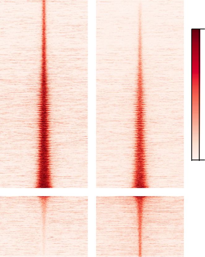
Downloaded from genesdev.cshlp.org on December 1, 2020 - Published by Cold Spring Harbor Laboratory Press

                                                                                                     Coordination of lineage choice by TET1

              A

              B

              C                           D                                           E

              F

Figure 3. Tet1 engagement at primed loci depends on Zic2. (A) Bubble plots indicating transcription factors associated with top enriched
motifs in Tet1-bound EpiLC-specific and ESC-specific peaks analyzed by HOMER (Heinz et al. 2010). (B) Bubble plots indicating transcrip-
tion factors associated with top enriched motifs in loss-in-KO and gain-in-KO ATAC-seq peaks in EpiLCs. Motif names highlighted in blue
are enriched in both Tet1-bound EpiLC-specific peaks and loss-in-KO ATAC-seq peaks in EpiLCs. (C) Western blot analysis of Zic2, Wt1,
and Sox3 expression in 2iL-cultured ESCs and EpiLCs. (D) Density aggregation plot of Tet1 ChIP-seq signal densities in EpiLCs and Zic2
ChIP-seq signal densities in EpiSCs (Matsuda et al. 2017) at the overlap of Tet1 EpiLC-specific peaks and loss-in-KO ATAC-seq peaks in
EpiLCs. (E) Knockdown efficiency in nontargeting (NT) siRNA or Zic2 siRNA treated EpiLCs. Data shown are mean values ± SEM (n = 3)
from three independent cell lines. (F ) ChIP-qPCR analysis of Tet1 binding at the Lhx5(-91.6), Lhx5(-21.8) (left panel), and Foxp1(+42.5)
(right panel) in Zic2 knockdown EpiLCs. (Top) IGV snapshots of Tet1 ChIP-seq tracks in EpiLCs and 2iL-cultured ESCs, ATAC-seq in
WT and Tet1 GT/GT (KO) EpiLCs, and Zic2 ChIP-seq tracks in EpiSCs at Lhx5 (left panel) and Foxp1 (right panel). Gene structure annota-
tions are shown below each track set. Regions where EpiLC-specific Tet1 peaks (BED annotations of statistically significant peaks shown
as blue bars below tracks) coincide with loss-in-KO ATAC-seq peaks in EpiLCs are highlighted in blue. (Bottom) ChIP-qPCR data shown
are mean ± SEM (n = 4) from three independent cell lines. Position coordinates in parenthesis are with respect to TSS in kilobases. The
enrichments of ChIP samples are normalized to the values of the corresponding input.

                                                                                                         GENES & DEVELOPMENT             7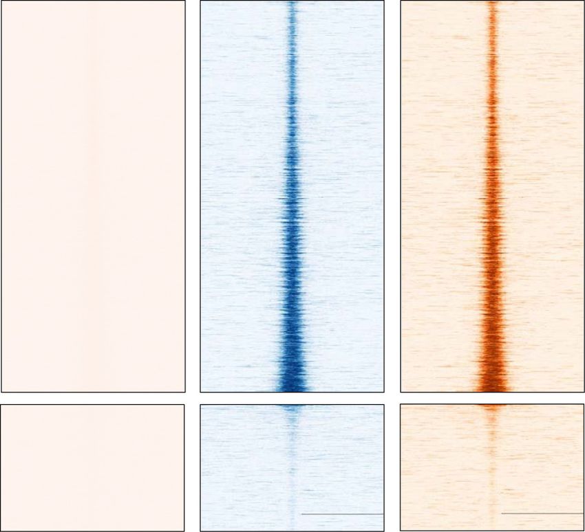
Downloaded from genesdev.cshlp.org on December 1, 2020 - Published by Cold Spring Harbor Laboratory Press

Luo et al.

   When comparing normalized binding intensities cen-              higher in Tet1 KO, 93 (7.5%) similar in WT and KO, but
tered at Tet1-bound peaks, we observed similarly strong            none lower in KO. Normalized read densities of all 1244
Oct4 co-occupancy at ESC-specific Tet1-bound peaks in              Oct4 ChIP-seq peaks showed significantly increased
2iL ESCs; at these loci, both Tet1 and Oct4 binding inten-         Oct4 occupancy upon loss of Tet1 (Fig. 4F). These peak re-
sities diminished in EpiLCs (Fig. 4A). Venn analysis incor-        gions were lowly accessible sites engaged by Oct4 in WT
porating ESC-specific Oct4-bound peaks showed an                   cells and associated with 1602 genes, many of which are
overlap with 47% of ESC-specific Tet1-bound peaks (Fig.            involved in lineage development (Supplemental Table
4B). At EpiLC-specific Tet1 peaks, Oct4-binding intensi-           S4). The most striking examples of enhanced Oct4-bind-
ties in EpiLCs were similar to pre-existing Oct4 levels in         ing sites include a proximal promoter element of Oct4 it-
ESCs, but comparatively weaker relative to those of                self (Fig. 4G), Nodal targets including Foxh1, Lefty1, Skil,
Tet1 in EpiLCs (Fig. 4C). Only 8% of these EpiLC-specific          and Agpat3 (Fig. 4H), and distal sites of Wnt signaling
Tet1-bound peaks overlapped with EpiLC-specific Oct4-              components Axin2, Lef1, and Fzd7 (Supplemental Fig.
bound peaks (Fig. 4D). Because of the larger size of Buecker       S4E). Similar enhanced Oct4 genomic occupancies were
et al.’s (2014) Oct4 ChIP-seq peaks, we reclassified those         recapitulated in Oct4 ChIP-seq analysis of B6.129P2-
by their genome distribution either at promoters or inter-         Tet1 GT/GT EpiLCs (Fig. 4G,H; Supplemental Fig. S4E).
genic regions. When centered on ESC-specific Oct4-bound               Focusing on the 1151 up-in-KO Oct4 peaks in EpiLCs,
peaks, colocalization of Tet1 and Oct4 binding was evi-            we found minimal overlap with EpiLC-specific (only 49
dent only at promoter but not intergenic regions in ESCs           or 4.3%) or with common Tet1-bound regions (72 or
(Supplemental Fig. S4B). At EpiLC-specific Oct4-bound              6.3%) (Fig. 4I). Therefore, at the majority of these up-in-
peaks, Tet1-binding intensities were relatively weak at            KO Oct4 ChIP-seq peak regions, Oct4 is not replacing
both promoters and intergenic regions. Collectively, these         Tet1. GO analysis of these up-in-KO Oct4 ChIP-seq peaks
data suggest that Tet1 and Oct4 coregulate genes at pro-           showed enrichment for somatic stem cell population
moter regions in the naive state but diverge to engage dis-        maintenance, but region and gene set coverages were
tinct sets of promoters and distal enhancers in the primed
Downloaded from genesdev.cshlp.org on December 1, 2020 - Published by Cold Spring Harbor Laboratory Press

                                                                                                      Coordination of lineage choice by TET1

             A                                                                      E

             B

                                                                                     F
             C

             D

             G                                  H                                               I

             J                                                        K

Figure 4. Distinct engagement of Tet1 and Oct4 at primed enhancers. (A) Heat map and density aggregation plots of Tet1 and Oct4 ChIP-
seq signal densities in 2iL-cultured ESCs and EpiLCs centered at Tet1-bound ESC-specific peaks. (B) Venn diagram indicating the overlap
of Tet1-bound ESC-specific peaks and Oct4-bound ESC-specific peaks. (C) Same as in 3A, but centered at Tet1-bound EpiLC-specific
peaks. (D) Same as in 3B for Tet1-bound and Oct4-bound EpiLC-specific peaks. (E) Heat map of Oct4 ChIP-seq signal densities in EpiLCs
(Buecker et al. 2014) and ATAC-seq signal densities in WT and Smad2;Smad3 KO EpiLCs (Senft et al. 2018) at differential accessibility
peaks defined in Figure 2A. (F) Heat map and density aggregation plot of Oct4 ChIP-seq signal densities in WT and Tet1 KO EpiLCs of
B6129S6F1-Tet1 tm1Koh strain at total 1244 Oct4-bound peaks defined by MACS2 bdgdiff module. See Supplemental Table S4 for an anno-
tated list of all peaks. (G,H) IGV snapshots of Tet1 ChIP-seq tracks in EpiLCs and 2iL-cultured ESCs, and Oct4 ChIP-seq tracks WT and
Tet1 KO EpiLCs, at indicated loci located at proximal promoters of Oct4 (G) and Nodal targets Lefty1 and Foxhl (H). Gene annotations are
shown below each track set. Blue highlights indicate statistically differential peaks of Oct4 binding identified by MACS2 in two strains of
Tet1-deficient EpiLCs (BED annotations shown as blue bars below tracks). (I) Venn diagram indicating the overlap of Oct4-bound up-in-
KO ChIP-seq peaks in EpiLCs with Tet1-bound EpiLC-specific (top panel) or Tet1-bound common (bottom panel) ChIP-seq peaks. (J,K)
Functional annotation of GO terms (J) and phenotype annotation of mouse genome informatics (MGI) ontology (K ) enriched in 388
Oct4-bound up-in-KO ChIP-seq peaks in EpiLCs defined by R/Bioconductor package DiffBind at FDR < 0.1. See Supplemental Table S5
for an annotated list of ranked GO terms.

                                                                                                          GENES & DEVELOPMENT             9
Downloaded from genesdev.cshlp.org on December 1, 2020 - Published by Cold Spring Harbor Laboratory Press

Luo et al.

       A                                                                               C

       B

       D
                                                                              F

       E

       G                                                        H

        I

Figure 5. Tet1 coordinates ME and NE fate choice via regulation of the Oct4–Nodal–Wnt axis (A) Schematics of serum-free embryoid
bodies (EB) differentiation assay. Activin A (AA) was supplied at day 2 as a positive control to stimulate ME differentiation. Arrowheads
denote cell splitting or EB reaggregations. (B) Time-course expression analysis of ME markers (Brachyury, Eomes, Mixl, Foxa2, and Goose-
coid) in WT and Tet1 tm1Koh (KO) EBs. Data shown are mean ± SEM of n = 3 biological replicate ESC lines per genotype. Normalized expres-
sion values are relative to values of WT EBs at day 4 without AA stimulation for ME markers and relative to values of WT EBs at day 0 for
NE markers. Statistical test: two-way ANOVA. (∗ ) P < 0.05; (∗∗ ) P < 0.01; (∗∗∗ ) P < 0.001. (C) Immunofluorescence staining of Brachyury (red)
in sectioned WT and Tet1 KO EBs at day 4. DAPI (blue) indicates the nuclei. Scale bar, 50 µm. (D) Same as in 5B for NE markers (Sox1, Pax6,
and Lhx5) in WT and Tet1 KO EBs. (E) Expression of Oct4, Nodal, and Wnt signaling target Sp5 in WT and Tet1 KO EBs. Normalized ex-
pression values are relative to values of WT EBs at day 0. Data shown are mean ± SEM (n = 4) from three biological replicate ESC lines per
genotype. (F) Western blot analysis of phospho-Smad2, total Smad2/3, active-β-catenin, and total β-catenin in whole-cell lysates of day 0,
2, and 4 WT and Tet1 KO EBs. (4+) WT EBs at day 4 treated with exogenous AA (25 ng/mL). Biological replicate lines per genotype are
presented. (G) Time-course expression analysis of Oct4 and Tet1 in WT and Tet1 KO EBs. Data shown are mean ± SEM (n = 3) from three
biological replicate ESC lines per genotype. Normalized expression values are relative to the values of WT EBs at day 0. (H) Western blot
time-course analysis of Oct4 and Tet1 in whole-cell lysates from WT and Tet1 KO EBs. (4+) WT EBs at day 4 stimulated with AA (25 ng/
mL). Relative values of Oct4 and Tet1 band intensities normalized to β-actin levels are shown below the respective signal bands. (I) ChIP-
qPCR analysis of Smad2/3 binding to the Lefty1 (−1.3) and Oct4 (−2) proximal promoters, and Eomes (−10), and Goosecoid (+6) distal en-
hancers in day 4 WT and Tet1 KO EBs. Position coordinates in parenthesis are with respect to TSS in kilobases. The enrichments of ChIP
samples are normalized to the values of the corresponding input.

10          GENES & DEVELOPMENT
Downloaded from genesdev.cshlp.org on December 1, 2020 - Published by Cold Spring Harbor Laboratory Press
Coordination of lineage choice by TET1
(Fig. 5B; Supplemental Fig S5A). A homogeneous pattern Lhx5 in WT EBs, but cannot restore expression in Tet1
of ME induction in KO EBs was verified by enhanced Bra- KO EBs to WT levels (Fig. 6D). Oct4 depletion in ESCs re-
chyury immunofluorescence staining in almost all cells in sults in spontaneous loss of self-renewal and differentia-
EB sections (Fig. 5C). This skewing phenotype toward ME tion into extraembryonic lineages (Niwa et al. 2000;
differentiation recapitulates our previous observations Radzisheuskaya et al. 2013). In our approach, in which
following acute Tet1 depletion by siRNA in v6.5 ESCs we depleted Oct4 after induction of EB differentiation, si-
(Koh et al. 2011). Furthermore, nondirected differentiation lencing of naive markers (Klf4, Esrrb, and Rex1) was not
was associated with decreased expression of NE (Pax6, affected in WT EBs (Supplemental Fig. S6A); among tro-
Sox1, and Lhx5) genes by day 4 (Fig. 5D). phoblast markers (Cdx2, Hand1, and Mash2) tested,
Naive pluripotency markers, including Klf4, Esrrb, and only Cdx2 expression was increased by Oct4 depletion.
Rex1, were fully silenced by day 4 in WT and KO EBs (data In Tet1 KO EBs, further loss of Oct4 resulted in small in-
not shown), verifying that exit from pluripotency was not creases in Klf4 and Rex1 expression, and more pronounced
inhibited by loss of Tet1. However, we consistently ob- differential up-regulation of Hand1 and Mash2 by day 4
served sustained expression of Oct4 in KO cells compared (Supplemental Fig. S6B). Collectively, these results reveal
with its fully repressed levels in WT EBs by day 4, along the contribution of Oct4 in embryonic and extraembryon-
with elevated expression of Nodal and the Wnt target ic differentiation in the context of Tet1 loss of function.
Sp5 (Fig. 5E). By performing Western blot analysis during Next, we added chemical inhibitors during EB reaggre-
the differentiation time course, we verified that following gation at day 2 to block signaling components, namely,
removal of BMP4 and LIF at day 0, active phosphorylated SB431542 to inhibit Nodal–Smad2/3 pathway, IWP-2 to
Smad2/3 proteins were extinguished by day 4 in WT EBs inhibit Wnt palmitoylation, and XAV939 to stabilize the
unless Activin A was added; in contrast, Smad2/3 proteins Wnt negative regulator Axin2 (Anastas and Moon 2013).
sustained their phosphorylation status in KO EBs between To access whether these treatments provided effective
days 2 and 4 (Fig. 5F). In concert with these differences, ac- blockade of relevant signaling, we examined gene expres-
tive β-catenin proteins were detectable at higher levels in sion of Nodal and the Wnt targets Axin2 and Sp5. As ex-
KO compared with WT EBs throughout the differentiation pected, hyperactivated Nodal expression levels in Tet1
time courses. Oct4 transcripts were down-regulated by KO EBs were fully abrogated by SB431542, but also by
day 3 in WT EBs, following down-regulation of Tet1 by the Wnt inhibitors, consistent with an action of Wnt up-
day 2 (Fig. 5G). In contrast, Tet1 KO EBs sustained Oct4 stream of Nodal-Smad2/3 (Fig. 6E). On the other hand,
protein expression even when Oct4 transcripts were the Wnt target genes were effectively repressed by IWP-2
down-regulated by day 4, suggesting that Tet1 may regu- and XAV939, but variably enhanced in WT and KO EBs
late Oct4 levels during germ layer differentiation by a by SB431542, suggesting that Nodal signals may provide
posttranslational mechanism (Fig. 5H). Similar observa- negative feedback regulation of Wnt (Fig. 6E). The effects
tions were made using incipient congenic B6-Tet1 GT/GT of inhibitor treatment on Brachyury, Eomes, and Mixl ex-
ESCs (Supplemental Fig. S5B,C). Moreover, we often ob- pression phenocopied the effects on Nodal, in agreement
served Oct4 signals to appear as doublet bands of about with these ME genes being downstream targets of collab-
50 kDa, higher than the expected molecular mass of 37 orative Wnt and Nodal inputs (Fig. 6F). In contrast, expres-
kDa, which may suggest the presence of posttranslational sion of Pax6, Sox1 and Lhx5 in KO EBs can be rescued to
modifications such as O-GlcNAcylation (Jang et al. 2012). nearly WT levels by SB431542, but not by the Wnt inhib-
To further validate elevated Nodal signals at key ME lin- itors, suggesting that hyperactivated Nodal signals in Tet1
eage genes in Tet1 KO EBs, we performed Smad2 ChIP KO EBs cross-inhibits these NE markers while promoting
and interrogated Smad2 recruitment to known Smad2/3 ME fate (Fig. 6G). An additional treatment with inhibitors
target loci (Mullen et al. 2011; Wang et al. 2017). At prox- at day 0 in this assay produced no further changes (data not
imal promoter sites of Oct4 and Lefty1 and distal sites of shown), in agreement with the action of extracellular cues
Eomes and Goosecoid, increased Smad2 occupancy were occurring between days 2 to 4 of differentiation (Fig. 5F;
detected in Tet1 KO compared with WT EBs (Fig. 5I). Gadue et al. 2006).
Previous studies have shown collaborative interactions
between Wnt/β-catenin, Nodal–Smad2/3, and Oct4 in the
Loss of Tet1 impairs differentiation into anterior
regulation of ME differentiation of mouse and human
neuroectoderm lineage
ESCs (Funa et al. 2015; Wang et al. 2017). To clarify
how these effectors modulate differentiation phenotypes Because expression of NE marker genes can be variable in
downstream from Tet1, we inhibited each of these three the nondirected differentiation assay described above, we
components in Tet1 KO EBs (Fig. 6A). First, we silenced tested our ESC lines upon inductive neuronal differentia-
Oct4 expression in EBs by applying two rounds of Oct4 tion. In the first differentiation protocol based on supple-
SMARTpool siRNA transfection during serum-free EB mentation with retinoic acid (RA) (Bibel et al. 2007), we
differentiation at days 0 and 2 (Fig. 6B). In line with a con- collected EBs in serum media after a 4-d treatment with
tribution of Oct4 toward ME skewing in Tet1 KO EBs, RA at 5 μM and replated the cells for terminal neuronal
depletion of Oct4 expression in Tet1 KO EBs reduced differentiation (Fig. 7A). Tet1 GT/GT ESCs generated EBs
expression of Brachyury, Mixl1, and Eomes to near WT with severely impaired expression of NE markers Pax6,
levels by day 4 (Fig. 6C). However, Oct4 depletion can en- Sox1, NeuroD1, and Nestin, concomitantly with elevated
hance expression of the neural lineage marker Sox1 and Oct4 levels (Supplemental Fig. S7A,B). Because these
GENES & DEVELOPMENT 11Downloaded from genesdev.cshlp.org on December 1, 2020 - Published by Cold Spring Harbor Laboratory Press
Luo et al.
A
B
C D
E
F
G
Figure 6. Oct4 knockdown or Wnt/Nodal signaling blockade rescue ME skewing in Tet1 KO. (A) Schematics of serum-free EB differen-
tiation assays including treatment with Oct4 siRNA or signaling inhibitors. To silence the expression of Oct4 completely, two rounds of
Oct4 siRNA were added at day 0 and day 2. Signaling inhibitors, including SB431542, XAV939, and IWP2, or DMSO as control were added
at day 2 only. (B) RT-qPCR (left panel) and Western blot (right panel) analysis of Oct4 expression in days 0, 2, and 4 WT and Tet1 tm1Koh KO
EBs treated with Oct4 siRNA. RT-qPCR data shown are mean ± SEM of n = 3 independent experiments from two lines per genotype. Rel-
ative values of Oct4 band intensities normalized to β-actin levels are shown below the signal bands. (C,D) Expression of ME markers
(C ) and NE markers (D) in WT and Tet1 tm1Koh KO EBs treated with Oct4 siRNA. Data shown are mean values ± SEM of n = 3 independent
experiments from two lines per genotype. (E–G) Expression of Nodal and Wnt target genes (E), ME markers (F ), and NE markers (G) in WT
and Tet1 tm1Koh (KO) EBs treated with signaling inhibitors. Schematic at the right in E indicates the inferred Wnt and Nodal signaling cas-
cade. Data shown are the mean ± SEM of n = 3 independent experiments from three biological replicate lines per genotype. Normalized
expression values are relative to values of WT EBs at day 0.
genes are normally activated to high levels in WT EBs af- by absence of Tet1. We performed ChIP-qPCR analysis
ter treatment with RA at day 4, when Tet1 expression is for the presence of histone H3 Lys27 trimethylation
no longer detected, we asked how the chromatin status (H3K27me3) and acetylation (H3K27ac), which are mutu-
at their gene promoters or enhancers were altered ally exclusive marks for poised/silenced and active
12 GENES & DEVELOPMENTDownloaded from genesdev.cshlp.org on December 1, 2020 - Published by Cold Spring Harbor Laboratory Press
Coordination of lineage choice by TET1
A B C
D E
F
Figure 7. Loss of Tet1 impairs differentiation into anterior neuroectoderm lineage. (A) Schematics of retinoic acid (RA)–directed EB dif-
ferentiation assay. (B) Immunofluorescence staining of the neural marker β-tubulin III (green) in terminal neurons differentiated from WT
(WT17 and WT34) and KO (KO1 and KO15) ESCs of strain B6129S6F1Tet1 tm1Koh. DAPI (blue) indicates the nuclei. Scale bars, 100 µm.
(C) Schematics of anterior neural progenitor cell (antNPC) monolayer differentiation assay. ESCs are cultured in serum-free medium sup-
plied with bFGF for 3 d. Inhibitors or DMSO control were added from day 2. (D) Gene expression of neural markers in WT and Tet1 tm1Koh
KO ESCs following differentiation into antNPCs treated with the Wnt signaling inhibitor XAV939, Nodal signaling inhibitor SB431542, or
DMSO (vehicle control). Except for hindbrain markers Mafb and Hoxa2, all other genes are markers of anterior fate. Data shown are the
mean ± SEM of two independent differentiation experiments in which a total of n = 5 or 6 biological replicate lines per genotype were an-
alyzed. Fold induction is expressed relative to values of a WT cell line at day 0 on a log10 scale, except Wnt1 for which values are relative to
a WT line at day 5 on a linear scale. P-values are obtained by multiple t-tests. (E) Western blot analysis of phospho-Smad2, total Smad2/3,
active-β-catenin, total β-catenin, and Oct4 in whole-cell lysates of days 0, 3, and 5 WT and Tet1 tm1Koh (KO) antNPCs. Biological replicate
lines per genotype are presented. Relative values of Oct4 band intensities normalized to β-actin levels are shown below the signal bands.
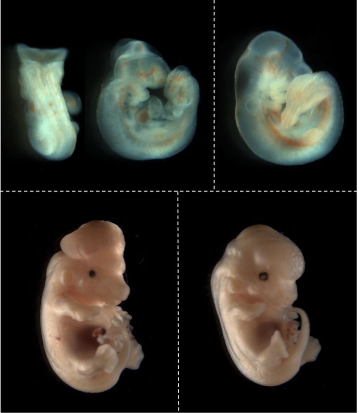
(F) Embryos at E10.5 (Theiler stage 16) and E13.5 (Theiler stage 22) that are homozygous or heterozygous for the Tet1 tm1Koh allele on a
C57BL/6 background. Failed neuropore closure is indicated with a white arrow. Scale bar, 1 mm.
regulatory regions, respectively (Creyghton et al. 2010; induced gains in H3K27ac at the activated NE gene loci in
Rada-Iglesias et al. 2011), at distal regulatory loci of WT cells; these chromatin changes were completely
Pax6, Sox1, and Nestin (Supplemental Fig. S7C). At day abolished in Tet1 GT/GT EBs, indicating a severe defective
0 of EB differentiation onset, we did not observe any differ- cellular response to RA stimulation. Because both WT
ences in the presence of these marks at all three loci be- and Tet1 GT/GT ESCs of the B6.129P2 strain cannot adhere
tween WT and Tet1 GT/GT ESCs, arguing against any well to substrates during replating, we switched to com-
effect of Tet1 on pre-existing histone status prior to activa- paring WT and KO ESCs of strain B6129S6F1-Tet1 tm1Koh
tion by differentiation cues. By day 6 of EB aggregation, RA to test further monolayer differentiation into terminal
GENES & DEVELOPMENT 13Downloaded from genesdev.cshlp.org on December 1, 2020 - Published by Cold Spring Harbor Laboratory Press
Luo et al.
neurons expressing β-tubulin III (also known as Tuj1). We Tet1-dependent primed loci appear distinct from those
observed extensive loss of Tuj1-expressing neurons in KO engaged by the essential pluripotency factor Oct4 and in-
lines compared with WT lines (Fig. 7B). These phenotypes dependent of Smad2/3 regulation. However, absence of
were reproduced during terminal neuronal differentiation Tet1 enhances Oct4 binding to a subset of its genomic
of E14Tg2a ESC lines (129 strain) in which Tet1 is stably targets, including Oct4 itself and Nodal and Wnt targets,
depleted (Supplemental Fig. S7D,E) and also during differ- unleashing a feed-forward loop that amplifies Smad2/3
entiation of induced pluripotent stem cells (iPSCs) repro- and canonical Wnt signaling recruitment to ME genes
grammed from KO mouse embryonic fibroblasts of mixed during differentiation when Tet1 expression is no longer
B6;129S6-Tet1 tm1Koh strain (Supplemental Fig. S7F; Bar- sustained. We propose a model in which Tet1 safeguards
toccetti et al. 2020). both DNA methylation fidelity and chromatin accessi-
To further dissect the effects of Tet1 loss of function bility at neural fate developmental loci and indirectly
in NE lineage development, we used a more recently modulates occupancy by Oct4 at key ME-fate loci in post-
established serum-free protocol that directed mouse implantation epiblast cells. Entry into the primitive
ESCs to differentiate into anterior neural progenitor cells streak then requires rapid loss of Tet1 expression to allow
(antNPCs) on monolayer cultures by supplementation Oct4 to collaborate with Wnt and Nodal signals to induce
with fibroblast growth factor-basic (bFGF) (Fig. 7C; Gouti ME while suppressing NE differentiation.
et al. 2014; Cruz-Molina et al. 2017). In this system, Tet1 While Oct4 exhibits enhancer repatterning during con-
transcripts and protein expression were lost by day 3, but version of naive ESCs to primed EpiLCs, its dynamic geno-
Oct4 expression persisted until day 4 (Supplemental Fig. mic occupancy appears to be dependent on other cell
S7G,H). In the absence of small molecule inhibitors, sev- state-specific TFs, including Zic2/3 and Otx2 (Buecker
eral anterior NPC fate markers Lhx5, Sim2, and Wnt1 et al. 2014). Similarly, we observed that EpiLC-specific
were significantly compromised in Tet1 KO compared Tet1 bound regions are enriched for consensus motifs
with WT cells (Fig. 7D). Upon blockade of posteriorization for Wt-1, Zic, and Sox family factors, but not Otx2, sug-
activities provided by Wnt signaling, or Nodal inhibition, gesting that a distinct set of partner TFs may facilitate en-
expression of Lhx5, Sim2, Sox1, Pax6, and En1 were res- gagement of Tet1 at primed enhancers separately from
cued in Tet1 KO ESCs to WT levels (Fig. 7D). In agreement those engaged by Oct4. Accessibility of these loci are
with hyperactivated Nodal and canonical Wnt signaling, lost in the absence of Tet1 but cannot be restored by res-
we observed increased expression of active β-catenin and cued re-expression of Tet1, arguing against a pioneer func-
phosphorylated Smad2/3 in KO cells by day 3 of differen- tion for Tet1 as a “de novo” DNA demethylase that can
tiation (Fig. 7E). These results suggest that impaired neu- open up closed chromatin. Consistent with having a high-
ronal differentiation potential in Tet1 KO ESCs is not the ly generic DNA sequence specificity, the N-terminal
result of inherent NE gene defects, but largely a response CXXC zinc finger domain in Tet1 confers an affinity for
to altered extrinsic signaling inputs. unmethylated CpG motifs, enriched in the majority of
Finally, we examined mouse embryos in midgestation mammalian gene promoters (Zhang et al. 2010; Frauer
using our B6-Tet1 tm1Koh strain. From heterozygous-inter- et al. 2011), such that Tet1 by itself binds predominantly
cross timed pregnancies, we consistently observed failure to CpG-rich regions in ESCs where it preserves methyla-
of midbrain–hindbrain neuropore closure in E10.5 Tet1- tion fidelity by preventing aberrant de novo DNA methyl-
null embryos, which manifested as exencephaly by E13.5 ation activity (Williams et al. 2011; Wu et al. 2011; Xu
(Fig. 7F). We previously described that the first morpholog- et al. 2011). Here, we showed that Zic2 may be a key cofac-
ical defects in Tet1-deficient embryos were detected as tor recruiting Tet1 to its primed enhancer sites, where
deformities at E8.5 after gastrulation, coincident with Tet1 can then promote an open chromatin state.
initiation of neuropore closure, and failed anterior neuro- Defined basal media conditions in ESC differentiation
pore closure at E9.5 (Khoueiry et al. 2017). Therefore, reg- models favor development along the neural lineage in
ulatory control of anterior neuroectodermal lineage the absence of extrinsic signals, supporting a “neural de-
potential by Tet1 is critical for proper neural tube closure. fault” fate in epiblast cells (Ozair et al. 2013). In support
of this model, a recent study demonstrated that Polycomb
proteins facilitate a permissive regulatory topology in
Discussion ESCs at a set of poised enhancers that are necessary for
the induction of anterior neural genes during differentia-
Our results provide a molecular framework for under- tion (Cruz-Molina et al. 2017). Our study adds Tet1 as an-
standing how a pre-existing epigenetic landscape shaped other important component of the “neural default” wiring
by Tet1 in the epiblast influences the integration of cell- in primed epiblast cells by regulating the chromatin states
intrinsic pluripotency factor expression and extrinsic sig- at neurodevelopmental loci, possibly as a “maintenance”
naling cues during subsequent germ layer specification. DNA demethylase. A recent study showed that TET1,
While Tet1 is highly expressed in both naive and primed TET2, and TET3 triple-KO in human ESCs results in
pluripotent states, the genomic occupancy patterns are DMNT3B-mediated DNA hypermethylation predomi-
dynamic and related to distinct biological functions rele- nantly at bivalent promoters and a neural differentiation
vant to each cell state. Moreover, de novo Tet1 occupancy defect associated with failure of PAX6 induction during
during lineage priming affects chromatin accessibility at differentiation, which can be partially rescued by re-
developmental loci comprising neural fate genes. These expression of TET1 (Verma et al. 2018). These studies,
14 GENES & DEVELOPMENTDownloaded from genesdev.cshlp.org on December 1, 2020 - Published by Cold Spring Harbor Laboratory Press
Coordination of lineage choice by TET1
much in agreement with our observations in this study 2016). Moreover, we observed loss of Sox2 in both WT
and previously in Tet1 KO mice (Khoueiry et al. 2017), and KO EBs by similar extents and kinetics during EB dif-
suggest that disruption of the epigenetic landscape in ferentiation (data not shown); thus, dysregulation of Sox2
primed pluripotency can occur without corresponding expression is unlikely to account for loss of NE potential
effects on gene expression at pregastrulation, but affect in Tet1 KO EBs. However, inhibition of Nodal signals res-
activation of neural genes later during development. Pos- cued differentiation skewing toward ME and restored NE
sible effects of Tet1 KO on recruitment of Polycomb- gene expression. The latter results are consistent with pre-
repressive components to primed enhancers remain to vious observations in human ESCs, where Wnt/β-catenin
be investigated, although we had not observed any chang- induction of neural crest markers are sustained only if
es in H3K27me3 status at distal enhancers of Pax6, Sox1, Nodal signaling is inhibited (Funa et al. 2015). Thus, the
and Nestin when comparing WT and KO ESCs at the ini- impaired NE potential of Tet1 KO cells is primarily a re-
tiation of EB differentiation. sponse to aberrant extrinsic cues that repress TF gene
In Tet1 KO EpiLCs, the altered chromatin landscape is expression.
accompanied by increased genomic occupancy by Oct4 at NE genes most affected in Tet1 KO EBs are anterior neu-
several low-affinity sites at developmental gene loci. ronal progenitor markers (Lhx5, Sim2, and Wnt1), which
These observations are consistent with a previous study are coincidently targets of repression by the posterioriza-
that described Oct4 binding at a large set of low-accessi- tion activity of Wnt/β-catenin signaling during in vitro
ble and differentiation-related genomic regions, potential- generation of neuromesodermal progenitors (Gouti et al.
ly priming cell fate enhancers (Simandi et al. 2016). 2014). Consistent with evidence of hyperactive β-catenin
Because Tet1 occupancy sites and Tet1-dependent chro- in Tet1 KO EBs, inhibiting Wnt restores expression of an-
matin accessibility in EpiLCs are largely disparate from terior neuronal genes in KO cells during neuronal differen-
Oct4-bound regions, functional interactions between tiation. It remains to be investigated whether modulating
Tet1 and Oct4 at the genome-scale are likely to be indi- Wnt/β-catenin and graded Nodal signals may produce dif-
rect. However, we observed an exceptional coincidence ferential effects in WT and Tet1 KO EBs on the anterior
of an EpiLC-specific Tet1-bound site that showed en- versus posterior identities of differentiating neuronal
hanced Oct4 binding in the absence of Tet1 at the proxi- progenitors. Since loss-in-KO accessibility regions are en-
mal primed enhancer of Oct4 (Fig. 4G), suggesting that riched in the GO term for paraxial mesoderm develop-
Tet1 may evict Oct4 from an autoregulatory element at ment (Supplemental Fig. S2E), Tet1 may potentially also
the Oct4 promoter. Using a POU5f1-luciferase reporter regulate the bipotency of Sox2+/T+ neuro-mesoderm pro-
construct transfected in KO EpiLCs, we did not observe genitors (Gouti et al. 2014; Li et al. 2016; Koch et al.
any competitive effect of rescued Tet1 expression on 2017). Nonetheless, the dysregulation of anterior markers
OCT4 promoter-reporter activity (data not shown); how- including Lhx5, Sim2, and Wnt1 by loss of Tet1 is consis-
ever, we cannot preclude direct functional antagonism tent with failed anterior neuropore closure observed in
between Tet1 and Oct4 in the native chromatin environ- Tet1-null embryos (Khoueiry et al. 2017) and in tuft mu-
ment of the Oct4 autoregulatory site or posttranslational tant mice in which a nonsense mutation in the Tet1
regulation of Oct4 by Tet1. Nodal and Wnt targets with gene has been detected (Fong et al. 2016), suggesting
enhanced Oct4 binding also include genes encoding that Tet1 primarily regulates anterior neural identity.
negative feedback regulators (Lefty1, Lefty2, Skil, and Our results position Tet1 as an upstream negative regu-
Axin2); however, these effects are likely downstream lator of Wnt/β-catenin signaling. Possible modes of action
from collaborative Oct4 and extracellular signals. In naive include gene activation of Wnt repressors by DNA deme-
ESCs in vitro and the epiblast in vivo, loss of TET proteins thylation or noncatalytic repression of signal effectors.
causes loss of expression of neighboring Lefty1 and Lefty2 As precedents for the former mechanism, promoter
gene loci in association with hypermethylation of their hypomethylation and derepression of Wnt pathway gene
promoters and enhancers (Koh et al. 2011; Dai et al. inhibitors by TET enzymes have been previously report-
2016; Khoueiry et al. 2017). Collectively, these studies ed—Sfrp4 by Tet3 during serum-free neural EB differenti-
suggest that a fundamental role of TET catalytic activity ation (Li et al. 2016); DKK3 and DKK4 by TET1 in human
at pregastrulation stages is to keep epiblast cells in “neu- colon cancer cells to suppress tumor growth (Neri et al.
ral default” mode by promoting accessibility of TFs to 2015). Here, we observed that the axis of regulatory con-
neural loci, modulating Oct4 expression and genomic oc- trol involves modulation of Oct4 by Tet1 in the primed
cupancy, and sustaining expression of Nodal and Wnt an- epiblast, since several gene targets of elevated Oct4 bind-
tagonists to prevent precocious signaling activation of ing in Tet1 KO EpiLCs encode canonical Wnt components
primitive streak entry. such as Fzd1, Fzd7, and Fzd8. Moreover, de novo occupan-
Stochastic differences in expression levels of pluripo- cy sites of Tet1 in EpiLCs are enriched in motifs for candi-
tency factors may influence germ layer fate: Higher ratios date TFs that are potentially negative regulators of Wnt/
of Oct4 to Sox2 suppresses NE while promoting ME differ- β-catenin, such as Wt-1 and Zic2/3 (Kim et al. 2009; Pour-
entiation and vice versa (Thomson et al. 2011). We ob- ebrahim et al. 2011). Future investigations will identify
served that elevated Oct4 expression in Tet1 KO EBs Tet1 partners with potential pioneering activities in or-
increased ME gene expression, but did not directly inhibit chestrating the primed state chromatin, which in turn
NE potential, in line with a positive role of Oct4 in inte- controls the tripartite axes of differentiation cues involv-
grating ESC response to external cues (Simandi et al. ing Oct4, Nodal and Wnt.
GENES & DEVELOPMENT 15You can also read