Exercise training effects on natural killer cells: a preliminary proteomics and systems biology approach
←
→
Page content transcription
If your browser does not render page correctly, please read the page content below
125 Exercise and natural killer cells

Exercise training effects on natural killer cells: a preliminary proteomics and
systems biology approach
Francisco Llavero 1 *, Lidia B. Alejo 2,3 *, Carmen Fiuza-Luces 3 *, Alejandro López Soto 4, Pedro L. Valenzuela 2, Adrián
Castillo-García 5, Javier S. Morales 2, David Fernández 2, Itziar Pagola Aldazabal 2,3, Manuel Ramírez 6, Alejandro Santos-
Lozano 3,7, José L. Zugaza 1,8,9 †, Alejandro Lucia 2,3 †

*First three authors contributed equally
† José L. Zugaza and Alejandro Lucia share senior authorship

1
  Achucarro Basque Center for Neuroscience, Science Park of the UPV/EHU, Leioa, Spain
2
  Faculty of Sport Sciences, European University of Madrid, Madrid, Spain
3
  Research Institute Hospital 12 de Octubre (‘imas12’), PaHerg, Madrid, Spain
4
  Department of Biochemistry and Molecular Biology, Faculty of Medicine, Instituto Universitario de Oncología del Principado de
Asturias (IUOPA), Instituto de Investigación Sanitaria del Principado de Asturias (ISPA), University of Oviedo, Spain
5
  Fissac - Physiology, Health and Physical Activity, Madrid, Spain
6
  Oncohematology Department, Children’s Hospital Niño Jesús, Madrid, Spain
7
  i+HeALTH, European University Miguel de Cervantes, Valladolid, Spain
8
  IKERBASQUE, Basque Foundation for Science, Bilbao, Spain
9
  Department of Genetics, Physical Anthropology and Animal Physiology, Faculty of Science and Technology, UPV/EHU, Leioa, Spain

                         ABSTRACT                                      The proteins showing a higher differential expression
                                                                       (Log2 fold-change>10 and false discovery rate [FDR]
Background: Regular exercise, particularly moderate-intensi-           q-value
Exercise and natural killer cells 126

cise-induced mobilization of natural killer (NK) lymphocytes          the potential immunosuppressive effects of intense exercise
to the circulation, which might be accompanied by an improved         training (of which HIIT is a good example), versus the doc-
cytotoxicity (or ‘killing capacity’) of these cells (28).             umented beneficial effect of MICT for host immune defense
      MICT has been shown to increase NK cytotoxic activity           (61). To the best of our knowledge, only one study found that
in preclinical models (13, 34) and human studies (for a review        an acute session of HIIT (performed at the end of a 6-week in-
see Zimmer et al (80)); for instance, in women with mild obe-         tervention of this training modality) increased NK cell count
sity (45), in patients (52) or survivors of breast cancer (15), in    and activation (as assessed by the percentage of circulating
patients with stomach cancer after curative surgery (38), or in       CD56+CD3−NK cells) compared with pre-session levels in
children with pediatric cancer undergoing hematopoietic stem          women with overweight/obesity (n=3) (4). It thus remains to
cell transplantation (9). Improvements have also been reported        be determined whether the effects of MICT on NK function
after resistance training in older women (35) or in women re-         are altered with the addition of subsequent vigorous train-
covering from breast cancer (22). In addition, some cross-sec-        ing sessions (e.g., HIIT). An acute session of MICT (80% of
tional studies have reported that NK cytotoxicity is greater in       VO2peak) induced a rise in NK cell activity in well-trained
young/old athletes (36), trained marathon runners (40) and            young men that was not corroborated at lower intensities
cyclists (40), female elite rowers (44), and endurance-trained        (50% of VO2peak) (43). Yet, how this result translates to high-
older women (67 to 85 years) (42) than in their non-athletic/         er (‘HIIT-like’) intensities, especially with regard to chronic
sedentary peers. And in general, NK cell cytotoxicity has been        (i.e., training-induced) rather than acute effects, is unknown.
shown to be higher among older people who are active than             One way to address this is to conduct a study where the same
in their less active age-matched controls (27, 61, 76). In oth-       participants are assessed longitudinally along different train-
er studies, however, no improvement was reported in patients          ing states. This might help to draw more conclusions about
with breast cancer after MICT alone (41) or combined with a           the effects of exercise training on NK function, particularly in
healthy eating intervention (56), or in older women after 12-         light of the dearth of comparative studies currently available
week MICT (42). Furthermore, 3 months of training with mul-           and the heterogeneity among them (particularly with regard
ticomponent exercises (strength, balance, walking, stretching)        to model [preclinical or human], participants’ characteristics
of gradually increasing intensities reduced NK cytotoxic activ-       such as age and training status, or type of training programs).
ity in frail older adults (54) and one month of heavy exercise        Mechanistically, proteomic analysis might provide important
training during the pre-competition season had a similar effect       information on causal links as the proteome reflects the in-
in female volleyball players (65).                                    teraction of both inherent (genetics) and environmental fac-
      The underlying mechanisms by which MICT may increase            tors in the responses and adaptations of tissues and cells to
NK number or function remain to be clearly elucidated. One            different stimuli (3). In addition, systems biology can help to
pre-clinical study using tumor-bearing mice found that exer-          understand the protein networks involved (55).
cise training (wheel running) for six weeks induced the mobi-              The purpose of the present study was to determine, in
lization of NK cells and increased their infiltration into tumors     healthy adults, the effects of an exercise training intervention
(51). Mechanistic analyses revealed that NK cell mobilization         of progressive intensities (MICT followed by HIIT) on NK
to the circulation was dependent on epinephrine, the hormonal         cells compared to a pre-training (baseline) state, using an inte-
effector of the ‘fight-or-flight’ response that is activated during   grative proteomics and systems biology approach.
exertion, whereas tumor homing of NK cells was dependent
on interleukin (IL)-6, a myokine released from contracting
muscles. Although exercise did not enhance NK cell cytotox-
icity per se, it ‘prepared’ the tumor environment for their in-                               METHODS
filtration by enhancing the expression of ligands for some NK
cell-activating receptors (e.g., NKG2D and NKp46). This is            Participants and experimental design
consistent with human studies reporting acute transcriptomic
(53) or epigenetic modifications (79) in NK cells after a single      The study was approved by the local ethics committee and was
exercise session in humans, although a recent study found that        performed during 2018 in accordance with the Declaration of
a 12-week resistance training intervention failed to induce sig-      Helsinki. Inclusion criteria were as follows: (i) middle-aged
nificant changes in the NK cell transcriptome of patients with        (30–50 years) man/woman; (ii) not diagnosed with any ma-
breast cancer undergoing adjuvant therapy (48).                       jor cardiorespiratory or systemic disease contraindicating
      A type of exercise training that is gaining popularity in       exercise; (iii) not taking any medication; (iv) not having an
Western societies is high-intensity interval training (HIIT),         infectious condition; and (v) not having performed structured
because it is thought to stimulate aerobic fitness and muscle         exercise training (< 2 sessions/week of < 30 minutes) or prac-
molecular adaptations that are comparable (if not superior)           ticed regularly any sport in the last 3 months. Of the 20 poten-
to those elicited by MICT despite a lower time commitment             tially eligible subjects originally contacted (University staff at
(i.e., lower total exercise volume) (19). The HIIT training           two Faculties of the Universidad Europea de Madrid [UEM]),
model typically involves short, repeated bouts of intense ef-         nine subjects meeting all the aforementioned criteria agreed
fort (e.g., fast running or intensive bicycling), interspersed        to participate. One subject declined to continue in the study
with short recovery periods (each lasting a few minutes or            despite performing all the baseline assessments because he
less). There is a paucity of data about the effects of HIIT on        changed jobs and moved to a different city. Thus, eight appar-
immune function, especially on NK cells, and the chronic              ently healthy individuals (3 male, 5 female; mean ± standard
effects of this training modality are unknown. Yet, this is an        deviation [SD] age: 40 ± 6 years [range: 32, 50]; body mass
interesting question in light of the ongoing debate regarding         index: 24.0 ± 2.1 kg·m-2) were finally enrolled in the study.

                                                                                                                            EIR 27 2021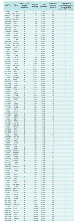
127 Exercise and natural killer cells

     All the participants were assessed at three time points: (i)        5–10-minute cool-down period (at the preferred PO). For the
at baseline (untrained); (ii) after a 4-week MICT phase (mid-in-         Monday sessions, after a ~15-minute warmup slightly below
tervention); and (iii) after a subsequent 4-week HIIT phase              the VT, participants performed three (in the first week) to
(post-intervention). The study design is summarized in Figure 1.         five (in the remainder of weeks) 3-minute intervals at a PO
                                                                                                           above the RCP (starting
                                                                                                           from just above the RCP
                                                                                                           in the first week and in-
                                                                                                           creasing the PO by 2-3%
                                                                                                           per week thereafter) inter-
                                                                                                           spersed with 3-minute rest
                                                                                                           periods. For the Wednes-
                                                                                                           day sessions, participants
                                                                                                           performed 10 (in the first
                                                                                                           week) to 15 (in the last
                                                                                                           two weeks) 30-second
                                                                                                           sprints (at maximal or
                                                                                                           near-maximal intensity)
                                                                                                           interspersed with 80-sec-
                                                                                                           ond rest periods. Rating of
                                                                                                           perceived exertion (RPE,
                                                                                                           on a 0 to 10 scale) were
                                                                                                           recorded in each session,
                                                                                                           with subjects instructed to
                                                                                                           aim at an RPE value ≥ 9
                                                                                                           during the intervals versus
                                                                                                           4 and 2 in the recovery pe-
                                                                                                           riods between the 3-min-
                                                                                                           ute intervals and 30-sec-
Figure 1. Study design. Protocol and intervention (a) and assessments (b).
                                                                                                           ond sprints, respectively.

Intervention                                                        Measurements

All exercise-training sessions were individually supervised         Exercise tests
(I.P.A. and L.B.A.), and were performed indoors with the same       All the participants performed a graded cycle ergometer test
bicycle-ergometer (Ergometrics Ergoline 800; Jaeger, Bitz,          until volitional exhaustion, at baseline and at mid- and post-in-
Germany) that was used during the exercise tests for determi-       tervention, respectively (after a 48-hour rest period from the
nation of VO2peak, ventilatory threshold (VT) and respiratory       last training session). All tests were performed between 9:00
compensation point (RCP), which are described later.                am and 12:00 pm at the exercise physiology laboratory of the
                                                                    UEM. Before the tests (~08:00 am), venous blood samples
Moderate-intensity continuous training                              were collected from the antecubital vein into VacutainerR
This first phase started the week after baseline assessments and    (sodium-heparin) tubes (BD Biosciences, San José, CA) for
lasted four weeks in total, including four sessions per week        performing all the studies on NK cells that are described in the
(Monday, Wednesday, Friday and Sunday). The total duration          following sections.
of all the sessions was the same (~50 minutes), barring grad-            Gas exchange data were recorded ‘breath-by-breath’ with
ual build-up to this duration during the first week. The core       a metabolic cart (Vmax 29C; SensorMedics Corp., Yorba Lin-
part of the session lasted 40 minutes and the workload (power       da, CA). The test started at 20 watts and the load was increased
output [PO]) was gradually increased as follows: PO corre-          in a ramp-like fashion (5 watts/12 sec [20 watts/minute on av-
sponding to the VT (first week), 5% above the VT (second            erage]) while cadence was kept constant at 60 to 70 rpm. Par-
week), midpoint between VT and RCP (third week), and 10%            ticipants were verbally encouraged to continue pedalling until
below the RCP (fourth week). All the sessions were preceded         volitional exhaustion, and were continuously monitored elec-
and followed by a 5–7-minute warmup below the VT and five           trocardiographically. The VO2peak was defined as the highest
minutes of cool-down (at the preferred PO), respectively.           value (mean of 30 seconds) reached during the test. We also
     The subjects refrained from doing any exercise sessions        determined the VT and RCP (also termed ‘second ventilatory
for one week after the MICT intervention and then started the       threshold’) using the following criteria: VT, the point at which
HIIT program.                                                       the ventilatory equivalent for oxygen (ventilation [VE]∙VO2-1)
                                                                    starts to increase with no concomitant increase in the venti-
High-intensity interval training                                    latory equivalent for carbon dioxide (VE∙VCO2-1) and with
This second phase also lasted four weeks in total, and in-          departure from linearity of VE; RCP, the point at which both
cluded two sessions per week (Monday and Wednesday).                VE∙VO2-1 and VE∙VCO2-1 increase together with a decrease in
The total duration of all the sessions was ~40 minutes. Each        end-tidal pressure of carbon dioxide (31).
session started with a ~15-minute warmup and ended with a

EIR 27 2021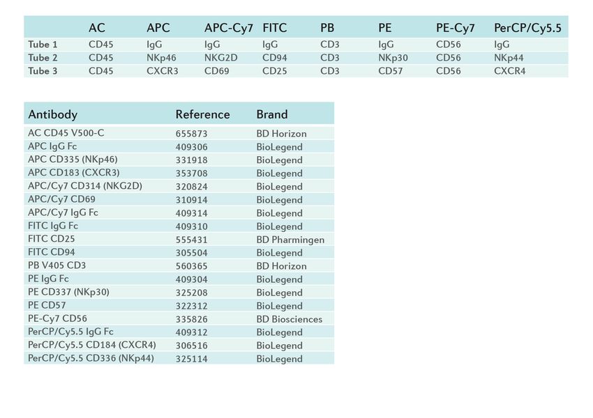
Exercise and natural killer cells 128
Flow cytometry with 355 µL of buffer UA (8 M urea in 0.1 M Tris/HCl, pH
Flow cytometry analyses (FACSCantoTM II, BD Bioscienc- 8.5). Spin unit were then centrifuged at 14,000 × g for 15 min-
es) were conducted to assess the presence of various cell sur- utes and washed again with 375 µL of buffer UA. Alkylation
face markers related to cellular activation processes (Table 1). of cysteine residues in the samples was carried out by adding
The gating strategy was based on dead/live cells and doublets 100 μL of freshly prepared iodoacetamide solution (50 mM
discrimination. When possible, a minimum of 10,000 events in UA buffer) into the spin filters and incubating the samples
of the population of interest was analyzed. FACSDiva™ soft- in the dark for 20 minutes. Filters were washed three times
ware (BD Biosciences) was used for analysis. with 200 μL of UA followed by three washes with 100 μL of
50 mM ammonium bicarbonate. On-filter digestion was per-
Natural killer cell function formed overnight with trypsin at 37°C. The released peptides
Peripheral blood mononuclear cell purification and cul- were collected by centrifugation at 14,000 × g for 10 minutes
tures. Heparinized blood was diluted two-fold with phosphate followed by an additional wash with 50 μL of 50 mM ammo-
buffered saline (PBS) and layered on top of Ficoll-Paque in nium bicarbonate. The peptide-containing eluates were finally
a 50 mL conical polypropylene tube (BD Biosciences). After desalted using C-18 spin columns (C-18 Micro Spin Column;
centrifugation, peripheral blood mononuclear cells (PBMC) Harvard Apparatus, Holliston, MA) and dried in a speed vac-
were recovered and washed twice in calcium- and magne- uum (Thermo Scientific). Samples were resuspended in 0.1%
sium-free PBS. Cells were seeded in culture flasks containing formic acid and peptide concentration was measured with the
Rosewell Park Memorial Institute 1640 medium and 10% fe- Qubit Protein Assay Kit (Thermo Scientific). Before the liquid
tal bovine serum (FBS) (both from Gibco/Thermo Scientific, chromatography-mass spectrometry (LC-MS/MS) analysis,
Flow cytometry
Waltham, MA). After 48 hours, cell suspensions were stim- 200 ng of each biological replicate (baseline, MICT, HIIT) and
Flow cytometry
ulated with 10analyses
µg/mL(FACSCantoTM
phytohemagglutinin II, BD Biosciences)
(PHA) (Sigma were conducted
0.66 µg to assess
sample thewas
presence
analyzed.
of variousSt
Aldrich, cellLouis,
surfaceMO) markers
(18,related
49) andto cellular activation
functional assaysprocesses 1). The gating
were (Table Liquid strategy
chromatography-tandem mass spectrometry.
was based on dead/live cells and doublets discrimination. When possible, a minimum of 10,000
performed as indicated below. LC-MS/MS
events of the population of interest was analyzed. FACSDiva™ software (BD Biosciences) was used
analysis was performed using a Q Exactive
Natural killer cell isolation. NK cells were purified from mass spectrometer interfaced with an Easy-nLC 1000 na-
for analysis.
PBMC using the NK Cell Isolation Kit (Miltenyi Biotech, Ber- noUPLC System (both from Thermo Scientific). Digested
gisch
Natural Gladbach,
killer cellGermany).
function Briefly, non-NK cells (T, B, stem, peptides were loaded onto an Acclaim PepMap100 pre-
Peripheraland
dendritic blood mononuclear
erythroid cells,cell purification
monocytes, andgranulocytes)
and cultures. Heparinizedcolumn blood(75wasµm diluted
× 2two-
cm) connected to an Acclaim PepMap
fold with
were phosphate labeled
magnetically buffered using
saline a(PBS)
cocktailand of
layered on top of Ficoll-Paque
biotin-conjugat- in a
RSLC (50 µm × 15 50 mL conical
cm) analytical column (Thermo Scien-
polypropylene tube (BD Biosciences). After centrifugation, peripheral blood mononuclear cells
ed antibodies and the NK Cell MicroBead Cocktail (Miltenyi tific). Peptides were eluted with a 180 minute linear gradi-
(PBMC) were recovered and washed twice in calcium- and magnesium-free PBS. Cells were seeded
Biotech). Isolation
in culture flasks of highly
containing pure Park
Rosewell NK cells was Institute
Memorial achieved by medium
1640 ent ofand 3–30%
10% fetalacetonitrile
bovine in 0.1% formic acid at a flow rate
depletion of magnetically-labeled cells.
serum (FBS) (both from Gibco/Thermo Scientific, Waltham, MA). Afterof48300 nL/minute
hours, directly onto the nanoES Emitter (Ther-
cell suspensions
wereNatural
stimulated killer
with cell cytotoxicity
10 µg/mL assay. K562
phytohemagglutinin (PHA)cells wereAldrich,
(Sigma mo Scientific).
St Louis, MO) The(18,mass
49) spectrometer was operated in a top
and functional
used assayscells,
as NK target were performed
and wereascultured
indicatedinbelow.
Iscove’s Mod- 10 data-dependent mode. Survey scans were acquired at a
ified Dulbecco’s Medium supplemented with 10% FBS. To resolution of 70,000 (m/z 200) and fragmentation spectra
Natural killer cell isolation. NK cells were purified from PBMC using the NK Cell Isolation Kit
assess NK cytotoxic capacity, K562 cells were loaded with at 17,500 (m/z 200). Peptide selection was done with an
(Miltenyi Biotech, Bergisch Gladbach, Germany). Briefly, non-NK cells (T, B, stem, dendritic and
bis[acetoxymethyl]
erythroid cells, monocytes, 2,2´:6´,2´´-terpyridine-6,6´´-dicarboxylate
and granulocytes) were magnetically labeledisolation window
using a cocktail of of 2.0 Th and normalized collision energy
biotin-
(BATDA),
conjugated antibodies and the ligand
a hydrophobic NK Cellthat penetrates
MicroBead the plasma
Cocktail of 28 was
(Miltenyi Biotech). applied
Isolation of for peptide fragmentation. The maximum
highly
membrane
pure NK cellsquickly, according
was achieved to the of
by depletion DELFIA® EuTDA Cy-
magnetically-labeled cells. injection time was 120 milliseconds for survey, and MS/MS
totoxicity Kit (Perkin Elmer, Waltham, MA). The assay was scans and automatic gain control target values of 3e6 for
Natural killer
performed withcell5cytotoxicity assay. K562 cells
× 103 BATDA-loaded K562were usedand
cells as NK100targetsurvey
cells, and wereand
scans cultured
5e5 forin MS/MS scans were used. Acquired
Iscove’s Modified Dulbecco’s Medium supplemented with 10% FBS. To assess NK cytotoxic
μL of primary NK cells at various concentrations (2 × 10 , 1 × raw data files were processed with MaxQuant (10) software
4
capacity, K562 cells were loaded with bis[acetoxymethyl] 2,2´:6´,2´´-terpyridine-6,6´´-dicarboxylate
10 4
, 5 × 10³ and 2.5 × 10³ cells). After cytolysis, the released (v1.6.0.16) using the internal search engine Andromeda,
(BATDA), a hydrophobic ligand that penetrates the plasma membrane quickly, according to the
ligand
DELFIA® (TDA)
EuTDA reacts with a europium
Cytotoxicity Kit (Perkin solution to constitute
Elmer, Waltham, MA).a Theand assaysearched
was performed againstwiththe UniProt database restricted to
fluorescent chelate. The
5 × 10³ BATDA-loaded fluorescent
K562 cells and signal
100 μL correlates
of primary directly Homo concentrations
NK cells at various sapiens entries (2 ×(release 2018_11). Carbamidometh-
with
104, 1the amount
× 10 4
, 5 × 10of3 lysed
and 2.5cells:
× 103 cells). After cytolysis, the released ylation ligand (TDA) was setreactsaswith
fixeda modification, whereas methionine
europium solution to constitute a fluorescent chelate. The fluorescent signal correlatesoxidation directly with and protein N-terminal acetylation
the amount of lysed cells: were defined as variable modifications. Mass
!"#$%&'$()*+ %$+$*-$ (/01()-)34#0()*($01- %$+$*-$ (/01()-)
% Specific release = x 100 tolerance was set to 8 and 20 ppm at the MS and
5*"&'1' %$+$*-$ (/01()-)34#0()*($10- %$+$*-$ (/01()-)
MS/MS level, respectively. Enzyme specificity
Mass spectrometry-based proteomics was set to trypsin, allowing for a maximum of
Samplespectrometry-based
Mass processing and protein digestion. Frozen sample aliquots of 1–3two
proteomics × 10missed
6
NK cells were lysed The “match between runs” option
cleavages.
in 150 µL SDT-lysis buffer (4% SDS and 0.1 M DTT in 0.1 M Tris/HCl, pH 7.6) using 1:10 sample
Sample processing and protein digestion. Frozen sample al- was enabled with a 1.5-minute time window and a 20-min-
to buffer ratio, at 95°C for 5 minutes. Samples were briefly sonicated to reduce the viscosity of the
iquots
lysates ofand1–3then× centrifuged
106 NK cells were lysed
at 16,000 × g forin 150 µL SDT-lysis
10 minutes. ute alignment
Sample processing was based window
on theto match identification across sam-
buffer
procedure(4%ofSDS and 0.1 et
Wisniewski Mal.
DTT (75)inwith
0.1 M Tris/HCl,
minor pH 7.6) Aliquots
modifications. us- ples. (100The
µL) minimum
of lysates were peptide length was set to seven amino
ing
loaded1:10 sample
onto to buffer ratio,
spin ultrafiltration at (Amicon
units 95°C forUltra-0.5
5 minutes. Samplesmembrane;
Ultracel-30 acids. The Merck,false discovery rate (FDR) for peptides and pro-
Kenilworth,
NJ) and
were washedsonicated
briefly with 355 µL to of bufferthe
reduce UA viscosity
(8 M urea in of0.1theMlysates
Tris/HCl, pHteins8.5). Spinset
was unit
towere
1%.thenNormalized spectral protein label-free
centrifuged at 14,000 × g for 15 minutes and washed again with 375
and then centrifuged at 16,000 × g for 10 minutes. Sample quantification intensities µL of buffer UA. Alkylation of were calculated using the Max-
cysteine residues in the samples was carried out by adding 100 μL of freshly prepared iodoacetamide
processing was based on the procedure of Wisniewski et al. LFQ algorithm.
solution (50 mM in UA buffer) into the spin filters and incubating the samples in the dark for 20
(75) withFilters
minutes. minor weremodifications. Aliquots
washed three times with (100
200 μL µL) of lysates
of UA followed by three MaxQuant
washes with output
100 μLdata was analyzed with the Perseus
were
of 50 mMloaded onto spin
ammonium ultrafiltration
bicarbonate. On-filterunits (Amicon
digestion Ultra-0.5 overnight
was performed modulewith (v1.6.5.0)
trypsin at(67).
37°C.Proteins only identified by site, con-
Ultracel-30 membrane;
The released peptides wereMerck,
collectedKenilworth, NJ) and
by centrifugation washed
at 14,000 × g fortaminants
10 minutesand reverse
followed hits were removed. Changes in protein
by an
additional wash with 50 μL of 50 mM ammonium bicarbonate. The peptide-containing eluates were
finally desalted using C-18 spin columns (C-18 Micro Spin Column; Harvard Apparatus, Holliston,
MA) and dried in a speed vacuum (Thermo Scientific). Samples were resuspended in 0.1% formic EIR 27 2021
6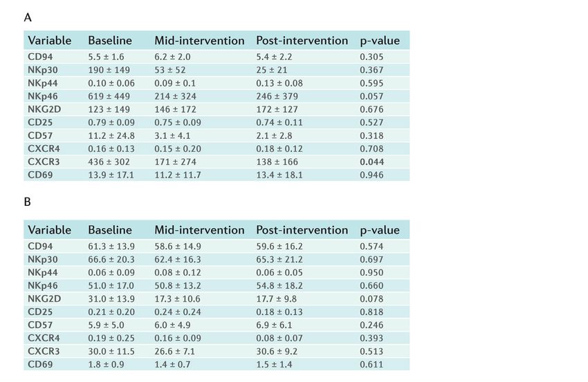
129 Exercise and natural killer cells

abundance higher than 4-fold in proteins identified with more             (focusing on log2FC values) at the top end of the list. The FDR
than two unique peptides were considered as relevant protein              cutoff was set at 25% to maximize hypothesis generation. Specif-
differences.                                                              ically, the enrichment was run over the following databases: Gene
                                                                          Ontology (GO) terms (Biological Process, Cellular Component,
Statistical analyses                                                      Molecular Function) and Kyoto Encyclopedia of Genes and Ge-
Exercise capacity and natural killer cell numbers/cytotoxicity            nomes (KEGG) pathways.
Data are presented as mean ± SD. Normal distribution (Sha-                Visualization as a network of the relationship between en-
piro-Wilk test) and homoscedasticity (Levene’s test) of the               riched pathways. Cytoscape 3.5.1. software was used to repre-
data were checked before any statistical treatment. A one-way             sent the relationship between the enriched pathways identified by
repeated-measures ANOVA was used to examine differences                   GSEA (based on the number of common annotated genes between
across the three conditions (i.e., baseline, mid-intervention,            datasets).
and post-intervention). Bonferroni’s test was performed post
hoc when a significant condition (or ‘time’) effect was present.
The Greenhouse-Geisser correction was applied when Mauch-                                            RESULTS
ly’s test of sphericity was violated. Statistical analyses were
conducted using a statistical software package (SPSS v23;                 No adverse effect was noted during the exercise sessions and
IBM, Amonk, NY) setting the significance level at α=0.05.                 no participant reported any medical condition (e.g., infection/s)
                                                                          during the study that could have altered the results.
Proteomics
Candidate proteins. To identify proteins in plasma that were              Exercise capacity
differentially expressed across the three conditions (baseline,           No significant time effect (p = 0.455) was found for partic-
mid-intervention and post-intervention) the magnitude of the              ipants’ body mass (baseline: 69.5 ± 9.5 kg; mid-interven-
changes was calculated by taking the base 2 logarithm of the              tion: 69.1 ± 9.0 kg; and post-intervention: 69.4 ± 9.1 kg). A
mean fold change (log2FC). For proteins with no expression                significant time effect (p = 0.016) was found for VO2peak,
in either of the three samples, log2FC values were adjusted by            which showed an increasing trend during the study (baseline:
adding one to each mean and then calculating the ratio. Negative          34.9 ± 4.8 mL·kg-1·min-1; mid-intervention: 38.7 ± 7.6 mL·kg-
values indicate down-regulation whereas positive values indicate          1
                                                                           ·min-1; and post-intervention: 39.5 ± 7.5 mL·kg-1·min-1). In
up-regulation. Multi-test correction was performed according to           post hoc pairwise comparisons, a quasi-significant difference
the Benjamini-Hochberg method, that is, p-values were adjusted            was found for post-intervention versus baseline (p = 0.071).
with FDR correction (5).                                                  The peak PO (PPO) attained during the tests (p = 0.004 for
Processing of protein expression data. Proteins identified in the         time effect; baseline: 2.4 ± 0.2 watts·kg-1; mid-intervention:
plasma profiles were filtered to unique human-reviewed protein            2.8 ± 0.4 watts·kg-1; and post-intervention: 2.9 ± 0.4 watts·kg-1), as
entries according to the UniProt Knowledgebase (UniProtKB)                well as the PO eliciting the RCP (p = 0.006 for time effect; baseline:
(67). To reduce potential interference for high signals, keratin          2.1 ± 0.2 watts·kg-1; mid-intervention: 2.4 ± 0.4 watts·kg-1; and
protein entries were excluded from the analysis. In addition, the         post-intervention: 2.5 ± 0.3 watts·kg-1) also showed an increas-
protein entries that were traced to an unreviewed UniProtKB en-           ing trend during the study, with significant post hoc differences
try were manually curated to find a valid reviewed entry. When            for post-intervention versus baseline (p = 0.028 for PPO and
more than one protein entry was traced to the same reviewed Un-           0.044 for RCP). No significant time effect (p = 0.154)
iProtKB entry, the following criteria were applied to prevent du-         was, however, found for the PO eliciting the VT (baseline:
plications: (i) to prioritize protein entries with an identical protein   1.1 ± 0.1 watts·kg-1; mid-intervention: 1.1 ± 024 watts·kg-1; and
ID to the one noted in UniProtKB; (ii) to prioritize protein entries      post-intervention: 1.2 ± 1.2 watts·kg-1).
that were automatically traced to the reviewed UniProtKB entry
over those that were first traced to an unreviewed or deleted one         NK number and cytotoxicity
and then manually curated; (iii) to prioritize protein entries with       A significant time effect was found for PBMC (p < 0.001,
valid signals; (iv) to prioritize whole protein entries over those        Figure 2A) and NK (p < 0.001, Figure 2B) cell counts, with both
referring to fragments; and (v) to apply an alphabetical criterion        cell counts considerably increasing at both mid- and post-inter-
when all the criteria above were not applicable.                          vention compared with baseline, and with a clear training time-de-
Contextualization of the differentially expressed proteins                pendent effect (i.e., a further, significant increase was found at
within “natural killer cell” protein network. Effector proteins           post-intervention as compared with mid-intervention).
were used to focus the analysis on the biological condition of in-        A significant time effect was also found for NK cell cytotoxici-
terest in the human biological network. The relationships between         ty (p < 0.001, Figure 2C); which significantly increased at both
the differentially expressed proteins and the immune system were          mid- and post-intervention compared with baseline; however, no
assessed. Different publicly available databases were consulted           training time-dependent effect was found (i.e., no significant dif-
for the human protein network generation (Reactome, Molecular             ferences were found for mid- versus post-intervention).
INTeraction database (MINT) and Biological General Repository
for Interaction Datasets [BioGrid]) (25, 26).                             Flow cytometry
Gene set enrichment analysis. Proteomic data were analyzed                No significant condition effect was found except for the sur-
using the Gene Set Enrichment Analysis (GSEA) tool (63) to                face expression of the chemokine receptor CXCR3 expressed as
compare the differential pathways and molecular processes be-             mean fluorescence intensity (p = 0.044, with a trend towards a
tween conditions. This approach categorizes genes according to            decrease at mid- and post-intervention compared with baseline),
a specific metric, ranking the most statistically significant genes       although no significant differences were found in post hoc pair-

EIR 27 2021
Exercise and natural killer cells 130

                                                                            wise comparisons (p = 0.244 and 0.093 for baseline versus mid-
                                                                            and post-intervention, respectively; and p = 1.000 for mid- versus
                                                                            post-intervention) (Table 2).

                                                                            Proteomics
                                                                            A total of 2538 human proteins were identified by LC-MS/MS,
                                                                            of which 2448 valid protein entries were used in the next steps
                                                                            of the study and 90 were filtered out (i.e., potential contaminants,
                                                                            reverse and only identified by site).

                                                                            Protein candidates
                                                                            Mid-intervention versus baseline. The expression of 74 plasma
                                                                            proteins was significantly different between mid-intervention and
                                                                            baseline (FDR q-value < 0.01), of which 49 and 25 were upregu-
                                                                            lated and downregulated, respectively, at mid-intervention (Table
                                                                            3).
                                                                                 Moreover, one and nine unique proteins were identified (FDR
                                                                            q-value < 0.01) at baseline and at mid-intervention, respectively.
                                                                            Among the candidate proteins, 17 are known to be potentially re-
                                                                            lated to immune function (Figure 3):
                                                                            Proteins upregulated at mid-intervention (versus baseline):
                                                                            N-acylsphingosine amidohydrolase 1 (ASAH1); V-type proton

                                                                            Figure 3. Visual protein interaction network of the proteins related to
                                                                            the immune system that were differentially expressed (upregulated or
                                                                            downregulated) at mid-intervention compared to baseline.

                                                                            ATPase catalytic subunit A (ATP6V1A); CD44; cathepsin B
                                                                            (CTSB); integrin beta chain-2 (ITGB2, also known as ‘CD18’);
                                                                            nucleoporin 88 (NUP88); phosphoinositide-3-kinase regulatory
                                                                            subunit 1 (PIK3R1); cAMP-dependent protein kinase catalytic
                                                                            subunit beta (PRKACB); proteasome (prosome, macropain) sub-
                                                                            unit, alpha 1 (PSMA1); glycogen phosphorylase, liver isoform
                                                                            (PYGL); RAN binding protein 2 (RANBP2, also known as ‘nu-
                                                                            cleoporin 358’); Rho-associated coiled-coil containing protein
                                                                            kinase 1 (ROCK1); and WAS/WASL interacting protein family
                                                                            member 1 (WIPF1).
                                                                            Proteins downregulated at mid-intervention (versus baseline):
                                                                             dedicator of cytokinesis 2 (DOCK2); hexosaminidase subunit
Figure 2. Within-subject comparison of the number of peripheral              beta (HEXB); minor histocompatibility protein HA-1 (HMA1,
blood mononuclear cells ([PBMC], panel A), and of the number (panel
                                                                             also known as ‘Rho GTPase activating protein 45’); and neutro-
B) and cytotoxicity (panel C, N=8) of natural killer (NK) cells. Cytotox-
icity was expressed as % specific fluorescence release (see text for         phil cytosolic factor 2 (NCF2).
more details) at a ratio of effector (NK) to target (K562) cells of 1:1,     Post-intervention versus baseline. The expression of 76 plas-
with the results replicated (i.e., same p-values) at different E:T ratios    ma proteins was significantly different between post-interven-
(1:2, 2:1 and 4:1).
                                                                             tion and baseline, of which 22 and 54 were upregulated and
                                                                             downregulated, respectively, at post-intervention (Table 4).
                                                                             No unique proteins were identified (FDR q-value < 0.01) at
                                                                             post-intervention compared with baseline.

                                                                                                                                      EIR 27 2021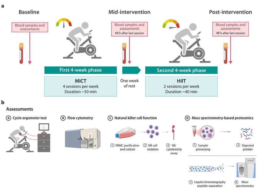
Table
131     1. Antibodies
    Exercise and naturalper  tube
                         killer    and
                                cells         per peripheral blood sample used for flow cytometry analysis.

Table 1. Antibodies per tube and per peripheral blood sample used for flow cytometry analysis.

Abbreviations: AC, AmCyan; APC, Allophycocyanin; APC-Cy7, Allophycocyanin cyanin-7; Ig, Immunoglobulin; FITC, Fluorescein isothiocya-
nate; PB, Pacific blue; PE, Phycoerythrin; PE-Cy7, Phycoerythrin cyanin-7; PerCP-Cy7, Peridinin chlorophyll protein.

 Table footnote. Abbreviations: AC, AmCyan; APC, Allophycocyanin; APC-Cy7, Allophycocyanin
Table              Table
      2. Flow cytometry     2. Flow
                        results       cytometry
                                         results in NK cells.
                                in NK cells.
 cyanin-7;    Ig, Immunoglobulin;           FITC,
                                          Fluorescein isothiocyanate; PB, Pacific blue; PE,
 Phycoerythrin; PE-Cy7, Phycoerythrin cyanin-7; PerCP-Cy7, Peridinin chlorophyll protein.

                                                                  19

Table footnote. Values are mean ± SD of fluorescence intensity (A) or of percent positive (B) for each NK cell receptor. The only significant
p-value for the time (condition) effect is in bold. Abbreviations: CXCR3, C-X-C chemokine receptor type 3; CXCR4, C-X-C chemokine receptor
type 4 (also known as fusin or CD184 [cluster of differentiation 184]).
 Table footnote. Values are mean ± SD of fluorescence intensity (A) or of percent positive (B) for
 each NK cell receptor. The only significant p-value for the time (condition) effect is in bold.
 Abbreviations: CXCR3, C-X-C chemokine receptor type 3; CXCR4, C-X-C chemokine receptor type
EIR 27 2021

 4 (also known as fusin or CD184 [cluster of differentiation 184]).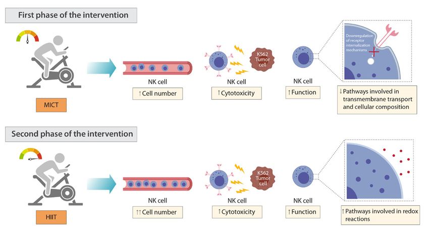
Exercise and natural killer cells 132
e 3. List of proteins
Table 3. List differentially expressed
of proteins differentially at mid-intervention
expressed versusbaseline.
versus
at mid-intervention baseline.
Table footnote. Symbols: ↑ upregulation at mid-intervention com-
pared to baseline; ↓ downregulation at mid-intervention compared
to baseline. Abbreviations (other than protein names): FDR, false
discovery rate; Log2FC, log 2 fold change. Abbreviations (pro-
teins): AARS, alanyl-tRNA synthetase 1; ACADSB, acyl-coA
dehydrogenase short/branched chain; ARHGEF1, Rho guanine
nucleotide exchange factor 1; ASAH1, N-acylsphingosine amido-
hydrolase 1; ATP2B4, ATPase plasma membrane Ca2+ transporting
4; ATP5L, ATP synthase membrane subunit G; ATP6V1A, ATPase
H+ transporting V1 subunit A (also known as V-type proton AT-
Pase catalytic subunit A); CD44, CD44 molecule; COPS3, COP9
signalosome subunit 3; CORO7, CORO7-PAM16 readthrough;
CTSB, cathepsin B; DDX5, DEAD-box helicase 5; DNAJB11,
DnaJ heat shock protein family (Hsp40) member B11; DOCK11,
dedicator of cytokinesis 11; DOCK2, dedicator of cytokinesis 2;
EIF2S1, eukaryotic translation initiation factor 2 subunit alpha;
EIF3H, eukaryotic translation initiation factor 3 subunit H; EN-
DOD1, endonuclease domain containing 1; ERO1L, endoplasmic
reticulum oxidoreductase 1 alpha; FLII, FLII actin remodeling pro-
tein; GLG1, Golgi glycoprotein 1; GLIPR2, GLI pathogenesis re-
lated 2; GOT2, glutamic-oxaloacetic transaminase 2; GRHPR, gly-
oxylate and hydroxypyruvate reductase; GSR, glutathione-disulfide
reductase; H3F3A, H3.3 histone A; HARS, histidyl-TRNA synthe-
tase 1; HEATR1, hEAT repeat containing 1; HEXB, hexosamini-
dase subunit beta; HIST1H1D, H1.3 linker histone, cluster mem-
ber; HIST1H1E, H1.4 linker histone, cluster member; HMHA1,
minor histocompatibility protein HA-1 (also known as Rho GTPase
activating protein 45 ); IPO5, importin 5; ITGB2, integrin subunit
beta 2 (also known as ‘CD18’); LMAN2, lectin, mannose bind-
ing 2; MAP7D1, MAP7 domain containing 1; MTA2, metastasis
associated 1 family member 2; NCF2, neutrophil cytosolic factor
2; NEK9, NIMA related kinase 9; NHP2L1, NHP2-like protein 1
(also known as ‘small nuclear ribonucleoprotein 13) [SNU13]);
NUMA1, nuclear mitotic apparatus protein 1; NUP88, nucleoporin
88; PARK7, parkinsonism associated deglycase; PDS5B, PDS5 co-
hesin associated factor B; PGD, Phosphogluconate dehydrogenase;
PIK3R1, phosphoinositide-3-kinase regulatory subunit 1; PMP-
CB, mitochondrial-processing peptidase subunit beta; PRKACB,
protein kinase CAMP-activated catalytic subunit beta; PRPF6,
pre-MRNA processing factor 6; PRPF8, pre-MRNA processing
factor 8; PSMA1, proteasome (prosome, macropain) subunit alpha
1; PYGL, glycogen phosphorylase L; RAB1B, RAB1B, member
RAS oncogene family; RAD50, RAD50 double strand break repair
protein; RANBP2, RAN binding protein 2; RANGAP1, Ran GT-
Pase activating protein 1; RCC2, regulator of chromosome conden-
sation 2; RNF40, ring finger protein 40; ROCK1, Rho associated
coiled-coil containing protein kinase 1; RPS7, ribosomal protein
S7; RPS9, ribosomal protein S9; SAFB, scaffold attachment factor
B; SEPT9, septin 9; SH3BGRL3, SH3 domain binding glutamate
rich protein like 3; SHMT2, serine hydroxymethyltransferase 2;
SLC25A3, solute carrier family 25 member 3; SMC1A, structural
maintenance of chromosomes 1A; SRRM2, serine/arginine repeti-
tive matrix 2; TAOK3, TAO kinase 3; TBL2, transducin beta like 2;
UQCRFS1, ubiquinol-cytochrome C reductase, Rieske iron-sulfur
polypeptide 1; VIP36, lectin, mannose binding 2; WIPF1, WAS/
WASL interacting protein family member 1; ZC3HAV1, zinc finger
CCCH-type containing, antiviral 1; ZYX, zyxin.
. Symbols: ↑ upregulation at mid-intervention compared to baseline; ↓ downregulation at mid-intervention
seline. Abbreviations (other than protein names): FDR, false discovery rate; Log2FC, log 2 fold change.
proteins): AARS, alanyl-tRNA synthetase 1; ACADSB, acyl-coA dehydrogenase short/branched chain;
o guanine nucleotide exchange factor 1; ASAH1, N-acylsphingosine amidohydrolase 1; ATP2B4, ATPase
ne Ca2+ transporting 4; ATP5L, ATP synthase membrane subunit G; ATP6V1A, ATPase H+ transporting
(also known as V-type proton ATPase catalytic subunit A); CD44, CD44 molecule; COPS3, COP9
21
EIR 27 2021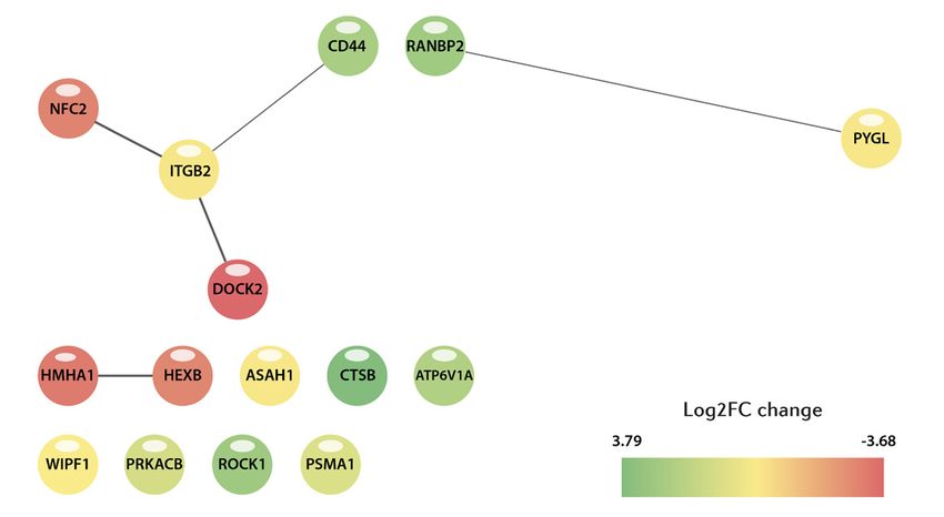
133 Exercise and natural killer cells
le 4. List of proteins differentially expressed at post-intervention versus baseline.
          Table 4. List of proteins differentially expressed at post-intervention versus baseline.

                                                                                      Table footnote. Symbols: ↑ upregulation at post-intervention com-
                                                                                      pared to baseline; ↓ downregulation at post-intervention compared
                                                                                      to baseline. Abbreviations (other than protein names): FDR, false
                                                                                      discovery rate; Log2FC, log 2 fold change. Abbreviations (pro-
                                                                                      teins): ACSL4, acyl-CoA synthetase long chain family member 4;
                                                                                      ACTN1, actinin alpha 1; APOBR, apolipoprotein B Receptor; AR-
                                                                                      HGEF1, Rho guanine nucleotide exchange factor 1; ARPC5, actin
                                                                                      related protein 2/3 complex subunit 5; ATP2A2, ATPase sarcoplas-
                                                                                      mic/endoplasmic reticulum Ca2+ Transporting 2; ATP2B4, ATPase
                                                                                      plasma membrane Ca2+ transporting 4; CD48, CD48 molecule;
                                                                                      CNDP2, carnosine dipeptidase 2; CORO7, CORO7-PAM16 read-
                                                                                      through; COX4I1, cytochrome C oxidase subunit 4I1; EEF1B2,
                                                                                      eukaryotic translation elongation factor 1 beta 2; EMILIN1, elastin
                                                                                      microfibril interfacer 1; EPRS, glutamyl-prolyl-tRNA synthase 1;
                                                                                      FCER1G, Fc fragment of IgE receptor Ig; G6PD, glucose-6-phos-
                                                                                      phate dehydrogenase; GBP1, guanylate binding protein 1; GDI1,
                                                                                      GDP dissociation inhibitor 1; GOT2, glutamic-oxaloacetic trans-
                                                                                      aminase 2; GSTO1, glutathione S-transferase omega 1; H1FX,
                                                                                      H1.10 linker histone; H2AFX, H2A.X variant histone; HARS,
                                                                                      histidyl-tRNA synthetase 1; HDGF, heparin binding growth factor;
                                                                                      HIST1H1D, histone cluster 1 H1 family member D; HIST1H1E,
                                                                                      histone cluster 1 H1 family member E; HIST1H4A, H4 clustered
                                                                                      histone 1; HIST2H2AC, H2A clustered histone 20; HNRNPH1,
                                                                                      heterogeneous nuclear ribonucleoprotein H1; HNRNPUL1, het-
                                                                                      erogeneous nuclear tibonucleoprotein U Like 1; ILK, integrin
                                                                                      linked kinase; ITGA2, integrin subunit alpha 2; ITGAL, integrin
                                                                                      subunit alpha L; KIF5B, kinesin family member 5B; LTA4H, leu-
                                                                                      kotriene A4 hydrolase; LTBP1, latent transforming growth factor
                                                                                      beta binding protein 1; MDH1, malate dehydrogenase 1; MTDH,
                                                                                      metadherin; MVP, major vault protein; NCF2, neutrophil cytosolic
                                                                                      factor 2; OGDH, oxoglutarate dehydrogenase; OXCT1, 3-oxoacid
                                                                                      CoA-transferase 1; PARVB, parvin beta; PFKP, phosphofructoki-
                                                                                      nase, platelet; PGAM1, phosphoglycerate mutase 1; PGD, phospho-
                                                                                      gluconate dehydrogenase; PPP2R1A, protein phosphatase 2 scaf-
                                                                                      fold subunit alpha; PRDX6, peroxiredoxin 6; PTMA, prothymosin
                                                                                      alpha; RAD50, RAD50 double strand break repair protein; RALY,
                                                                                      RALY heterogeneous nuclear ribonucleoprotein; RCC1, regulator
                                                                                      of chromosome condensation 1; ROCK1, Rho associated coiled-
                                                                                      coil containing protein kinase 1; RPS15A, ribosomal protein S15a;
                                                                                      SACM1L, SAC1 like phosphatidylinositide phosphatase; SART1,
                                                                                      spliceosome associated factor 1, recruiter of U4/U6.U5 tri-SnRNP;
                                                                                      SCP2, sterol carrier protein 2; SH3BGRL3, SH3 domain binding
                                                                                      glutamate rich protein like 3; SHMT2, serine hydroxymethyltrans-
                                                                                      ferase 2; SMARCA5, SWI/SNF related, matrix associated, actin
                                                                                      dependent regulator of Chromatin, subfamily A, member 5; SRC,
                                                                                      SRC proto-oncogene, non-receptor tyrosine kinase; STIP1, stress
                                                                                      induced phosphoprotein 1; STK4; serine/threonine kinase 4; STX7,
                                                                                      syntaxin 7; SUN2, Sad1 and UNC84 domain containing 2; TBX-
                                                                                      AS1, thromboxane A synthase 1; TOR1AIP1, torsin 1A interacting
                                                                                      protein 1; TPI1, triosephosphate isomerase 1; TUBB1, tubulin beta
                                                                                      1 class VI; UGGT1, UDP-glucose glycoprotein glucosyltransferase
                                                                                      1; USO1, USO1 vesicle transport factor; VWF, Von Willebrand fac-
                                                                                      tor; WTAP, WT1 associated protein; YARS, tyrosyl-tRNA synthe-
                                                                                      tase 1; YWHAH, tyrosine 3-monooxygenase/tryptophan 5-mono-
                                                                                      oxygenase activation protein Eta; ZC3HAV1, zinc finger CCCH-
                                                                                      type containing, antiviral 1

e. Symbols: ↑ upregulation at post-intervention compared to baseline; ↓ downregulation at post-intervention
aseline. Abbreviations (other than protein names): FDR, false discovery rate; Log2FC, log 2 fold change.
 (proteins): ACSL4, acyl-CoA synthetase long chain family member 4; ACTN1, actinin alpha 1; APOBR,
                                             23

          EIR 27 2021
Exercise and natural killer cells 134
Among the candidate proteins, 16 proteins are known to be poten- coil containing protein kinase 1 (ROCK1); SRC proto-onco-
tially related to immune function (Figure 4): gene, non-receptor tyrosine kinase (SRC); tubulin beta 1 class
VI (TUBB1); and Von Willebrand factor (VWF).
Post-intervention versus mid-intervention. The expression of
106 plasma proteins was significantly different between mid- and
post-intervention (FDR q-value < 0.01) (Table 5), with 26 and 80
upregulated and downregulated, respectively, at post-intervention.
Among the candidate proteins, 24 proteins are known to be
potentially related to immune function (Figure 5):
Figure 4. Visual protein interaction network of the proteins related to Figure 5. Visual protein interaction network of the proteins related to
the immune system that were differentially expressed (upregulated or the immune system that were differentially expressed (upregulated or
downregulated) at post-intervention compared to baseline. downregulated) at post-intervention compared to mid-intervention.
Proteins upregulated at post-intervention (versus baseline): Proteins upregulated at post-intervention (versus mid-in-
integrin subunit alpha L (ITGAL); and Fc fragment of IgE re- tervention): malectin (MLEC); proteasome 26S subunit
ceptor Ig (FCER1G). (PSMD3); and zeta chain of T cell receptor associated protein
Proteins downregulated at post-intervention (versus baseline): kinase 70 (ZAP70).
ARPC5 actin related protein 2/3 complex subunit 5 (ARPC5); Proteins downregulated at post-intervention (versus
guanylate binding protein 1 (GBP1); glutathione S-transferase mid-intervention: ATPase H+ transporting V1 subunit A (AT-
omega 1 (GSTO1); kinesin family member 5B (KIF5B); leu- P6V1A); calcium/calmodulin dependent protein kinase II delta
kotriene A4 hydrolase (LTA4H); major vault protein (MVP); (CAMK2D); catalase (CAT); CD44 molecule (CD44); cathep-
neutrophil cytosolic factor 2 (NCF2); phosphoglycerate mu- sin B (CTSB); cathepsin S (CTSS); kinesin family member
tase 1 (PGAM1); protein phosphatase 2 scaffold subunit alpha 5B (KIF5B); leukotriene A4 hydrolase (LTA4H); major vault
(PPP2R1A); peroxiredoxin 6 (PRDX6); Rho associated coiled- protein (MVP); nucleoporin 88 (NUP88); P21 (RAC1) acti-
EIR 27 2021oteins differentially expressed
135 Exercise and atcells
natural killer post-intervention versus mid-intervention.
Table 5. List of proteins differentially expressed at post-intervention versus mid-intervention.
Table footnote. Symbols: ↑ upregulation at post-intervention compared to
mid-intervention; ↓ downregulation at post-intervention compared to mid-in-
tervention. Abbreviations (other than protein names): FDR, false discovery
rate; Log2FC, log 2-fold change. Abbreviations (proteins): ACSL4, acyl-CoA
synthetase long chain Family member 4; AIFM1, apoptosis inducing factor
mitochondria associated 1; ANXA5, annexin A5; APOBR, apolipoprotein B
receptor; ATL3, atlastin GTPase 3; ATP2A2, ATPase sarcoplasmic/endoplas-
mic reticulum Ca2+ transporting 2; ATP6V1A, ATPase H+ transporting V1 sub-
unit A; BASP1, brain abundant membrane attached signal protein 1; CAM-
K2D, calcium/calmodulin dependent protein kinase II delta; CAT, catalase;
CCT3, chaperonin containing TCP1 subunit 3; CD44, CD44 molecule; CD48,
CD48 molecule; CNDP2, carnosine dipeptidase 2; COPS3, COP9 signalo-
some subunit 3; COX4I1, cytochrome C oxidase subunit 4I1; CTSB, cathep-
sin B; CTSS, cathepsin S; CUL4B, cullin 4B; DDX5, DEAD-box helicase 5;
DKC1, dyskerin pseudouridine synthase 1; DNAJB11, DnaJ heat shock pro-
tein family (Hsp40) member B11; EEF1B2, eukaryotic translation elongation
factor 1 beta 2; EIF2S1, eukaryotic translation initiation factor 2 subunit alpha;
ELAVL1, ELAV like RNA binding protein 1; EMILIN1, elastin microfibril
interfacer 1; EPRS, glutamyl-prolyl-tRNA synthetase 1; FAM120A, family
with sequence similarity 120A; G6PD, glucose-6-phosphate dehydrogenase;
GDI1, GDP dissociation inhibitor 1; GOT2, glutamic-oxaloacetic transami-
nase 2; GPS1, G protein pathway suppressor 1; H1FX, H1.10 linker histone;
H2AFX, H2A.X variant histone; H3F3A, H3.3 histone A; HDGF, heparin
binding growth factor; HIST1H1D, H1.3 linker histone, cluster member;
HIST1H1E, H1.4 linker histone, cluster member; HIST1H4A, H4 clustered
histone 1; HIST2H2AC, H2A clustered histone 20; HMGN4, high mobili-
ty group nucleosomal binding domain 4; HNRNPA0, heterogeneous nuclear
ribonucleoprotein A0; HSD17B4, hydroxysteroid 17-beta dehydrogenase 4;
ILF3, interleukin enhancer binding factor 3; ITGA2, integrin subunit alpha
2; KIF5B, kinesin family member 5B; LTA4H, leukotriene A4 hydrolase;
LTBP1, latent transforming growth factor beta binding protein 1; MAP7D1,
MAP7 domain containing 1; MDH1, malate dehydrogenase 1; MLEC, malec-
tin; MTDH, metadherin; MVP, major vault protein; NEK9, NIMA related ki-
nase 9; NUP88, nucleoporin 88; OGDH, oxoglutarate dehydrogenase; PAK2,
P21 (RAC1) activated kinase 2; PDS5B, PDS5 cohesin associated factor B;
PFKP, phosphofructokinase, platelet; PGD, phosphogluconate dehydroge-
nase; PHB2, prohibitin 2; PIK3R1, phosphoinositide-3-kinase regulatory
subunit 1; PPP2R1A, protein phosphatase 2 scaffold subunit alpha; PRDX6,
peroxiredoxin 6; PREX1, phosphatidylinositol-3,4,5-trisphosphate dependent
Rac exchange factor 1; PSMD3, proteasome 26S subunit, non-ATPase 3;
PTMA, prothymosin alpha; RANBP2, RAN binding protein 2; RBM15, RNA
binding motif protein 15; RCC2, regulator of chromosome condensation 2;
RDH11, retinol dehydrogenase 11; RECQL, RecQ like helicase; RNF40, ring
finger protein 40; RNH1, ribonuclease/angiogenin inhibitor 1; ROCK1, Rho
associated coiled-coil containing protein kinase 1; RPLP2, ribosomal protein
lateral stalk subunit P2; RPS5, ribosomal protein S5; RPS9, ribosomal pro-
tein S9; RSU1, Ras suppressor protein 1; S100A11, S100 calcium binding
protein A11; SACM1L, SAC1 like phosphatidylinositide phosphatase; SCP2,
sterol carrier protein 2; SEC61B, SEC61 translocon beta subunit; SEPTIN5,
septin 5; SEPTIN9, septin 9; SF3B1, splicing factor 3b subunit 1; SFXN1,
sideroflexin 1; SLC25A3, solute carrier family 25 member 3; STIP1, stress
induced phosphoprotein 1; STK4, serine/threonine kinase 4; TAOK3, TAO
kinase 3; TBL2, transducin beta like 2; TCOF1, treacle ribosome biogenesis
factor 1; TCP1, T-complex 1; TES, testin LIM domain protein; THRAP3, thy-
roid hormone receptor associated protein 3; TMX1, thioredoxin related trans-
membrane protein 1; TPI1, triosephosphate isomerase 1; TUBB1, tubulin beta
1 class VI; UGGT1, UDP-glucose glycoprotein glucosyltransferase 1; USO1,
USO1 vesicle transport factor; VWF, Von Willebrand factor; YWHAG, ty-
rosine 3-monooxygenase/tryptophan 5-monooxygenase activation protein
gamma; YWHAH, tyrosine 3-monooxygenase/tryptophan 5-monooxygenase
activation protein Eta; ZAP70, zeta chain of T cell receptor associated protein
kinase 70; ZYX, zyxin.
↑ upregulation at post-intervention compared to mid-intervention; ↓ downregulation at post-
EIR 27 2021
mid-intervention. Abbreviations (other than protein names): FDR, false discovery rate; Log2FC,Exercise and natural killer cells 136
vated kinase 2 (PAK2); phosphoinositide-3-kinase regulatory after only four weeks of regular exercise (MICT in this case).
subunit 1 (PIK3R1); protein phosphatase 2 scaffold subunit Importantly, because immune function was assessed after a
alpha (PPP2R1A); peroxiredoxin 6 (PRDX6); Ran binding 48-hour rest upon termination of both MICT and HIIT phases,
protein 2 (RANBP2); Rho associated coiled-coil containing controlling for an acute exercise effect, our results point to a
protein kinase 1 (ROCK1); S100 calcium binding protein chronic benefit of regular exercise training on NK function.
A11 (S100A11); SEC61 translocon beta subunit (SEC61B); Flow cytometry analysis showed that exercise training
T-complex 1 (TCP1); tubulin beta 1 class VI (TUBB1); and benefits do not necessarily involve upregulation of NK activat-
Von Villebrand Factor (VWF). ing receptors. A trend towards a decrease in CXCR3 expres-
Selection of the ten best candidate proteins. The proteins sion (determined as mean fluorescence intensity) was found
that showed a higher differential expression (all with a Log- at both mid- and post-intervention compared with baseline,
2FC > 10 and FDR q-value < 0.001) were as follows: COP9 yet there were no significant differences in post hoc pairwise
signalosome subunit 3 (COPS3); DnaJ heat shock protein fam- comparisons. Using proteomics coupled to systems biology,
ily (Hsp40) member B11 (DNAJB11); histidyl-TRNA syn- we identified some candidate proteins and pathways (or ‘pro-
thetase 1 (HARS); NIMA related kinase 9 (NEK9); NUP88; tein networks’) enriched by the training intervention. Thus,
PIK3R1, regulator of chromosome condensation 2 (RCC2); compared with baseline, the MICT phase downregulated the
TAO kinase 3 (TAOK3); transducin beta like 2 (TBL2); and expression of proteins related to transmembrane transport and
ring finger protein 40 (RNF40). All of them were upregulated cellular composition, whereas the expression of proteins re-
with MICT versus baseline, except HARS, which was down- lated to redox reactions was upregulated after the subsequent
regulated with MICT versus baseline. Of these ten candidate HIIT phase. Another interesting finding was that while the
proteins only two, PIK3R1 and NUP88, are currently known increment in NK cytotoxicity was similar at both mid- and
to play a documented role in immune function. post-intervention, the candidate proteins and pathways in-
Gene set enrichment analysis. A total of four enriched path- volved were different between the two time points. Exercise
ways (FDR < 25%) were found, of which two related to trans- studies like the one described here might contribute to identify
membrane transport and cellular composition were downreg- new proteins with a previously unreported role on NK func-
ulated at mid-intervention versus baseline, and two related to tion.
oxidation-reduction reactions were upregulated at post-inter-
vention vs baseline (Table 6). Given the increasingly important role of NK cells, it is
fundamental to unravel the effects of regular exercise on NK
cell function and the potential mechanisms involved. Indeed,
NK cells, which share features of both innate and adaptive
DISCUSSION immunity, have a powerful killing function against diseased
target cells that can compromise organismal homeostasis. NK
Our study reports several novel findings. We found that the cells are constantly on high alert for malignant cell-transfor-
first 4-week phase of the program (MICT) increased the num- mation and bacterial/viral infection, and monitor target cells
ber of PBMC and NK cells in blood over baseline, and this for surface expression of stress-regulated self-molecules that
was further increased by the second 4-week phase (HIIT). are ligands for NK cell activating receptors (37). Engagement
However, the second part of the intervention did not lead to of these receptors triggers the exocytic release by NK cells of
additional improvements over the first one for NK cell func- granules with cytotoxic mediators – granzymes and perforins
tion with respect to cytotoxicity (or ‘killing capacity’). In fact, – to destroy such abnormal cells. A high presence of NK cells
a ceiling effect was observed, where both training phases in- in tumor microenvironments has been reported to be a positive
duced a comparable and remarkable (~10-fold) improvement prognostic factor for patients with a variety of malignancies,
in NK cell cytotoxicity over the untrained, baseline state. Of including during metastatic disease (30). However, despite the
note, such a noticeable benefit on NK function was observed rapid and efficient capacity of NK cells to recognize and kill
Table 6. Processed differentialy expressed after endurance exercise training (moderate-intensity
continuous
Table training
6. Processed followed
differentialy by high-intensity
expressed interval
after endurance exercise training)
training compared with
(moderate-intensity baseline.
continuous training followed by
high-intensity interval training) compared with baseline.
EIR 27 2021137 Exercise and natural killer cells
tumor cells, their function is impaired by the tumor microen- suggesting that, if exercise were able to change the expression
vironment, even leading to their dysfunction or exhaustion levels of these receptors in humans, it would be more an acute
(78). Thus, numerous strategies to improve NK cell cytotoxic than a chronic effect. Indeed, increases in the secretion of epi-
capacity have been attempted, including activation with cyto- nephrine and of the myokine IL-6 occur during (and in the
kines or analogs (78). In this context, our results showing an hours following) an acute exercise bout whereas in the present
increase in NK cytotoxicity of one order of magnitude with study we aimed at assessing the chronic effects of regular ex-
both training programs provide support to the notion that a ercise bouts, and as such we studied the participants’ NK cells
non-pharmacological intervention – regular exercise – could under resting conditions (after a previous 48-hour rest).
be considered as a coadjuvant to cancer immunotherapies and
cancer treatment in general (6, 7, 60). Transcriptomic (53) and epigenetic (79) modifications
have been reported in NK cells after an acute exercise session.
Our findings that MICT enhances NK cell cytotoxicity in Just two-minute bouts of MICT (cycle ergometer exercise at
healthy adults is in agreement with several prior studies in adults 77% of VO2peak) interspersed with one-minute rest result
(15, 38, 45, 52), although others did not report an improvement in the upregulation of genes related to pathways involved in
(41, 42, 56). In this regard, a novelty of our study was that, at cancer and cell communication (p53 signaling pathway, mel-
least in healthy non-immunocompromised adults, the addition anoma, glioma, prostate cancer, adherens junction, and focal
of heavy exercise (HIIT) following MICT is essentially as bene- adhesion) in healthy young men (53). Also, running a half
ficial as prior MICT in terms of improving NK function. In fact, marathon was found to induce global histone modifications in
a further increase in the release of NK cells to the bloodstream NK cells and a subsequent increase in the expression of the ac-
was observed at post-intervention compared with mid-interven- tivating NK cell receptor NKG2D in patients with cancer and
tion. Whether this result is due to an additional benefit of HIIT their controls (80). Another study found that acute exercise
per se (i.e., higher work intensities) or to the accumulation of can provoke epigenetic modifications in NK cells, in this case
more exercise bouts in general (i.e., longer duration of the in- affecting the balance between the activating immunoglobu-
tervention) remains to be determined. That addition of HIIT did lin-like receptor KIR2DS4 and the inhibiting KIR3DL1 recep-
not negatively affect NK cell function and might actually con- tor, with potential benefits on NK cell function (57). Finally,
siderably improve the number and function of these cells is an acute exercise could also have a stimulating effect on NK cell
important finding because intense exercise has been traditional- cytotoxicity through intracellular signaling, for instance by
ly viewed as a stressor to immune function that could potentially mediating an increase in perforin levels inside these cells (23),
lead to a certain state of immunosuppression with subsequent with perforin-mediated cytolysis playing a key role in the con-
increase in the risk of infections, at least of the upper respiratory trol of acute viral infections by NK cells (64).
tract (8). In fact, one month of heavy exercise training during
the pre-competition season was reported to reduce NK cytotox- The effects of chronic exercise on NK cells at the molec-
icity in female volleyball players (65), and another study found ular level are much less clear. Dias et al. (12) found that 18
reductions in resting NK cell numbers and proportions after a weeks of aerobic endurance training changed the expression of
7-month training season in elite swimmers (20). It remains to 211 gene transcripts involved in cell cycle regulation, prolifer-
be determined, however, whether a HIIT intervention with no ation, and development of immune cells in PBMC. Similarly,
previous MICT in untrained subjects elicits similar adaptations a study comparing young endurance athletes and non-athlete
to those observed here. controls identified 72 candidate transcripts in PBMC involved
in encoding ribosomal proteins and oxidative phosphorylation
The mechanisms by which exercise might increase NK (29). It should be borne in mind, however, that PBMC are a
number or function remain to be clearly elucidated. A pre- heterogeneous mix of immune cells, and changes in gene ex-
clinical murine study by Pedersen et al. found that exercise pression over time may be driven by alterations in immune cell
training did not enhance NK cell cytotoxicity per se, but rather proportions and not necessarily in NK cells per se. In contrast
‘prepared’ the tumor microenvironment for their infiltration to the aforementioned studies in PBMC, a recent study showed
by enhancing the expression of ligands for NK cell-activat- that a 12-week resistance training intervention had negligible
ing receptors such as NKG2D and NKp46 (51). Of note, no effects on the NK cell transcriptome (48). Slight increases
training effects were noted in our study for surface expression were found for some candidate gene transcripts, of which only
of NKG2D or NKp46 in flow cytometry analyses. NK cells – one of them, ten-eleven translocation methylcytosine dioxy-
especially CD56dim cells, which make up 90% of the total cell genase 1 (TET1, involved in DNA demethylation) is actually
population and are classified as cytotoxic (68) – can respond known to play a relevant role with regard to NK cell function.
to exercise-induced epinephrine (51). In this context, the Ped-
ersen et al. study showed that NK cells were mobilized by ex- The ten candidate proteins that showed a higher differen-
ercise-induced epinephrine and were redistributed to tumors in tial expression in the present study were COPS3, DNAJB11,
an IL6-dependent manner (51). More recently, another mech- HARS, NEK9, NUP88, PIK3R1, RCC2, TAOK3, TBL2 and
anistic study showed that the acute mobilization of NK cells RNF40 (all FDR values < 0.001). All of them were upregulat-
in response to a 30-minute exercise bout at an intensity above ed at mid-intervention versus baseline, except HARS, which
the lactate threshold – and thus also above a typical MICT was downregulated at mid-intervention versus baseline. Of
session – in cyclists was largely dependent on epinephrine the ten candidate proteins two – PIK3R1(p85a) and NUP88
signaling through β2-adrenergic receptors (21). In this regard, – have bona fide roles in immune function. The PI3K signal-
our proteome analyses failed to find significant changes in NK ing pathway is involved in a broad range of cellular processes,
b2-adrenergic or IL-6 receptors at mid- or post-intervention, including growth, metabolism, differentiation, proliferation,
EIR 27 2021You can also read