Progress in Lipid Research - SFB 824
←
→
Page content transcription
If your browser does not render page correctly, please read the page content below
Progress in Lipid Research 74 (2019) 18–30

                                                                  Contents lists available at ScienceDirect

                                                              Progress in Lipid Research
                                                     journal homepage: www.elsevier.com/locate/plipres

Review

Hsp70 interactions with membrane lipids regulate cellular functions in                                                                                        T
health and disease
Zsolt Balogia,b, Gabrielle Multhoffc, Thomas Kirkegaard Jensend, Emyr Lloyd-Evanse,
                                                          ⁎              ⁎
Tetsumori Yamashimaf, Marja Jäätteläg, John L. Harwoodh, , László Víghi,
a
  Department of Biochemistry and Medical Chemistry, University of Pécs Medical School, Pécs, Hungary
b
  Hungarian Academy of Sciences, Institute of Experimental Medicine, Budapest, Hungary
c
  Center of Translational Cancer Research, Radiation ImmunoOncology Group, Campus Klinikum rechts der Isar, Technische Universität München, Munich, Germany
d
  Orphazyme A/S, Copenhagen, Denmark
e
  School of Biosciences, Cardiff University, Sir Martin Evans Building, Cardiff, UK
f
  Department of Psychiatry and Behavioral Science, Kanazawa University Graduate School of Medical Science, Kanazawa, Japan
g
  Apoptosis Department and Centre for Genotoxic Stress, Danish Cancer Society, Institute for Cancer Biology, Copenhagen, Denmark
h
  School of Biosciences, Cardiff University, Cardiff, UK
i
  Biological Research Centre, Hungarian Academy of Sciences, Institute of Biochemistry, Szeged, Hungary

A B S T R A C T

Beyond guarding the cellular proteome the major stress inducible heat shock protein Hsp70 has been shown to interact with lipids. Non-cytosolic Hsp70 stabilizes
membranes during stress challenges and, in pathophysiological states, facilitates endocytosis, counteracts apoptotic mechanisms, sustains survival pathways or
represents a signal that can be recognized by the immune system. Disease-coupled lipid-associated functions of Hsp70 may be targeted via distinct subcellular
localizations of Hsp70 itself or its specific interacting lipids. With a special focus on interacting lipids, here we discuss localization-dependent roles of the membrane-
bound Hsp70 in the context of its therapeutic potential, particularly in cancer and neurodegenerative diseases.

1. Introduction                                                                              [11,12], as well as to the extracellular space [13] in pathophysiological
                                                                                             states, such as cancer. Importantly, the unusual localization of Hsp70 is
    The stress-inducible heat shock protein 70, HSPA1A or Hsp70.1                            associated with a series of tumor specific functions such as counter-
(Hsp70 hereafter) [1] is expressed at low or undetectable levels in                          acting lysosomal membrane permeabilization (LMP) and subsequent
unstressed, healthy cells. Upon different stresses its expression is ra-                      lysosome-dependent cell death [14] or immunomodulatory and inva-
pidly induced through mitogen-activated protein kinase/extracellular                         sion promoting roles of cell surface and extracellular Hsp70 [15]. Given
signal-regulated kinase (MAPK/ERK) and stress-activated protein ki-                          that normal cells do not show these specific features, Hsp70 unusually
nase (SAPK) signaling cascades, which activate heat shock factors                            localized in endosomes, lysosomes and at the extracellular side re-
(HSFs) [2–5]. Hsp70 restores the balance of the cell's proteome by as-                       presents therapeutically targetable functions.
sisting in refolding of denatured proteins. Importantly, Hsp70 is fre-                           In fact, membrane association and lipid interactions have also been
quently upregulated in disease states, including cancer. The tumor                           reported for several other members of the ubiquitous heat shock protein
microenvironment, where cells are subjected to free radicals, acidosis,                      family, e.g. small heat shock proteins, Hsp60, Hsp70 and Hsp90, in
hypoxia and nutrient deprivation, and where high levels of mutant                            different organisms [16–20]. As an indication of a functional interplay
proteins are present, causes stressful conditions challenging cancer cells                   between Hsps and membranes, expression of Hsps is controlled by the
[5]. The resultant high levels of Hsp70 in various cancer cells [6,7]                        physical state of the membrane through activation of the Rac1-medi-
enhances cell growth, suppresses senescence and confers resistance to                        ated heat shock response [21–26]. Following specific lipid changes,
stress-induced apoptosis [8].                                                                membrane reorganization and interaction of Hsps with cellular mem-
    Hsp70 is commonly known as a cytosolic molecular chaperone that                          branes stabilize membrane structure and function during stress chal-
translocates to the nucleus upon stress conditions [9]. However, it has                      lenges [27–30]. Membrane-controlled initiation and stopping of the
been documented that Hsp70 also localizes to the luminal side of the                         heat shock response has led to the concept of regulating heat shock
endosomal-lysosomal system [10] and to the plasma membrane                                   protein expression by modulating the membrane's lipid phase through

    ⁎
        Corresponding author.
        E-mail addresses: Harwood@cardiff.ac.uk (J.L. Harwood), vigh@brc.hu (L. Vígh).

https://doi.org/10.1016/j.plipres.2019.01.004
Received 15 December 2018; Received in revised form 18 January 2019; Accepted 28 January 2019
Available online 30 January 2019
0163-7827/ © 2019 Published by Elsevier Ltd.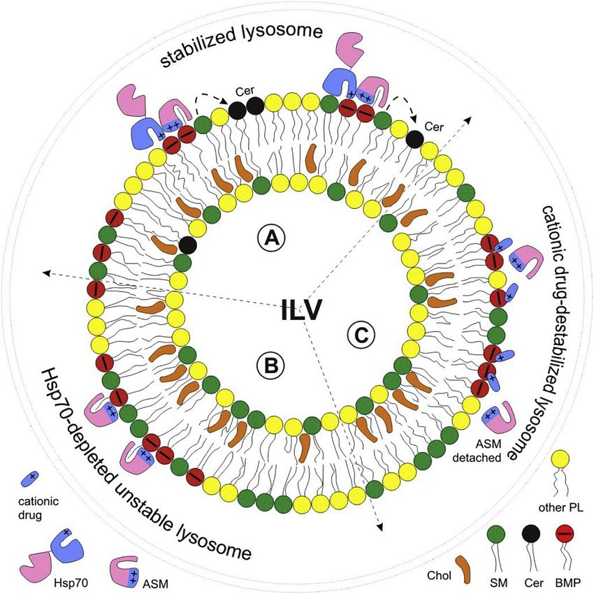
Z. Balogi et al.                                                                                                           Progress in Lipid Research 74 (2019) 18–30

“membrane lipid therapy” [31,32]. The heat shock protein co-inducer
hydroximic acid derivatives, such as Bimoclomol and BGP-15, are small
multi-target molecules that intercalate into membranes and stabilize
their lipid rafts by modulating membrane composition and structure
[33,34]. Several studies have shown beneficial effects of BGP-15 on
various disease models [35]. It is noted that such Hsp co-inducer
compounds potentiate the response to a pre-existing stress without
exhibiting effects in nonstressed environments. Dihydropyridine deri-
vatives, another recently explored family of Hsp co-inducers, such as
LA1011 and LA1044, improve the spatial learning and memory func-
tions in wild type mice, and eliminate neurodegeneration by increasing
dendritic spine density and reducing tau pathology and amyloid plaque
formation in APPxPS1 double mutant mouse model of Alzheimer's
disease [36,37]. Recently it was shown that binding of these dihy-
dropyridines to Hsp90 compromises Hsp90's chaperone activity [36],
which consequently induces the heat shock response in diseased cells.
Furthermore, xenohormetic plant compounds with a general beneficial
effect on animals also induce Hsp expression, and therefore have been
applied for the treatment of neurodevelopmental delay [38]. Further               Fig. 1. Lipid interacting and post-translational modified regions of Hsp70 (A)
modulators of Hsp expression with respect to neurological diseases have           Full length crystal structure and domains of Hsp70 shown for the prokaryotic
been described elsewhere [39,40].                                                 Hsp70 DnaK (PDB: 2KHO). Hsp70 has an N-terminal nucleotide binding domain
                                                                                  (NBD: pale brown), a short linker region (red) that couples to the substrate
                                                                                  binding domain (SBD) consisting of a substrate-binding subdomain (SBSD:
2. Membrane crossing and post-translational modifications of
                                                                                  purple) and a helical lid subdomain (HLS: coral). (B) Residues of the “lysine-
HSP70
                                                                                  arginine cluster” interacting with the lipid phosphatydilserine (PS) (PDB: 4PO2
                                                                                  of the linker and SBD of human Hsp70). Positively charged R533, R535, K569,
    Despite the high therapeutic potential of Hsp70 – membrane in-                K573, K589, K597 shown in red are proposed to specifically bind to PS at the
teraction, the mechanism by which Hsp70, lacking a leader sequence, is            cytoplasmic leaflet of endosomes, allowing Hsp70-cargo entry to endosomes via
capable of crossing the endosomal-lysosomal or the plasma membrane                autophagy [45]. (C) Example residues that are post-translational modified
is not well understood. In vitro studies with reconstituted protein-lipid         (PTM) and functionally relevant (PDB: 4PO2 of the linker and SBD of human
systems have unraveled a specific interaction between Hsp70 and                    Hsp70). Regions involved in oligomerization of Hsp70 are shown in green.
phosphatidylserine (PS) [41,42] and proposed that Hsp70 oligomers                 Further residues that are exposed to PTMs are shown (in red) as relevant for
generate pores in the cell membrane [43]. PS indeed confers a negative            Hsp70 dimerization and client protein interaction (T504, K561, K568, K569
charge to the cytosolic leaflet of the plasma membrane and also to the             and K507, K512, K526), E3 Ub ligase CHIP interaction (K561, Y611). Different
                                                                                  PTMs of K561 (in yellow) were found to be important for substrate interaction,
endosomal membrane, allowing the recruitment of proteins with strong
                                                                                  oligomerization, client and self-ubiquitination, cell growth and survival. (For
or moderate positive charges, respectively [44]. More recently, it has
                                                                                  interpretation of the references to colour in this figure legend, the reader is
been shown that a cluster of positively charged Lys and Arg residues              referred to the web version of this article.)
(R533 to K601/K597) anchor Hsc70/Hsp70 to the endosomal mem-
brane, which enables entry of Hsc70/Hsp70-cargo complexes to endo-
somes through microautophagy [45]. Interestingly, this lipid inter-               regulation of interrelated or interfering Hsp70 functions such as sub-
acting region has been identified to be important for other functions as           strate or lipid binding (Fig. 1). To dissect the impact of post-transla-
well. Hsp70 is composed of a nucleotide-binding domain (NBD) and a                tional modifications on Hsp70 localization and function necessitates
substrate-binding domain (SBD), which are connected by a linker                   further in-depth studies using subcellular fractions that can then be
(Fig. 1A). The linker domain (aa 384-397) and a fraction (aa 557-641)             rendered to a specific function.
of the helical lid subdomain (HLS) of SBD, which overlaps with the lipid
interacting region (R533, R535, K569, K573, K589, K597 of human                   3. HSP70 trafficking and tumor invasion
Hsp70), are involved in oligomerization [46,47] (Fig. 1B). More spe-
cifically, Morgner et al. identified Lys rich regions throughout the                    Cell surface, endosomal, lysosomal and extracellular pools of Hsp70
whole molecule, but mostly in the SBD (K108-K561/569), that direct                are interconnected in a highly dynamic fashion (Fig. 2). Plasma mem-
Hsp70 monomers in an antiparallel orientation [48]. T504, K561,                   brane-bound Hsp70 enters the endosomal route via clathrin dependent
K568, K569 and K507, K512, K526 residues of the SBD in ATP and ADP                and independent mechanisms, and a fraction of internalized protein is
bound state allow not only dimerization but also interaction with the             recycled back to the surface. When excess Hsp70 is present in the cell,
co-chaperones Hsp40, Hsp90, HopGR and client proteins (Fig. 1C).                  Hsp70 is further trafficked to late endosomes and lysosomes [54]. Cy-
Importantly, phosphorylation and acetylation of these residues stabilize          tosolic Hsp70 may also enter the endo-lysosomal system via an autop-
protein-protein interactions and, therefore, they are likely to also affect        hagic mechanism as implicated above [45]. Importantly, Hsp70 is re-
lipid interactions of this region. Further, trimethylation of K561 of the         sistant to proteolytic cleavage (and is, hence distinguishable from its
Hsp70 family members by METTL21A methyltransferase alter the af-                  cargos which are destined for lysosomal degradation) thus allowing it
finity of Hsp70 towards monomeric and fibrillar α-synuclein [49], and               to exert its anti-apoptotic role. A large body of evidence describes
phosphorylation and methylation of HLS residues including K561 and                Hsp70 present in both membrane-bound and soluble forms in the endo-
Y611 are necessary for proper ubiquitination by E3 ubiquitin ligase               lysosomal system, which are released by multivesicular bodies [55–58]
CHIP [50]. Hsp70 is ubiquitinated at 12 out of its 39 Lys residues in-            and secretory lysosomes [54,59], respectively. These mechanisms not
cluding K561 [51]. Hot-spots of phosphorylation in Ssa1, the yeast                only supply plasma membrane bound Hsp70, but also result in a con-
homologue of Hsp70, at T36-S38 and T492-S495-T499 are important                   siderable amount of exosomal membrane-bound or soluble Hsp70
for normal growth and survival [52]. A large number of multiple post-             which has immunomodulatory potential.
translational modification sites point to a combinatorial code for a                   Upregulated expression levels of Hsp70 is a diagnostic measure in
specific function [53]. Overlapping patterns of motives and post-                  several cancers, indicating increased cancer cell proliferation, ‘clinical
translational modifications, in particular in the SBD, imply tight                 stage’, or ‘increased grade’ together with shorter overall survival

                                                                             19
Z. Balogi et al.                                                                                                             Progress in Lipid Research 74 (2019) 18–30

                                                                                     4. Plasma membrane bound and extracellular HSP70

                                                                                         Global cell surface protein profiling of membranes of tumor and
                                                                                     normal cells revealed a tumor-specific, plasma membrane localization
                                                                                     of a variety of different Hsps [12,69,70]. Although lacking a classical
                                                                                     consensual transmembrane sequence, Hsp70 also has been found on the
                                                                                     cell surface [12,69,71] and in the extracellular milieu of intact tumor
                                                                                     cells [72–74]. Membrane localization of Hsps appears to be restricted to
                                                                                     malignantly transformed cells [12,69,75,76], bacterial/viral/fungal/
                                                                                     parasite-infected cells and spermatogenic cells [77–79]. In normal cells,
                                                                                     Hsp70 is only found inside the cell but not on the plasma membrane.
                                                                                     Therapeutic interventions such as radiochemotherapy, Hsp90 inhibi-
                                                                                     tion and hyperthermia have been found to further increase the levels of
                                                                                     cytosolic and membrane-bound Hsps [12,15,80] in tumor cells. The
                                                                                     presence of Hsp70 in the extracellular milieu of viable cells [81–83] is
                                                                                     currently explained by an alternative lysosomal/endosomal pathway
                                                                                     (Fig. 2), which does not involve the classical ER-Golgi compartment
                                                                                     [59]. These findings concur with those from Asea and colleagues who
                                                                                     demonstrated that drugs which perturb ER-Golgi transport, including
                                                                                     monensin and brefeldin A, do not influence membrane expression and
                                                                                     release of Hsp70 [84].

                                                                                     5. Membrane anchorage of HSPS in tumor cell membranes

Fig. 2. Intracellular trafficking and secretion of Hsp70 Hsp70 (bucket                     Approximately 15 to 20% of the total cellular Hsp70 is found on the
symbol) is bound to the extracellular leaflet of the plasma membrane (PM).            plasma membrane of some tumor cells [85]. Since neither high-salt
Surface Hsp70 is internalized to early endosomes (EE), a fraction of which is        conditions nor changes in the extracellular pH affect the Hsp70 mem-
recycled back to the PM through sorting and recycling endosomes (SE, RE).
                                                                                     brane expression density on tumor cells, it is unlikely that Hsp70 is
Provided sufficient intracellular Hsp70, internalized Hsp70 is further trafficked
                                                                                     bound to proteinous cell surface receptors [86]. Already in 1989,
to late endosomes/multivesicular bodies (MVB), where BMP enriched in-
traluminal vesicles (ILV) are formed with Hsp70 attached to the membrane.
                                                                                     Hightower and Guidon noted that Hsp71/Hsp73 could bind fatty acids
Fusion of MVBs with the PM exposes Hsp70 at the cell surface and releases            and suggested possible direct interactions with membrane lipids [87].
exosomes containing Hsp70. Hsp70 may further be targeted to lysosomes (L),           Further on it has been proposed that Hsps accumulate in glyco-
which upon lysosomal exocytosis expose Hsp70 at the cell surface and release         sphingolipid and cholesterol-rich microdomains (CRMs) [83,88–90].
their soluble Hsp70 content to the extracellular space. Mechanisms of mem-           CRMs were originally defined as regions within the plasma membrane
brane crossing and supply of Hsp70 to the endolysosomal system are not               that are enriched in cholesterol, glycosphingolipids, glycosylpho-
known, but autophagy and direct membrane crossing mechanisms have been               sphatidylinositol-anchored proteins and some other acylated proteins
implicated.                                                                          [91,92]. As super-resolution cell imaging techniques are now suitable
                                                                                     for investigating membrane lipid domains [93,94] these early findings
[60–63]. Given the correlation between excess Hsp70 levels and its                   should be revisited. A more recent effort has confirmed strong binding
lysosomal, cell surface and extracellular appearance (Fig. 3), unusual               of Hsp70 to cholesterol and sphingomyelin domains in model mem-
localization of Hsp70 appears to be an attractive target for therapeutic             branes, and importantly high resolution atomic force microscopy re-
interventions. These targets include but may not be limited to lysosomal             vealed nano-domain size (up to 200 nm in diameter) of Hsp70 clusters
membrane-bound Hsp70, which protects against lysosome-dependent                      on the cellular membrane (see Fig.4). These results may point to pos-
cell death [10,14,64], and plasma membrane- bound Hsp70, which                       sible Hsp70-membrane lipid platforms formed [47]. Glycosphingolipids
promotes invasion [15,65] and endocytosis [47,66]. These features that               that are enriched in tumor cell membranes, provide neoplastic and
would give rise to survival benefit for cancer patients may provide                   normal stem cell markers with immunogenic potential [95]. However,
unique possibilities to fight tumor progression and metastasis. More-                 glycosphingolipid-mediated immunoreactivity is often limited by a
over, surface localized and extracellular Hsp70 serve as potent stimuli              cholesterol-induced reorientation of glycosphingolipid head groups in a
for the innate immune system and can therefore be exploited as an                    parallel rather than perpendicular conformation, which in turn hinders
effective adjuvant therapy [67,68]. These targets and their therapeutic               their recognition by the immune system [95]. Therefore, one could
potential are detailed in the following sections.                                    assume that cholesterol depletion by methyl-beta-cyclodextrin might
                                                                                     improve immunogenicity of tumor cells. A comparative lipidomic
                                                                                     analysis of the glycosphingolipid content revealed significantly greater

                                                                                                    Fig. 3. Model for excess Hsp70-mediated tumor invasion
                                                                                                    Low grade tumor cells with lower levels of intracellular Hsp70
                                                                                                    (left side, bucket symbol used) also express low levels of
                                                                                                    Hsp70 in the endo-lysosomal system and at the cell surface,
                                                                                                    which correlate with a non-invasive phenotype. Contrary,
                                                                                                    high grade tumor cells often with high levels of intracellular
                                                                                                    Hsp70 (right side) express high levels of Hsp70 in the endo-
                                                                                                    lysosomal system and at the cell surface, as well as displaying
                                                                                                    an invasive phenotype. Anti-apoptotic effects of cytosolic or
                                                                                                    lysosomal Hsp70 and the tumor-promoting effect of surface
Hsp70 are involved in facilitating tumor invasion as reviewed in [63]. Blue and red symbols correspond to basal (low level) and excess Hsp70, respectively. For
trafficking routes refer to Fig. 2. MVB (late endosome, multivesicular body), L (lysosome), PM (plasma membrane). (For interpretation of the references to colour in
this figure legend, the reader is referred to the web version of this article.)

                                                                                20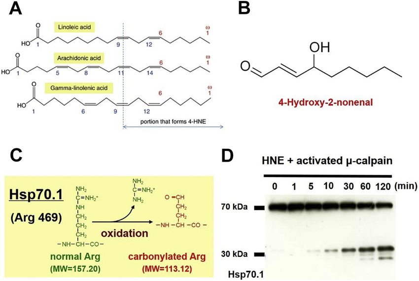
Z. Balogi et al.                                                                                                              Progress in Lipid Research 74 (2019) 18–30

Fig. 4. Hsp70 clustering at the tumor cell surface Hsp70 forms larger size of nano-domains in the cell membrane of tumor cells expressing higher level of
intracellular Hsp70. (A) Model of plasma membrane-bound Hsp70, where blue and red symbols correspond to basal (low level) and excess Hsp70, respectively. (B)
Topography, atomic force microscopy recognition and overlay images. Note that only red pixels above the recognition threshold (rec) are shown in overlay images.
These areas are found Hsp70 positive [47]. Data are displayed with courtesy of Dr. Lilia Chtcheglova and Prof. Peter Hinterdorfer, Johannes Kepler Univertity, Linz,
Austria. (For interpretation of the references to colour in this figure legend, the reader is referred to the web version of this article.)

amounts of globotriaosylceramide Gb3 [96] in tumor cells with a high                 Hsp70 demonstrated that plasma membrane bound Hsp70 forms large
compared to a low Hsp70 membrane expression. Gb3 is a receptor for                   clusters and rings potentially surrounding endocytic sites in the cell
Verotoxin [97,98] and AB5-Shiga toxin, an enterotoxin produced by                    membrane at higher intracellular and cell surface Hsp70 concentrations
Shigella dysenteria and enterohemorrhagic Escherichia coli. It is fre-               (Fig. 4). Shown in both the cell membrane and reconstituted systems
quently found in the plasma membrane of germinal center B cells and                  clustering was found to depend on the ability of Hsp70 to oligomerize,
Burkitt's lymphoma cells and solid tumors [99–104] but it is not present             and larger nano-domains (above 70 nm in diameter) of surface Hsp70
in most normal cells. Staining of Gb3 and Hsp70 on the plasma mem-                   correlated with its ability to facilitate endocytosis in cancer cells
brane of Hsp70-positive tumor cells revealed their co-localization.                  [47,66].
Moreover, cholesterol depletion results in a loss of Hsp70 from the
plasma membrane of tumor cells [85]. Previous work by Lingwood
et al. has demonstrated that Hsp70 also binds to 3`-sulfogalactolipids               6. Immunological role and therapeutic exploitation of membrane-
via its ATPase domain (NBD) (Fig. 1A) [105]. Based on binding patterns               bound and extracellular HSP70
of antibodies that detect different epitopes of Hsp70 in the ATPase and
the C-terminal substrate binding domain, the orientation of Hsp70 in                     Significant amounts of membrane-associated Hsp70 are often in-
Gb3 containing membrane domains appears to support the above result.                 dicative of highly aggressive tumors, metastatic potential and resistance
Together with the finding that recombinant Hsp70 specifically interacts                to therapy [11,65,110]. However, Hsps with molecular weights ranging
with artificial lipid vesicles containing Gb3, this supports the hypoth-              from 70 to 90 kDa also elicit protective anti-tumor immune responses if
esis that Gb3 might be one of the tumor-enriched lipid components that               expressed on the plasma membrane or in the extracellular milieu.
enables the integration of Hsp70 in the plasma membrane of tumor cells               Previous work of Multhoff and colleagues reported that in the presence
[85].                                                                                of interleukin-2 (IL-2), plasma membrane-bound Hsp70 acts as a tumor-
    Apart from the glycosphingolipid Gb3, Hsp70 has been found to                    specific recognition structure for natural killer (NK) cells pre-activated
interact with artificial lipid bilayers in the presence of phosphati-                 with Hsp70 protein [11,111,112] or a peptide derived thereof (TKD)
dylserine (PS) [41,43]. The group of DeMaio has shown that the in-                   [113]. In contrast, resting NK cells of tumor patients are unable to kill
teraction of Hsp70 with PS is largely based on the negative charge of                Hsp70 membrane-positive tumor cells. Since the induction of the cy-
phospholipids [106]. PS residing in liposomes enables the insertion of               tolytic activity of NK cells with TKD/IL-2 is dose-dependent and sa-
Hsp70 into the lipid bilayer and thereby can form higher molecular                   turable, it has been assumed that the stimulation of NK cells with Hsp70
weight oligomers that facilitate ion conductance in artificial lipid bi-              peptide might be mediated via receptors. Blocking experiments re-
layers [107]. Assuming that PS serves as the natural binding partner for             vealed that the C-type lectin receptor CD94 in combination with the
Hsp70 in vivo, a higher PS content would be expected in Hsp70                        activatory co-receptor NKG2C as well as other activatory receptors such
membrane-positive tumor sublines. In non-stressed cells, PS is pre-                  as the homodimeric receptor NKG2D and natural killer receptors
dominantly found on the inner membrane layer, whereas, upon stress                   (NKp30, NKp44, NKp46, NKp80) can act as mediators of the interaction
PS can switch to the outer membrane leaflet, where it can be de-                      of NK cells with Hsp70 membrane-positive tumor cells [114–117].
termined by a specific cell surface staining using the Ca2+-dependent                 Following binding of these NK cell receptors to membrane-bound
phospholipid binding protein Annexin A5. PS on the outer membrane                    Hsp70, the production and release of the serine protease granzyme B
leaflet is considered as an early marker for apoptotic cell death in many             and perforin is initiated which, in turn, results in apoptotic cell death of
cell types where it acts as an “eat-me” signal for macrophages [108].                the tumor cell [56,118]. Even in the absence of perforin, granzyme B
However, in the case of tumor cells PS can also be present on the outer              has been found to interact with membrane-bound Hsp70 on tumor cells.
membrane leaflet of viable, therapy-resistant, hypoxic cells [109]. It                Following binding and uptake of granzyme B into tumor cells via
appears that under non-stressed conditions, Hsp70 predominantly re-                  Hsp70-mediated endocytosis, apoptosis can thus be induced [119]. It
sides in membrane clusters whereas following stress Hsp70 often co-                  remains a matter of debate how granzyme B induces tumor cell apop-
localizes with PS outside these clusters. In line with this, atomic force            tosis after endo-lysosomal transfer via an Hsp70 pathway.
microscopy combined with antigen specific recognition of surface                          Depending on the Hsp profile of the lipid surface of actively released
                                                                                     exosomes derived from tumor cells [56,120] either stimulatory or

                                                                                21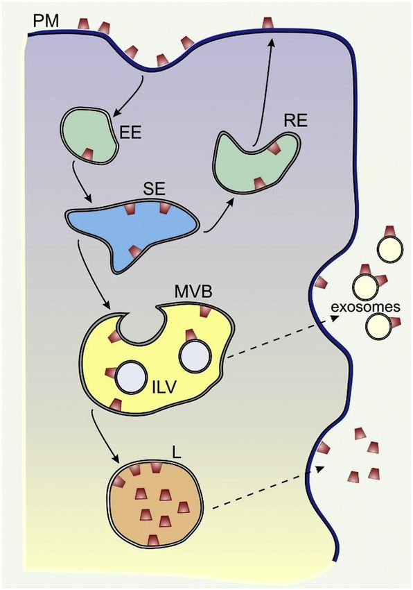
Z. Balogi et al.                                                                                                        Progress in Lipid Research 74 (2019) 18–30

inhibitory NK-mediated immune responses can be elicited. In the pre-             7. HSP70 as a regulator of lysosomal lipid catabolism and
sence of immunogenic peptides that are chaperoned by extracellular               membrane stability
Hsps also adaptive immune responses can be initiated following peptide
cross-presentation via antigen presenting cells [121,122]. Another                    As discussed above, ample amounts of Hsp70 are found on the
mechanism whereby extracellular Hsp70 might be able to stimulate                 surface of cancer cells [11,136]. Since high endocytic activity being a
tumor cell death is the complex formation of the innate immunity                 characteristic of cancer cells, it is, therefore, not surprising that their
protein Tag7 with Hsp70. It has been shown that the interaction of the           lysosomal membranes also contain this protein [59,137]. More sur-
Tag7-Hsp70 complex with TNFR1 triggers the activation of RIP1-ki-                prisingly and contrary to most other proteins ending up in the lyso-
nase, an increase in intracellular concentration of Ca2+ and an acti-            somal lumen, Hsp70 is capable of resisting lysosomal hydrolases and of
vation of calpains, a family of Ca2+ dependent cytoplasmic cysteine              remaining functional in this hostile environment [10,14]. The re-
proteases, which result in the permeabilization of lysosomal mem-                sistance to hydrolysis is likely due to the effective, pH-dependent an-
branes [123]. The lysosome-induced release of cathepsins B and D can             chorage of Hsp70 to the lysosomal membranes via its high-affinity
depolarize mitochondrial membranes and induce ROS production                     binding to bis(monoacylglycero)phosphate (BMP, lysobisphosphatidic
which eventually initiates tumor cell necroptosis [123]. In contrast to          acid), an anionic phospholipid abundant in lysosomes [14]. BMP ac-
tumor cells, Hsp70 which is released by normal human monocytes in                cumulates predominantly in the membranes of intraluminal vesicles
response to granulocyte monocyte-colony stimulating factor (GM-CSF)              (ILV) of the endolysosomal system, and is critical for the formation of
can prevent the formation of gap-junction intercellular communication            ILVs [138]. Fluorescence spectroscopy-based analyses of BMP-Hsp70
between capillary cells and monocytes, and thus could affect in-                  interactions suggest that BMP attaches to both the ATP- and the sub-
flammation and tumor growth [124]. An anti-inflammatory cardio-                    strate-binding domain of Hsp70 in an extended conformation with acyl
protective effect could be shown by plasma exosomes expressing CD63,              chains inserting into hydrophobic crevices within Hsp70 [139]. This
CD81 and Hsp70 derived from healthy donors [125]. This protective                anchorage is expected to cause a stringent orientation of Hsp70 on the
effect has been found to be dependent on Hsp70/Toll-like receptor 4               membrane surface and to induce a transition of its substrate-binding
(TLR4) interactions and an activation of kinases that stimulate Hsp27.           domain into an intermediate conformational state, which may be es-
In summary, depending on the source of the releasing cell type (tumor            sential to retain substrate interactions within the hydrophobic bilayer
or normal cells) and the micromilieu (e.g. hypoxia [126]) Hsp-bearing            interior. The functionality of lysosomal Hsp70 is supported by accu-
exosomes can exert contradictory immunological responses.                        mulating data showing that not only Hsp70 expressed in cells, but also
    Patients with highly aggressive tumors have elevated levels of               extracellularily added recombinant Hsp70 taken up by endocytosis and
serum exosomes, which regulate cell-cell communication by transfer-              accumulating in lysosomes, regulates lysosomal lipid catabolism and
ring molecules such as cytosolic proteins (including Hsps), lipids,              lysosomal membrane integrity [14,137,140–149]. As discussed below,
microRNAs and mRNAs [127]. Hsp70 membrane-positive tumor cells                   these cytoprotective, lysosomal functions of Hsp70 open new possibi-
secrete exosomes carrying Hsp70 on their membranes [56]. Extra-                  lities to inhibit and promote cell death in the treatment of various de-
cellular as well as membrane-bound Hsp70 fulfil dual functions of                 generative diseases and cancer, respectively.
mediating therapy resistance [65] and playing pivotal roles in anti-
tumor immune responses [11]. Hsp70 membrane-positive tumor cells                 8. Lysosomes and lysosome-related disorders
have been found to be significantly more susceptible to the lysis of
Hsp70-peptide and IL-2 activated NK cells as compared to their Hsp70                 Lysosomes are cytosolic vesicles that function as cellular recycling
membrane-negative counterparts [11,12]. At present the capacity of ex            stations, where over 50 acid hydrolases digest all major macro-
vivo TKD/IL-2-stimulated NK cells to kill autologous tumor cells is              molecules of the cell to breakdown products available for metabolic
being tested in a clinical phase II trial in patients with non-small cell        reutilization [150]. Additionally, they serve as major endocytic, Ca2+
lung cancer after radiochemotherapy [128,129].                                   signaling and more recently as metabolic hubs that sense the nutrient
    Furthermore, surface Hsp70 positive exosomes derived from tumor              availability and translate it to appropriate signaling pathways
cells have been found to stimulate the migratory and cytolytic capacity          [151–154]. Lysosomal membranes can be divided into the limiting
of NK cells [56]. In line with this finding, an intratumoral injection of         membrane and any internal membranes of ILVs [155]. These differ
recombinant Hsp70 into patients with glioblastoma has been shown to              significantly in their function and composition. The internal mem-
induce an increased cytolytic activity of NK cells and a cytokine shift          branes are the sites of lipid degradation. As previously mentioned, they
towards a T helper 1 (Th1)-mediated immune response in preclinical               are characterized by high levels of an anionic phospholipid, BMP,
models [130] and a pilot study in human patients [131]. Apart from               whose negative charge serves as a docking site for positively charged
recombinant Hsp70 protein that interacts with membrane Hsp70                     domains of lysosomal lipases (e.g. acid sphingomyelinase) or their co-
through its oligomerization domain [132], the serine protease gran-              factors (e.g. saposin) [155]. At the same time, the limiting membrane
zyme B has been found to interact with membrane Hsp70 on tumor                   serves as a barrier that inhibits lethal leakage of lysosomal hydrolases
cells. Following binding and Hsp70-mediated recycling endosomes,                 into the cytosol while controlling the proper exchange of ions and the
granzyme B induces tumor-specific apoptosis via perforin-independent              export of metabolites [150]. Heavily glycosylated luminal tails of ly-
pathway [119]. Regarding these findings EGFR targeting granzyme B                 sosomal-associated membrane proteins (e.g. LAMP-1 and LAMP-2) form
which is overexpressed in NK cells has been found to enhance tumor               a protective glycocalyx shield to the inner face of the membrane [156],
apoptosis [133]. The presence of perforin oligomers induces a rapid              and numerous channel-forming proteins transport ions and metabolites
plasma membrane flip-flop of phospholipids that facilitate the translo-            across the lysosomal membrane [154,157].
cation of granzyme B across plasma membrane bilayers [134]. HS-1                     Deficiency or malfunction of various lysosomal hydrolases or their
associated protein X-1 (HAX-1), a protein that is involved in the                co-factors, transport proteins or membrane proteins leads to chronic,
maintenance of the mitochondrial membrane potential also serves as a             often lethal, lysosomal storage disorders that affect many organs, most
target for granzyme B. After granzyme B-mediated HAX-1 cleavage, the             critically brain [158,159]. In addition to the classic lysosomal storage
N-terminal part stimulates mitochondrial depolarization and sub-                 disorders that are the most common cause of childhood neurodegen-
sequent lysosomal degradation [135].                                             eration [160], milder lysosomal dysfunction may contribute to
                                                                                 pathologies of more common human diseases, such as neurodegenera-
                                                                                 tion [160–162]. Moreover, lysosomal hyper-activation has recently
                                                                                 emerged as a hallmark of metastatic cancer [163–165]. Although ly-
                                                                                 sosomal storage disorders can be of mutation origin in over 50 different

                                                                            22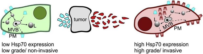
Z. Balogi et al.                                                                                                         Progress in Lipid Research 74 (2019) 18–30

lysosomal or lysosome-regulating genes, also the accumulation of sto-              alter membrane properties [187] Interestingly, Hsp70 could enhance
rage material and the resulting dysfunction of lysosomes results in                also the catabolism of several less abundant sphingolipids [148], whose
overlapping tissue pathology and clinical symptoms, with cell death                role in the maintenance of lysosomal membrane integrity remains to be
and neuronal loss being marked features in critically ill patients. Loss of        studied. Of special interest is the enzyme galactosylceramidase, whose
lysosomal membrane integrity and release of lysosomal hydrolases to                loss of activity results in accumulation of galactosylsphingosine (a.k.a.
the cytosol can be acutely lethal to cells. As the primary point of no             psychosine) that disrupts lysosomal pH [188], possibly destabilizing the
return in a wide variety of cell death cascades [166–169], lysosomal               lysosomal membranes by interaction with the pH sensitive ion channel
leakage may, in turn, cause cellular and organ dysfunction developed               TDAG8 [189]. Finally, it should be also noted that Hsp70-facilitated
during chronic lysosomal dysfunction. This view is supported by the                sphingomyelin degradation and concomitant ceramide formation al-
demise of cells observed in samples from patients with some lysosomal              lows a Niemann-Pick C2 (NPC2) mediated cholesterol egress from the
storage disorders [14,170–172], and cancer cell death following lyso-              lysosome [190,191], which is expected to affect membrane integrity
some-targeting therapies (reviewed in [168,173]). Notably, cancer cells            and cell survival in multiple ways.
either overexpressing Hsp70 or treated with recombinant Hsp70 are                      In addition to regulating lysosomal lipid catabolism, Hsp70 may
significantly protected against lysosomal leakage and subsequent cell               regulate lysosomal membrane stability by protecting the membranes
death, whereas those depleted of Hsp70 undergo spontaneous lyso-                   from oxidative stress. Inside the lysosomes, iron and other chemically
somal membrane permeabilization, or become more susceptible to ly-                 reactive metals (e.g. copper, zinc and cobalt) can generate reactive
sosome-disruptive stimuli [14,137,140–147,174–176].                                oxygen species through Fenton-type chemical reactions, which can lead
                                                                                   to oxidization and destabilization of membrane lipids [192,193]. In-
9. Lysosomal membrane integrity is regulated by HSP70                              terestingly, one of the common pathologies in various lysosomal sto-
                                                                                   rage diseases is the marked elevation of oxidative stress providing a
    Maintenance of the lysosomal membrane integrity is of utmost im-               possible mechanistic clue to the loss of lysosomal integrity and cell
portance for cellular homeostasis and survival. Yet, our knowledge on              death occurring in these diseases [194–197]. Importantly, the well
the mechanisms regulating lysosomal membrane permeability is only                  documented protective effect of Hsp70 against oxidative stress is pre-
beginning to emerge. Among the emerging lysosomal membrane de-                     served inside the lysosomes. In the case of photo-oxidation of acridine
stabilizers are certain lipids and reactive oxygen species [167,169],              orange-loaded lysosomes, real-time high-resolution imaging has de-
both of which can be regulated by Hsp70. Sphingomyelin, arachidonic                monstrated that Hsp70 localized in the lysosomal lumen effectively
acid and possibly high concentrations of sphingosine promote lyso-                 protects lysosomal membranes and thereby mitigates their destabili-
somal leakage, cell death and enhanced pathology in cells and tissues              zation upon local oxidative stress [14]. Furthermore, Hsp70 is cyto-
from lysosomal storage disease patients [14,147,175,177–179]. The                  protective in other lysosomal oxidative stress models, including age-
ability of Hsp70 to stabilize lysosomal membranes has been largely                 related macular degeneration of retinal pigment epithelium and lyso-
attributed to its ability to enhance sphingolipid catabolism in the ly-            somal iron accumulation [10,143,176]. It remains to be studied, whe-
sosomes through its high-affinity binding to BMP [14,148,175]. As                    ther Hsp70 has a direct antioxidant effect or whether the protection of
discussed above, this anionic phospholipid is an essential cofactor for            lysosomal membranes against oxidative stress is due to indirect effects,
lysosomal sphingolipid catabolism [180]. Via its negative charge, it               such as changes in the lipid composition of the membranes.
tethers several sphingolipid-degrading enzymes to the internal lyso-
somal membranes where their substrates are located, thereby in-                    10. Therapeutic exploitation of lysosomal HSP70 function
creasing their activity and protecting them from lysosomal degradation.
The high-affinity association of Hsp70 and BMP, which protects Hsp70                     After the initial discoveries of the role of Hsp70 in lysosomal
from lysosomal degradation as discussed above, also facilitates the BMP            membrane stability and lysosomal lipid catabolism [10,14], a number
binding of sphingolipid-degrading enzymes and, in so doing, further                of recent publications have reported improved lysosomal enzyme ac-
enhances their activity and inhibits their degradation [14,139,148].               tivities and lysosomal function through the induction of heat shock
The lysosomal membrane-stabilizing effect of Hsp70 may rely in par-                 proteins in various lysosomal storage disorders [148,149,198–201]. As
ticular on its enhancing effect on the enzyme acid sphingomyelinase                 a consequence, the induction of the heat shock response is emerging as
that hydrolyses sphingomyelin to ceramide and phosphocholine                       an attractive therapeutic approach to treat these devastating diseases.
(Fig. 5). Hsp70-induced conversion of ILV-sphingomyelin to ceramide                The power of this approach is supported by recent data showing that
counteracts lysosomal aggregation and membrane permeabilization,                   recombinant Hsp70 can reverse lysosomal pathology in primary fibro-
which are hallmarks of stress-induced cell death and may contribute to             blasts from eight different lysosomal storage disorders and has sig-
cellular pathophysiology in some lysosomal storage disorders                       nificant therapeutic effects on both substrate accumulation and neu-
[14,170,175,181]. The mechanism by which accumulating ceramide                     rological manifestations in murine models of three of them, i.e. Fabry,
stabilizes lysosomes remains largely unknown. Nevertheless, levels of              Sandhoff and Niemann-Pick type C diseases [148]. Notably, these
very long chain ceramide species (C24:0, C24:1, C24:2) were significantly           therapeutic effects of recombinant Hsp70 can be recapitulated by oral
increased in Hsp70 transgenic mouse embryonic fibroblast (MEF) cells                administration of arimoclomol, a small molecule co-inducer of heat
as compared to their controls [14]. While short to long chain ceramides            shock proteins, currently in clinical trials for Niemann-Pick disease type
are frequently considered as mediators of cellular death, very long                C [148]. It should be noted that the therapeutic effects of Hsp70 in
chain ceramide species may protect membrane integrity and confer                   lysosomal storage diseases are not confined to its direct effects in ly-
survival benefit on cells [182–184]. It is possible that ceramides gen-             sosomes, but are likely to depend also on the classic chaperone func-
erated in the lysosome eventually influence other cellular membranes,               tions of Hsp70.
therefore affecting lysosomal stability indirectly as well [185]. If the                Contrary to lysosomal storage disorders and degenerative disease,
plasma membrane integrity should be severely impaired lysosomal                    where increased lysosomal Hsp70 activity appears to have a beneficial
ASM, facilitated by Hsp70 may be also exposed to the cell surface,                 effect, the inhibition of lysosomal Hsp70 function is emerging as an
where ceramide-enriched platforms seal the membrane. This is                       attractive approach to treat cancer. Whereas the direct inhibition of
achieved by conical shaped ceramides capable of inducing membrane                  Hsp70 in the lysosomal lumen may be technically challenging, the in-
invaginations hence facilitating vesicle budding and fission [186]. In-             hibition of its target, acid sphingomyelinase, can be easily achieved. In
creased concentration of lysosomal ceramide counteracts aggregation                fact, over a hundred commonly used, FDA-approved drugs are func-
of lysosomes with other intracellular vesicles and membranes, and                  tional inhibitors of this enzyme [202]. These drugs are characterized by
perhaps strengthen the lysosomal limiting membranes by its ability to              a hydrophobic ring structure and a hydrophilic side chain with a

                                                                              23
Z. Balogi et al. Progress in Lipid Research 74 (2019) 18–30
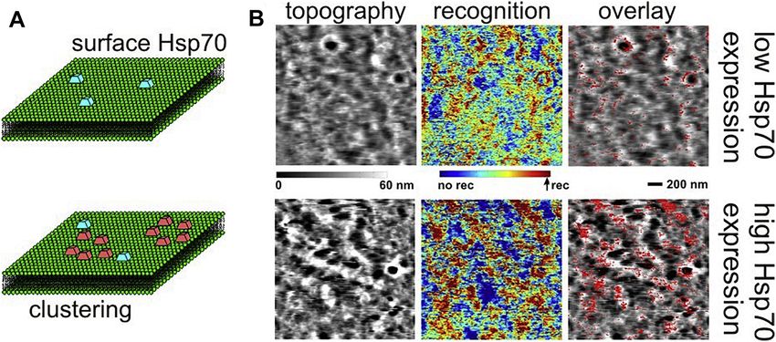
Fig. 5. Hsp70-mediated preservation of lyso-
somal membrane integrity Lysosomal membranes,
in particular those of intraluminal vesicles (ILVs) are
enriched in bis(monoacyl)glycerophosphate (BMP)
of inverted conical shape that allows high curvature
membrane formation. (A) Negative charge of the
head group of BMP recruits acid sphingomyelinase
(ASM) as well as Hsp70 to the membrane surface.
ASM converts sphingomyelin (SM) to membrane
stabilizing ceramide (Cer), which is largely depen-
dent on Hsp70 bound to the luminal side of ILVs.
Hsp70 dependent activation of ASM and other lyso-
somal lipases eventually changes the lipid composi-
tion of all cellular membranes. (B) Hsp70 depletion
generates lysosome instability, triggering in turn ly-
sosomal membrane permeabilization (LMP)-medi-
ated cell death. (C) Alternatively, cationic lysoso-
motropic drugs neutralize the negative charge of bis
(monoacyl)glycerophosphate (BMP), that ASM and
Hsp70 are anchored to, therefore causing lysosomal
instability, LMP and cell death. PL: glyceropho-
spholipid, Chol: cholesterol, double circle: limiting
membrane.
cationic amine group. In the acidic pH of lysosomes, the basic amine the brain and neurons are regularly exposed to different kinds of acute
groups are protonated resulting in an up to 1000-fold accumulation and chronic environmental stresses. The brain contains high levels of
[203]. The incorporation of such cationic amphiphilic drugs into polyunsaturated fatty acids and redox transition metal ions, especially
membranes in the lysosomal lumen neutralizes the negative membrane iron. In spite of its high oxygen consumption, however, levels of lower
charge and inhibits the function of several lysosomal lipases, including molecular weight and enzymatic antioxidants are relatively low in the
acid sphingomyelinase [204]. Thus, they have exactly the opposite ef- brain. Accordingly, the brain with poor antioxidant defense appears
fect to lysosomal Hsp70 (Fig. 5). Importantly, cancer cells are especially particularly susceptible to lipid peroxidation by reactive oxygen species
sensitive to the accumulation of sphingomyelin [175,205,206], which [226]. Peroxidation of membrane lipids may show numerous effects
may explain why these functional inhibitors of acid sphingomyelinase such as increased membrane rigidity, decreased membrane-bound en-
display selective cytotoxicity towards transformed cells both in vitro zyme activity, altered membrane receptor activity, and altered mem-
and in various cancer models in mice [175,207–212]. Their putative brane permeability. Therefore, it is not surprising that the role of lipid
efficacy in cancer treatment is further supported by a recent pharmaco- peroxidation has been widely investigated in the pathogenesis of a
epidemiological register-based cohort study showing a statistically variety of neurodegenerative diseases including Alzheimer's disease,
significant association between cationic amphiphilic antihistamine use Parkinson's disease, amyotrophic lateral sclerosis, Huntington's disease,
and reduced mortality among Danish cancer patients [212]. Down syndrome [227].
Importantly, lipid peroxidation yields a variety of bioactive pro-
ducts and one of the most extensively studied examples is hydro-
11. HSP70 disorder and lysosomal-mediated neuronal death
xynonenal [228,229]. The most common source of hydroxynonenal is
an endogenous one, when it is produced by peroxidation of membrane
Lysosome-dependent cell death is characterized by the destabiliza-
phospholipids or plasma low-density lipoproteins. Hydroxynonenal
tion of its limiting membrane (Fig. 5) followed by the leakage of ca-
generation in the brain has been associated with exposure to drugs,
thepsins from the lysosomal lumen into the cytoplasm [167,213–218].
ethanol or irradiation, and with ischemia or inflammation. In contrast,
Using the monkey experimental systems of transient brain ischemia,
exogenous hydroxynonenal is generated during food processing, i.e.,
Yamashima et al. [219–221] formulated the ‘calpain-cathepsin hy-
heating, especially deep-frying, of ω-6 vegetable oils (Fig. 6 A,B) [228].
pothesis’ as a mechanism of programmed neuronal necrosis. They de-
Because of its chemical reactivity, hydroxynonenal can exert pleiotropic
monstrated that the lysosomal membrane of hippocampal CA1 neurons
effects notably cell death. After the ischemia/reperfusion sequence in
is disrupted by the activated μ-calpain after transient ischemia, which
myocardial infarction, accumulated reactive oxygen species promote
causes the release of lysosomal cathepsins B and L. Thereafter, the
generation of hydroxynonenal, which disrupts actin cytoskeleton, alters
‘calpain-cathepsin hypothesis’ has been confirmed, using a variety of
Ca2+ homeostasis, and triggers cardiomyocyte cell death [230]. Hy-
experimental paradigms from C. elegans to rodents [222–225]. The role
droxynonenal induces signaling for apoptosis via both the Fas-mediated
of lysosomal enzyme cathepsins in initiation and execution of the ne-
extrinsic and the p53-mediated intrinsic pathways [228,231]. Thus,
crotic cell death program has become clear [149,166,167]. Moreover,
24
Z. Balogi et al.                                                                                                     Progress in Lipid Research 74 (2019) 18–30

                                                                                                       Fig. 6. Generation of hydroxynonenal (HNE),
                                                                                                       HNE-mediated carbonylation and calpain-medi-
                                                                                                       ated cleavage of carbonylated Hsp70 (A, B)
                                                                                                       Generation of hydroxynonenal (4-hydroxy-2-
                                                                                                       nonenal) from ω-6 polyunsaturated fatty acids such
                                                                                                       as linoleic, arachidonic and gamma-linolenic acids.
                                                                                                       (C) Carbonylation of Hsp70 occurs at the key site
                                                                                                       Arg469 in post-ischemic CA1 neurons. (D) Time-de-
                                                                                                       pendent μ-calpain-mediated cleavage of carbony-
                                                                                                       lated-Hsp70 by hydroxynonenal (HNE) in monkey
                                                                                                       CA1 tissue. For further details see text.

hydroxynonenal can trigger β- cell apoptosis in the pancreas, and in-              Hsp70 is known to recycle altered proteins, stabilize lysosomal
duce glucose intolerance and type 2 diabetes [232]. Since hydro-               membranes and protect cells from diverse oxidative stresses.
xynonenal can impair Na+/Ca2+ pumps and glucose and glutamate                  ‘Hydroxynonenal-induced Hsp70 carbonylation’ (Fig. 6C) followed by
transporters by modifying membranes, the resultant ionic and energetic         ‘calpain-mediated cleavage of carbonylated Hsp70’ (Fig. 6D) may be
disturbances cause neuronal cell death [233,234]. However, the de-             crucial for the execution of both ischemic and degenerative neuronal
tailed mechanism of how hydroxynonenal can lead to cell death has              death. Calpain activation and Hsp70 disorder, combined together, at
been controversial until recently.                                             the lysosomal membranes may bring about programmed neuronal
    Accumulated data suggest dual roles of Hsp70 not only as a mole-           death by releasing hydrolytic cathepsin enzymes [219–221]. The
cular chaperone for altered (misfolded/aged/damaged) proteins but              pathway of cerebral ischemia and/or oxidative stresses, either acute (in
also as a guardian of lysosomal integrity [14,235–237]. Hsp70 con-             case of stroke) or chronic (in case of degeneration) could result the
tributes to lysosomal stabilization (Fig. 5) by binding to the en-             following sequence of μ-calpain activation → excessive intake of ω-6
dolysosomal anionic phospholipid BMP, a co-factor essential for                vegetable oils → increase of hydroxynonenal in the brain → hydro-
sphingomyelin catabolism [14]. Membranes of ILVs of the functioning            xynonenal-mediated Hsp70 carbonylation → activated μ-calpain-
lysosomes are characterized by abundant BMP [155,238]. Then, Hsp70-            mediated cleavage of carbonylated Hsp70 → lysosomal membrane de-
BMP binding enhances activity of acid sphingomyelinase, which med-             stabilization → cathepsin release → breakdown of the cell constitutive
iates the sphingolipid degradation at the internal membrane in the             proteins, which may in turn represent a central role not only for is-
acidic (pH 4.5) compartment to generate ceramide [180,239,240].                chemic neuronal death [221,236,244] but also for Alzheimer neuronal
Ceramide protects lysosomal membrane integrity as discussed above              death [237,245].
[14,235,241] (Fig. 5A). Thus, in cases of Hsp70 depletion, not only                A continuum of abnormalities of the lysosomal system can be
failure of protein trafficking and degradation but also lysosomal de-            identified in ischemic and Alzheimer neurons [160,246]. The common
stabilization or rupture may occur (Fig. 5B). In the monkey hippo-             characteristic is that functional Hsp70 is indispensable for lysosomal
campal CA1 neurons after transient ischemia, Oikawa et al. [242]               autophagy that is crucial for the homeostasis of neurons. The control of
previously found by proteomic analysis that Hsp70 can become an in             protein turnover is particularly important in post-mitotic cells such as
vivo target of carbonylation by hydroxynonenal (Fig. 6C). Intriguingly,        neurons, where accumulation of altered proteins may be highly detri-
carbonylation of Hsp70 increased more than ten-fold in the post-is-            mental to cell survival [247]. Neurons must maintain large volumes of
chemic CA1 neurons, compared to the non-ischemic controls. Subse-              membrane and cytoplasm, and continually traffic autophagy-related
quently, in the in vitro experiments, Hsp70 being carbonylated by hy-          garbage long distances from distal ends of dendrites and axons back to
droxynonenal was found to become susceptible to cleavage by activated          the cell body where lysosomes are most active for catabolite clearance
μ-calpain (Fig. 6D) [243]. ‘Calpain-mediated cleavage of carbonylated          [248]. Hsp70 is the most structurally and functionally conserved cha-
Hsp70’ can lead to both autophagy failure and lysosomal destabiliza-           perone that plays a principal role in the trafficking and degradation of
tion with the resultant release of cathepsins and neuronal death [244].        altered proteins and their quality control for the cytoprotection of
    Although neuronal death in Alzheimer's disease has been thought to         neurons under a number of different conditions [245]. Accordingly, in
be caused by the initial cerebral accumulation of amyloid β for half a         case of Hsp70 dysfunction, failure of lysosomal autophagy may occur,
century, it still remains enigmatic because the underlying mechanism of        which leads to accumulation of autophagic vacuoles in both ischemic
Alzheimer neuronal death due to amyloid β still remains unknown.               and Alzheimer neurons [246]. Since the proteotoxic stress in ischemia/
Thus, research of this disease is moving away from the simple as-              reperfusion during stroke is severe, neurons die within hours or days
sumption of linear causality as proposed in the ‘amyloid hypothesis’.          after the insult. On the contrary, the proteotoxic stress in Alzheimer's
Recently, hydroxynonenal has been shown to be involved in a great              diseases is extremely mild, and neurons can battle it for months or
number of pathologies such as neurodegenerative diseases, metabolic            years, perhaps by raising pro-survival defenses such as Hsp70. Previous
diseases, and cancers [232]. Yamashima recently put forward a per-             studies [249–253] indicated increased levels of protein oxidation in the
spective view that the causative substance for Alzheimer neuronal              Alzheimer brains, and suggested a possible involvement of hydro-
death is actually ω-6 vegetable oil-derived hydroxynonenal [245].              xynonenal-mediated protein carbonylation for the progression of

                                                                          25
Z. Balogi et al. Progress in Lipid Research 74 (2019) 18–30
Alzheimer's disease. When sub-threshold levels of Hsp70 carbonylation and exponentially growing mammalian cells, FEBS Lett. 324 (2) (1993) 191–195.
are coupled with sub-threshold levels of calpain activation, for ex- [4] V. Adler, A. Schaffer, J. Kim, L. Dolan, Z. Ronai, UV irradiation and heat shock
mediate JNK activation via alternate pathways, J. Biol. Chem. 270 (44) (1995)
ample, due to long-standing mild cerebral ischemia and/or amyloid β 26071–26077.
accumulation, programmed neuronal death becomes steadily sig- [5] K. Xie, S. Huang, Regulation of cancer metastasis by stress pathways, Clin. Exp.
Metastasis 20 (1) (2003) 31–43.
nificant year by year. Not only in cerebral ischemia but also in Alz- [6] M. Santarosa, D. Favaro, M. Quaia, E. Galligioni, Expression of heat shock protein
heimer's disease, ‘calpain-mediated cleavage of carbonylated Hsp70’ 72 in renal cell carcinoma: possible role and prognostic implications in cancer
may cause lysosomal membrane rupture/destabilization, which leads to patients, Eur. J. Cancer 33 (6) (1997) 873–877.
[7] K. Nanbu, I. Konishi, M. Mandai, H. Kuroda, A.A. Hamid, T. Komatsu, et al.,
the release of cathepsins into the cytoplasm, which can then trigger Prognostic significance of heat shock proteins HSP70 and HSP90 in endometrial
progressive neuronal death. Nowadays, researchers of Alzheimer's dis- carcinomas, Cancer Detect. Prev. 22 (6) (1998) 549–555.
[8] V.L. Gabai, J.A. Yaglom, T. Waldman, M.Y. Sherman, Heat shock protein Hsp72
ease are gradually but steadily moving away from the classical amyloid
controls oncogene-induced senescence pathways in cancer cells, Mol. Cell. Biol. 29
hypothesis, and speculate that another age-related, disease-promoting (2) (2009) 559–569.
factor and/or substance probably interact with the core mechanisms of [9] E.A. Nollen, F.A. Salomons, J.F. Brunsting, J.J. van der Want, O.C. Sibon,
H.H. Kampinga, Dynamic changes in the localization of thermally unfolded nu-
the disease. Accordingly, targeting Hsp70 might be a promising strategy clear proteins associated with chaperone-dependent protection, Proc. Natl. Acad.
for both elucidating the mechanism of Alzheimer neuronal death as Sci. U. S. A. 98 (21) (2001) 12038–12043.
well as developing novel therapeutic agents for Alzheimer's disease [10] J. Nylandsted, M. Gyrd-Hansen, A. Danielewicz, N. Fehrenbacher, U. Lademann,
M. Hoyer-Hansen, et al., Heat shock protein 70 promotes cell survival by in-
where defects in lysosomal proteolysis and lipid accumulation have hibiting lysosomal membrane permeabilization, J. Exp. Med. 200 (4) (2004)
been observed [254]. 425–435.
[11] G. Multhoff, C. Botzler, L. Jennen, J. Schmidt, J. Ellwart, R. Issels, Heat shock
protein 72 on tumor cells: a recognition structure for natural killer cells, J.
12. Concluding remarks Immunol. 158 (9) (1997) 4341–4350.
[12] G. Multhoff, C. Botzler, M. Wiesnet, E. Muller, T. Meier, W. Wilmanns, et al., A
stress-inducible 72-kDa heat-shock protein (HSP72) is expressed on the surface of
It is now widely accepted that some of the heat shock protein mo- human tumor cells, but not on normal cells, Int. J. Cancer 61 (2) (1995) 272–279.
lecular chaperones have additional biological functions over their basic [13] A. Asea, S.K. Kraeft, E.A. Kurt-Jones, M.A. Stevenson, L.B. Chen, R.W. Finberg,
role in cellular proteostasis, i.e. acting as ‘moonlighting proteins’. The et al., HSP70 stimulates cytokine production through a CD14-dependant pathway,
demonstrating its dual role as a chaperone and cytokine, Nat. Med. 6 (4) (2000)
moonlighting Hsp70 has been emerging an important therapeutic 435–442.
target. However, efforts targeting essential chaperone activity or the [14] T. Kirkegaard, A.G. Roth, N.H. Petersen, A.K. Mahalka, O.D. Olsen, I. Moilanen,
et al., Hsp70 stabilizes lysosomes and reverts Niemann-pick disease-associated
interacting complexes of Hsp70 with proteins have not yet resulted in lysosomal pathology, Nature 463 (7280) (2010) 549–553.
excellent specific and efficient drug candidates of low toxicity [15] M. Gehrmann, J. Marienhagen, H. Eichholtz-Wirth, E. Fritz, J. Ellwart, M. Jaattela,
[255–257]. Drug design is hampered by the facts that Hsps are highly et al., Dual function of membrane-bound heat shock protein 70 (Hsp70), Bag-4,
and Hsp40: protection against radiation-induced effects and target structure for
conserved [258] and that Hsp70, specifically Hsp70.1 has multiple natural killer cells, Cell Death Differ. 12 (1) (2005) 38–51.
functions. Hsp70 is in fact more than simply a cytosolic chaperone [16] Z. Torok, I. Horvath, P. Goloubinoff, E. Kovacs, A. Glatz, G. Balogh, et al., Evidence
for a lipochaperonin: association of active protein-folding GroESL oligomers with
[19,63] and considered a major regulator of signaling pathways
lipids can stabilize membranes under heat shock conditions, Proc. Natl. Acad. Sci.
[8,259,260]. As reviewed here, non-cytosolic localization, membrane U. S. A. 94 (6) (1997) 2192–2197.
crossing and lipid interactions of Hsp70 are associated with membrane [17] Z. Torok, P. Goloubinoff, I. Horvath, N.M. Tsvetkova, A. Glatz, G. Balogh, et al.,
Synechocystis HSP17 is an amphitropic protein that stabilizes heat-stressed mem-
resistance, facilitation of endocytosis, counteracting apoptotic me- branes and binds denatured proteins for subsequent chaperone-mediated re-
chanisms and sustaining survival signaling at pathophysiological states. folding, Proc. Natl. Acad. Sci. U. S. A. 98 (6) (2001) 3098–3103.
Unlike roles in the cytosol these unique functions may not only be [18] N.M. Tsvetkova, I. Horvath, Z. Torok, W.F. Wolkers, Z. Balogi, N. Shigapova, et al.,
Small heat-shock proteins regulate membrane lipid polymorphism, Proc. Natl.
targeted via Hsp70 itself or its interacting proteins, but also via specific Acad. Sci. U. S. A. 99 (21) (2002) 13504–13509.
lipids that either interact with or can be modulated by Hsp70. In order [19] I. Horvath, G. Multhoff, A. Sonnleitner, L. Vigh, Membrane-associated stress
proteins: more than simply chaperones, Biochim. Biophys. Acta 1778 (7–8) (2008)
for rational drug design of membrane/lipid-mediated Hsp70 functions, 1653–1664.
we need further mechanistic and structural studies of Hsp70 membrane [20] M. Zhang, D. Wang, P. Li, C. Sun, R. Xu, Z. Geng, et al., Interaction of Hsp90 with
interactions and lipid modulation. phospholipid model membranes, Biochim. Biophys. Acta 1860 (2) (2018)
611–616.
[21] I. Horvath, A. Glatz, V. Varvasovszki, Z. Torok, T. Pali, G. Balogh, et al., Membrane
Acknowledgement physical state controls the signaling mechanism of the heat shock response in
Synechocystis PCC 6803: identification of hsp17 as a “fluidity gene”, Proc. Natl.
Acad. Sci. U. S. A. 95 (7) (1998) 3513–3518.
ZB is a Bolyai Research Fellow and supported by UP MS KA-2018-05 [22] L. Vigh, B. Maresca, J.L. Harwood, Does the membrane's physical state control the
and ÚNKP-18-4-PTE-26 new national excellence program of the expression of heat shock and other genes? Trends Biochem. Sci. 23 (10) (1998)
369–374.
Ministry of Human Capacities. This work has been supported by also [23] L. Vigh, I. Horvath, B. Maresca, J.L. Harwood, Can the stress protein response be
GINOP-2.3.2-15- 2016-00049, GINOP-2.3.3-15-2016-00025 and controlled by 'membrane-lipid therapy'? Trends Biochem. Sci. 32 (8) (2007)
GINOP-2.3.2-15-2016-00040. Work in the Multhoff laboratory has been 357–363.
[24] G. Balogh, I. Horvath, E. Nagy, Z. Hoyk, S. Benko, O. Bensaude, et al., The hy-
supported by DFG (SFB824-3), STA 1520/1-1 BMBF 01GU0823, BMWi perfluidization of mammalian cell membranes acts as a signal to initiate the heat
(AiF) ZF4320102CS7. Work in the Lloyd-Evans lab is supported by the shock protein response, FEBS J. 272 (23) (2005) 6077–6086.
[25] E. Nagy, Z. Balogi, I. Gombos, M. Akerfelt, A. Bjorkbom, G. Balogh, et al.,
MRC (MR/P007651/1), BBSRC (BB/S002774/1), EU Horizon 2020
Hyperfluidization-coupled membrane microdomain reorganization is linked to
BATcure consortium grant (666918) and Action Medical Research. The activation of the heat shock response in a murine melanoma cell line, Proc. Natl.
related research in the Jäättelä laboratory has been supported by Acad. Sci. U. S. A. 104 (19) (2007) 7945–7950.
[26] B. Gungor, I. Gombos, T. Crul, F. Ayaydin, L. Szabo, Z. Torok, et al., Rac1 parti-
Danish National Research Foundation (DNRF125), European Research cipates in thermally induced alterations of the cytoskeleton, cell morphology and
Council (AdG 340751), Danish Cancer Society (R167-A11061), Danish lipid rafts, and regulates the expression of heat shock proteins in B16F10 mela-
Council for Independent Research (DFF-7016-00360), and Novo noma cells, PLoS One 9 (2) (2014) e89136.
[27] Z. Balogi, Z. Torok, G. Balogh, K. Josvay, N. Shigapova, E. Vierling, et al., "Heat
Nordisk Foundation (NNF15OC0016914). shock lipid" in cyanobacteria during heat/light-acclimation, Arch. Biochem.
Biophys. 436 (2) (2005) 346–354.
[28] Z. Balogi, O. Cheregi, K.C. Giese, K. Juhasz, E. Vierling, I. Vass, et al., A mutant
References small heat shock protein with increased thylakoid association provides an elevated
resistance against UV-B damage in Synechocystis 6803, J. Biol. Chem. 283 (34)
[1] J. Hageman, H.H. Kampinga, Computational analysis of the human HSPH/HSPA/ (2008) 22983–22991.
DNAJ family and cloning of a human HSPH/HSPA/DNAJ expression library, Cell [29] G. Balogh, G. Maulucci, I. Gombos, I. Horvath, Z. Torok, M. Peter, et al., Heat
Stress Chaperones 14 (1) (2009) 1–21. stress causes spatially-distinct membrane re-modelling in K562 leukemia cells,
[2] R.I. Morimoto, Cells in stress: transcriptional activation of heat shock genes, PLoS One 6 (6) (2011) e21182.
Science 259 (5100) (1993) 1409–1410. [30] G. Balogh, M. Peter, A. Glatz, I. Gombos, Z. Torok, I. Horvath, et al., Key role of
[3] M.F. Dubois, O. Bensaude, MAP kinase activation during heat shock in quiescent lipids in heat stress management, FEBS Lett. 587 (13) (2013) 1970–1980.
26You can also read