Novel Approaches for the Treatment of Alzheimer's and Parkinson's Disease
←
→
Page content transcription
If your browser does not render page correctly, please read the page content below
International Journal of
           Molecular Sciences

Review
Novel Approaches for the Treatment of Alzheimer’s
and Parkinson’s Disease
Michiel Van Bulck 1,2 , Ana Sierra-Magro 1,2 , Jesus Alarcon-Gil 1 , Ana Perez-Castillo 1,2 and
Jose A. Morales-Garcia 1,2,3, *
 1    Instituto de Investigaciones Biomédicas (CSIC-UAM), Arturo Duperier, 4. 28029 Madrid, Spain;
      mvbulck@iib.uam.es (M.V.B.); anasierra@iib.uam.es (A.S.-M.); jalarcon@iib.uam.es (J.A.-G.);
      aperez@iib.uam.es (A.P.-C.)
 2    Centro de Investigación Biomédica en Red sobre Enfermedades Neurodegenerativas (CIBERNED),
      Valderrebollo, 5, 28031 Madrid, Spain
 3    Departamento de Biología Celular, Facultad de Medicina, Universidad Complutense de Madrid (UCM),
      Plaza Ramón y Cajal s/n, 28040 Madrid, Spain
 *    Correspondence: jmorales@iib.uam.es

 Received: 31 December 2018; Accepted: 3 February 2019; Published: 8 February 2019

 Abstract: Neurodegenerative disorders affect around one billion people worldwide. They can arise
 from a combination of genomic, epigenomic, metabolic, and environmental factors. Aging is the
 leading risk factor for most chronic illnesses of old age, including Alzheimer’s and Parkinson’s
 diseases. A progressive neurodegenerative process and neuroinflammation occur, and no current
 therapies can prevent, slow, or halt disease progression. To date, no novel disease-modifying therapies
 have been shown to provide significant benefit for patients who suffer from these devastating
 disorders. Therefore, early diagnosis and the discovery of new targets and novel therapies are of
 upmost importance. Neurodegenerative diseases, like in other age-related disorders, the progression
 of pathology begins many years before the onset of symptoms. Many efforts in this field have led to the
 conclusion that exits some similar events among these diseases that can explain why the aging brain is
 so vulnerable to suffer neurodegenerative diseases. This article reviews the current knowledge about
 these diseases by summarizing the most common features of major neurodegenerative disorders,
 their causes and consequences, and the proposed novel therapeutic approaches.

 Keywords: aging; Alzheimer; Parkinson; neurodegeneration; neuroinflammation; neurogenesis;
 novel approaches

1. Introduction
     Neurodegenerative diseases are incurable and debilitating conditions that result in progressive
degeneration and/or death of nerve cells. The huge variety of neurodegenerative diseases, in terms
of pathological characteristics, symptoms, and treatments, makes it very difficult to classify them in
general terms. Taking into account the different neurodegenerative diseases, one of the parameters
we can considerer is prevalence. According to this, there are two most prevalent neurodegenerative
diseases: Alzheimer’s disease (AD) and Parkinson’s disease (PD) [1,2]. This affirmation is substantiated
in the literature. In 2015, there were 46.8 million AD patients worldwide with direct and indirect
costs to society of 81,800 million USD [2], and in 2016 there were 6.1 million individuals with PD
worldwide [1]. Dementia is a major symptom of AD, consisting of memory impairment accompanied
by dysfunction, which is responsible for the inability to develop daily life activities [3]. Vascular
dementia is quite important since it is considered the second most common cause of dementia in the
aging population and also is thought to underlie AD. This disorder consists of a decline in cognitive

Int. J. Mol. Sci. 2019, 20, 719; doi:10.3390/ijms20030719                       www.mdpi.com/journal/ijms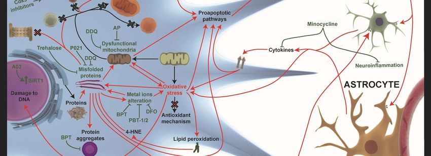
Int. J. Mol. Sci. 2019, 20, 719                                                                   2 of 36

skills caused by blocking or reduction of blood flow to the brain [4]. On the other hand, PD is
characterized by motor symptoms such as bradykinesia/akinesia, resting tremor, rigidity, and postural
abnormalities, and non-motor symptoms such as dementia, hyposmia, depression, and emotional
changes [5].
      Behavioral and psychological symptoms of dementia (BPSD) represent neuropsychiatric
symptoms occurring in subjects with dementia. They are clinically relevant as cognitive symptoms as
they strongly correlate with the degree of functional and cognitive impairment. BPSD include anxiety,
elation, hallucinations, agitation, irritability, abnormal motor behavior, apathy, depression, and sleep
or appetite changes [6]. Although some of these BPSDs could be alleviated by the use of atypical
antipsychotics, these compounds have been described to induce clinically significant metabolic adverse
events, increasing mortality in patients with dementia [7,8].
      Despite the high prevalence of these diseases, the treatments available today are not able to
significantly modify the progress of the disease, since these treatments only treat the symptoms of it.
A substantial part of traditional treatments for neurodegenerative diseases including AD, cerebral
amyloid angiopathy, frontotemporal dementia, mild cognitive impairment, and PD are based on
immunotherapy, most of them active and passive immunotherapy trials conducted based on amyloid,
tau, and α-synuclein targeting (Alzforum.org; https://www.alzforum.org/therapeutics). Some other
studies have suggested the use of small molecules for AD treatment, able to cross the BBB, like statins.
These include simvastatin, a HMG-CoA reductase inhibitor, approved by FDA for the treatment of
hypercholesterolemia and diabetic cardiomyopathy. In that sense retrospective clinical studies directed
by Wolozin and colleagues, demonstrated a significant reduction (at least 50%) in the risk incidence of
suffering AD and PD after simvastatin administration [9,10]. Now, research is focusing on finding new
disease-modifying therapies to slow disease progression [11,12].
      Better understanding of the triggering factors involved in the onset and progression of the
disorders are crucial for developing novel therapies. The main characteristic of these diseases is
the existence of two subtypes: Familial and idiopathic forms. The familial form, which an average
of 5% of patients will develop before 65 years of age, is associated with various genetic mutations.
The idiopathic form of the disease, which accounts for 95% of cases, is associated with aging and
has some other unrevealed risk factors [2,13]. In the case of AD, the familial form is caused by
mutations of genes coding for amyloid precursor protein (APP), presenilin-1 (PSEN-1), and presenilin-2
(PSEN-2) [2]. In familial PD, there is a longer list of related gene mutations, for example, α-synuclein
(SNCA), Leucine-rich repeat kinase 2 (LRRK2), PTEN induced kinase 1 (PINK1), ATPase cation
transporting 13A2 (ATP13A2), and other genes, like PARK [13]. The importance of identifying and
understanding the roles of these genes in the underlining pathological mechanism could reveal new
therapeutic approaches.
      The presence of common pathological pathways involved in various neurodegenerative diseases
that are associated with genes mutations that provoke familial forms could be promising approaches
for treatments in AD and PD [14]. At the same time, different etiologies share similar underlying
pathological pathways. Unfortunately, this is not a coincidence, since mutations at different steps
of the same cellular processes, in the case of familiar disease and aging, share common risk factors.
Understanding the relations and distinctions between these pathological processes at the molecular,
cellular, and physiological levels need to be described carefully. One example of this concept can be
explained with the gene coding for the triggering receptor expressed on myeloid cells 2 (TREM2) and
genetic risk factors in different neurodegenerative diseases [15]. The TREM2 mutation exacerbates
dysfunction in molecular receptor-mediated pathways related to inflammation, which downregulates
good cellular responses, which provokes dysregulation of immune responses in the brain [15]. This
example indicates how a pathological process can be studied and used as a targeting approach at
different biological levels.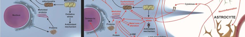
Int. J. Mol. Sci. 2019, 20, 719 3 of 36
In general, the most important pathological processes that underline these diseases are: misfolding
proteins and protein aggregates, mitochondria dysfunction, oxidative stress, ER stress, autophagy
impairment, alteration of intracellular calcium homeostasis, inflammation, and neurogenesis
Int. J. Mol. Sci. 2019, 20, x FOR PEER REVIEW 3 of 34
impairment [14,16–20]. We know that there are similar pathological processes involved at different
biological levels that might provoke neurodegeneration. The attempt to search for new targets
involved at different biological levels that might provoke neurodegeneration. The attempt to search
involved
for newintargetsneurodegeneration could bring us closer
involved in neurodegeneration to disease-modifying
could bring us closer to drug compounds. For
disease-modifying drugthis
reason,
compounds. For this reason, the aim of this review is to summarize the new research strategies thatto
the aim of this review is to summarize the new research strategies that are being used
identify
are being disease-modifying
used to identify therapies through thetherapies
disease-modifying knowledge of the common
through pathological
the knowledge of theprocess
common that
underlines neurodegenerative diseases. Here, we focus on two of the most prevalent neurodegenerative
pathological process that underlines neurodegenerative diseases. Here, we focus on two of the most
prevalent
diseases: neurodegenerative
Parkinson’s diseases:
disease and Parkinson’s
Alzheimer’s disease and Alzheimer’s disease.
disease.
2.2.Pathological
PathologicalTargets
Targetsin
inNeurodegenerative
Neurodegenerative Diseases
Diseases
ToTounderstand
understandhowhowthese
thesedisease-modifying therapies act,
disease-modifying therapies act, we
we need
needto
toknow
knowthe
thepathological
pathological
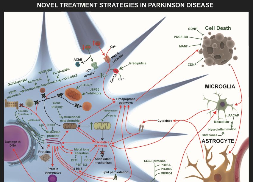
events that characterize these diseases (Figure 1).
events that characterize these diseases (Figure 1).
Figure 1.
Figure 1. TheThe
general pathways
general involved
pathways in neurodegenerative
involved diseases. Physiological
in neurodegenerative diseases. processes like
Physiological
endosomal-lysosomal
processes autophagy, neuroinflammatory
like endosomal-lysosomal responses, mitochondrial
autophagy, neuroinflammatory homeostasis,
responses, mitochondrial
proteostasis, proteostasis,
homeostasis, and metabolic profiling profiling
and metabolic (proteome(proteome
and lipidome) are dysregulated
and lipidome) are dysregulated in
neurodegenerative diseases (red arrows). Alterations in homeostasis
in neurodegenerative diseases (red arrows). Alterations in homeostasis mechanisms like themechanisms like the
endosomal–proteosomal–autophagy pathway
endosomal–proteosomal–autophagy pathway and
and an
an increase
increasein inmisfolded
misfoldedprotein
proteinaggregation
aggregationare are
major factors in Alzheimer’s disease (AD) and Parkinson’s disease (PD). The oxidative
major factors in Alzheimer’s disease (AD) and Parkinson’s disease (PD). The oxidative stress stresscaused
causedby
by mitochondrial
mitochondrial dysfunction
dysfunction and dysregulation
and dysregulation of endogenous
of endogenous antioxidantantioxidant
mechanismsmechanisms
is influencedisby
influenced by the level of free radicals. The positive feedback loop between oxidative
the level of free radicals. The positive feedback loop between oxidative stress, misfolded proteins, stress,
and
misfolded proteins, and mitochondrial dysfunction is crucial in therapeutic interventions.
mitochondrial dysfunction is crucial in therapeutic interventions. Furthermore, pre- and post-synaptic
Furthermore,
integrity pre- to
loss due and post-synaptic
alterations integrityhomeostasis
in calcium loss due to alterations
together in calcium
with homeostasis
the above pathwaystogether
is an
with the above pathways is an important mechanism involved in proapoptotic pathway
important mechanism involved in proapoptotic pathway activation. In addition, the remains of dead activation.
In addition, the remains of dead cells and the misfolded proteins released into the extracellular
cells and the misfolded proteins released into the extracellular environment provoke glia-activation,
environment provoke glia-activation, which releases cytokines and free radicals, exacerbating
which releases cytokines and free radicals, exacerbating neuronal death, which establishes another
neuronal death, which establishes another negative feedback loop between neurodegeneration and
negative feedback loop between neurodegeneration and neuroinflammation. Finally, these alterations
neuroinflammation. Finally, these alterations also affect neurogenesis in Alzheimer’s disease (AD)
also affect neurogenesis in Alzheimer’s disease (AD) and Parkinson’s disease (PD).
and Parkinson’s disease (PD).
2.1. Misfolded Proteins and Protein Aggregates
Most neurodegenerative diseases are characterized by protein aggregates formed mainly by a
specific protein that varies in each disease. In general, all these proteins are characterized by very large
disoriented domains without a defined secondary structure. When these proteins are prone to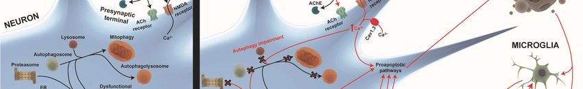
Int. J. Mol. Sci. 2019, 20, 719                                                                    4 of 36

2.1. Misfolded Proteins and Protein Aggregates
     Most neurodegenerative diseases are characterized by protein aggregates formed mainly by
a specific protein that varies in each disease. In general, all these proteins are characterized by
very large disoriented domains without a defined secondary structure. When these proteins are
prone to aggregation (oligomers to fibrils) in pathology, rich β-sheets are promptly available as
secondary structures [21]. In the case of AD, two types of protein aggregate appear: soluble
intracellular (monomer to oligomers) aggregates and insoluble extracellular (proto-fibril to fibrils)
aggregates, which are mainly formed by beta amyloid (Aβ) [22], and other intracellular aggregates
called neurofibrillary tangles (NFT) formed by hyperphosphorylated tau protein (tau) [23]. In PD,
an intracellular protein aggregate named the Lewy Body appears, which is formed by misfolded
α-synuclein (α-Syn) proteins [24].
     The causes of neurofibrillary tangles and Lewy Body formation are unknown. However,
there are several hypotheses, including oxidative stress and mitochondrial dysfunction, for their
starting point [25,26]. In the case of Aβ plaques, which involve conformational and aggregate
changes, the causes are unclear. Besides this, possible pathways have been discovered for Aβ
formation. Aβ is formed by the differential processing of amyloid precursor protein (APP). APP
can be processed by the α-secretase enzyme, giving rise to the soluble amyloid precursor protein-α
(sAPPα) fragment, which follows the “physiological” non-amyloidogenic pathway or by the enzyme
β-secretase (BACE1), prompting the soluble amyloid precursor protein-β (sAPPβ) fragment, which
follows the “pathophysiological” amyloidogenic pathway. Finally, both types of fragment are processed
by γ-secretase, where sAPPα does not result in molecules with pathogenic potential, while sAPPβ
gives rise to the Aβ peptide species, the main component of Aβ plaques [27].
     Despite this, cells have physiological mechanisms to maintain proteostasis and degradation
of protein aggregates, such as macroautophagy, chaperone-mediated autophagy, or the
ubiquitin-proteasome system which appears to be altered in AD and PD. However, many of these
aggregates and proteins are partially resistant to degradation by these mechanisms, progressively
losing their effectiveness due to the sequestration of chaperones by the aggregates [21,27]. In addition,
the appearance of misfolded proteins and aggregates that cannot be degraded will follow a prion-like
propagation mechanism involving misfolding protein aggregates between cells [21].
     The importance of these misfolded proteins and aggregates lies in their cytotoxic capacity through
different mechanisms. However, it should be noted that only the oligomers and fibrils that precede
the formation of the aggregates are toxic; therefore, the aggregates are proposed to be a protective
mechanism against toxic forms [28]. Thus, the cytotoxic effects of these misfolded proteins, apart from
the effects on the machinery for proteostasis [21], are mitochondrial damage, ER stress, autophagy,
and calcium homeostasis impairment [17,27].

2.2. Mitochondrial Dysfunction
     Nowadays, it is well known that mitochondria are involved in several important cell functions
besides ATP generation, such as the maintenance of calcium (Ca2+ ) homeostasis, intrinsic apoptosis
activation through cytochrome c release, modification of the reduction-oxidation potential of cells,
and oxidative stress regulation [29]. However, mitochondria can also contribute to neuronal
death in the context of age-related neurodegenerative disorders through a number of mechanisms
including the alteration of mitochondrial dynamics (fission/fusion and organelle trafficking) and
biogenesis/mitophagy processes; impairment of Ca2+ homeostasis; mutations on mitochondrial
DNA (mtDNA); incorrect activation of apoptosis; oxidative stress; and alteration of cellular
metabolism [30]. These mitochondria malfunctions can be due to different reasons. For example,
as mentioned above, misfolded proteins are a common feature in neurodegenerative diseases,
and there are a number of ways in which these proteins can alter mitochondrial activity, such
as their direct association with mitochondrial structures, damage to mtDNA, impairment to
organelle trafficking and dynamics, deregulation of bioenergetics and quality control pathways,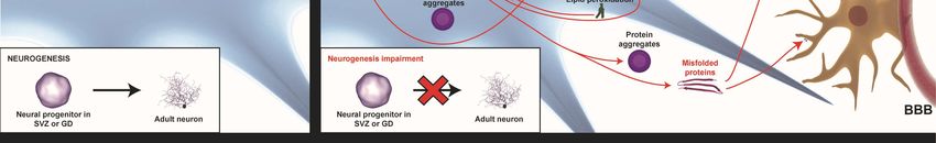
Int. J. Mol. Sci. 2019, 20, 719                                                                    5 of 36

and stimulation of mitochondria-dependent cell death [31]. On the other hand, some evidence
suggests that the accumulation of α-Syn in PD and phospho-tau and Aβ in AD may be direct
consequences of mitochondrial dysfunction [32,33]. Also, mutations of some nuclear genes that occur
in neurodegenerative disorders affect proteins involved in mitochondrial activity which can trigger
mitochondrial dysfunction in such pathologies. For instance, PD-related mutations in phosphatase
and tensin homolog (PTEN)-induced putative kinase 1 (PINK1) and Parkin proteins have been
implicated in mitochondrial quality control [34] and elicit mitophagy decrease and mitochondrial
dysfunction [35–37]. Another mutation that occurs in some PD cases is in DJ1 protein, a chaperone
involved in protection from oxidative stress [38]. This produces increased sensitivity to this stress
in cells, impairs mitochondrial respiratory chain complexes, decreases ATP production, and alters
mitochondrial morphology [39,40]. Similarly, some AD related mutations can alter mitochondrial
activity. This is the case for PSEN-1 and PSEN-2 proteins which are localized on the endoplasmic
reticulum (ER) mitochondrial associated membranes (MAMs), and their mutation triggers increased
cytosolic Ca2+ levels in these regions, inducing mitochondrial Ca2+ overload and stimulation of
mitochondrial respiration, thereby increasing reactive oxygen species (ROS) generation [41].

2.3. Oxidative Stress
      The intracellular balance between oxidants and antioxidants is regulated by the production of free
radicals by the mitochondria (electron transport chain and different enzymes), the ER, peroxisomes,
or different enzymes (e.g., NAPDH oxidases or xanthine oxidases), and the reduction of these free
radicals by different antioxidant mechanisms, such as glutathione, superoxide dismutase, catalase,
and peroxiredoxins. However, in neurodegenerative diseases, this balance is broken and there is
a situation of oxidative stress, mainly due to the mitochondrial dysfunction that occurs in these
diseases, which causes a high increase in the production of free radicals, and the cellular antioxidant
mechanisms cannot confront it [42]. In addition, in the case of AD, there are several pathological
mechanisms that cause an increase in oxidative stress, such as the activation of NADPH oxidases by
Aβ peptides with the consequent production of free radicals by this enzyme, the overactivation of
N-methyl-D-aspartate receptors (NMDAR) (excitotoxicity) that promotes an influx of Ca2+ to the cell,
causing an increase in the production of free radicals [43], or the binding of the Aβ peptide to metals,
which leads to the production of free radicals [42,44]. In the case of PD, oxidative stress also comes
from auto-oxidation of dopamine that, when free in the cytoplasm of dopaminergic (DA) neurons [45],
provokes the constant flow of Ca2+ in these neurons, which causes them to have a basal firing rate,
but also makes them more susceptible to oxidative stress [46,47]. The high energy demand suffered
by this type of neuron also triggers higher production of free radicals due to greater mitochondrial
respiratory function [45,47]. In both cases, we must bear in mind that the neuroinflammation present
in these diseases also aggravates oxidative stress [14].
      The problem with oxidative stress is the intracellular cytotoxic consequences that mainly derive
from the modification of molecules due to the free radicals—highly reactive molecules that alter all
the molecules they find in their case [42]. Thus, at a molecular level, this oxidative stress causes the
peroxidation of lipids and formation of 4-hydroxynonenal (4-HNE), which can react with proteins
to generate adducts or alter their conformation; carbonylation and nitration of proteins with the
consequent loss of their conformation; and oxidation of nitrogenous bases, which leads to mutations
and destabilization of nucleic acids [48]. These biochemical alterations to certain biomolecules trigger
high consequences at the cellular level such as alteration of the integrity of the membranes; loss of
conformation of numerous proteins, which can cause, among other problems, alterations to metabolism
or alterations in the mechanisms of mutation correction in the DNA; a decrease in ATP levels; alterations
in mitochondrial function, such as high production of free radicals by the electron transport chain,
which establishes a positive feedback loop for the production of free radicals; and an increase in the
misfolding of the Aβ, tau, and α-Syn proteins [48,49]. Finally, due to the action of all these cytotoxic
processes in the different compartments of the cell, oxidative stress is able to activate proapoptotic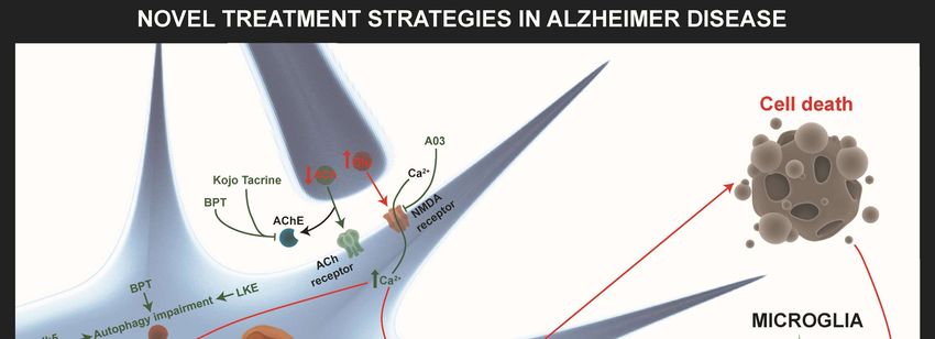
Int. J. Mol. Sci. 2019, 20, 719                                                                     6 of 36

pathways that lead to the death of neurons, including the intrinsic pathway of apoptosis, due to
damage to the mitochondria [50].

2.4. Autophagy Impairment
      Autophagy is a cellular mechanism that carries out the degradation and recycling of different
cellular components, including the degradation of misfolded aggregates and proteins. However,
we can differentiate three different ways in which it is performed: Chaperone-dependent autophagy,
microautophagy, and macroautophagy [51]. The first process is carried out through the ubiquitination
of the misfolded proteins and their subsequent treatment by chaperones. However, as previously
commented, when the function of the chaperones in these diseases is altered, this option cannot
be performed [52]. On the other hand, the other two autophagy methods are also altered in most
neurodegenerative diseases for various reasons that can be related to all the steps necessary to carry
them out, including the biogenesis of autophagosomes or lysosomal function. An example of the
relationship between autophagy and neurodegenerative diseases is the fact that numerous genetic risk
factors for them are related to autophagy, as is the case of ATP13A2 and VPS35 for PD or the case of
PICALM and PSEN-1 for AD [53].
      Thus, as indicated, a primary function of autophagy is the degradation of misfolded proteins
and aggregates. However, this is not the only function of autophagy related to neurodegenerative
diseases, since, for example, autophagy has also been considered an antioxidant mechanism [54]
or, specifically, because macro-autophagy is also responsible for the degradation of dysfunctional
mitochondria (mitophagy), [54,55]. Therefore, the alteration of the macroautophagy present in these
diseases can trigger a poor withdrawal of the dysfunctional mitochondria present in the cell, which
exacerbates the negative consequences of mitochondrial dysfunction [54,55]. In addition, in the case of
PD, mutations in the PINK1 gene, a genetic risk factor for this disease, are related to one of the main
pathways of mitophagy [55].

2.5. Intracellular Ca2+ Homeostasis Alteration
     The intracellular homeostasis of calcium is extremely important in the neural cells present in
the Central Nervous System (CNS) because it acts as a second messenger in many pathways, such
as the release of neurotransmitters by neurons, synaptic plasticity, astrocyte Ca2+ waves, activation
of proapoptotic enzymes, etc. [56]. That is why the concentration of this ion is highly controlled,
being the main storage center of this ion in the ER [57]. However, Ca2+ homeostasis is altered
in neurodegenerative diseases, which leads to strong cytotoxic and dysfunctional consequences
for neurons [17], which usually lead to alterations in the functions of enzymes regulated by Ca2+
concentrations, thereby provoking an increase in protein aggregation; alteration of lysosomal function
and, therefore, autophagic function [57]; a decrease in long-term potentiation (LTP); and increase in
long-term depression (LTD) [56], among others.
     Nowadays, there are several compatible hypotheses to explain the dysregulation of the Ca2+
concentration that occurs in neurodegenerative diseases, including the possibility that it is due to
the effects of other cellular alterations present in these diseases. One of these hypotheses is that it is
because of the process of excitotoxicity, which occurs on account of an overactivation of glutamate
NMDAR, which is due to different reasons depending on the pathology, but, in general, provokes an
overload of Ca2+ inside the cell, which has cytotoxic consequences for the neuron. For example, this
can cause the activation of proapoptotic enzymes such as calpains [17]. In addition, in the case of AD,
some of the hypotheses that explain this dysregulation are the ability of the Aβ peptide to cause pores
in the ER that release Ca2+ from its interior to the cytoplasm or the effect of mutations in presenilins
that provoke this alteration in Ca2+ concentration [56].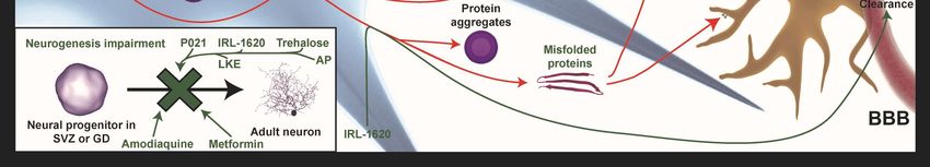
Int. J. Mol. Sci. 2019, 20, 719                                                                     7 of 36

2.6. Neuroinflammation
     Glial cells (astrocytes and microglia) have been recognized to have a clear function in
neurodegenerative diseases since past decades. However, these cells are extremely important because
they have immune functions, which, under physiological conditions, result in controlled reactions,
but, under pathophysiological conditions, such as neuronal death, give rise to exacerbated reactions in
the midterm. In this way, in neurodegenerative diseases, a positive feedback loop is created between
neuronal death and neuroinflammation [14].
     The reasons for this neuroinflammation are diverse, such as the activation of astrocytes and
microglia by the binding of the misfolded proteins of each disease to the toll-like receptors (TLR) of
these cells [58] and the activation of other damage-associated molecular pattern (DAMPS) receptors of
these cells by the damage signals present in these diseases. The exposure of these cells to misfolded
proteins and to cell damage signals in chronic diseases leads to an exacerbated immune reaction
by the glia cells. In this way, this reaction provokes the chronic production of free radicals and
pro-inflammatory cytokines that activates neuronal death pathways, in addition to activating other
astrocytes and microglia cells [59].

2.7. Neurogenesis Impairment
      Adult neurogenesis in humans is a mechanism of cerebral plasticity and, therefore, is fundamental
for the correct functioning of the nervous system. Although several years ago it was believed that
this neurogenesis process only occurred at the early stages, many studies so far have shown it to take
place in adults, specifically in the subventricular zone and the subgranular cells of the dentate gyrus
of the hippocampus [60]. In some neurodegenerative diseases, it is clearly the pathogenic decrease of
neurogenesis, but the reason why it happens is unclear and depends on the disease. Nonetheless, adult
neurogenesis is generally affected by neuroinflammation, which causes a decrease in this process [61,62].
      It has been shown that impaired neurogenesis is associated with Alzheimer’s and Parkinson’s
diseases. Firstly, this is because adult neurogenesis in humans is necessary to maintain certain
cognitive functions that are mainly affected in these diseases [60]. In addition, although there is still
some controversy in the results in humans and in animal models of this disease, it has been shown
that neurogenesis is altered and that the deposition of the extracellular Aβ peptide (in the case of AD)
is involved in this alteration [63]. On the other hand, PD is also closely related to the process of adult
neurogenesis. As in the previous case, in humans, there is still controversy regarding the results [62],
but in animal models, it has been observed that, in general, there is a decrease in neurogenesis in brain
zones where this process occurs physiologically [64].

2.8. Metal Ions Homeostasis Alteration
      It has been previously demonstrated an alteration of metal ions homeostasis, including iron (Fe),
copper (Cu), and zinc (Zn) in AD and PD. However, at this time, the reasons for this are unclear.
In general, it is characteristic an increase and site redistribution of these metal ions levels, together
with the appearance of these ions in Aβ plaques and Lewy bodies, the typical protein aggregates of
AD and PD, respectively. There are some clues that indicate that this could due to the altered function
of regulator homeostasis proteins; it appears that APP, α-syn, neuromelanin, and other proteins not
related with these diseases act as indirect or direct regulators of some metal ions homeostasis [65–67].
      These metal ions homeostasis alteration has cytotoxic consequences on cell survival. On the one
hand, the reaction of some metal ions with free radicals or molecular oxygen increases oxidative stress
by Haber-Weiss and Fenton reaction. Moreover, some pro-oxidant enzymes (like xanthine oxidase or
nitric oxide synthase) can be activated in an indirect way by these metal ions [65]. Additionally, some
of these metal ions can interact with the archetypal proteins involved in these diseases and promote
their aggregation [66,67]. Lastly, among other cytotoxic consequences, iron’s capacity to activate a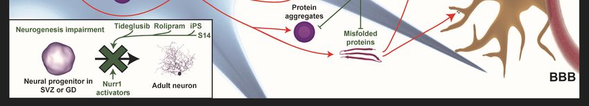
Int. J. Mol. Sci. 2019, 20, 719                                                                    8 of 36

programmed cell death known as ferroptosis, which is independent of the oxidative stress and other
cell death pathways, is noteworthy [66].

3. Novel Treatment Strategies in AD
       Most of the novel treatments are based on multi-facet strategies against familiar or idiopathic
AD. Here, we focus on the recent preclinical targets and novel treatment approaches that are under
investigation (Figure 2, Table S1). Nowadays, only five Food and Drug Administration (FDA) approved
drugs are on the market and these only diminish the progression of the disease. Four of them, donepezil,
galantamine, rivastigmine, tacrine, are based on acetylcholinesterase inhibition, and one of them,
memantine, has an antagonist influence on NMDAR [68–70]. At present, three clinical trials for AD are
still ongoing: gantenerumab (ClinicalTrial.gov identifier: NCT03443973, NCT03444870, NCT02051608,
NCT01224106, NCT01760005), crenezumab (NCT02353598, NCT01998841, NCT02670083, NCT03443973,
NCT03491150), and aducanumab (NCT01677572, NCT02484547, NCT02477800, NCT03639987) (Table S1).
Earlier immunotherapies caused the failure of immunization of these monoclonal antibodies in AD due
to unsuccessful clinical efficacy and major safety problems (e.g., amyloid-related imaging abnormalities)
when used at high doses. Other underlining factors are variation in their antibodies epitopes as well
as a high variability in the recognition of the structural conformation of Aβ species. Accessibility
of N-terminus antibodies immunization of Aβ is more prone to success, compared to hydrophobic
C-terminus immunization. Also, N-terminus antibodies have more efficient clearing of the aggregates,
since bapineuzepam, gantenerumab, and aducanumab provoke microglial activation and phagocytosis.
New trials targeting prodromal and early stage of the disease (e.g., gantenerumab, crenezumab, BAN2401,
aducanumab) are in the pipeline, since most of previous trails failed. The main reason of this failure
is a late intervention in patients when too much Aβ has been accumulated and the Aβ cascade is
irreversible [71–73]. Therefore, novel treatments are urgently needed, including both single target and
multi target drugs therapies that could act on the molecular pathway links to misfolded proteins (Aβ
and tau), synaptic integrity, cognitive impairments, autophagy, and mitochondrial dysfunctions (e.g.,
oxidative stress, peroxidase induced cytotoxicity), as well as pro- and anti-inflammatory responses
related to AD. However, non-invasive administration methods for these drug compounds have been
taken into account.

3.1. Targeting the Excitotoxicity and Misfolding Protein Aggregations
      A few newly developed compounds in preclinical research been proven to have similar properties
as acetylcholinesterase inhibitor and NMDAR antagonist/agonist that have established use in the
clinic. However, they might have beneficial effects on other dysregulated molecular pathways in AD.
Here, we focus on the molecular drug targets that have additional activity to that already assumed by
the FDA approved drugs.

3.1.1. Novel Acetylcholinesterase Inhibitors for Multi-Target Drug Therapy
     In AD, a low level of acetylcholine is an important factor in cognitive impairment. Inhibition
of acetylcholinesterase (AChE) has been shown to increase cognitive impairment and most
4-(1-benzylpiperidin-4-yl) thiosemicarbazone (BPT) analogues (except 2,3,4-OH-BBPT) have shown
potential to act as moderate AChE inhibitors compared to those that are already clinical available
AChE inhibitors, like donepezil. Besides this, various 4-(1-benzylpiperidin-4-yl) thiosemicarbazone
(BPT) analogues were tested on the other five major hallmarks related to AD: anti-proliferative activity,
metal chelation, oxidative stress, dysfunction of autophagy, and protein aggregation. The pyridoxal
4-(1-benzylpiperidin-4-yl)thiosemicarbazone (PBPT) analogue proved to be the best for all of the
five major AD hallmarks. A sixth factor was assessed to determine the feasibility of crossing the
blood–brain barrier (BBB) by oral administration following the “Lipinski’s Rule of Five” [74].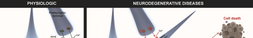
patients when too much Aβ has been accumulated and the Aβ cascade is irreversible [71–73]. Therefore,
novel treatments are urgently needed, including both single target and multi target drugs therapies that
could act on the molecular pathway links to misfolded proteins (Aβ and tau), synaptic integrity,
cognitive impairments, autophagy, and mitochondrial dysfunctions (e.g., oxidative stress, peroxidase
induced cytotoxicity),
Int. J. Mol. Sci. 2019, 20, 719 as well as pro- and anti-inflammatory responses related to AD. However,
9 of 36
non-invasive administration methods for these drug compounds have been taken into account.
Figure
Figure 2.2. Novel
Novel treatment
treatment strategies
strategies for
for AD.
AD. The
The most
most relevant
relevant approaches
approaches to to restore
restore altered
altered
pathways
pathways in in AD
AD are
are shown
shown withwith green
green arrows
arrows when when processes
processes areare improved
improved or or T-bars
T-bars when
when
inhibited.
inhibited. The
The novel
novel acetylcholinesterase
acetylcholinesterase (AChE)
(AChE) inhibitors
inhibitors BPT
BPT and
and Kojo
Kojo tacrine
tacrine affect
affect autophagy
autophagy
impairment,
impairment, misfolded
misfolded proteins,
proteins, and
and their
their aggregates
aggregates in in the intracellular and
the intracellular extracellular brain
and extracellular brain
environment.
environment. A03 A03compound
compound hashas an antagonist
an antagonist influence
influence on theon the NMDAR-mediated
NMDAR-mediated pathwayspathways
involved
involved in increasing
in increasing SIRT1 expression.
SIRT1 expression. Other
Other target target strategies
strategies of autophagyof autophagy
impairment impairment
are CDK5 are CDK5
inhibitors
inhibitors
and small and small lanthionine
lanthionine ketamine-ethyl
ketamine-ethyl ester (LKE)ester (LKE) against
molecules molecules againstCRMP2
increased increased CRMP2
expression.
expression.
Neurogenesis Neurogenesis
impairment impairment
is improved is byimproved by P021,
P021, IRL-1620, IRL-1620,
trehalose, trehalose,
metformin, metformin,
LKE, LKE,
amodiaquine,
amodiaquine, and allopregnanolones
and allopregnanolones (AP) (BR297) (AP) (BR297) IRL-1620
treatments. treatments. IRL-1620the
increases increases
clearancetheofclearance
Aβ in the of
Aβ in the bloodstream
bloodstream by the
by influencing influencing
Endothelin the Endothelin
B (ETB) B (ETB)
receptor. P021 andreceptor.
trehaloseP021
haveand trehalose
positive have
influences
positive
on misfolded influences on misfolded proteins.
proteins. Diethyl(3,4-dihydroxyphenethylamino) Diethyl(3,4-dihydroxyphenethylamino)
(quinolin-4-yl)methylphosphonate
(quinolin-4-yl)methylphosphonate (DDQ) and AP
(DDQ) and AP (BR297) increase mitochondrial (BR297) increase
biogenesis and Aβ mitochondrial biogenesis
clearance. Minocycline and Aβ
increases
anti-inflammatory responses and decreases pro-inflammatory responses. Metal ions homeostasis
alteration can be rescued by PBT-1, PBT-2, 4-(1-benzylpiperidin-4-yl)thiosemicarbazones (BPT)
derivatives, and deferoxamine (DFO).
The other major factor in AD is the dysregulation of metal ion chelation (copper (Cu2+ ), zinc
(Zn2+ ),iron (Fe3+ )). These ions accumulate within the Aβ plaques, of which Cu2+ and Zn2+ facilitate
the self-aggregation of Aβ(1–40) and Aβ(1–42) peptides [75]. Overall, this study demonstrated (in order
of decreasing efficacy) that 2,3-OH-BBPT, 8-OH-QBPT, PCBPT, 2,3,4-OH-BBPT, SBPT, QBPT, and PBPT
display the ability to inhibit Cu2+ -mediated Aβ(1–40) and Aβ(1–42) aggregation. Nevertheless, PBPT
analogue and its metal chelators Cu2+ and Fe3+ have demonstrated low anti-proliferative efficacy,
which is a desirable characteristic for a long-term AD treatment. Besides this, PBPT showed a greater
ability to inhibit 59 Fe cellular uptake from 59 Fe-transferrin complexes by at least 41% compared to
the controls. A look at the effect of iron complexes on ascorbate oxidation demonstrated that Fe3+Int. J. Mol. Sci. 2019, 20, 719 10 of 36
complexes of PBPT, NBPT, 8-OH-QBPT, and 2,3-OH-BBPT analogues inhibited ascorbate oxidation
more greatly than control samples. These observations suggest that these ligands have the potential to
alleviate the Fe-mediated oxidative stress observed in AD. Also, PBPT and SBPT analogues were able
to alleviate hydrogen peroxide-mediated cytotoxicity which shows their potential to prevent oxidative
stress in AD [74]
Despite this, the autophagy mechanism, which is dysregulated in AD, plays an important function
by eliminating misfolding proteins. However, it was demonstrated that the BPT analogues PBPT,
PCBPT, 8-OH-QBPT, and 2,3,4-OH-BBPT increase autophagy flux, while NBPT, 2,3-OH-BBPT, and SBPT
inhibit the autophagy degradation pathway. Otherwise, no significant effect of QBPT was observed on
the autophagy pathway. This modulation could be a crucial function in increasing the clearance of Aβ
aggregated species which may be one of the major problems in AD [74].
Another acetylcholinesterase inhibitor, tacrine, a lost multi-target drug entity, was FDA approved.
Unfortunately, it was discontinued after other more prone acetylcholinesterase inhibitors were
discovered, due to its severe liver toxicity [70]. New insights have shown that Kojo tacrine (KT2D),
a tacrine isoform with similar acetylcholinesterase inhibition was developed. This KT2D was
synthetized with antioxidant properties and is less hepatotoxic than tacrine, fully completely selective
against AChE, and significantly neuroprotective against Aβ. Further properties of various KT2D
racemate mixes need to be tested extensively both in vitro and in vivo to determine their clinical
translation properties [76].
3.1.2. Novel NMDA-Receptor Antagonist as an AD Idiopathic Treatment Strategy
The major genetic risk factor in AD is apolipoprotein-E4 (ApoE4), which has a significant potential
to reduce Sirtuin 1 (Sirt1) expression. This sirt1 reduction leads to decreases in the FOXO3-mediated
oxidative stress response, Coactivator 1-α (PGC1α)-mediated ROS sequestration, and RARβ-mediated
ADAM10 expression and increases in P53-mediated apoptosis, NFκB-mediated Aβ toxicity, as well
as the acetylation of tau, which leads to microtubule instability and tau pathology [77]. Various Sirt1
enhancers have been identified, such as alaproclate, resveratrol, quercetin, fisetin, SRT1720, SRT1460,
and A03 racemates [78–84]. Nevertheless, A03 was described as a non-competitive NMDAR antagonist.
A03 has a similar effect on reducing the excitotoxicity as the FDA approved drug memantine. Besides
this, memantine does not influence Sirt1 expression. However, A03 has shown beneficial orally
pharmacokinetics profiles in the treatment of Sirt1 related pathogeneses in AD. In the amyloidogenic
pathway, cleavage fragments of sAPPβ as well as toxic Aβ species have been shown to decrease
after A03 treatment in transfected cell cultures either expressing ApoE4 or ApoE3. The decrease of
sirt1 expression was greater in ApoE4 compared to ApoE3 transfected cultures and was increased
by A03 treatment. No similar effect was seen for sAPPα. Unfortunately, in vivo A03 treatment of
E4FAD transgenic animals did not show any significant effect on the cleavage fragments Aβ(1–42) ,
sAPPα, and sAPPβ or the sAPPα/sAPPβ ratio in the hippocampal area, which is crucial area for
AD pathology. Besides this, long term oral A03 treatment of AD transgenic animals increased Sirt1
expression in the hippocampus, but not in the frontal cortex, which has been associated with memory
improvement. The importance of the above-mentioned compound needs to be further investigated
in terms of the relation between sirt1 and sAPPα in dose dependent cases in both preclinical and
clinical trials. The Sirt1 function has a major role related to tau pathology, which is an additional
risk for progression during AD. The first-in-class ApoE4 targeted therapeutic, A03, which influences
Sirt1 levels might be a good candidate for preclinical trails in MCI and AD due to its excellent brain
bioavailability and promising efficacy after chronic oral treatment [77].
3.1.3. Other Target-Receptor Mediated Treatment Strategies
The endothelin B (ETB) receptors is abundant in the CNS and has been shown to play a role in
development and neurogenesis. To influence the receptor-mediated function, a highly specific agonist
IRL-1620 was used to target the ETB receptor. Endothelin-1 isopeptide (ET-1) plays a central roleInt. J. Mol. Sci. 2019, 20, 719 11 of 36
in the regulation of cardiovascular functions and regional blood flow [69,85,86] and may be part of
the mechanism by which Aβ interferes with vascular function in AD. It has been shown that Aβ
upregulates endothelin converting enzymes 1 and 2 (ECE-1 and ECE-2), which results in increased
production and release of ET-1 [69,87–89]. ECE-1 helps the clearance of Aβ by fragmentation of the
peptide [90]. The compound IRL-1620 stimulates the clearance of both ET-1 and Aβ as well as cerebral
blood flow which might have a positive influence on the clearing mechanisms of toxic aggregates.
Besides this, the compound improves memory deficiency and reduces oxidative stress, which are both
caused by Aβ toxicity [69,91,92]. IRL-1620 has been demonstrated to increase neural growth factor
(NGF) and synapsin I expression, which are both factors involved in neurogenesis and synaptogenesis
and which are altered in MCI and AD pathologies [69,92–94].
3.2. Targeting Mitochondrial Dysfunction and Related Pathways Involved in AD
Recently, the involvement of mitochondrial dysfunction in AD has been investigated
by using the pharmacologically developed compound diethyl(3,4-dihydroxyphenethylamino)
(quinolin-4-yl)methylphosphonate (DDQ). DDQ has demonstrated positive effects on mRNA and
protein levels related to mitochondrial dysfunction and synaptic dysregulation, which are both related
to AD. Besides this, DDQ has an effect on the mitochondrial dynamics, related to fission proteins
(DRP1 and Lis1), fusion proteins (Mfn1 and 2), and Aβ interactions. DDQ has shown a better docking
score than other single existing molecules, like MitoQ, Mdivi1, and SS31. One advantage of DDQ as a
novel target is that it binds to the active sites of Aβ and DRP1, inhibiting the Aβ and DRP1 complex
formations. The mRNA and protein levels of mitochondrial (PGC1α, Nrf1, Nrf2, TFAM, DRP1, Fis1,
Mfn1 and 2) and synaptic activity (Synaptophysin, PSD95, synapsin1 and 2, synaptobrevin1 and 2,
synaptopodin, and GAP43) were investigated after Aβ-induced pre-treatment or post-treatment with
DDQ. The mRNA and protein levels (PGC1α, Nrf1, Nrf2, and TFAM) were significantly increased after
Aβ incubation followed by DDQ treatment. Besides this, reductions of mitochondrial fission proteins
(DRP1 and Fis1) and increases of mitochondrial fusion proteins (Mfn1 and 2) were observed after DDQ
treatment. Despite this, pre-treatment with DDQ followed by Aβ treatment increased mitochondrial
biogenesis mRNA (PGC1α, Nrf1, Nrf2, and TFAM) levels, which suggests that DDQ pre-treatment
could serve as prevention agent in AD. DDQ pre-or post-treatment induced Aβ incubation led to a
downregulation in mitochondrial fission protein activity (DRP1 and Fis1) and upregulation of fusion
activity (Mfn1 and Mfn2). This led to the conclusion that DDQ pre-treatment reduces fission activity
(DRP1 and Fis1) and enhances fusion activity (Mfn1 and 2) in the presence of Aβ. However, DDQ
treatment-induced Aβ incubation also has a potential enhancing effect on synaptic activity which
is downregulated by Aβ pathology. The reduction of DRP1 and Aβ complexes is stronger in DDQ
pre-treated compared to post-treated cells. The reduction between Aβ and DRP1 interaction due to
DDQ treatment leads to a reduction in mitochondrial fragmentation and maintains the normal count,
normal length and normal function of mitochondria, and it might be protective against the Aβ plaque
load. Nevertheless, DDQ treatment has been demonstrated to play a neuroprotective role in Aβ toxicity
by significantly reducing Aβ(1–42) levels and increasing Aβ(1–40) levels. Despite this, DDQ enhances
mitochondrial function and increases cell viability, which leads to an increase in mitochondrial ATP
and cytochrome oxidase activity, as well as a reduction in free radicals and oxidative stress, which is
dysregulated in AD [95].
3.3. Targeting Autophagy
Autophagy dysregulation has been linked indirectly to AD using microtubule-associated protein
CRMP2 (collapsin response mediator protein-2) modulation. CRMP2 seems to have same features
as tau protein, but, besides this, CRMP2 undergoes profound posttranslational modifications in the
brain. CRMP2 is an important adaptor protein that is involved in vesicle trafficking, amyloidogenesis,
and autophagy. Tau protein did not show similar involvement in these molecular pathways [96].
Actually, CRMP2 was discovered to be a mediator of neurite retraction and neuron polarizationInt. J. Mol. Sci. 2019, 20, 719 12 of 36
during semaphorin signaling [97]. Besides this, tau has a direct effect and CRPM2 has an indirect
effect on the stabilization of actin-based microfilament networks. The difference between CRMP2
and tau is that CRMP2 is involved in endosomal-lysosomal trafficking and autophagy [98]. Although
CRMP2 has a highly binding affinity to endocytic adaptor protein (Numb) and MICAL-like protein
1 (MICAL-L1), which are involved in intracellular vesicle movement, as well as the amyloidogenic
processing of APP which occurs by endosomal trafficking, it has been speculated that CRMP2 becomes
functionally depleted in AD pathology due to both combination of the hyper-phosphorylation of tau;
sequestration into nascent neurofibrillary tangles; and oxidative post-translational modification of
CRMP2. This includes different multi-facet CRMP2 dependent processes, including amyloidogenic
APP trafficking through early and recycled endosomal compartments. Also, it has been demonstrated
that the knock-down of CRMP2 expression influences the autophagy flux [99]. Nevertheless,
the CRMP2-binding small molecules lanthionine ketamine-ethyl ester (LKE) normalize CRMP2
phosphorylation, reducing the Aβ burden and phosphorylating tau in AD in vitro and in vivo models.
Even some derivations of LKE can functionally enhance CRMP2 to promote growth factor-dependent
neurite outgrowth as well autophagy-related processes [96]. Despite this, long term LKE treatment
increases beclin-1 protein, which is another autophagy-related protein that is downregulated in MCI
and AD, which negatively influences the Aβ flux. Besides this, Glia-derived neurotrophic factor
(GDNF) can increase CRMP2 expression, which results in microtubule stabilization and enhanced
neurite outgrowth [99,100].
The activity of mTORC1, the gate-keeper of autophagy, can increase CRMP2 expression
which shows that CRMP2 might be a good therapeutic target [101,102]. Nevertheless, the CRMP2
phosphorylation signaling pathway could be modified by a pharmacological inhibitor of Cdk5, which
is under development and which can target the glycogen synthase kinase-3 (GSK3), which is the major
compound for regulating the hyper-phosphorylation of either tau or CRMP2 [103,104]. An upstream
therapeutic target that might influence CRMP2 expression, as can be seen by axon repulsion factor
semaphoring 3A (Sema3A), which binds to the neuropilin-1 receptor (NRP1) and plexin-A co-receptors;
its relation in AD needs to be further exploited [105,106]. Besides that, tau was discovered earlier
than CRPM2, and tau is more prone to form non-dissociable high molecular weight complexes than
CRMP2. Also, CRMP2 and phosphorylated-CRPM2 (pCRMP2) are not prone to aggregate or form
filaments like tau, which later on accumulate to form or associate with NFT-related proteins. Despite
this, tau mutations are neuropathic in humans, whereas a CRMP2 mutation has not been discovered
yet [96]. Nevertheless, CRMP2 hyper-phosphorylation is a very early event that occurs prior to Aβ
accumulation or tau hyper-phosphorylation [107]. This effect of CRMP2 hyper-phosphorylation is a
downstream consequence of altered non-amyloidogenic APP processing and perhaps been a more
prominent function than tau dysregulation in disorders involving both amyloidopathy and NFT
aggregation [108].
3.4. Targeting Neuroinflammation
Neuroinflammation is seen as an early event in AD, and drug agents involved in targeting
this process could slow down disease progression. Recently, Minocycline, a tetracycline antibiotic,
which interferes with the symptoms of neuropsychiatric disorder related to AD. Minocycline has been
shown to decrease proinflammatory cytokines, like tumor necrosis factor α (TNF-α) and interleukin
1β (IL-1β), and increase anti-inflammatory cytokines, like interleukin 10 (IL-10), in Aβ(1–42) -treated
animals compared to control rats [109]. This neuropsychiatric compound can be considered a new
treatment possibility to act against the early effects of AD neuropathology.
3.5. Activation of Neurogenesis and Neuronal Survival Pathways
Neurogenesis pathways are downregulated in AD, and various attempts have been made to
improve the neurogenic survival pathways in neurodegeneration by interfering with Aβ and tau
related mechanisms, boosting dendritogenesis or synaptogenesis and relevant metabolic processesInt. J. Mol. Sci. 2019, 20, 719 13 of 36
involved in AD pathology [86,110]. Beneath, some novel compounds that are under preclinical research
as possible new treatment strategies are discussed. Nowadays, interest is growing in developing
more orally bioavailable compounds as a non-invasive therapeutic tool. These compounds are easily
degraded and have the ability to cross the BBB.
3.5.1. Orally Bioavailable Compounds
The first orally bioavailable neurotrophic factor is a tetra peptide (P021), which is derived from
ciliary neurotrophic factor (CNTF), and which has a suitable biodegradation level and is able to cross
the BBB easily. The general functions of P021 are to inhibit leukemia inhibitory factor (LIF) signaling
and increase brain-derived neurotrophic factor (BDNF) expression. However, P021 treatment has been
able to rescue dendritic and synaptic deficits by boosting neurogenesis, preventing neurodegeneration,
Aβ, and tau pathologies, rescuing cognitive impairment in AD transgenic mice, as well as markedly
reducing age-related mortality in rat. Besides this, P021 inhibits the GSK3β pathway through the
phosphorylation of ser-9 by BDNF. This inhibition has direct effects on Aβ and Tau pathology.
Both P021 as well as its parent molecule peptide 6 can increase neurogenesis and synaptic plasticity,
which improve cognitive performance in AD transgenic animal models, even in the late stage of the
disease progression. Nevertheless, no major side effects have been reported. P021 treatment could
serve as an additional therapy strategy to act against AD pathology [110].
The second orally bioavailable compound which can be classified as a natural disaccharide, also
called trehalose, seems to have a protective role in denaturation and conformational protein changes
in neurological disorders [111]. Unfortunately, there is a lot of controversy about trehalose and its
derivatives in neurological pathologies. Nevertheless, the major factor in AD is the dysregulation
of Aβ protein. It has been reported that Aβ aggregation is inhibited by trehalose and other
derivatives [112]. The other major factor involved in AD that was described earlier is metal ion
chelation dysregulation. It has been reported that trehalose and its derivatives seem to have a
positive influence on Aβ aggregation [75]. Due to this previous relevance, trehalose was studied
in the transgenic Tg2576 mouse model for AD [112]. It was shown that intracerebral injection of
trehalose improves cognitive impairment in the APP/PSEN-1 model [111]. Unfortunately, this
was based on the invasive administration route. However, no statically significant improvement
of cognition has been shown by the non-invasive oral administration of trehalose and its derivatives.
Also, trehalose treatment did not show any significance in reducing metal ion induced Aβ aggregation,
amyloidogenic APP levels, and Aβ fragments, as well as showing no influence on autophagy flux in
both the cortex and hippocampus. Nevertheless, trehalose treatment seems to significantly increase
the neuromigrating protein doublecortin (DCX), a surrogate indicator for neurogenesis, as well as
increasing synaptic activity, especially presynaptic vesicle marker synaptophysin, in the hippocampus
and in the cortex [112]. Trehalose treatment seems to increase progranulin expression in both the
hippocampus and the cortex. In AD, progranulin, a regulator of neuronal growth and survival,
has shown a protective effect against Aβ neurotoxicity [112–114].
3.5.2. Other Pro-Neurogenic Compounds Related to Metabolic Disorder Linked to AD
Metabolic disorders like type II diabetes could be genetically linked to AD. In both of these
disorders, alterations in neurogenesis are seen. Besides this, the roles of insulin and insulin
growth factor, which are important factors in diabetes, have been demonstrated to influence
neurogenesis in ex vivo and hippocampal cultured cell lines and might play a crucial role in
AD pathology [115]. Compared to an FDA approved drug (donepezil), metformin, a type II
anti-diabetic drug, has demonstrated both pro-neurogenic potential as well as higher spatial
improvement in cognitive impairment in an aluminium chloride (AlCl3 )-induced (AlCl3 ·6H2 O)
mouse model, a metabolic model for neurodegeneration [68,116]. Metformin-treated aluminium
chloride (AlCl3 )·6H2 O-induced animals seems to have an irreversible increase in neurogenesis factors
(DCX and Neuronal Nuclei (NeuN)), and this can be explained by the effect of metformin onYou can also read