The Microbiota and Gut-Related Disorders: Insights from Animal Models - MDPI
←
→
Page content transcription
If your browser does not render page correctly, please read the page content below
cells
Review
The Microbiota and Gut-Related Disorders: Insights
from Animal Models
Layla Kamareddine 1 , Hoda Najjar 1 , Muhammad Umar Sohail 1,2 , Hadil Abdulkader 1
and Maha Al-Asmakh 1,2, *
 1    Department of Biomedical Science, College of Health Sciences, QU Health, Qatar University,
      P.O. Box 2713 Doha, Qatar; lkamareddine@qu.edu.qa (L.K.); Hnajjar@qu.edu.qa (H.N.);
      msohail@qu.edu.qa (M.U.S.); ha1305202@qu.edu.qa (H.A.)
 2    Biomedical Research Center, QU Health, Qatar University, P.O. Box 2713 Doha, Qatar
 *    Correspondence: maha.alasmakh@qu.edu.qa; Tel.: +974-4403-4789

 Received: 3 October 2020; Accepted: 29 October 2020; Published: 2 November 2020

 Abstract: Over the past decade, the scientific committee has called for broadening our horizons in
 understanding host–microbe interactions and infectious disease progression. Owing to the fact that
 the human gut harbors trillions of microbes that exhibit various roles including the production of
 vitamins, absorption of nutrients, pathogen displacement, and development of the host immune
 system, particular attention has been given to the use of germ-free (GF) animal models in unraveling
 the effect of the gut microbiota on the physiology and pathophysiology of the host. In this review,
 we discuss common methods used to generate GF fruit fly, zebrafish, and mice model systems
 and highlight the use of these GF model organisms in addressing the role of gut-microbiota in
 gut-related disorders (metabolic diseases, inflammatory bowel disease, and cancer), and in activating
 host defense mechanisms and amending pathogenic virulence.

 Keywords: animal models; germ-free; gut microbiota; gut-related disorders; host–defense; pathogen virulence

1. Introduction
     Humans develop a symbiotic relationship with microbes at an early age [1]. This relationship
progresses, in turn, into a gradual edification of an elaborate ecosystem known as the human microbiota
which contains more than 100 trillion microorganisms that crucially tip the balance towards health or
disease [2,3]. Diverse microbial communities are involved in several physiological processes in a host,
including immune cell development, fermentation and food digestion, maintenance of metabolic
homeostasis, and angiogenesis [4]. As such, considerable attention has been given to understanding
the association between the microbiota and a broad range of human pathologies, including metabolic
diseases like obesity and diabetes [5,6], inflammatory bowel disease (IBD), and cancer [7–9]. The use of
pertinent model systems with varying levels of microbial complexity provides a convivial opportunity
for a better insight into the effect of host–microbiota interaction on dictating a state of health or
disease in a host, an approach that could not be easily implemented in humans and other higher
complex organisms alone. Manipulation of the gut microbiota composition of both –vertebrates
and invertebrates allows functional and mechanistic studies directed towards determining the causality
in disease-associated alterations [10]. The gut microbiota-provoked immune maturation has been
presented as a chief alignment in perpetuating gut homeostasis by safeguarding the host from
injury [11], detrimental inflammation [12,13], and infections [14]. The role of the commensal microbiota
in maintaining gut morphology and homeostasis in both vertebrates and invertebrates has been
partly attributed to its involvement in modulating cell composition, epithelial renewal, and tissue
structure [15–17]. The symbiotic gut microbiota has been also profoundly implicated in various

Cells 2020, 9, 2401; doi:10.3390/cells9112401                                       www.mdpi.com/journal/cells
Cells 2020, 9, 2401                                                                                  2 of 25

aspects of host physiology including lipid and carbohydrate metabolism [18–27]. Interestingly, the gut
microbiota also exhibits a complex bidirectional relation with non-antibiotic drugs. Although the gut
microbiota composition is affected by the uptake of some commonly used drugs [28,29], the commensal
microbiota itself influences an individual’s response to drugs by enzymatically altering the drug’s
structure, therefore affecting its toxicity, bioactivity, and bioavailability [30]. The gut microbiota can
also indirectly impinge a cancer patient’s response to immunotherapy by affecting the patient’s general
immune status [31].
      Germ-free (GF) model organisms present one of the fundamental means to study the effect of
an altered microbial community on the physiological status of a host. Among different model organisms,
fruit flies, zebrafish, and mice have been extensively employed in such studies. The advantage of
using such model organisms is chalked up to their ease in maintenance, strong genetic coherence,
physiological and signaling pathways resemblance to humans, powerful manipulatable genetic systems,
and culturable microbiota.
      In this review, we shed more light on the use of these model systems to divulge the role of the gut
microbiota in activating host immunity and amending pathogenic virulence, and to unravel the role of
the gut microbiota in the development and progression of gut-related disorders.

2. Provisos of Selecting Animal Models to Study Gut-Related Disorders
      Several international laws governing animals0 use in research and testing mandate replacing
animal experiments, wherever reasonably feasible, by humane alternatives. To address a scientific
question, the primary choice of researchers could be initially directed towards employing non-animal
technologies, such as in silico or in vitro approaches [32]. Recently, research is underway to develop
computer simulations and gastrointestinal organoids as one approach to further refine the use of
animal models [33,34]. The use of animal models to address complex biological problems should not
merely be viewed as a technical utility and is therefore generally paralleled with ethical implications
considered with risks, animal and public health safety, and interpretability and reproducibility of
the results. It is indeed the researcher’s moral duty to honor animals and give due consideration to
their capacities for distress when being manipulated. For example, the production and maintenance of
GF animal colonies and the induction of metabolic diseases by oral or invasive procedures greatly
aggravate animals’ suffering. Therefore, it is extremely desired to follow standard provisions to protect
the animals used in such practices from discomfort, misery, trauma, or permanent harm, and to ensure
that, where possible, their suffering is kept to a minimum. The use of animals shall only be permissible
if the work promises to contribute to a fundamental understanding of important biological concepts or
to the advancement of knowledge that can reasonably be expected to benefit both humans and animals.
As such, the 3Rs (Replacement, Reduction, and Refinement) set by Russell and Burch [35] shall be
strictly followed to minimize animal suffering and usage.
      If the scientific objective raised can be only accomplished by using an animal model,
then a comprehensive assessment of the pros and cons should be conducted to identify and appraise
the most suitable animal model. Turner et al. [36], for instance, reviewed the human gut microbiota
model’s application in rats, mice, guinea pigs, minipigs, conventional swine, dogs, and non-human
primates and concluded that murine models bear the closest microbiota resemblance to humans.
In this context as well, fecal microbial transplant studies of humanized germ-free murine models have
shown that rats have more similar microbiota with humans than mice [37]. One reasoning behind that
could be attributed to the belief that rats have more human baseline microbiota than mice, enabling
the inoculated bacteria to be more stable in expression and development [38]. The lack of genetic
variants in rats might also restrict their utility in studying specific models of diseases [39]. Therefore,
genetic background, baseline microbiota resemblance, and phenotypic expression of the diseases
studied is generally considered while selecting the most suitable animal model. The decision-making
process regarding which animal model is most appropriate should also emphasize which species
and modelsare most likely to provide the most translatable and valid data and results. Some of the most
Cells 2020, 9, 2401 3 of 25
common justifications provided for the selection of animal models such as ease of maintenance and low
cost might not be always perceived as adequate, and therefore additional thought should be given
when deciding on the choice of the animal model with the most translatable outcomes.
3. Fruit Fly
Drosophila, commonly known as the fruit fly, is one of the most powerful model organisms used in
various research areas. Fruit flies are easily cultured and maintained in a laboratory, have a relatively
short life cycle, and yield many offspring in a short period of time [40]. Drosophila’s sequenced
genome has additionally allowed its genetic manipulation and the discovery of a number of genes
whose homologs cause diseases in humans [41]. In recent years, the use of Drosophila has been most
commonly implicated in genetics, drug screening and toxicity, immunology, and metabolism related
studies [42]. The fruit fly has also gained particular attention for its use in understanding gut-associated
disorders due to the structural and functional similarities between a fly and a vertebrate gut [43,44].
Into the bargain, the adjacent concordance between the fly gut-brain axis advocated the powerful
contribution of the gut microbiota to the fly’s behavior and its physiological processes [45]. Dysbiosis of
the fly gut has been associated with cancer [46], IBD occurrence [47], lifespan alteration, and overall
developmental progression [42]. Several studies have also employed Drosophila as the model of choice to
understand the host defensive mechanisms against different intestinal pathogens like Vibrio cholerae [48],
Serratia marcescens [49], Pseudomonas aeruginosa [50], and Enterococcus faecalis [51]. Many of these studies
also correlated metabolic adaptations with the host’s response to infection [52], and depicted that
alterations in the intestinal microbiota can have a profound impact on the host’s normal physiological
processes and may contribute to disease progression [53].
3.1. The Fruit Fly Gut and Microbiota
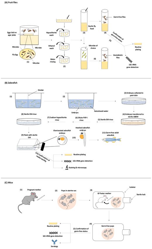
The Drosophila gut consists of three main regions: the foregut, the midgut, and the hindgut.
The foregut includes the oral cavity, esophagus, and crop in adult flies where food is ingested
and initially processed by the enzymes released. Further digestion and assimilation of the food by
enzymes including carbohydrases, proteases, and lipases occurs in the midgut. The hindgut regulates
the absorption of water, ions, and other nutrients, specifically those released from the Malpighian
tubules, a structure equivalent to the kidneys in vertebrates [54] (Figure 1A). The epithelial cell
lining of the fly gut is composed of four cell types including enterocytes (ECs), enteroendocrine
cells (EEs), intestinal stem cells (ISCs), and immature progenitor enteroblasts (EBs), which mainly
differentiate as ECs or possibly as EEs [55–57] (Figure 1B). The microbiota of a fly is separated from its
epithelial cells by a chitinous layer, instead of a mucus one, its hindgut is not a fermentation cavity
similar to a mammalian colon, and its gut is well-thought-out as an unsuitable milieu for obligate
anaerobic. As such, the Drosophila gut may not be colonized with most gut microbes needed to develop
a humanized microbiota model. Several standing points based on this could argue that a humanized
Drosophila-microbiota model might not be very informative in the interpolation of complex metabolic
diseases, and is instead more relevant to basic screening studies of host–pathogen interactions [54,58].Cells 2020, 9, 2401 4 of 25
Figure 1. The gut structure and normal flora of different model organisms including (A–C) the
fruit fly, (D–F) zebrafish, and (G–I) mouse. (A,D,G) Anatomy of the gut; (B,E,H) cell types present,
including enterocytes (EC), enteroendocrine cells (EE), intestinal stem cells (ISC), enteroblasts (EB),
goblet cells, stem cells (SC), vacuolated cells (VC) and, in the mouse, chemosensory tuft cells in the villi
and paneth cells, transit amplifying cells (TAC), and quiescent ISC in the crypts; (C,F,I) composition of
the major phyla according to [59–61]. BM = basement membrane.
Studies on the composition of the gut microbiota have primarily focused on the identification
of bacterial species and operational taxonomic units (OTUs) through laboratory cultures and 16S
rRNA gene amplicon sequencing [53]. The diversity of the microbial community in a Drosophila gut is
relatively less as compared to that of mammals, which is not a miniaturized version of the human gut
microbiota. Although a sole reason behind such a simplicity in fly’s microbial diversity has not been
delineated; yet, it is thought that a higher microbial diversity may be attributable to the type of food [62]
and to the presence of an adaptive immune system in mammals but not in Drosophila [63]. Several
factors including changes in gut oxygen tension or pH, host factors including genotype, development
and age, physiology and immune response, geographical locations, disturbance, and environmental
encounters might also explain the low-diversity of the microbiota community in the Drosophila
gut [59,64–68]. The microbiota of laboratory-reared flies fed complex polysaccharides diet such as
cornmeal and soy flour, for instance, have high abundance of Lactobacillus (Firmicutes of the order
Lactobacillales), while those fed sugar-rich diets have Acetobacteraceae (α-Proteobacteria), specifically
Acetobacter and Gluconobacter species, as the dominant bacterial group [54,68]. Some fly cultures
are also highly abundant with γ-Proteobacteria or Enterococcus to the point of eliminating or nearly
eliminating Acetobacteraceae and Lactobacillus. Several taxonomic differences in the microbiota also
exist between wild Drosophila populations and those reared in a laboratory. Although Acetobacteraceae,
Lactobacillales, and γ-Proteobacteria also dominate the gut microbiota of natural Drosophila populations,
the gut microbiota of wild flies is much more diverse [69–72]. Additionally, the number of Lactobacillus
tends to be low or undetected in natural Drosophila populations, with only Leuconostoc, Enterococcus,
and Weissella representing Lactobacillales being frequently abundant [73]. Though poorly studied,
yeasts, mainly Hanseniaspora, Pichia, and Candida, have been also detected in the gut of both wild-caught
and laboratory-reared Drosophila, particularly those fed rotting fruits [74–76] (Figure 1C).Cells 2020, 9, 2401 5 of 25
3.2. Generation of GF Fruit Flies
The generation of GF (axenic) and gnotobiotic model organisms, including flies, has greatly
contributed to our understanding of the role of the gut microbiota in maintaining a normal physiological
status of a host [77]. Generating GF flies can be achieved in different ways including egg dechorionation
(removal of the eggshell layer), rearing flies on a broad-spectrum antibiotic cocktail-containing diet
or placing flies on an axenic diet. In egg dechorionation, the first step involves egg laying of cage
flies on an agar plate. Eggs are then collected with a sterile paintbrush and embryos are subsequently
washed with hypochlorite solution, followed by water. The time of hypochlorite treatment varies
between protocols and some dechorionation procedures also include ethanol washes post hypochlorite
rinse [78–82]. Embryos are then carefully transferred with a paintbrush to axenic media. Since removing
endosymbionts like Wolbachia and Spiroplasma by bleaching remains challenging, the tetracycline
antibiotic is usually added to the axenic media to aid in this removal [82]. Flies serially transferred to
axenic media can remain GF for multiple generations [80,82].
Gnotobiotic or conventionalized flies can be produced following the same procedure of axenic
flies; yet, after the hypochlorite wash and aseptic transfer of dechorionated eggs to sterile food,
microbes of choice are added to the vials housing GF flies. Upon larval and adult feeding on microbe(s)
the association between the fly and the microbe(s) gets established and microbes readily colonize the fly
gut [54,80] (Figure 2A). Supplementing fly food with a broad spectrum antibiotic such as streptomycin
or tetracycline or a combination of antibiotics is another approach to alter the gut microbial community
and eliminate the microbiota [83–85]. Besides egg dechorionation and antibiotic containing diet,
placing flies on axenic food is also another approach used to suppress or eliminate the gut microbiota.
The most common methods used to verify a GF status of a fly include routine culture on selective
media like de Man, Rogosa, and Sharpe (MRS) [86], and PCR amplification and sequencing of 16S
rRNA gene regions [68,86].Cells 2020, 9, 2401 6 of 25
Figure 2. Generation of Germ-Free Model Organisms. (A) Generation of Germ-Free Fruit Flies.
Flies trapped in a cage lay their eggs on the agar plate. The surface of the laid eggs harbors microorganisms
from maternal fecal deposits (1). Using a sterile paint brush, eggs are collected from the agar plate before
larval formation, dechorionated with hypochlorite solution, and rinsed with water. An ethanol washCells 2020, 9, 2401 7 of 25
between the hypochlorite dechorionation step and the water rinsing step could be done (2). Dechorionated
eggs are transferred to sterile fly food (3) and germ-free flies are generated (4). The most common methods
used to confirm the germ-free status of flies include routine culturing on selective media permissive to
the growth of gut bacteria and 16S rRNA gene detection via PCR amplification and sequencing (5). To generate
gnotobiotic flies, microbe of choice is added to the sterile fly food vial containing dechorionated eggs (6).
(B) Generation of Germ-Free Zebrafish. Adult zebrafish are set up in breeding cages overnight with dividers
to prevent breeding and spawning (1). The next day, adults are transferred to tanks with autoclaved
water and dividers are removed to allow breeding and spawning for less than an hour. Embryos are
deposited in the tanks (2) and are then collected into petri dishes and rinsed thoroughly to remove any
debris (3). In a biosafety cabinet, zebrafish embryos are transferred to petri dishes with sterile antibiotic
embryo media (ABEM) (4) and undergo a sterile embryo media (EM) rinse (3 times) (5), followed by a dilute
polyvinylpyrrolidone-iodine (PVP-I) rinse (6), and finally a sodium hypochlorite rinse (7). Embryos are then
rinsed with sterile EM (3 times) again (8) before being transferred into sterile tissue culture flasks containing
sterile EM where they will be raised (9). If hatching (10) has not occurred within 3 days post-fertilization,
gentle flicking of the flask can mechanically dechorionated the embryos. Embryos eventually develop into
germ-free adult zebrafish (11). Commonly used methods to test the sterility of the zebrafish media are routine
culturing on various selective media under aerobic and anaerobic conditions, 16S rRNA gene detection via
PCR amplification and sequencing, and staining and microscopy procedures such as fluorescence in situ
hybridization and Gram stain (12). (C) Generation of Germ-Free Mice. Pups are removed from their mother
by Caesarean section (1) and while still in the uterine sac (2) transferred to a germ-free foster mother (also
raised germ-free) in an isolator (3). The first generation is not used for experiments, since the mother was not
germ-free and may have transmitted microbes transplacentally to the fetus. Further generations of pups are
delivered and cleaned inside this isolator for experimental use (4). The germ-free status of the animals is
monitored by routine plating of stool material, detection of 16S rRNA by PCR amplification and sequencing,
and serological techniques (5).
3.3. The Role of the Gut Microbiota in Fruit Fly–Pathogen Interactions
A fruit fly serves as an important model organism in host–pathogen interaction studies [87].
Although details on the relationship between the microbiota and pathogen virulence are still
emerging, studies clearly designate a role of commensals in host resistance to infection. Blum et al.
has shown that flies with an intact microbiota are less susceptible than GF flies to Serratia marcescens
and Pseudomonas aeruginosa infections. Captivatingly, augmenting the normal microbiota with higher
populations of the Lactobacillus plantarum commensal further protects the fly from infection [59].
In response to pathogenic infection, Drosophila induces several immune responses, one of which is
the production of reactive oxygen species (ROS) to fight off invading pathogens [55,88]. Besides its
role against pathogens, the production of ROS also contributes to wound healing, tissue repair,
and haematopoiesis by acting as signaling modulators or second messengers [89–91]. ROS production
is induced by the two NADPH enzymes: dual oxidase (Duox) and NADPH oxidase (Nox) [92].
Studies in Drosophila melanogaster have shown that both commensals and pathogenic microorganisms
induce ROS production by activating Nox and Duox, respectively [88,93,94]. Flies lacking Duox
activity are less resistant to enteric pathogenic infections and succumb to death faster when ingesting
microbe-contaminated food [88]. Similarly, ingesting commensals by Drosophila and mice, particularly
Lactobacillus spp., induce Nox1-dependent ROS production and consequently promote intestinal stem
cell proliferation [94]. Lactobacillus platarum has been presented as a strong activator of the ROS-sensitive
CncC/Nrf2 signaling pathway within enterocytes [95]. CncC-dependent gut expression of the cytokine
upd2, the gene product involved in regulating intestinal homeostasis via JAK/STAT signaling [96],
is up-regulated in the midgut tissue of Lactobacillus plantarum-fed larvae. Consistently, reduction of
Upd2 revokes the ability of Lactobacillus plantarum to induce epithelial proliferation in the Drosophila
midgut [95]. This is verily supported by the notion that indigenous bacteria condition the basal level of
epithelium renewal by stimulating intestinal stem cell division, plausibly through elevating JAK–STAT
and JNK activity. The JAK–STAT ligand upd3 is not expressed in the gut of GF flies [16,97].Cells 2020, 9, 2401 8 of 25
ROS and AMPs production are thought to complement each other during host defense, where AMPs
production contributes to the control of ROS-resistant bacteria [98]. Systemic control of AMP production
is mainly nuclear factor-κB (NF-κB) dependent, unlike the regulation of AMPs production in the gut,
which is more complex [92]. The gut commensal microbiota plays a role in eliciting IMD-Relish gut
immune response. The nuclear translocation of Relish detected in the intestinal cells of conventionally
raised flies (with and without gut Erwinia carotovora carotovora-15 infection) is almost entirely eliminated
in GF, antibiotic-treated, and IMD pathway mutant flies. Moreover, the expression levels of, PGRP-SC
and PGRP-LB, is also higher in the gut of conventionally reared wild-type flies as compared to
antibiotic-treated and IMD pathway mutant flies [81]. Interestingly, the short chain fatty acid acetate
produced by the gut microbiota activates the IMD NF-κB signaling pathway in enteroendocrine cells.
The absence of the intestinal microbiota, and therefore the production of dietary acetate, hinders
the nuclear translocation of Relish in the fly gut, mimicking the phenotype of relE20 mutant flies.
Down this route, acetate-induced nuclear localization of Relish elevates the expression of several IMD
pathway-regulated AMPs including Diptericin, Attacin, Cecropin, and Drososin [99]. It is worth noting
here that besides its protective effect, the intestinal microbiota may indirectly contribute to the virulence
of certain enteric pathogens. The consumption of microbiota produced acetate by V. cholera, for instance,
affords the pathogen a growth advantage, disrupts metabolic homeostasis, and promotes intestinal
steatosis in the infected host [100], a phenotype similar to that seen in GF flies and reversed by acetate
supplementation [99,100].
Upon gastrointestinal tract fungal infection, the commensal microbiota aids in host survival plausibly
by reducing fungal colonization. This interference in pathogen colonization could be mainly attributed
to the adjacent concordance between resident microbiota and host immunity, a reasoning tailored to
the observation that Candida albicans infection of GF larvae compromises the life span of the cytokine
Spätzle or IMD mutant larvae [95]. Although Toll signaling appears to be uninvolved in gut immunity
and is rather confined to hemocyte and fat body during systemic infection [101], it has been recently
shown that the interplay between microbiota-derived peptidoglycan (translocated from gut lumen into
systemic circulation) and constitutive Toll pathway activation in Klf15 mutants lacking nephrocytes-cells
and renal filtration of microbiota-derived peptidoglycan promotes resistance to infection [102]. Besides its
contribution to bacterial and fungal intestinal immunity, the microbiota also takes part in priming
antiviral gut immunity. To activate antiviral ERK signaling in the intestinal epithelium, two signal inputs
are required, one of which is dependent on priming the NF-kB-dependent induction of the secreted
factor Pvf2 upon microbiota (particularly Acetobacter pomorum) peptidoglycan recognition. This however
is followed by a second virus-initiated Cdk9 kinase-dependent signaling required for Pvf2 production
and intestinal ERK response [103]. In tsetse flies (Glossina spp.), the obligate mutualist Wigglesworthia
promotes the up-regulation of odorant binding protein (obp) six expression in the gut of intrauterine
tsetse larvae. Such an up-regulation is necessary and sufficient to induce the systemic expression
of the hematopoietic RUNX transcription factor lozenge and promote the subsequent production of
crystal cells involved in the melanotic immune reaction. Enthralling, the indigenous microbiota of
Drosophila larvae regulates an orthologous hematopoietic pathway. The expression levels of obp28a
(Drosophila’s orthologue of tsetse obp6) and lozenge is higher in conventionally reared larvae as compared
to axenic ones. Conventionally reared larvae also manifest more cuticular sessile crystal cells (a class
of hemocytes) and prophenoloxidase, the zymogen form of phenoloxidase, the rate limiting enzyme
in the melanotic immune response [104]. Producing hemocyte-free larvae by depleting plasmatocytes
and crystal cells causes a shift in immune effector pathways designated by massive lamellocyte
differentiation, Toll induction, and IMD repression. This, in turn, drives to a pro-inflammatory state
characterized by the formation of melanotic nodules in the hemolymph and to substantial developmental
defects. Interestingly, such phenotypes are microbiota-mediated, appear to be modulated by nitric oxide
levels, and could be ameliorated by antibiotic administration and change of food source [105,106].Cells 2020, 9, 2401 9 of 25
3.4. The Role of the Fruit Fly Gut Microbiota in Gut-Related Disorders
An omnipresent facet of all living beings with an open digestive tract is the commensal microbiota
colonization of their gastrointestinal swathe [27]. The gut microbiota community flourishes on nutrients
from the host’s diet and gut secretions and is sculptured by the gut environs, penchant for certain
types of food, and dietary patterns of the host [27,107]. This indigenous microbial community, in turn,
takes part in host growth, development, immune regulation, and maintenance of metabolic balance in
various ways including metabolites production, modulation of hormonal signals, secretion of essential
nutrients, and alteration of nutrient availability [23,27,92,99,108,109]. In Drosophila melanogaster, the gut
microorganisms (particularly Acetobacter pomorum and Lactobacillus plantarum) participate in growth
and development through insulin signaling [23,24,92,110], a process that is altered by gut microbiota
dysbiosis. Interestingly, such a dysbiosis could translate into metabolic disorders in flies, a phenotype
similar to the one reported in humans [111]. GF flies exhibit prolonged development time, disrupted insulin
signaling and lipid metabolism, a status that could be reversed by the supplementation of the microbial
metabolite acetate [24,99,100] or by the generation of gnotobiotic flies [22] colonized with different
bacterial taxa. Compellingly, the bacterial taxon needed to recapitulate glucose elevation differs from
that needed to maintain normal development rates and triglyceride levels. While the colonization of
any one of the five Acetobacter and Lactobacillus species (A. pomorum, A. tropicalis, L. brevis, L. fructivorans,
and L. plantarum) restores glucose levels to normal, the development rates and triglyceride content in
monocolonized flies differ according to the taxon present, where Acetobacter species braces the largest
reductions, unlike the majority of the Lactobacillus species that have no effect. Only flies with both Acetobacter
and Lactobacillus manage to restore triglyceride content to the level detected in conventional flies [22].
By the same token, the microbiota influences the gut morphology by impacting epithelial renewal
rate, epithelial cell type composition, and cellular spacing [15]. This contribution of the gut microbiota
to the positive regulation of midgut epithelium renewal happens via stem cell proliferation [15,16,24].
Indeed, the induction of intestinal stem cell proliferation and differentiation during steady-state conditions
(by microbiota) or to repair epithelium infectious damage (by pathogens) promotes homeostatic
responses [16,24,112,113]. Flies lacking such a compensatory proliferation behavior and therefore
an active tissue repair process succumb to infection faster. Interestingly, microbes are also involved in
skewing cell lineage in the gut. The absence of the gut microbiota results in a decrease in enteroblasts
and an increase in enteroendocrine cells in a GF fly gut [15,16,24].
In point of fact, such an alignment between the gut microbiota and epithelial cell proliferation explains
listing the microbiota as one aetiological factor for colorectal cancer development [114]. As much as the gut
microbiota facilitates several aspects of health and development, a dysbiotic microbiota committee can
promote hyperplasia and inflammation [115–119]. In humans for example, Helicobacter pylori, a resident
bacterium of the stomach, induces gastritis and elevates the risk of developing gastric cancer. Helicobacter pylori
infections are also associated with alterations in the gastric and colonic microbiota [120]; yet, the contribution
of Helicobacter pylori-induced dysbiosis to tumor development has not been established. Previous studies
have highlighted a role of Helicobacter pylori cytotoxin-associated gene A (CagA), a potent virulence factor
that modulates several host signaling pathways, including the Ras/ERK MAPK pathway, in disrupting
receptor tyrosine kinase signaling and promoting cell proliferation [121]. Besides, CagA was shown
to promote pro-inflammatory cytokines expression and alter the host response to infection by inducing
inflammatory processes via NF-κB signaling [122]. Interestingly, a recent related study in Drosophila delineated
the involvement of the gut microbiota in Helicobacter pylori-induced tumor development by showing that CagA
promotes microbial dysbiosis, contributing to unrestrained epithelial cell proliferation in the fly gut [123].
Several lines of evidence ascribe the disruption of intestinal NF-κB signaling and AMP production
to the etiology and pathology of IBD development [124–126]. Strikingly, a study by Ryu et al.,
shows that a defective regulation of AMP levels in flies lacking the developmental master control
gene, Caudal, exerts a selection pressure that favors the dominance of the pathogenic commensal,
Gluconobacter sp. strain EW707, promoting gut pathology [81]. This inception of a disease-causingCells 2020, 9, 2401 10 of 25
commensal organism in an immune-defective genotype setting advocates presenting a gut microorganism
as a plausible cause of chronic inflammatory disorders.
4. Zebrafish
Zebrafish (Danio rerio) is another key animal model organism whose use in host–microbe
interaction studies is being rapidly adopted. This comparatively simple vertebrate organism has
several advantages over higher mammals including its rapid growth and high fertility rates, small size,
and the economic costs needed for its rearing and maintenance in a laboratory setting. The growing
interest of employing zebrafish as the model organism of choice in research is also tailored to its
comprehensive genetic analysis, delayed maturation of adaptive immunity enabling innate immunity
focused studies, and optical transparency at embryonic and larval stages permitting high-resolution
in vivo imaging of developing gut and commensal microbiota [36,127–131].
Like in other animal models, the translation of findings from zebrafish to higher organisms
including humans poses several limitations due to many factors, one of which is that zebrafish
acquire their commensal microbiota from their aquatic habitats only, rather than from atmospheric
and terrestrial sources. Moreover, they are poikilothermic animals that are essentially maintained at
28 ◦ C, limiting their colonization by several microbes detected in homeothermic hosts. Zebrafish also
lack mammary glands and lungs which prevents modeling of host–microbiota interactions involving
these organs [132]. Nonetheless, and despite all of these limitations, zebrafish still serves as a powerful
model to explore interactions between animal hosts and microbiota in states of health and disease.
4.1. The Zebrafish Gut and Microbiota
Although the digestive tract of zebrafish lacks a stomach, it resembles that of mammals in
many other aspects, including the presence of a liver, gall bladder, pancreas, and intestine with
proximal-distal functional specialization. An adult zebrafish intestine consists of one long tube divided
into three morphologically distinct sections: the anterior intestine (intestinal bulb), mid-intestine,
and caudal intestine (Figure 1D). The anterior segment connects with the esophagus, where the digestion
and absorption of nutrients occurs. As in mammals, this absorption of nutrients diminishes gradually in
the distal gut, and transportation of essential ions, reabsorption of water, and fermentation occurs only
in the middle and posterior segments [133]. The wall of the zebrafish gut is covered by an epithelium
containing a single layer of mucus-producing goblet cells, absorptive enterocytes, enteroendocrine cells,
and M-like vacuolated cells (Figure 1E), although the submucosal glands, Peyer’s patches, and crypts
of lieberkuhn present in mammals are absent in these fish [131,133]. Although paneth cells are also not
present in the gut epithelium of zebrafish [134], several β-defensins are expressed at high levels in
this tissue [135]. At the molecular level, transcriptomics and gene expression profiling have revealed
considerable functional similarity between the digestive tracts of zebrafish, mice, and humans [136,137].
Colonization of the zebrafish gut by bacteria, many of which are detected in the mammalian
gut [138], appears to begin at around three days post fertilization (dpf), when the larvae hatch from
the axenic environment within their protective chorion [133]. Yet, vertical transmission of the microbiota
during oviposition has been also proposed [139]. The core microbiota in zebrafish undergoes specific
changes with time and much like Drosophila, is dominated by Proteobacteria (76–82%) (Figure 1F).
This reflects a considerable difference between its gut microbiota and that of humans and rodents,
where Firmicutes and Bacteroidetes represent the dominant phyla [140].
Qualitatively, six of the eleven bacterial phyla found in zebrafish are detected in the gut microbiota
of mice and five of these are also shared by the adult human microbiota [138]. However, at higher
phylogenetic resolution, the members of these shared phyla are quite different between zebrafish
and mammals. When murine microbes (dominated by Firmicutes and Bacteroidetes) get transplanted
into GF zebrafish, the small number of Proteobacteria among the bacteria in the mouse intestine
grows disproportionately. When GF mice are colonized with the zebrafish microbiota; however,
the small numbers of Bacteroidetes and Firmicutes present become dominant. This clearly indicatesCells 2020, 9, 2401 11 of 25
that the microbiota of these guts is subject to specific selective pressure [138], presumably due to
differences in diet, stress, and habitat (which may also vary considerably between different aquaculture
facilities in which zebrafish are kept and raised) [127,140–143]. On the other hand, the microbiota
of zebrafish and mammals respond in a similar fashion to a variety of stimuli, including toxins or
high dietary levels of fat [144,145]. Despite these differences, much can be learned from studying
the zebrafish microbiota.
4.2. Generation of GF Zebrafish
It is relatively easy and inexpensive to maintain zebrafish under GF conditions or colonize these
animals with specific bacterial species or complex communities (conventionalized). Human commensals
including Bacillus, Escherichia, Enterococcus, Prevotella, and Roseburia, were shown to be able to successfully
colonize a GF zebrafish larval gut, providing “humanized” larvae for investigation [138,146]. Furthermore,
the zebrafish gut can be colonized by several probiotic bacterial species, including Bifidobacterium
and Lactobacilli [147–149].
Zebrafish embryos are generally obtained through natural breeding, or by in vitro fertilization
of gametes collected by laparotomy or abdominal squeezing. Laparotomy usually involves minimal
exposure of the gametes to the intestinal contents, unlike in squeezing where gametes are subject to
transient exposure. To obtain GF embryos using both approaches, in vitro fertilization in a sterile
environment is required. Alternatively, the higher number of fertilized embryos obtained through
natural breeding can be cleaned, washed, and maintained under GF conditions using well-established
procedures. In brief, adult zebrafish breed in a clean cage filled with freshly autoclaved water,
and the formed embryos are then transferred to petri dishes containing sterile antibiotic embryo
medium (ABEM). After removal of infertile embryos, the viable ones are further incubated for
4–6 h, rinsed thoroughly with ABEM to remove visible debris, and subjected to a series of baths in
iodine, antibiotic, and bleach (with intermittent washing in sterile ABEM) to sterilize the surface
of the chorion [150]. Subsequently, the embryos are maintained in sterile ABEM, half of which is
replaced daily to provide fresh nutrients and eliminate waste, at 28 ◦ C and 14/10 h light/dark cycle
(Figure 2B). Axenic larvae eventually develop into adult zebrafish in culture flasks or in a GF isolator
fitted with HEPA-filters under positive pressure. Fish are generally maintained in sterile beakers
filled with gnotobiotic zebrafish media (GZM; made with 1 L distil water, 7.5 mL sea-salt mixture
stock solution, and 1.25 mL neutral pH buffer). Sterile food, water, and other supplies are also placed
inside the isolator. Procedures are performed inside the isolator using attached gloves with minimum
exposure to the external environment [151]. Otherwise, GF zebrafish can be also maintained in GZM
filled in sterile tissue culture flasks inside an air incubator. Flasks are moved into a biosafety cabin to
perform any procedure, adapting sterile techniques [152]. By comparing both methods, the use of GF
isolator is considered more robust and rigorous, while the tissue culture flask approach is easier to
manipulate with many different microbial conditions over a short time course.
4.3. The Role of the Gut Microbiota in Zebrafish–Pathogen Interactions
Studies on gnotobiotic animals have revealed that commensal microbes enhance the mass of
gut associated lymphoid tissue (GALT), train the host’s immune system, improve the integrity of
the intestinal barrier, and modulate the enteric nervous system [129,130]. However, less is known
about the effect of microbes on the general physiology of their host. In zebrafish, the growth of the gut
wall and expression of genes related to various populations of lymphocytes caused by inoculation of
GF zebrafish with conventionalized microbiota represent evolutionarily conserved responses [153].
Zebrafish have innate and adaptive immune systems that resemble those of humans.
Innate immunity develops quickly, with macrophage-like cells appearing at approximately 25 h
post-fertilization (hpf) [154]. Soon after hatching, the presence of commensal microbes is recognized
and promotes immune priming and infiltration of immune cells into the gut. A zebrafish adaptiveCells 2020, 9, 2401 12 of 25
immune system takes longer to mature; however, providing a unique opportunity to examine initial
reactions to intestinal infection [155].
Several studies have used different pathogens such as Salmonella Typhimurium, Shigella flexneri,
Vibrio anguillarum, Staphylococcus aureus, Pseudomonas aeruginosa, Mycobacterium marinum,
Escherichia coli, and Bacillus subtilis to infect the gut of GF zebrafish [156]. Infection with
E. coli, for example, was shown to upregulate the expression of genes related to innate immunity,
the proliferation of intestinal cells, and metabolism of nutrients. B. subtilis infection, in turn, enhanced
the expression of genes related to stress responses and innate immunity. Interestingly, a dual infection
of both bacterial species triggers macrophages and neutrophils-dependent protection against these
infections 22 to 36 hpf. Similarly, during the early stages of infection of GF zebrafish with Vibrio
anguillarum, the expression of genes encoding interleukin 1 beta (IL-1β), toll-like receptor 4 (TLR4),
NF-κB, and transferrin (TRF) decreased significantly [157]. Such findings illustrate the advantages
of infecting zebrafish with one specific pathogen at a time. Galindo-Villegas et al. [158] observed
that inoculation of GF zebrafish larvae with commensal microbes activates neutrophils and elevates
the expression of several pro-inflammatory cytokines via TLR/MyD88 signaling. As in humans that
develop typhoid fever, inoculation of zebrafish with S. Typhimurium activates the inflammasomes in
neutrophils [159]. Neutrophils recruited through chemotaxis engulf the pathogen, activate cytosolic
phospholipase A2, and release prostaglandins. The innate immune system of zebrafish senses
commensal microbes by detecting microbe-associated molecular patterns (MAMPs), which bind
primarily to toll-like (TLRs) and nucleotide oligomerization domain (NOD)-like receptors (NLRs) [160].
In addition to these cellular immune responses, microbial colonization of zebrafish also activates
intestinal alkaline phosphatase (IAP) at the brush border of the intestinal lumen. This enzyme plays
an essential role in maintaining gut homeostasis and mucosal defenses, mitigating inflammatory
disorders mediated by lipopolysaccharide via dephosphorylating and thus detoxifying thisendotoxin.
Moreover, commensal microbiota prevent inflammation through Myd88 and TNRF, which helps in
maintaining normal levels of neutrophils in the gut [161].
4.4. The Role of the Zebrafish Gut Microbiota in Gut-Related Disorders
The optical clarity of zebrafish during their early development allows fluorescent dyes to be
used in monitoring disease-associated changes. In addition, genetic approaches, such as mutations
and knockdowns, have been applied to zebrafish in attempt to identify changes associated with
human maladies. Several studies have presented zebrafish as a model of choice for studying
diabetes, obesity, atherosclerosis, and fatty liver disease [162]. Furthermore, these animals have
emerged as a pre-eminent model for studying gastrointestinal diseases and entero-neuroendocrine
complications, such as IBD, colitis, Hirschsprung disease, autism spectrum disorder, and chronic
intestinal pseudo-obstruction [163,164].
Induction of intestinal inflammation by administering oxazolone to adult zebrafish, for instance,
results in IBD [165]. Similarly, trinitrobenzene sulfonic acid impairs intestinal homeostasis in zebrafish
larvae, thereby inducing an inflammatory condition resembling human IBD [166]. Fluorescent
imaging of zebrafish larvae treated with trinitrobenzene sulfonic acid reveals changes in the peristaltic
movements and architecture of the gut [163]. As in human and rodent IBD, zebrafish IBD is associated
with over-expression of proinflammatory cytokines (IL-1β, IL10, and TNF-α), infiltration of the gut by
lymphocytes and granulocytes, and microbial dysbiosis [165,166]. A zebrafish gut with IBD-like colitis
contains an increased proportion of Proteobacteria and relatively few Firmicutes [167]. This microbial
dysbiosis affects the composition of infiltrating leukocytes and is necessary for the induction of
enterocolitis, a phenotype observed in human and rodent IBD as well [168].
Over the years, much emphasis has been laid on microbiota manipulation in metabolic diseases.
To understand the role of the microbiota in the development of diabetes, Zang et al. developed diet-induced
diabetes type 2 (DT2) models by overfeeding zebrafish for 4 weeks. It has been observed that despite offering
the same diet, overfeeding in DT2 group induced hyperglycemia and insulin resistance, a phenotype alsoCells 2020, 9, 2401 13 of 25
associated with a decline in the gut microbiota richness in the DT2 zebrafish. Furthermore, several amino acid
metabolic pathways (arginine, phenylalanine, and proline) were also downregulated in the DT2 zebrafish,
depicting taxonomic and functional similarities in the gut microbiota of DT2 zebrafish and humans [169].
Similarly, Semova and colleagues [141] elucidated the role of the microbiota in intestinal absorption
and metabolism of nutrients via in vivo imaging of fluorescent-labeled fatty acids in GF and conventional
obese zebrafish. The gut microbiota, dominated by Firmicutes, was shown to enhance fatty acid absorption,
along with the accumulation of fat droplets in the lacteal and hepatic portal circulations.
5. Mice
The house mouse, Mus musculus, has long served as a key model for investigating human biology
and the pathology of infection [170]. The advantage of using mice over other mammals is tailored
to their small body size, large litters, short generation time, and ease of breeding and maintenance.
Moreover, genetic analyses have revealed that a variety of their physiological and biochemical
processes are similar to those in humans. The ability to create transgenic and knockout mice strains
has contributed enormously to our understanding of the roles of specific gene products in both
physiological and pathological processes [171].
5.1. The Murine Gut and Microbiota
Murine models provide excellent tools for studying human diseases associated with the gut
microbiota. The anatomy of the murine gastrointestinal (GI) tract is similar to that of humans, apart from
the differences that do exist between both. For instance, the cecum of mice occupies a relatively larger
part of the total GI tract, and plays essential roles in plant fermentation and the production of vitamins
B and K. Furthermore, the intestinal villi of mice are longer than those in humans, increasing the surface
area for nutrient absorption and compensating for the absence of mucosal folds in the small intestine.
In addition, the murine colon is smooth, with no division (Figure 1G,H) [172].
Although the mouse and human gut microbiota are both dominated by the Bacteroidetes and Firmicutes
phyla [173], they differ at the species level, where many bacterial species that colonize the murine gut are
absent in humans [129]. Indeed, Ley and co-workers [174] reported that the gut microbiota of the mouse
can differ by as much as 85% from that of humans at the genus level. Moreover, by comparing the murine
and human caecal and stool microbiota, Krych et al. [175] concluded that the microbiota profiles of all of mice
strains studied so far are unique and share relatively few features with that of humans. At the same time,
both the mouse and human microbiota are dominated by the same phyla and share a substantial number
of genera, primarily Lactobacillus, Alistipes, Turicibacter, Clostridium, Bacteroides, and Blautia [176–180].
More recently, and based on three sets of published data [181–183], Wang and colleagues [184] characterized
the presence of 37 core bacterial genera in 101 healthy male and female mice of different ages and strains
and found that in 50% of these animals the microbiota is dominated by Anaerostipes, Parabacteroides,
and Anaerotruncus (Figure 1I). Thus, even though the core gut microbiota of mice and humans share several
features and are yet dissimilar in many ways [185], mice remain the most commonly used mammal for
studies on the gut microbiota [173].
5.2. Generation of GF Mice
The use of GF mice has been essential for studying host–microbe interactions [186]. Briefly, pups are
removed aseptically by caesarean section of the pregnant dam, transferred to a GF foster mother in
an isolator while still in the uterine sac, and maintained and bred in the isolator. Thus, the mother
of the first generation of mice is not axenic and viruses, bacteria, and microbial metabolites can be
transmitted trans-placentally from the mother to the fetus. Further generations of pups are delivered
and cleaned inside the isolator to be used for experimental work (Figure 2C). The GF status of mice is
monitored regularly using a combination of serology, culturing, and molecular techniques for the detection
of viruses, bacteria, and fungi [129,187,188]. An alternative approach couldinvolve the repeated use
of broad-spectrum antibiotics to deplete the gut bacteria. In addition, pregnant dams can be alsoCells 2020, 9, 2401 14 of 25
treated with antibiotics in order to minimize the possibility of microbial transfer to the fetus. However,
this approach does not eliminate viruses, fungi, or antibiotic-resistant bacterial species. Furthermore,
while GF mice generated by the procedure described above exhibit general impairment of their immune
system and entero-neuroendocrine development, such animals created by antibiotic treatment maintain
several neuroendocrine and metabolic functions and developmental pathways [189,190].
To determine the contribution of gut bacteria to disease development, gnotobiotic mice can be
colonized with one or two specific bacteria or a conventional mixed microbiota [129]. For example,
colonization with Bacteroides fragilis have shown that this bacterium plays a role in immune homeostasis
by maintaining a balance between the populations of CD4+ T helper (Th1) and Th2 lymphocytes,
as well as by directing FoxP3+ T regulatory (T reg) cells to the intestine [191–193]. In addition, GF mice
can be colonized successively with different bacterial strains over the course of several generations,
with an observed response to this varying condition [129]. For example, Atarashi et al. [12] inoculated
a cocktail of spore-forming Clostridia strains orally into GF mice to determine their potential role in
the development and activation of T reg cells. Such inoculation during the early stages of life prevented
the development of colitis in adult mice, indicating that the microbiota may play a role in averting
intestinal inflammation and autoimmune reactions.
In this context, humanized microbiota mice (HMM) are generated by inoculating GF mice with
human gut bacteria or feces [194]. Transplanting the microbiota of obese donors or women during
the third trimester of their pregnancy into GF mice induces some of the host’s characteristics in
the recipient mice, including weight gain, elevated adiposity, and resistance to insulin [195,196].
Similarly, when Smith et al. [197] performed a study wherein a HMM was created by inoculation
from a Malawian twin cohort suffering from severe malnutrition, mice initially lost a considerable
amount of weight, a phenotype that was recovered through dietary interventions. This suggests that
the microbiota may also play an essential role in energy homeostasis.
5.3. The Role of the Gut Microbiota in Mice–Pathogen Interactions
As in zebrafish, MAMPs are recognized by the host innate immune system via pattern recognition
receptors (PRRs) including TLRs and NLRs. Petnicki-Ocwieja and co-workers [198] reported that Nod2
is expressed at a significantly reduced level in the intestine of GF mice and that normal expression
could be restored by microbial recolonization. Furthermore, the intestine of Nod2-deficient mice
was more susceptible to colonization by pathogenic microbes. NLRs are activated by the intestinal
microbiota and contributes to formation of the inflammasome and to NF- κB signaling. Induction
of the inflammasome leads to the activation of caspase-1 and, subsequently, to the maturation of
proinflammatory cytokines IL-1β and IL-18 [199]. The gut microbiota of mice in which the NLRP6
inflammasome had been knocked out exhibit an overabundance of Prevotellaceae, which enhanced
the severity of chemically induced colitis [200]. Likewise, and similar to what has been reported in
zebrafish, TLR/MyD88 signaling in mice is essential for the activation of innate immune responses
and for the expression of proinflammatory cytokines. Inoculation of MyD88-/- GF mice with segmented
filamentous bacteria increases the numbers of intestinal IgA-producing plasma cells and Th17 cells [201].
5.4. The Role of the Mice Gut Microbiota in Gut-Related Disorders
Recent scientific insights have highlighted potential associations between the gut microbiota
and various disease conditions, such as metabolic [202], cardiovascular, and inflammatory
disorders [203], as well as cancer [204]. Among the several murine models of IBD, animals that
have been genetically modified and/or had colitis induction chemically exhibit alterations in their
gut microbiota. Such alterations have been shown to resemble those observed in humans with IBD,
with a reduction in diversity and significant shifts in Enterobacteriaceae (Escherichia), Bacteroidaceae
(Bacteroides), and Ruminococcaceae profiles [205–207]. A20 is an inhibitor of NF-κB and apoptotic
signaling and has been associated with increased susceptibility to IBD in humans [208,209]. In a mouse
model susceptible to spontaneous colitis, deletion of A20 in intestinal epithelial and myeloid cellsCells 2020, 9, 2401 15 of 25
(A20IEC/myel-KO ) stimulated apoptosis in the epithelium, loss of Paneth and goblet cells, change in
the composition of the microbiota, and reduction in bacterial diversity. Interestingly these changes
have been shown to be driven by the commensal Mucispirillum schaedleri, a bacteria known to colonize
the mucus layer of the GI tract of mice [210]. Hudcovic et al. [211] demonstrated that immunodeficient
(SCID) GF mice do not develop colitis in response to treatment with dextran sodium sulfate (DSS),
whereas corresponding immunocompetent animals do. Similarly, GF IL-10 deficient mice fail to
develop colitis, unlike IL-2 deficient GF mice that exhibit spontaneous colitis when colonized with
E. coli, but not Bacteroides vulgatus or both bacterial species together [212]. Additionally, IL-10-deficient
mice exposed to Helicobacter hepaticus, rodentium, or typhlonious develop severe colitis [213].
The ability of the microbiota of obese mice to extract energy from the diet is greater than that of
non-obese animals. Turnbaugh and colleagues found that, in comparison to lean mice, the microbiota of
leptin-knockout (ob/ob) animals produced more monosaccharides and short-chain fatty acids that could
provide the host with extra energy from otherwise indigestible foods [195]. Mice fed a high-fat diet
have a reduced ratio of Bacteroides:Firmicutes in their gut microbiota, as well as a larger population of
Proteobacteria [214]. Furthermore, transplantation of the microbiota from mice in which obesity had
been induced by dietary means into lean GF mice increases the adiposity of the recipients to a greater
extent than transplantation from lean mice.
GF mice have also been employed in attempts to understand potential correlations between
the development of tumors and gut microbiota. Indeed, the microbiota was shown to reduce the severity
of intestinal tumors in GF mice as compared to conventional ones [215]. Moreover, knocking out genes
encoding p53 and the β chain of the T-cell receptor (TCR) in GF mice exhibit an elevated frequency of
spontaneous colorectal tumors [216]. Current investigations are ongoing to examine the potential role of
the intestinal microbiota in the development of adenocarcinomas in the ileocecum and cecum of β chain of
TCR and p53 deficient mice. Arthur and colleagues [217] examined the role of E. coli in inducing invasive
carcinoma in GF IL10-deficient mice, with or without a polyketide synthase genotoxic island, and reported
reductions in the number and invasiveness of tumors, in combination with microbial dysbiosis involving
expansion of microorganisms with genotoxic potential. Such observations demonstrate clearly that
the gut microbiota plays a role both in priming the immune system and in carcinogenesis.
6. Conclusions
Drawing parallels between vertebrate and invertebrate model systems has served as a foundation for
better understanding of conserved pathways utilized by both microbe and host during health and disease.
A complex symbiotic relationship exists between hosts and gut microbes, which—if altered—can lead
to immune activation, exacerbation of pathogenic virulence, and promotion of disease progression.
Although different in power and downside, the overlap in the microbial communities, host defense factors,
and host–microbiota interactions across animal models is remarkable. The advantage of Drosophila’s
simplicity in genetics and microbial diversity presents it as a feasibly manipulable host that could
be readily altered in a laboratory setting. Slow development of immune system makes zebrafish
an ideal organism for infection pathology and host immune response study. Mice have much genetic
and metagenomic resemblance with humans and are by far the most valuable models for analyzing
functional host–microbiota relationships. The utilization of these animal models to generate a GF internal
environment has paved the way to uncover the role of the microbiota in host–pathogen interactions and in
the development of gut-related disorders, an apprehension that could open up for the development of
plausible therapeutic approaches for gut dysbiosis-associated diseases.
Author Contributions: Conceptualization, M.A.-A. and L.K.; writing—original draft preparation, L.K. & H.N.
wrote the Drosophila section, M.A.-A. & M.U.S. wrote the Zebrafish section, M.A.-A., M.U.S., H.A. & wrote the mice
section; Figure design, H.N.; writing—review and editing M.A.-A. & L.K. All authors have read and agreed to
the published version of the manuscript.
Funding: The authors are supported by internal grants from Qatar University (QUCP-CHS-2019-1).
Conflicts of Interest: The authors declare no conflict of interest.You can also read