More Than Just Simple Interaction between STIM and Orai Proteins: CRAC Channel Function Enabled by a Network of Interactions with Regulatory Proteins
←
→
Page content transcription
If your browser does not render page correctly, please read the page content below
International Journal of
               Molecular Sciences

Review
More Than Just Simple Interaction between STIM and Orai
Proteins: CRAC Channel Function Enabled by a Network of
Interactions with Regulatory Proteins
Sascha Berlansky † , Christina Humer † , Matthias Sallinger and Irene Frischauf *

                                           Institute of Biophysics, Johannes Kepler University, 4020 Linz, Austria; sascha.berlansky@jku.at (S.B.);
                                           christina.humer_1@jku.at (C.H.); matthias.sallinger@jku.at (M.S.)
                                           * Correspondence: irene.frischauf@jku.at
                                           † These authors contributed equally to the work.

                                           Abstract: The calcium-release-activated calcium (CRAC) channel, activated by the release of Ca2+
                                           from the endoplasmic reticulum (ER), is critical for Ca2+ homeostasis and active signal transduction
                                           in a plethora of cell types. Spurred by the long-sought decryption of the molecular nature of the
                                           CRAC channel, considerable scientific effort has been devoted to gaining insights into functional and
                                           structural mechanisms underlying this signalling cascade. Key players in CRAC channel function are
                                           the Stromal interaction molecule 1 (STIM1) and Orai1. STIM1 proteins span through the membrane of
                                           the ER, are competent in sensing luminal Ca2+ concentration, and in turn, are responsible for relaying
                                           the signal of Ca2+ store-depletion to pore-forming Orai1 proteins in the plasma membrane. A direct
                                           interaction of STIM1 and Orai1 allows for the re-entry of Ca2+ from the extracellular space. Although
                                           much is already known about the structure, function, and interaction of STIM1 and Orai1, there is
                                           growing evidence that CRAC under physiological conditions is dependent on additional proteins to
                                 function properly. Several auxiliary proteins have been shown to regulate CRAC channel activity
                                    by means of direct interactions with STIM1 and/or Orai1, promoting or hindering Ca2+ influx in a
Citation: Berlansky, S.; Humer, C.;        mechanistically diverse manner. Various proteins have also been identified to exert a modulatory
Sallinger, M.; Frischauf, I. More Than     role on the CRAC signalling cascade although inherently lacking an affinity for both STIM1 and
Just Simple Interaction between STIM       Orai1. Apart from ubiquitously expressed representatives, a subset of such regulatory mechanisms
and Orai Proteins: CRAC Channel            seems to allow for a cell-type-specific control of CRAC channel function, considering the rather
Function Enabled by a Network of           restricted expression patterns of the specific proteins. Given the high functional and clinical relevance
Interactions with Regulatory Proteins.     of both generic and cell-type-specific interacting networks, the following review shall provide a
Int. J. Mol. Sci. 2021, 22, 471.
                                           comprehensive summary of regulators of the multilayered CRAC channel signalling cascade. It also
https://doi.org/10.3390/ijms22010471
                                           includes proteins expressed in a narrow spectrum of cells and tissues that are often disregarded in
                                           other reviews of similar topics.
Received: 10 December 2020
Accepted: 29 December 2020
                                           Keywords: Ca2+ ; SOCE; CRAC; STIM1; Orai1; positive/negative modulators of CRAC channel
Published: 5 January 2021
                                           activation; protein–protein interactions; indirect regulation; protein trafficking; ER-PM junctions;
Publisher’s Note: MDPI stays neu-          clustering; inactivation
tral with regard to jurisdictional clai-
ms in published maps and institutio-
nal affiliations.
                                           1. Introduction
                                                Alterations in the concentration of freely available cytosolic calcium (Ca2+ ) represent
Copyright: © 2021 by the authors. Li-
                                           a vital element of cellular signal transmission, allowing for the release of inflammatory me-
censee MDPI, Basel, Switzerland.
                                           diators, as well as of messengers like neurotransmitters or hormones, to relay information
This article is an open access article
                                           to distant target sites. On the single-cell level, Ca2+ transients are critical for the contraction
distributed under the terms and con-       of myocytes, the induction of changes in gene expression patterns, and cell proliferation
ditions of the Creative Commons At-        and differentiation, in addition to driving apoptotic cell death [1,2].
tribution (CC BY) license (https://             Ca2+ is an outstanding second messenger. The presence or absence of the active
creativecommons.org/licenses/by/           signal-transducing component is, given the chemical inertness of the ion, dependent
4.0/).                                     neither on an interplay of synthesis and turnover nor on covalent modifications. Instead,

Int. J. Mol. Sci. 2021, 22, 471. https://doi.org/10.3390/ijms22010471                                               https://www.mdpi.com/journal/ijms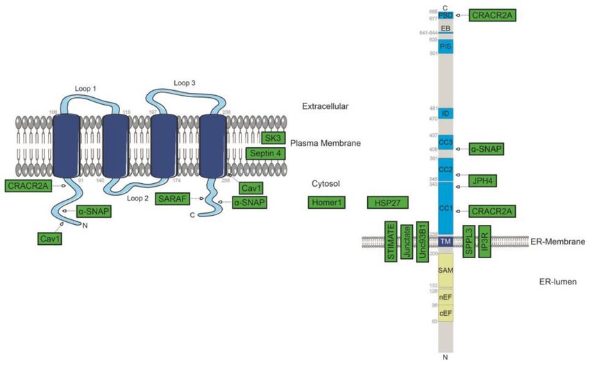
Int. J. Mol. Sci. 2021, 22, 471                                                                                             2 of 29

                                  globally or spatially restricted elevations in the free intracellular Ca2+ level depend on the
                                  entry of ions from the cells’ external pool upon the opening of Ca2+ ion channels within
                                  the plasma membrane (PM) or on the release from internal stores, primarily from the
                                  endoplasmic reticulum (ER). A mechanism that liaises both processes is store-operated
                                  Ca2+ entry (SOCE), interconnecting a drop in the level of Ca2+ within the ER-reservoir to
                                  the stimulation of PM-embedded channels. This mechanism is triggered by the engagement
                                  of cell surface receptors with their corresponding ligands and the subsequent generation
                                  of soluble inositol triphosphate (IP3 ) by enzymatic cleavage of phosphatidylinositol 4,5-
                                  bisphosphate (PIP2 ), a minor phospholipid of the cellular plasma membrane. After capture
                                  of the intracellular messenger by IP3 receptors that span the ER membrane and their
                                  subsequent activation, the exit of Ca2+ from the ER lumen is triggered [3–5]. The resulting
                                  increase in cytosolic Ca2+ is rather limited yet sufficient to trigger Ca2+ influx from the
                                  extracellular space. Importantly, this store-operated entry holds a dual role by allowing
                                  (i) for the induction of diverse Ca2+ -dependent cellular events and (ii) the re-establishment
                                  of cellular rest, as it provides a source of ions for the replenishment of internal stores [3].
                                        Among store-operated channels, the calcium-release-activated calcium (CRAC) chan-
                                  nel, initially described in cells of the immune system in the late 1980s and early 1990s, is the
                                  best characterized. The CRAC channel is distinguished by an extraordinary selectivity for
                                  Ca2+ , a low single-channel conductance of tens of femto-Siemens (fS) and, on the molecular
                                  level, by the indispensability of two proteins residing in spatially separate phospholipid
                                  bilayers: Stromal Interaction Molecule 1 (STIM1) within the ER-membrane and Orai1 in
                                  the plasma membrane. For a recent review, see Lewis et al., 2020 [6].
                                        CRAC Activation Cascade| The activation of CRAC channels follows a diffusion-trap
                                  mechanism. Thereby, a drop in ER-luminal Ca2+ levels is sensed by the N-terminal domain
                                  of STIM1. The STIM1 luminal portion contains a canonical EF-hand domain, a proto-
                                  typical Ca2+ binding structure that sequesters Ca2+ by means of electrostatic interactions
                                  with acidic residues that are responsible for capturing Ca2+ upon rest. Given constant
                                  equilibration between protein-bound and free fractions of the ion, store-depletion forces
                                  dissociation of Ca2+ from resting-state dimeric STIM1 [7–9]. This sets a series of conforma-
                                  tional changes in motion, accompanied by alterations in the folding state and interaction
                                  of luminal STIM1 domains and the transmembrane segment, as well as of the cytosolic
                                  protein domains. Once activated by ER store-depletion, STIM1 oligomerizes and translo-
                                  cates to sites within the ER membrane that are close to the PM, separated by distances
                                  of about 10–25 nm only [10,11]. While folded towards the ER-membrane upon rest, the
                                  activation of STIM1 includes an extension of its C-terminus vital to allow for interactions
                                  with cytosolic domains of pore-forming Orai1 proteins. This direct coupling between
                                  STIM1 C-terminal CRAC-activating domain/STIM-Orai-activating region (CAD/SOAR)
                                  domains and Orai1 culminate in opening of the CRAC channel [12–15]. Based on X-ray
                                  crystallographic and cryo-electron microscopic data on homologous Orai proteins of the
                                  fruit fly Drosophila melanogaster showing CRAC channels to be formed of six subunits, the
                                  human Orai1 protein is also likely to form hexameric complexes to constitute an active
                                  CRAC channel [16–18]. Apart from hexameric assemblies forming CRAC channels, Orai1
                                  proteins function as subunits in other channels as well. There, they either function in a
                                  store-operated and STIM-regulated manner if associated with members of the canonical
                                  type of transient receptor potential proteins (TRPC) or, upon forming pentameric assem-
                                  blies with the Orai3 isoform, give rise to arachidonate-regulated Ca2+ (ARC) channels. The
                                  latter are functionally detached from internal Ca2+ stores and modulated by a fraction
                                  of STIM1 proteins resident in the plasma membrane rather than the ER [19]. Although
                                  the series of events that culminate in CRAC channel opening is rather well established,
                                  inconclusiveness still must be clarified considering stoichiometric relations of STIM1 and
                                  Orai1, conformational transitions within the channel complex leading to the establishment
                                  of the conductive state, as well as molecular events of fast and slow Ca2+ -dependent in-
                                  activation. Moreover, a broad spectrum of proteins is believed to support the function of
                                  this signalling cascade. In particular, in a physiological context and endogenous levels of
Int. J. Mol. Sci. 2021, 22, 471 3 of 29
protein expression, the literature indicates that CRAC channel function relies on a reservoir
of positive and negative modulators for authentic CRAC currents to arise. Exemplifying
the presumptive multitude of regulatory proteins associated with the CRAC channel in a
direct manner, data of Várnai et al. indicated that Orai1 channels form a macromolecular
complex protruding 11-14 nm into the cell interior [20,21]. Analogously, HeLa cells stably
transfected with STIM1 and Orai1 led to the detection of Orai1 in extended complexes
upon rest (700 kDa), while STIM1 seems to engage lesser interactions in the quiescent state
(~200 kDa) but is captured in a complex with also Orai1 of 670 kDa upon store depletion—
an observed phenomenon that points to the presence of auxiliary partners within this
signalling cascade as well [21]. Interaction partners are eventually directly involved in any
step of the activation cascade or serve to establish signalling hubs critical for downstream
responses. Furthermore, proteinaceous modulators of SOCE functioning in an indirect
manner have been reported, for instance, by creating a distinct lipid microenvironment at
ER-PM junctions [22]. Given that interacting proteins and indirect regulators hold vital
roles in CRAC channel function but are often left in disregard in the rather STIM1/Orai1-
centered research field, the following review focuses on a compacted recapitulation of so
far published modulators of STIM1 and/or Orai1.
2. Regulators of CRAC Channel Function
2.1. Protein Trafficking and Dynamics
Ca2+ current amplitude depends on the absolute amount of channel proteins present
in the membrane. Consistently, modulation of protein expression and targeting to the
respective membrane delineates a primordial regulatory layer of CRAC channel function.
In this regard, Orai1 proteins are dynamically internalized from the plasma membrane,
whereby the proportion of Orai1 proteins present on the cell surface under resting state
and physiological conditions was reported to approximate 40%, while coupling of STIM1
interferes with internalization. In concert with the exocytotic machinery, this shifts the
equilibrium towards a preferential plasma membrane residence (~65%) [23–25]. The
importance thereof is further highlighted given that defective channel trafficking has been
shown to lead to serious clinical phenotypes [26–30]. For instance, patients suffering
from atopic dermatitis have recently been identified to show an increase in membrane-
resident Orai1, leading to a mismatch in the STIM1-Orai1 stoichiometry that phenotypically
culminates in the inhibition of Ca2+ entry and gene expression [31]. Proteins that are
believed to regulate trafficking and dynamics of STIM1 and Orai1 are discussed in the
following and are further summarized in Table 1. Apart from proteins involved in cell
surface expression of Orai1, endocytosis, and turnover, ER-sculpturing proteins and these
involved in the establishment/maintenance of ER-PM junctions are included as well as
others regulated by STIM1/Orai1 or those involved in downstream responses, as will be
treated in upcoming sections.
Table 1. Regulatory proteins within the calcium-release-activated calcium (CRAC) signalling cascade.
Interaction-Site within
Protein Function References
STIM1/Orai
CCT Orai1157–167 Promotes Orai1 endocytosis Hodeify et al., 2018 [23]
Involved in the increase in cell surface
SNAP-25 not reported Woodard et al., 2008 [25]
expression of Orai1 upon activation
Cross et al., 2013 [32]
Plasma membrane expression of Orai1,
Smaardijk et al., 2017 [33]
SPCA2 Orai1 STIM1-independent activation,
Peretti et al., 2019 [34]
regulation of SOCE (SPCA2C)
Feng et al., 2020 [35,36]Int. J. Mol. Sci. 2021, 22, 471 4 of 29
Table 1. Cont.
Interaction-Site within
Protein Function References
STIM1/Orai
Endocytosis of Orai1 in the proximal
Amnionless Orai1 Zeng et al., 2017 [37]
tubulus; critical for albumin re-uptake
Surveillance of Orai1; prevents
RHBDL2 Orai1 inappropriate activation and regulates Grieve et al., 2020 [38]
STIM1-Orai1 stoichiometry
Calnexin STIM1 Plasma membrane expression Saitoh et al., 2011 [39]
Microtubule + end tracking, Pozo-Guisado et al., 2013 [40]
EB1 STIM1642–645
ER-remodelling, STIM1 motion upon rest Sampieri et al., 2009 [20]
Asanov et al., 2013 [41]
APC STIM1650–685 STIM1 clustering at ER-PM junctions
Munemitsu et al., 1994 [42]
Promotes Orai1 ubiquitinylation,
UBQLN1 Orai1 correlates with a downregulation in Lee et al., 2013 [43]
CRAC channel activity
RTN4 not identified ER tubulation Jozsef et al., 2014 [44]
Hirata et al., 2006 [45]
JPH1/2 not identified ER-PM junctions
Woo et al., 2012 [46]
Stabilization of STIM1-Orai1 complex
Homer 1a STIM1 under conditions of rising intracellular Jardin et al., 2012 [47]
Ca2+ levels
Regulates interferon signalling
TM203 STIM1 Shambharkar, et al., 2015 [48]
(STING-pathway)
Phosphorylation of STIM1, suppression
CDK1 STIM1 Smyth et al., 2009 [49]
of SOCE during mitosis
Suppresses PMCA in its activity upon
PMCA STIM1 CRAC channel activation, contributes to Ritchie et al., 2012 [50]
the establishment of Ca2+ microdomains
Inhibits opening of CaV1.2 and leads to
VGCC STIM1 withdrawal of CaV1.2 from the plasma Park et al., 2010 [51]
membrane
negative modulator of energy-consuming
POST STIM1/Orai1 Krapivinsky et al. (2011) [52]
transmembrane Ca2+ transport
Formation of a signalling complex that
AKAP79 Orai1 sequesters downstream acting proteins Kar et al., 2014 [53]
close to Orai1-formed pores
Increases in ADCY8 activity upon SOCE
ADCY8 Orai1 N-terminus Willoughby et al., 2012 [54]
at cellular subdomains
Chaperonin-containing T-complex protein 1| Recycling of Orai1 proteins under
resting-state conditions is dependent on Rho proteins, as well as Rab5 and chaperonin-
containing T-complex protein 1 (CCT, also referred to as TRiC for T-complex protein 1 Ring
Complex). Thereby, CCT is considered as the main regulator of Orai1 endocytosis in a man-
ner relying on direct interactions of the concerning protein with a CCT-binding site within
the intracellular loop of Orai1 subunits (amino acids 157–167). Explicitly, this interaction
seems to promote internalization, given that inhibited CCT-binding was reported to foster
cell surface expression upon rest, imposing consequences on SOCE activation, duration, as
well as on downstream responses [23,30].
SNAP-25| In regard to protein targeting, the plasma membrane-associated SNARE
protein synaptosome associated protein 25 (SNAP-25) was shown to be intricate for
activation-related and Ca2+ -dependent plasma membrane accumulation of Orai1 [25].Int. J. Mol. Sci. 2021, 22, 471 5 of 29
SPCA2| Apart from proteins mastering generic reshuffling of Orai1, several mech-
anisms of Orai1 internalization or plasma membrane enrichment seem to diverge in a
cell-type-specific manner. In that context, the secretory pathway Ca2+ -ATPase SPCA2 was
reported to interact with Orai1 to accomplish its forward trafficking in mammary epithelial
tissues [32]. Physiologically, the interplay between SPCA2 and Orai1 is important for lacta-
tion. However, instead of constituting an actual regulator of SOCE, the interaction with
SPCA2 allows for constitutive channel activation irrespective of STIM1, thus accomplishing
store independent Ca2+ entry (SICE) instead. However, SPCA2 shall be highlighted herein
for its clinical relevance. In this regard, Orai1 and SPCA2 were reported to be expressed at
aberrant levels in some breast cancer tissues with overexpression patterns correlating with
cancer aggressiveness [32–35].
Findings of Fenech et al. suggest that SPC2C, a C-terminally shortened version of
SPCA2 expressed in cells of pancreatic origin, affects store-independent, as well as store-
dependent, Ca2+ influx. Although co-immunoprecipitation analysis indicated interactions
between Orai1 and SPCA2C that correlated with the patterns of STIM1 expression, its
modulation of SOCE was intriguingly reported to be independent of this association and to
occur in an Orai1-independent manner [36]. In addition, recent experimental data revealed
STIM1-independent activation of Orai1 by interactions with the ubiquitously expressed
SPCA1a isoform. This association apparently owns clinical relevance as well, considering
that impairments thereof relate to Hailey–Hailey disease, a disorder of the skin [55].
Contrasting SPCA2-dependent incorporation into the plasma membrane, CRAC chan-
nels are specifically withdrawn from the cell surface in the course of meiosis to hinder
store-operated currents from activating. This is accomplished by means of an endocytotic
pathway orchestrated by caveolin (Cav), dynamin, and Rab5 and is dependent on a con-
sensus caveolin-binding locus (amino acids 52–60) present in the intracellular N-terminus
of Orai1 [23,56]. Apart from meiosis, specific internalization of Orai1 is also observable in
epithelial cells of the proximal tubulus. Recent data indicate that the protein amnionless, a
critical element of receptor-mediated endocytosis, associates with STIM1-complexed Orai1
after store depletion, allowing for clathrin-mediated, yet caveolin-independent, uptake in
a manner correlating with the Ca2+ -dependent re-uptake of albumin from primary urine.
Consequently, downregulation of this process due to changes in the expression level of
Orai1 and elevations in the level of steady-state internalization were implicated to underlie
progressive losses of normal kidney function in diabetic patients [37].
RHBDL2| Recently, Grieve et al. identified Orai1 as a substrate for the rhomboid-like
protein 2 (RHBDL2), a membrane protein that belongs to the family of serine proteases.
The authors propose that RHBDL2 regulates Ca2+ signalling by optimizing the stoichiom-
etry between Orai1 and STIM1. In this study, the protein was supposed to prevent an
inappropriate activation of Orai1 in resting cells, based on a mechanism of conformational
surveillance. According to the study, a loss of RHBDL2 led to elevated levels of Orai1,
causing a disbalance in Ca2+ homeostasis [38].
Calnexin, Exportin1, and Transportin1| The presence of STIM1 within the ER-membrane
is vital for its function as Ca2+ sensor and SOCE activation. However, a fraction thereof is
expressed within the plasma membrane under steady-state conditions, hypothesized to
comprise a regulatory effect on Ca2+ currents conducted by store-operated channels [57].
Apart from discussions on glycosylation patterns as a decisive factor for cell-surface or ER-
membrane expression, a study by Keil and co-workers on hippocampal neurons concerns
the ubiquitin-proteasome system exerting a regulatory function on the presence of STIM1
on the cell surface and SOCE. Indeed, exposure of cells to inhibitors of the proteasome
elevated plasma membrane expression of STIM1 as well as peak Ca2+ currents upon store
depletion to a significant extent. In contrast, overexpression of the E3 ligase POSH reduced
plasma membrane levels of STIM1 [58]. In addition, Saitoh et al. found that STIM1 vari-
ants missing their C-terminal coiled-coil domains are extensively targeted to the plasma
membrane, eventually being an indication for a critical role of the concerning segments
in ER retention. The same study identified glycosylation-independent binding betweenInt. J. Mol. Sci. 2021, 22, 471 6 of 29
calnexin and STIM1, in addition to associations between the latter and a pair of proteins
functioning in protein transport, exportin1 and transportin1 [39].
EB1 and +TIP| Other auxiliary proteins relevant to CRAC channel function affect the
dynamics of STIM1/Orai1 within the ER- or plasma membrane rather than upstream traf-
ficking. In this regard, STIM1 dynamics are highly dependent on microtubules, cytoskeletal
components that are inherently essential for cell viability. The microtubule plus-end track-
ing protein (+TIP) end-binding 1 (EB1) associates with the STIM1642-645 segment, the latter
of which belongs to the class of +TIPs as well [59]. In addition to tracking emerging micro-
tubules and allowing for continuous movement of STIM1 throughout the ER membrane
if stores are replete, EB1 is involved in ER remodelling. The interaction with STIM1 is
hindered upon activation, based on the phosphorylation of a triad of serine residues (575,
608, and 621) that are located in the vicinity of the EB1 association motif, as is accomplished
by extracellular signal-regulated kinase 1/2 (ERK1/2) [40]. Thereby, a study indicated
that the release of EB-1 is followed by binding of the microtubule stabilizer adenomatous
polyposis coli (APC). The latter, inherently also representing an interaction partner of EB-1
and a potent tumour suppressor with genetic alterations correlating with colon cancer, as-
sociates with a rather distal STIM1 domain, including the amino acids from position 650 to
685 [41,42]. Knock-down of APC was shown to shift foci of STIM1 clustering from ER-PM
contact sites towards regions buried deep within the cell interior with the consequence of
impeded coupling to and activation of Orai1. Moreover, experimental findings indicate
that the activation-related relay between interactions of both EB-1 and APC with STIM1 is
reverted upon deactivation by store-replenishment, implying the dissociation of APC to
facilitate re-binding of EB-1 [20,41].
Heat shock protein: HSP27| Heat shock protein 27 (HSP27) is a chaperone that
interacts with various proteins. Huang et al. showed that knock-down of HSP27 leads to a
reduction of Ca2+ influx via a decrease of STIM1. The authors propose that the reduction
of STIM1 might be due to a loss of stability of the proteins and not due to transcription.
Immunoprecipitation assays indicate that HSP27 interacts with STIM1, but no interaction
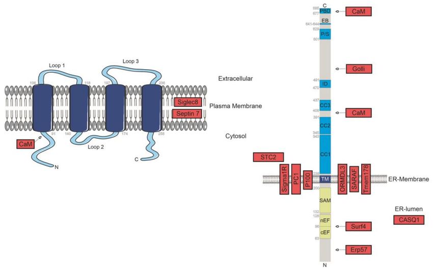
was observed for Orai1 (Figure 1) [60].
Figure 1. Identified positive regulators of SOCE. Known protein-localisation together with potential
identified interaction sites between STIM1/Orai1 and regulatory-interacting proteins are represented
in green boxes.
UBQLN1| Lee et al. discovered an interaction between Ubiquilin 1 (UBQLN1), a
protein that degrades presenilin and polyglutamine proteins, and Orai1. Ubiquilin 1 leads
to an increase of Orai1 ubiquitination and downregulation of channel activation. TheInt. J. Mol. Sci. 2021, 22, 471 7 of 29
authors suggest that Ubiquilin 1 downregulates intracellular Ca2+ mobilization by promot-
ing the ubiquitination and lysosomal degradation of Orai1. According to the study, the
UBL and UBA domains of Ubiquilin 1 seem to be non-relevant for the interaction with
Orai1 [43]. Moon et al. recently published a study indicating that upon ubiquitination,
Orai1 is degraded in a Crbn-dependent manner by the proteinase cereblon [61].
2.2. ER Sculpturing Proteins, ER-PM Tethers, and PM Microdomains
Reticulon 4| Proteins involved in the regulation of the CRAC signalling cascade yet
devoid of physical interactions with both STIM1 and Orai1 eventually possess a critical
role in the establishment of a proper ER morphology. Among them ranks the ubiquitously
expressed reticulon 4 (RTN4), retained in the ER on the basis of structural elements that map
to its C-terminal segment, namely the reticulon homology domain (RHD). This domain
comprises two hydrophobic stretches that are intercalated by a hydrophilic segment and
are thought to insert into the ER membrane as hairpins. Given that this integration
distorts the lipidic organization of the outer leaflet to a higher extent compared to the
inner layer, RTN proteins are, in addition to oligomerization in an arc-shaped fashion,
critical for the establishment/stabilization of membrane domains of high curvature, such
as ER tubules. Consistently, murine embryonic fibroblasts devoid of RTN4 expression
exhibited tubulation deficiencies in favour of an enrichment in the appearance of ER sheet
structures, while a normal ER function was nevertheless maintained in many respects.
However, exposure to thapsigargin (TG), an inhibitor of the sarco/endoplasmic reticulum
Ca2+ ATPase (SERCA), was associated with a lesser magnitude of Ca2+ mobilization, in
addition to a significant reduction in the consequent Ca2+ entry from the extracellular space.
This is mechanistically in line with an unavoidable shift in the presence of STIM1 from a
normally tubular distribution to sheets due to the overall changes in ER morphology. This
likely poses constraints on the diffusion-limited ability of STIM1 to reach ER–PM junctions
upon activation [44].
Rather than ensuring just close intermembrane contact to allow for physical STIM1–
Orai1 interactions, proteins contributing to the establishment and maintenance of ER-PM
junctions hold, eventually in conjunction with modulators of local lipidic compositions,
a multifarious regulatory potential on SOCE. While referring to a comprehensive review
of Cao et al. [62] or Zaman and colleagues [63] on this matter, a limited set of structural
components of ER-PM junctions shall be explicitly mentioned herein, including junctate
and junctophilins [45].
Junctate and Junctophilins| Knock-down of junctophilin 1 (JPH1) that bridges the
gap intercalated between transverse tubular invaginations of the plasma membrane of
skeletal muscle and the sarcoplasmic reticulum (SR), or analogous depletion of junctophilin
2 (JPH2), the latter of which is ubiquitously expressed in any muscle type, was reported to
interfere with SOCE and SR Ca2+ storage capacity even before the fundamental molecular
players of CRAC currents had been unravelled (Table 1) [45,46]. Later, JPH1 was reported to
assemble in puncta during store depletion, coinciding with these formed by STIM1, whereas
specific JPH2 mutants have been identified to lead to hypertrophic phenotypes of skeletal
myotubes in an Orai1-dependent manner [46,64]. Two further types of junctophillins,
JPH3 and JPH4, have also been identified, sharing their fundamental architecture with
the former variants, including the appearance of phospholipid-binding domains at the N-
terminal end and a single transmembrane domain defining the C-terminus to traverse the
phospholipid bilayer of the ER. However, these are mainly expressed in cells of the nervous
system, while junctophilin 4 was recently also identified to scaffold ER–PM junctions in T
lymphocytes. Nevertheless, JPH4 seems to be, at least in the latter cell type, dispensable
for the establishment and/or maintenance of ER–PM junctions, since deficiency in this
junctophilin was accompanied by neither numeric nor structural changes in the concerning
intermembrane contacts. Knock-down of JPH4 was shown to compromise resting-state
ER Ca2+ content and SOCE in primary T cells and Jurkat T lymphocytes, interfering with
effector functions. Consistent with findings that the latter are rescuable by overexpression ofInt. J. Mol. Sci. 2021, 22, 471 8 of 29
STIM1 together with observations that knock-down of the peculiar junctional component
truncates STIM1-clusters emerging successively to store depletion in size and number,
JPH4 was suggested to be vital for STIM1 relocation upon activation. This is eventually
dependent on direct couplings mapping to the first and second coiled-coil domain of STIM1
(CC1 and CC2) and JPH4, the latter of which seems to be constitutively clustered at ER–
PM junctions. Indeed, pronounced overexpression of the junctional protein is, according
to data of Woo et al. [65], capable of forcing STIM1 to accumulate at junctions even if
ER luminal Ca2+ reservoirs are replete, yet urging the sensor protein in a conformation
unable to activate CRAC channels (Figure 1). Moreover, STIM1 variants deprived of
their polybasic PIP2-binding domain, necessary to trap the sensor at ER-PM junctions by
means of electrostatic interactions, were shown to be capable of protein-interaction guided
clustering at ER–PM contact sites in the presence of junctophilin-4 or junctate. Junctate
represents an additional ER membrane-spanning protein and is a putative component of
ER-PM contact sites, also in cells devoid of endogenous Orai1 [65,66]. Taking all this into
account, it seems plausible that junctional proteins contributing to the recruitment of STIM1
are critical for SOCE under physiological conditions with expression levels of functional
Orai1 and STIM1 serving as limiting factors [66]. Direct coupling between junctate and
STIM1 is accomplished by ER-luminal sections of both, rather than transmembrane and/or
cytosolic segments (Figure 1) [65,66].
Interestingly, not only STIM1, but also the 33 kDa measuring junctate, originally
described in electrically excitable cells and later also in components of the immune system,
is equipped with an EF-hand domain, located at the C-terminus of the protein, able to sense
ER-luminal Ca2+ . In this regard, initial reports of Srikanth and colleagues linking junctate
to CRAC channel activation revealed that mutation of Ca2+ binding domains in junctate
leads to cluster formation of STIM1 under store-replete conditions [66]. Contrasting data on
JPH4 overexpression, store-independently clustered STIM1 proteins were indeed reported
to be able to induce gating of Orai1 channels [65,66]. In addition, junctate, depletion
of which was shown to significantly compromise store-operated Ca2+ influx in Jurkat T
lymphocytes, seems to comprise a moderate affinity for Orai1 in complex with STIM1,
with interactions being promoted successively to ER Ca2+ store emptying [66]. Apart from
direct associations with STIM1 and/or Orai1, the discussed junctional constituents seem to
engage in a complex to synergistically promote SOCE whereby the cytosolic domains of
both proteins are apparently critical [65,66]. Such an interplay with other junctional and/or
plasma-membrane-embedded proteins is essential for junctate to reside at ER–PM contact
sites since it shows a rather uniform distribution throughout the ER membrane upon sole
expression [66]. Apart from its role in bridging ER and PM, junctate is relevant for the
establishment of ER–phagosome contacts. Spatial increases in intracellular Ca2+ levels that
are achieved by the STIM1-guided opening of Ca2+ channels of the phagosomal membrane
boost phagocytic efficacy. In this regard, junctate was shown to be able to recover Ca2+
transients and phagocytosis in cells with knock-out of STIM1 or STIM2, respectively [67].
Caveolin-1| The clinically highly relevant Caveolin-1 (Cav1), already mentioned in
a previous Section 2.1 in the context of meiotic cell-surface clearance of Orai1, serves, as
indicated in Figure 1, as a positive regulator of SOCE in a series of cell types, given that
SOCE-specific currents were repeatedly shown to be attenuated upon treatment with Cav1-
specific siRNA (Figure 1) [68,69]. However, the molecular-scale function of Cav1 in SOCE
is controversial in some aspects. On the one hand, a recent study by Bóhorquez-Hernández
et al. [70] identified SOCE activation going along with an association of Orai1 and Cav1.
Here, the latter protein that inserts into the phospholipid bilayer in the form of a hairpin
was proposed to comprise an affinity to a segment of the fourth transmembrane helix of
Orai1, as well as the cytosolic N-terminus, explicitly residues intercalated between the
positions 250-258 and those from position 52 to 60, respectively [30,70,71]. Thereby, Cav1
expression might be vital to allow for further important regulatory mechanisms to come
into play, namely those imposed by lipids acting upon the accumulation of Orai1 within
dynamically assembled subregions of the membrane that are defined by peculiar lipidicInt. J. Mol. Sci. 2021, 22, 471 9 of 29
compositions [71,72]. This is explained by Cav1 constituting a major scaffolding element
of specialized microdomains of the plasma membrane characterized by heightened levels
of cholesterol and sphingolipids that form invaginations, termed caveolae [68,69,73,74].
Consistently, overexpression of Cav1 was shown to correlate with elevations in store-
operated currents in a manner paralleled by alterations in caveolae count in Hs578/T
breast cancer cells, while knock-down lowered the number of these microstructures and
silenced SOCE activity to a considerable extent [69]. In addition to its effect on channel
function itself, Cav1 plays a direct role in the induction of downstream responses. It serves
as an interaction platform for a series of signal-transducing proteins to allow, in concert with
the specific lipidic microenvironment, for the establishment of signalling hubs [71,72]. (For
review, see Pani et al., 2009 [73].) Thereby, structurally divergent domains of the scaffolding
protein have recently been suggested to be vital for the ability of entrant Ca2+ to allow for
the activation of specific transcription factors [70]. Contradictory to these functions, knock-
down of Cav1 in smooth muscle cells of the respiratory system was reported to vanquish
SOCE in a manner related to decreases in whole-cell and calveolar Orai1 expression.
Conversely, overexpression of Cav1 in the same cell type culminated in elevated levels of
Orai1 to be synthesized, in turn promoting Ca2+ entry upon store depletion. The same
report by Sathish and co-workers hypothesized that the regulatory potential of Cav1 on
SOCE was independent of STIM1, given that exposure to Cav1-targeted siRNA left STIM1
levels unaffected, while overexpression of the Ca2+ sensor increased SOCE in a manner only
partially reversible upon knocking down Cav1 [74]. However, it should be emphasized that
a potential effect of Cav1 on CRAC channel expression was not reproducible in other cell
types, such as HEK293, wherein neither STIM1 nor Orai1 levels diverged in the presence
or absence of Cav1, as found by another group. The promotion of SOCE by Cav1 was
alternatively proposed to be due to an enhanced stability of STIM1-Orai1 complexes after
store depletion [70].
Septins| Several subtypes of septin proteins serve, as initially revealed in RNAi-
based efforts of Sharma et al., as regulators of SOCE in a manner mechanistically related
to the initially stated function of Cav1. Following store depletion, septins dynamically
reposition from a widespread distribution throughout the plasma membrane to form
local accumulations in a manner temporally correlated with STIM1 redistribution and
co-clustering of both fundamental elements of the CRAC channel complex. Notably, the
foci enriched in septins are spatially separated from STIM1/Orai1 clusters and in relation
to the specific septin subform, impose different functional consequences on SOCE yet
accomplished in an indirect manner [75,76]. For a detailed review of the role of septins
in SOCE, see Deb et al., 2016 [77]. The septins SEPT2, SEPT4 and SEPT5, collectively
constituting filament-forming GTPases, were, according to analysis in Jurkat T lymphocytes
and HeLa cells, implicated as positive modulators of SOCE (Figure 1). This is mastered by
a septin-dependent enrichment of PIP2 and eventually PIP3 at sites of close ER-PM contact
that promotes, based on electrostatic interactions between the polybasic domain of STIM1
and the charged phospholipid head groups, arrest of extended-state STIM1 and in turn,
STIM1-dependent recruitment of Orai1 to ensure efficient Ca2+ influx [75]. This is consistent
with findings of other groups, including reports that depletion of SEPT4/5 correlates with
a downturn in the density of STIM1 clusters [78]. Apart from the reshuffling of PIP2
during activation, septins regulate the distribution of membrane lipids in the resting state,
accounting for observations that the ablation of specific forms correlates with alterations in
Orai1 distribution upon rest [75].
Moreover, Katz and co-workers proposed that SEPT4/5 indirectly modulate the num-
ber of ER–PM contact sites and slow the diffusion of Orai1 detaching from STIM1. Here,
TG-treatment was reported to locally confine motions of Orai1 that were in parts negoti-
ated by knock-down of SEPT4/5 and restorable by SEPT4 expression, whereas diffusion
characteristics of STIM1 upon rest or after store depletion were insensitive to changes
in SEPT4/5 expression. The lower mobility in the vicinity of ER-PM junctions would in
turn foster rebinding of CRAC channels based on elevations in their local concentration,Int. J. Mol. Sci. 2021, 22, 471 10 of 29
whereas initial recruitment of Orai1 to the junctions was stated to be exclusively accom-
plished in a STIM1-dependent manner but irrespective of septins [78]. Apart from the
former septin proteins, a modulatory role of SEPT7 on Ca2+ entry has been established
yet is divergent in several respects [76,79]. On the one hand, SEPT7 was shown to down-
regulate store-operated currents if overexpressed, whereas on the other, lessening of its
levels allows for store-independent Ca2+ entry in Drosophila neurons and elevations in
basal cytosolic Ca2+ levels (Figure 2). This is putatively explained by alterations in the
structure of residual septin filaments that serve as mimetic of another store-dependent
remodelling that is indispensable for PIP2-reorganizations [76]. Furthermore, a recent
study identified the human SEPT7 as a critical regulator of Orai1-driven Ca2+ entry into
neuronal progenitor cells and differentiated neurons. Although the underlying mechanism
remains to be elucidated, it seems to deviate from the one declared for the homolog of
fruit-fly, given that the polybasic segment of the protein was shown to be indispensable for
human SEPT7 to exert its regulatory function rather than the GTPase domain as relevant in
the case of the former [79].
Figure 2. Identified negative regulators of store-operated Ca2+ entry (SOCE). Known protein-localisation together with
potential identified interaction sites between STIM1/Orai1 and regulatory-interacting proteins are represented in red boxes.
2.3. Positive Modulators of SOCE
Apart from the previously treated proteins that regulate CRAC channel activity in a
rather indirect manner, for instance by modulating cell surface expression of Orai1, protein
turnover or membrane structuration, others directly assist in CRAC channel activation
at different stages of the multilayered activation cascade. These positive modulators, for
which a graphical summary that further includes indirect regulators is provided in Figure 1,
are discussed in the following section.
α-SNAP| By knock-down analysis in Drosophila Kc cells, Jurkat T lymphocytes,
HEK293 cells or U2OS cells, Miao and co-workers revealed a thereto unseen, direct role of
soluble N-ethylmaleimide sensitive fusion protein (NSF) attachment protein (SNAP) of
fruit-fly and the ubiquitously present mammalian α-SNAP in the onset of store-operated
Ca2+ influx that was in the case of the latter protein restorable by overexpressing the
closely related β-SNAP, expression of which is physiologically limited to the brain. Despite
the inherent function of SNAP in vesicle fusion, depletion of α-SNAP renders plasma-
membrane expression and ER-membrane localization of Orai1 and STIM1 untouched.
Instead, knock-down of α-SNAP was identified to lead to increases in the dimensionsInt. J. Mol. Sci. 2021, 22, 471 11 of 29
of STIM1-Orai1 clusters with normal STIM1 densities but unproportional elevations in
Orai1 count. Consistently, overexpression of the modulator was reported to augment Ca2+
influx in correlation with higher per-cluster densities of STIM1. Thereby, the regulatory
potential of α-SNAP seems to rely on direct protein–protein interactions, involving CAD of
STIM1, as well as both cytosolic tails of Orai1, yet in view of co-immunoprecipitation and
pull-down efforts, comparatively weak in the case of the N-terminus (Figure 1). Despite a
certain level of constitutive association, the proportion of α-SNAP bound to STIM1 elevates
in correlation with the emptying of ER Ca2+ reservoirs, whereas lacking, in the absence
of STIM1, co-localization with Orai1 irrespective of the filing state of the ER. Nonetheless,
under conditions of co-expression of STIM1 and Orai1, α-SNAP was observed to prefer-
entially co-localize with clusters containing both STIM1 and Orai1. Taken together, these
data seem to, with regard to α-SNAP, have functional significance for the establishment of
appropriate STIM1/Orai1 ratios co-agglomerating upon store depletion [80].
However, in a subsequent study by the same group, α-SNAP was proposed to function
in the association of dimeric Orai1 into multimeric, Ca2+ -selective channels. Thereby, Li
et al. reported that Orai1 formed dimers in mouse embryonic fibroblasts under resting cell
conditions, while dynamically engaging assemblies of dimeric, tetrameric or hexameric
stoichiometry following store depletion. Ablation of α-SNAP was therein found to lead
to a shift towards hexameric Orai1 associations and, concomitantly, to a downturn in the
proportion of dimeric and tetrameric complexes. The former assemblies were reported to
be capable of conducting sodium currents to considerable extents in a regime insensitive
to increases in the level of STIM1 interaction [81]. Indeed, further elucidation of the role
of α-SNAP in STIM1/Orai1 function recently demonstrated shortcomings in CD4 T cell
signal transduction due to reduced α-SNAP expression mechanistically related to sodium
currents conducted by Orai1 rather than SOCE [82]. Moreover, concatemers of Orai1 with
a pair of SOAR segments appended to the C-terminus, otherwise conducting constitutive
Ca2+ currents, were observed to significantly lose Ca2+ influx in favour of sodium entry in
cells hampered in α-SNAP expression. Seemingly contradicting this is that if expressed
as soluble fragments, CAD/SOAR was reported to be competent of re-establishing Ca2+
influx also in cells with compromised α-SNAP expression. In any case, STIM1–STIM1
interactions were, in the study of Li et al., devoid of any regulatory potential imposed
by α-SNAP, and similar to the preliminary report, depletion of the modulator was per
se found to allow for STIM1-Orai1 co-association, lacking apparent influences on STIM1
coupling to the C-terminal section of Orai1. Instead, cells knocked-down in α-SNAP
expression were significantly compromised in interactions between STIM1 and the N-
terminal Orai1 domain, hypothesized to account for the observed higher mobility of Orai1
in the region of ER–PM junctions. These data might indicate that α-SNAP is required
for conformational transitions of the Orai1 N-terminus indispensable for efficient CRAC
channel activation [81].
CRACR2A| A regulatory protein that has affinity to STIM1 and Orai1 is CRAC regu-
lator 2A (CRACR2A also referred to as EFCAB4B or FLJ33805) originally discovered by
Srikanth et al. According to bioinformatic analyses, CRACR2A, which predominantly
locates to the cytosol, is equipped with a pair of EF-hand domains in the N-terminal region
and contains, in addition to a leucine-rich element, a coiled-coil domain in its C-terminal
section. The homolog CRAC2B that was identified at the same time shows conserved
residue identities within the putative EF-hand motif and the C-terminal coiled-coil domain
and shares a sequence identity of 36% with human CRACR2A. The interactions among
CRAC2A and STIM1 or Orai1 involve the coiled-coil domain, as well as the proline/lysine
rich segment of the former, and considering Orai1, map to the N-terminal domain with K85
and K87 serving as critical interaction sites (Figure 1) [21]. It is worth mentioning that the
sites of CRACR2A-STIM1 and CRACR2A-Orai1 interaction overlap with these described
to bind calmodulin (CaM) in the presence of Ca2+ , explicitly including hOrai168-91 and
the lysin-rich domain of hSTIM1 [21,83]. CRAC2A associations with both basic compo-
nents of the CRAC channel complex are associated with a stringent dependence on theInt. J. Mol. Sci. 2021, 22, 471 12 of 29
activation state, as these are promoted successively to store depletion yet exclusively under
Ca2+ -free conditions, while otherwise strong binding between the mentioned domains
of STIM1/Orai1 and CRACR2A was reported to vanquish if exposed to buffer solutions
supplemented with 2 mM Ca2+ [21]. In view of its EF-hand domain, CRACR2A was
supposed to serve as a sensor of cytosolic Ca2+ levels, dissociating from the CRAC channel
complex if concentrations of the ion are elevated. Consistently, an EF-hand mutant devoid
of Ca2+ binding was curtailed from Ca2+ sensitive binding as well. Supplementing in vitro
analysis, total internal reflection fluorescence (TIRF) microscopic analysis of fluorescently
labelled STIM1 proteins or co-transfection of STIM1-mCherry and Orai1-GFP showed that
store-depletion dependent clustering of STIM1 at ER-PM junctions is significantly reduced
in CRACR2A-silenced Jurkat T cells compared to controls. Similarly, Orai1 clustering is
strictly dependent on the presence of CRACR2A, while hindered by its depletion. Con-
versely, expression of the Ca2+ -insensitive CRACR2A form, together with STIM1-YFP, led
to variable clustering already under resting state conditions, while resulting in extended
clusters after store depletion, and was in the longer term shown to considerably elevate the
proportion of Jurkat T cells undergoing apoptotic cell death compared to these express-
ing solely endogenous CRACR2A or counterparts transfected with wildtype CRACR2A.
Further, expression of CRACR2A in HeLa cells stably expressing STIM1 and Orai1 was
associated with a significant increase in the maximum of SOCE, while EF-hand mutants not
only led to elevations in peak [Ca2+ ]int , but also in persistency. Altogether, CRACR2A likely
stabilizes the association of Orai1 and STIM1 upon store depletion as long as intracellular
Ca2+ concentrations are low. However, further data are necessary to decipher whether
the cytosolic EF-hand protein is actively involved in the accumulation of STIM1 and/or
Orai1 at ER-PM junctions or recruited to already clustered proteins in a subsequent step.
Although CRACR2A was reported to be more potent in enhancing Orai1-dependent Ca2+
influx, CRAC2B seems to show a functional redundancy with the former [19,21]. However,
both proteins seem to differ in view of tissue-specific expression levels, with CRACR2A
constituting the prevailing form in thymus and spleen, as well as the Jurkat T cell line,
while CRAC2B is in greater abundance in HEK293 cells. Consistently, knock-down of the
expression of the 2B homolog showed solely mild effects on SOCE in Jurkat T cells com-
pared to CRACR2A knock-down, while outweighing the impacts on store-dependent Ca2+
entry in HEK293 cells [21]. In addition to the aforementioned cytosolic form of CRACR2A
(CRACR2A-c), a longer variant, CRACR2A-a, has been identified. However, this isoform
that is present in vesicles is equipped with a Rab GTPase domain at the C-terminal end
unseen in the shorter CRAC2A protein, accounting for functional differences and shall thus
remain neglected herein [84]. For review, see Srikanth et al., 2017 [85].
STIMATE| Through genome-wide RNAi screens to identify modulators of cellular
Ca2+ signalling, Quintana et al. found a protein in the ER membrane encoded by the
TMEM110 gene (Figure 1), which regulates the long-term maintenance and short-term
remodeling of ER–PM junctions [86]. This protein, later named STIMATE (STIM-activating
enhancer), acts as a positive regulator of SOCE in vertebrates and works in concert with
other proteins in the dynamic remodelling of the junctions during signalling (Figure 1).
STIMATE has been shown to directly interact with STIM1 to promote STIM1 conformational
switch, and depletion of STIMATE reduces STIM1 puncta formation and suppresses Ca2+
dependent NFAT signalling [87,88].
Homer| An additional protein that has a positive regulatory effect on SOCE is Homer
1a, as initially described in a study of Jardin-Polo et al. on platelet function (Table 1) [47].
In general, Homer proteins, encoded by three different genes whereof each gives rise to a
set of transcripts, serve as scaffolds, capturing proteins intermingled in Ca2+ -dependent
signal transduction and/or regulate these in an isoform-dependent fashion [89]. Homer
family members are collectively defined by an Ena/VASP homology 1 (EVH1) region
within the N-terminal domain, allowing for the binding to proline-rich segments (PPXXFR)
of interaction partners, including IP3R and TRPC proteins [90,91]. Among the proteins
derived from the Homer 1 gene, Homer 1a is smallest with the EVH1 domain of HomerInt. J. Mol. Sci. 2021, 22, 471 13 of 29
1a being only extended by a short C-terminal segment. Instead, elongated C-termini
of alternative gene products comprise a coiled-coil domain that allows for concomitant
interactions with equivalent domains of other Homer proteins and capture of targets to
be regulated. The shorter, only monomeric variant was in turn implicated as a negative
regulator of signalling lattices scaffolded by coiled-coil domain-containing forms, since it
recognized the same sequence motif within target proteins but lacked interactions with
other Homer proteins [89]. Consistently, the C-terminally extended Homer 1c was found
to regulate store-operated Ca2+ channels with different consequences on function [92,93].
In the aforementioned attempt of Jardin and colleagues, the Homer 1a isoform was
shown to interact with STIM1 upon store depletion and in a Ca2+ dependent manner, given
that chelation of cytosolic Ca2+ impeded this complexation. Although Orai1 proteins also
co-immunoprecipitated under the same conditions, the group suggested this apparent
association with Homer 1 to be indirect and accomplished by concomitant binding of
STIM1 (Figure 1). In conjunction with functional data revealing that interference with
Homer 1a binding lacked impacts on the activation of Ca2+ influx and initial current
characteristics, the hypothesis was raised that early steps of STIM1-Orai1 coupling occur
independently of Homer 1a. Instead, Ca2+ -promoted association of the specific regulator
was proposed to subsequently ensure that the complex formed by STIM1 and Orai1 is
maintained also under conditions of rising intracellular Ca2+ levels, given that ablation
of Homer 1a association correlated with considerable impairments in Ca2+ entry [47].
The regulatory role of Homer 1 on SOCE has recently been implicated as a promising
pharmacological tool for the treatment of conditions related to damages of the blood
vessel wall, as in the case of diabetic, atherosclerotic, and hypertensive conditions [94].
This is explained by Ca2+ -dependent dedifferentiation, proliferation, and relocation of
vascular smooth muscle cells into the lumen of blood vessels, accounting for the associated
pathological phenotypes. Orai1, STIM1, and several members of the TRPC family were
shown to be expressed at higher levels upon injury, whereas Homer 1 expression was
altogether just detected in injured cells [94–96]. In this regard, Jia et al. reported that any
of these components involved in store-operated Ca2+ entry interact in a direct manner
upon injury, including binding of Homer 1 to Orai1 and multiple TRPC-type proteins.
Thereby, knock-down of Homer 1 was shown to lead to a similar effect on SOCE in cultured
rat aortic vascular smooth muscle cells, manipulated to model injured counterparts, as
treatment with siRNA targeting STIM1, while concomitant ablation of the scaffolding
protein and the latter revealed an additive inhibitory effect on Ca2+ entry [94]. Apart from
pathogenic alterations of vasculature, Homer 1a function in SOCE seems to be relevant
for injured neurons as well in a manner related to STIM1 and TRPC channels rather
than Orai1. There, modulators of SOCE have been suggested as potential targets for the
treatment of Parkinson's disease (PD). Treatment of an in vitro model of PD neurons with
STIM1-targeted siRNA or pharmacological antagonists of SOCE increased cell viability
and lessened the rate of apoptotic cell death, either of which correlated with elevations
in the Homer 1a mRNA levels compared to untreated controls. Intriguingly, the positive
effects of SOCE inhibition on cell viability were in parts reverted upon ablating Homer 1a
expression [97]. Apparently contrasting with the latter, a study on a non-specific inhibitor
of SOCE (SKF-96365) is related to a significant downturn in Homer 1 expression levels
upon inhibitor application, whereby protective effects of SOCE inhibition on the same PD
model system were posed to relate to a regulatory role of Homer in ER Ca2+ levels [90].
This seeming contradiction in terms of Homer 1 expression might be explained by the
former study explicitly investigating the Homer 1a variant, while a closer specification of
the protein variant is lacking in the latter effort.
SPPL3| The signal peptide peptidase-like 3, an ER-localized intramembrane aspartyl
protease, is a member of the signal peptide peptidase (SPP) family, which is known for its
activity as a sheddase [98,99]. The latter includes proteases that are specifically responsible
for the withdrawal of ectodomains from membrane proteins, a post-translational process
that is vital for an appropriate function of many membrane standing proteins and mod-Int. J. Mol. Sci. 2021, 22, 471 14 of 29
ulation of their expression levels [100]. A study by Makowski et al. proposes SPPL3 as a
positive modulator of the SOCE pathway, as NFAT activation was apparently promoted in
the presence of SPPL3 (Figure 1). However, this seems to be accomplished by SSPL3 serving
as promoter of STIM1-Orai1 associations rather than related to its proteolytic function.
Intriguingly, the protein was even found to be indispensable for the activation of Orai1 by
constitutively active STIM1 mutants. These functions of SPPL3 were reported to rely on
its association with STIM1 at the transmembrane domain and, eventually, the cytosolic
CAD, ameliorating interactions between the latter and Orai1. STIM1 as well as cytosolic
CAD fragments of STIM1 were affected by SPPL3 knock-down and immunoprecipitation
studies showed associations of SPPL3 with STIM1 [99].
SK3| A growing number of experimental data enforce that ion channels composed
of Orai1 engage signalling complexes with other types of ion channels, allowing for a
reciprocal regulation in terms of activity [101–104]. A study by Guéguinou et al. identi-
fied a complex formation in lipid rafts between the small conductance calcium-activated
potassium channel 3 (SK3), also known as KCa2.3, with Orai1 as well as TRPC1 initi-
ated by activated STIM1 (Figure 1). The authors proposed the existence of a positive
feedback loop in which SOCE activates the Akt pathway as well as SK3 channel activity,
which leads to SOCE amplification [102]. This association of SK3 and Orai1 was identified
to be highly relevant for the migratory abilities of cancer cells [105]. Activation of the
cAMP-PKA cascade was shown to lessen the activities of sole and Orai1-complexed SK3,
hindering Ca2+ influx and in turn, mobility of cancer cells [101]. Apart from SK3, the
small-conductance Ca2+ -dependent potassium channel SK4, also referred to as KCa3.1,
relies on store-depletion-guided Ca2+ influx via Orai1 channels to gate open, as does the
large-conductance Ca2+ -activated potassium channel BKCa, in turn shifting membrane
potential towards hyperpolarized values. This ensures the continuation of Ca2+ influx
and replenishment of the ER stores by maintaining a high driving force of Ca2+ influx and
eventually regulates myocyte contraction [103,104].
Unc93B1| Unc93B1, located in the ER membrane of several cell types, is composed of
597 amino acids and shows 12 transmembrane domains. Upon cell stimulation, Unc93B1
can re-localize into endosomes or lysosomes and plays an important role in innate and
adaptive immunity by regulating Toll-like receptor (TLR) signalling [106,107]. Several
studies identified a loss-of-function mutation on position 412 (H412R), which leads to dis-
ruption of the interaction between Unc and Toll-like receptors TLR3, -7, and -9 in dendritic
cells but also stops antigen-cross presentation independently of TLR help [108]. Next to the
regulation of TLR-signalling, Maschalidi et al., as well as Beutler et al., identified Unc93B1
as a possible interaction partner of STIM1 (Figure 1). They postulated Unc93B1 as a further
regulator of SOCE and Ca2+ homeostasis, STIM1 Ca2+ signalling activity together with
Unc93B1 function being essential for MHCI antigen cross-presentation [109,110]. Reduced
interaction between STIM1 and Unc93B1 H412R mutants was observed in this regard, as
well as a potential wildtype Unc93B1 STIM1 EF-hand domain interaction. Maschalidi
et al. assume that Unc93B1 acts as chaperone, which helps STIM1 in the early stages of the
oligomerization process. Next to the help in oligomerization processes, Unc93B1 could play
an important role in the transfer of STIM1 to ER/PM junctions, activating Orai1 proteins,
as was shown by TIRF-microscopy experiments. The linkage between Unc93B1 and STIM1
together with Orai1, leading to Ca2+ influx from outside the cells and hence resulting in
phagosomal Ca2+ hotspots, which are needed for phagocytosis, phagosomal maturation,
and antigen-cross presentation, is suggested to be a potential target for further investigation
but is not known in detail [109].
2.4. Negative Modulators of SOCE
In addition to direct binding partners of STIM1/Orai1 fostering the activation or
maintenance of the active state of CRAC channels, a set of proteins has been identified
that is vital for the prevention of spontaneous activation, as well as for the cessation of
Ca2+ influx by aiding channel de-or inactivation, respectively. The next paragraphs reviewYou can also read


