Prospects for NK Cell Therapy of Sarcoma - MDPI
←
→
Page content transcription
If your browser does not render page correctly, please read the page content below
cancers
Review
Prospects for NK Cell Therapy of Sarcoma
Mieszko Lachota 1 , Marianna Vincenti 2 , Magdalena Winiarska 3 , Kjetil Boye 4 ,
Radosław Zagożdżon 5, * and Karl-Johan Malmberg 2,6, *
 1    Department of Clinical Immunology, Doctoral School, Medical University of Warsaw, 02-006 Warsaw,
      Poland; mieszko.lachota@wum.edu.pl
 2    Department of Cancer Immunology, Institute for Cancer Research, Oslo University Hospital, 0310 Oslo,
      Norway; marianna.vincenti@rr-research.no
 3    Department of Immunology, Medical University of Warsaw, 02-097 Warsaw, Poland;
      mwiniarska@wum.edu.pl
 4    Department of Oncology, Oslo University Hospital, 0310 Oslo, Norway; kjetil.boye@rr-research.no
 5    Department of Clinical Immunology, Medical University of Warsaw, 02-006 Warsaw, Poland
 6    Center for Infectious Medicine, Department of Medicine Huddinge, Karolinska Institutet,
      Karolinska University Hospital, 141 86 Stockholm, Sweden
 *    Correspondence: radoslaw.zagozdzon@wum.edu.pl (R.Z.); k.j.malmberg@medisin.uio.no (K.-J.M.)

 Received: 15 November 2020; Accepted: 9 December 2020; Published: 11 December 2020

 Simple Summary: Sarcomas are a group of aggressive tumors originating from mesenchymal tissues.
 Patients with advanced disease have poor prognosis due to the ineffectiveness of current treatment
 protocols. A subset of lymphocytes called natural killer (NK) cells is capable of effective surveillance
 and clearance of sarcomas, constituting a promising tool for immunotherapeutic treatment. However,
 sarcomas can cause impairment in NK cell function, associated with enhanced tumor growth
 and dissemination. In this review, we discuss the molecular mechanisms of sarcoma-mediated
 suppression of NK cells and their implications for the design of novel NK cell-based immunotherapies
 against sarcoma.

 Abstract: Natural killer (NK) cells are innate lymphoid cells with potent antitumor activity. One of
 the most NK cell cytotoxicity-sensitive tumor types is sarcoma, an aggressive mesenchyme-derived
 neoplasm. While a combination of radical surgery and radio- and chemotherapy can successfully
 control local disease, patients with advanced sarcomas remain refractory to current treatment regimens,
 calling for novel therapeutic strategies. There is accumulating evidence for NK cell-mediated
 immunosurveillance of sarcoma cells during all stages of the disease, highlighting the potential
 of using NK cells as a therapeutic tool. However, sarcomas display multiple immunoevasion
 mechanisms that can suppress NK cell function leading to an uncontrolled tumor outgrowth. Here,
 we review the current evidence for NK cells’ role in immune surveillance of sarcoma during disease
 initiation, promotion, progression, and metastasis, as well as the molecular mechanisms behind
 sarcoma-mediated NK cell suppression. Further, we apply this basic understanding of NK–sarcoma
 crosstalk in order to identify and summarize the most promising candidates for NK cell-based
 sarcoma immunotherapy.

 Keywords: Natural Killer (NK) cells; immunotherapy; sarcoma; cancer; chimeric antigen receptor
 (CAR); adoptive cell therapy; tumor microenvironment (TME); cell-mediated cytotoxicity; solid tumors

1. Introduction
     Natural killer (NK) cells are the first-discovered members of the innate lymphoid cell (ILC)
family, providing defense against tumors and pathogen-infected cells [1,2]. They express a remarkably

Cancers 2020, 12, 3719; doi:10.3390/cancers12123719                              www.mdpi.com/journal/cancers
Cancers 2020, 12, 3719                                                                                      2 of 31

diverse repertoire of inhibitory and activating surface receptors, regulating their responses [3].
NK cell-activating receptors recognize either stress-induced ligands, virus-encoded proteins,
or Ig-coated cells. In contrast, inhibitory receptors contribute to self/non-self-discrimination by
recognizing polymorphic major histocompatibility complex (MHC) class I ligands, also known as
human leukocyte antigen (HLA) (Table 1) [3–5].

              Table 1. A brief summary of natural killer (NK) cell activating and inhibitory receptors.

              Receptor              Known Ligands        Molecular Structure                Function
                                                                                  Stimulatory (short cytoplasmic
   Killer Immunoglobulin-like                              Immunoglobulin
                                    HLA-A, Bw, C, G                                   tail) or inhibitory (long
          receptors (KIR)                                    Superfamily
                                                                                          cytoplasmic tail)
                                                           Immunoglobulin
          CD16 (FcγRIII)            Fc portion of IgG                                      Stimulatory
                                                             Superfamily
                                                           Immunoglobulin
       CD2 receptor family
                                                             Superfamily
            2B4 (CD244)                  CD48                                              Stimulatory
                                    PVR (CD155) and
        DNAM-1 (CD226)                                                                     Stimulatory
                                    Nectin-2 (CD112)
            NTB-A                     Homophilic                                           Stimulatory
        CS1 (CRACC)                   Homophilic                                           Stimulatory
      NKG2 receptor family                                   C-type lectins
           NKG2D                    MICA/B, ULBPs                                          Stimulatory
        CD94/NKG2A                     HLA-E                                                Inhibitory
        CD94/NKG2C                     HLA-E                                               Stimulatory
        CD94/NKG2E                     HLA-E                                               Stimulatory
      Natural Cytotoxicity                                 Immunoglobulin
       Receptors (NCRs)                                      Superfamily
          NKp30a/b                    B7-H6, BAG6                                          Stimulatory
           NKp30c                     B7-H6, BAG6                                           Inhibitory
            NKp44                        PCNA                                              Stimulatory
            NKp46                       Vimentin                                           Stimulatory
            NKp80                         AICL                                             Stimulatory

     Different families of NK cell-activating receptors include NK Group 2 (NKG2) receptors,
natural cytotoxicity receptors (NCRs), DNAM-1, 2B4, and CD16 (FcRyIIIa). NKG2D is an activating
receptor belonging to the NKG2 family, recognizing MHC class I-related chain A/B (MICA/B) and
members of the UL-16 binding protein (ULBP) family [6,7]. The NCRs include NKp30, NKp44, NKp46,
and NKp80, which bind to B7-H6, AICL, or viral hemagglutinins [8–10]. DNAM-1 recognizes the viral
receptors PVR (CD155) and Nectin-2 (CD112). 2B4 binds other SLAM family proteins, whereas CD16 is
an Fc receptor for IgG, responsible for mediating antibody-dependent cell cytotoxicity (ADCC) against
antibody-opsonized cells [11–13]. The primary NK cell inhibitory receptors are the NKG2A and the
long-tailed killer cell immunoglobulin-like receptors (KIRs), which both bind to MHC class I molecules,
preventing NK-mediated lysis of cells with normal MHC expression [14]. Inhibitory KIRs are specific
for different MHC isotypes [14]. Acquisition of self-MHC class I binding KIRs during cell differentiation
tunes NK cells’ cytotoxic potential in a process termed education. Repeated interactions of inhibitory
KIRs with self-MHC class I molecules allow NK cells to acquire superior cytotoxic properties as well as
tolerance to self-MHC expressing cells [15–17]. The dynamic functional tuning of human NK cells
during NK cell differentiation and education and the implications for NK cell therapy are discussed in
detail in [18].
     As their name implies, NK cells can kill transformed or infected cells without the need for
earlier priming. Their cytotoxicity is executed either by degranulation, where the directional release
of perforin and granzymes induces apoptosis predominantly in a caspase-3-dependent manner,
or through death receptor ligands of the tumor necrosis factor (TNF) family, such as TNF, TNF-related
apoptosis-inducing ligand (TRAIL), and Fas ligand (FasL), acting primarily through caspase-8 [19,20].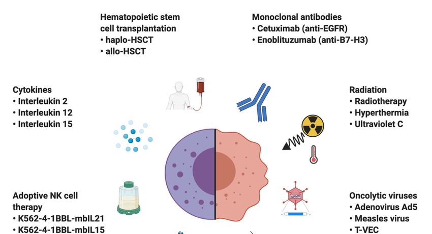
Cancers 2020, 12, 3719                                                                            3 of 31

Additionally, NK cells rapidly produce chemokines and cytokines upon activation, including interferon
(IFN)-γ, granulocyte-macrophage colony-stimulating factor (GM-CSF), interleukin (IL)-10, CCL3,
CCL4, CCL5, and CXCL8 that recruit and affect the function of hematopoietic and nonhematopoietic
cells in the tumor microenvironment (TME) [21].
      A growing amount of evidence suggests that the proper functioning of NK cells plays a
significant role in immune surveillance of cancer, prompting researchers to utilize NK cells in
cancer treatment [22,23]. Since NK cells do not express rearranged antigen receptors, they can be easily
transferred across MHC barriers without causing graft-versus-host disease (GvHD). Lack of MHC
restriction and their unique ability of cancer cell recognition through interactions of multiple surface
molecules impedes cancer immune evasion by MHC downregulation or a single antigen loss [24].
Because of these potent antitumor properties, intense studies are currently being carried out to use
NK cells, induced pluripotent stem cell (iPSC)-derived NK cells, and the NK cell line NK-92 as novel
therapeutic tools against cancer [25–27]. However, NK cell therapy faces many challenges, such as
inadequate homing properties, hostile TME, or tumor immunoevasion [28].
      Notably, one of the most NK cell-sensitive cancer types are sarcomas, a heterogeneous group of
aggressive mesenchyme-derived tumors with poor prognosis [29–32]. Sarcomas can originate from
different tissues such as bone, cartilage, muscle, adipose tissue, or blood vessels. Sarcoma’s yearly
incidence is approximately 5 per 100,000, accounting for less than 1% of malignant solid tumors
in adults but more than 20% in children [33,34]. The mainstay of sarcoma treatment based on a
combination of surgery and radiotherapy (RT) is able to control localized tumors; however, ~40% of
the patients experience tumor relapse and distant metastases [29,35]. Unfortunately, current treatment
regimens are ineffective in increasing overall survival in metastatic sarcomas, ranging from 11 to
20 months, creating a demand for novel and effective therapies [35,36]. The urgent nature of the
demand is further underlined by the fact that irrespectively of the stage, some sarcoma subtypes
have very few lines of systemic therapy with a clinically meaningful effect [35]. Both experimental
and clinical data support the immune system’s involvement in sarcoma tumorigenesis. Spontaneous
regressions and efficient immunosurveillance are observed in sarcomas, suggesting the prime role of
the immune system in tumor development and prompting researchers to explore the potential use of
immunotherapies in sarcoma treatment [22,37–39].
      Sarcomas are the first type of cancer for which immunotherapy was effectively applied. William
B. Coley injected streptococcal organisms into the tumors, based on observations of tumor regressions
in patients with concomitant streptococcal infections in the last decade of the 19th century. More than
half of the inoperable sarcoma patients treated by Coley were reported to respond completely [40].
Unfortunately, due to poorly characterized preparation and unpredictable toxicities, “Coley0 s toxins”
never became clinically useful.
      Because of the unique relationship between NK cells and sarcomas, we set out to review the
intimate crosstalk between NK cells and sarcoma cells during tumor initiation, promotion, progression,
and metastasis. Further, we discuss the current knowledge regarding sarcoma immunoevasion and
NK cell functionality. Finally, we review NK cell-based therapeutic approaches in sarcoma treatment,
tested in both preclinical and clinical settings.

2. NK Cell Immune Surveillance during Distinct Phases of Sarcoma Development
     NK cell-mediated immunosurveillance is an important factor in cancer development, especially
in metastasis control. Since sarcomas have been identified as one of the most NK-sensitive solid
tumors, they are a well-suited model to study NK cell-mediated cancer surveillance and tumor
immunoediting [30]. Different combinations of NK cell-activating ligands such as MICA/B ULBP1/2/3/5,
CD155, and CD112 are known to be expressed on both primary sarcoma samples and cell lines,
allowing for NK cell cytotoxicity [41–45]. The primary pathway of killing sarcoma cells appears to be
granule-dependent, with FasL-Fas interactions playing a minor role, possibly due to acquired FasL
resistance [43,46,47]. However, the significance of the FasL-Fas pathway may be underestimated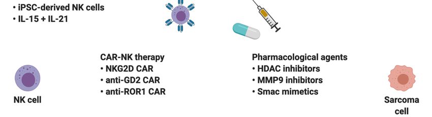
Cancers 2020, 12, 3719 4 of 31
because of technical limitations. A 4-h incubation time during a standard in vitro cytotoxicity assay is
insufficient to study death receptor-mediated apoptosis [48,49]. Indeed, NK cells were recently shown
to kill target cells by both mechanisms in a sequential manner, starting with granzyme B-dependent
killing and then
Cancers 2020,gradually
12, x transitioning to death-receptor killing during serial killing events 4 of 31[50].
A decrease in NK cell cytotoxicity in older adults is associated with an increased risk of cancer
granzyme
development [51].B-dependent killing and
Further, pediatric then gradually
osteosarcoma (OS)transitioning
patients have to death-receptor
a decreased numberkilling during
of circulating
serial killing events [50]. 0
NKs, together implicating NK cells potential role in controlling tumor initiation and progression [52].
A decrease in NK cell cytotoxicity in older adults is associated with an increased risk of cancer
Sarcomas have a scarce
development [51].immune
Further, infiltration compared (OS)
pediatric osteosarcoma to other solidhave
patients tumors [53]. Current
a decreased number evidence
of of
the prognostic roleNKs,
circulating of lymphocyte infiltration
together implicating NKin sarcomas
cells′ potentialis often
role incontradictory,
controlling tumor withinitiation
most of andthe studies
leaningprogression
towards the positive
[52]. Sarcomas effect
have of immune
a scarce effector
immune cell infiltration
infiltration compared toon disease
other solid prognosis
tumors [53]. [54–57].
Current evidence of the prognostic role of lymphocyte infiltration in sarcomas
NK cell abundance in the tumor infiltrate positively correlates with increased overall survival in is often contradictory,
several with most of the studies leaning towards the positive effect of immune effector cell infiltration on
sarcoma subtypes [58–60].
disease prognosis [54–57]. NK cell abundance in the tumor infiltrate positively correlates with
Additionally, early lymphocyte recovery after chemotherapy is associated with a better outcome
increased overall survival in several sarcoma subtypes [58–60].
in pediatricAdditionally,
OS [61,62].early Combining
lymphocyte surgery
recoveryandafterpolychemotherapy
chemotherapy is associated withwithsystemic IL-2 treatment
a better outcome
increasesinNK cell number
pediatric OS [61,62].andCombining
activity, with the magnitude
surgery of the increase
and polychemotherapy with correlating
systemic IL-2 with an improved
treatment
increases NK cell number and activity, with the magnitude of the
clinical outcome [63]. Finally, some studies have shown NK cells to be significant contributors increase correlating with an to the
improved clinical outcome
control of sarcoma metastases [64–68]. [63]. Finally, some studies have shown NK cells to be significant
contributors to the control of sarcoma metastases [64–68].
The aforementioned clinical data implicate the role of NK cells in controlling sarcomas0 growth.
The aforementioned clinical data implicate the role of NK cells in controlling sarcomas′ growth.
To summarize
To summarizethe available
the availableknowledge
knowledgeon onNKNK cell-mediated immunosurveillance
cell-mediated immunosurveillance of sarcomas,
of sarcomas, we we
divide the evidence into three sections, categorizing NK cell’s role in: initiation
divide the evidence into three sections, categorizing NK cell’s role in: initiation and promotion, and promotion,
progression, and metastases
progression, and metastases (Figure
(Figure1).1).
Figure Figure 1. Overviewofof the
1. Overview the central
centralrolerole
of natural killer (NK)
of natural cells(NK)
killer in all stages
cells of
in sarcoma evolution.
all stages of sarcoma
Through cell-mediated cytotoxicity, NK cells are able to inhibit tumor initiation, promotion,
evolution. Through cell-mediated cytotoxicity, NK cells are able to inhibit tumor initiation, promotion,
progression, and development of metastases. The key molecules necessary for NK cell-mediated
progression, and development of metastases. The key molecules necessary for NK cell-mediated tumor
tumor immunosurveillance are NKG2D, interferon (IFN)-γ, TNF-related apoptosis-inducing ligand
immunosurveillance are NKG2D,
(TRAIL), and perforin. Createdinterferon (IFN)-γ, TNF-related apoptosis-inducing ligand (TRAIL),
with BioRender.com.
and perforin. Created with BioRender.com.
2.1. Initiation and Promotion
2.1. Initiation and Promotion
Tumor initiation is the first step of cancer development during which, by rising mutational load,
Tumor initiation
healthy is the first
cells transform step of
into cancer cancer
cells. It candevelopment
be followed byduring which, bywhere
tumor promotion, rising mutational load,
transformed
cells undergo clonal proliferation and form a tumor. During these early
healthy cells transform into cancer cells. It can be followed by tumor promotion, where stages, it is up to the immune
transformed
system to eradicate the newly developed neoplastic cells before progression and dissemination occur.
cells undergo clonal proliferation and form a tumor. During these early stages, it is up to the immune
A common carcinogenesis model is based on methylcholanthrene (MCA), which induces
system to eradicate the newly developed neoplastic cells before progression and dissemination occur.
chemical mutagenesis and fibrosarcoma development upon inoculation. Smyth et al. evaluated
A common carcinogenesis
fibrosarcoma model
formation in mice is based
deficient on methylcholanthrene
in NK, Natural Killer T (NKT)(MCA), which
cells, or both induces
[69]. Both NK chemical
mutagenesis and
cells and NKTfibrosarcoma
cells seem to development upon
be essential for host inoculation.
protection againstSmyth et al. evaluated
MCA-induced sarcoma [69].fibrosarcoma
NK
formationprotective
in micefunction
deficientagainst sarcoma
in NK, couldKiller
Natural be enhanced
T (NKT) by cells,
IL-12 therapy
or both[69].
[69].Later,
BoththeNK group
cellshas
and NKT
cells seem to be essential for host protection against MCA-induced sarcoma [69]. NK protective functionCancers 2020, 12, 3719 5 of 31
against sarcoma could be enhanced by IL-12 therapy [69]. Later, the group has confirmed the crucial role
of NK cells in preventing the formation of MCA-induced sarcoma and studied the pathways responsible
for the recognition of transformed cells. Antibody-mediated neutralization of NK cell-activating
receptor NKG2D increased mice susceptibility to MCA-induced sarcoma formation. The importance
of the NKG2D was additionally underlined in IFN-γ−/− and TRAIL−/− mice, whereas mice depleted
of NK cells, T cells, or deficient for perforin did not display any NKG2D-dependent changes in
susceptibility. IL-12 therapy augmenting NK cell function and suppressing MCA-induced sarcoma
formation was also dependent on the NKG2D pathway. Although NKG2D ligand expression is
variable and often not detectable on sarcomas originating in wild type (WT) mice, sarcomas derived
from perforin-deficient mice were RAE-1+ and immunogenic when transferred into WT syngeneic
mice. These findings suggest an essential role of the NKG2D-perforin axis in control and shaping the
early events of tumor formation [70]. On the other hand, another NK cell receptor, NKp46, is not
associated with the surveillance of MCA-induced fibrosarcoma. However, tumors originating in
NKp46−/− mice implanted in WT mice induce a potent immune response suggesting a role of NKp46
in tumor immunoediting [71].
NRLP3 inflammasome, a cellular structure crucial for inducing and sustaining immune response,
promotes tumorigenesis in specific cancer types. The deletion of NRLP3 has a protective effect in
the MCA-induced fibrosarcoma model, dependent on NK cells and IFN-γ [72]. NRLP3 activation
was also shown to impede NK cell antimetastatic function by decreasing NK cell tumor homing [72].
The molecular mechanisms of pro-tumorigenic role of NRLP3 vary between cancer types and are
discussed in-depth in [73].
Carcinogenesis is also driven by oncogenic viruses such as HBV, HPV, EBV, and HHV-8 (KSHV).
The latter is known to induce Kaposi Sarcoma (KS), with acquired immunodeficiency syndrome (AIDS)
immunocompromised individuals being especially prone. Sirianni et al. showed that cells latently
infected with KSHV are efficiently lysed by NK cells from healthy individuals [74]. However, the study
yields specific limitations as the target cells were carefully selected based on their susceptibility to NK
cell cytotoxicity [74]. On the contrary, Matthews et al. reported average levels of classical MHC class I,
ICAM-1, HLA-E, and NKG2D ligands on latently infected primary fibroblasts, which caused a limited
activation of resting NK cells [75]. Interestingly, infected cells were efficiently cleared by IL-15-primed
NK cells [75].
A large study including over 1100 patients investigated the association between the HLA-KIR
polymorphism and KSHV and KS status, finding that, in patients with KIR3DS1 plus HLA-B Bw4-80I,
the KSHV seroprevalence was 40% lower, but the KS risk was two-fold higher. Similarly, the KSHV
seroprevalence was 40% lower, but the KS risk 80% higher with HLA-C group 1 homozygosity.
These data suggest that KIR-mediated NK cell activation may decrease KSHV infection’s risk but
enhance KS progression if infection occurs [76]. Peripheral blood (PB) NK cell counts, on the other
hand, do not correlate with the risk of KS development [77].
2.2. Progression
Cancer initiation and promotion can be followed by progression if not controlled by the immune
system. Progression is the last phase of localized tumor development, characterized by increased
growth speed and acquiring invasive potential.
Mice selectively depleted of NK1.1 positive cells demonstrated more rapid initial growth upon
injecting MCA207 sarcoma cells. In addition, large (20 mm) implanted MCA207 sarcomas were
rejected following cyclophosphamide and IL-12 treatment, but the time to tumor eradication was
significantly longer in mice with depleted NK cells [78]. Other groups confirmed that MCA-induced
sarcoma growth could be reduced by IL-12 treatment, with the effect being mediated by NK cells [69].
Takeda et al. provided evidence of NK cells playing a key role in limiting the L929 TRAIL-sensitive
fibrosarcoma progression in a subcutaneous murine model. The effect was mediated in a TRAIL andCancers 2020, 12, 3719 6 of 31
IFN-γ dependent fashion. The TRAIL pathway’s protective effect was dependent on NK cells and
IFN-γ, supported by gene knockout experiments [79].
One of the most critical NK cell functions is to enhance the infiltration of other immune cells into
the TME. In MCA-induced sarcomas, NK cells are known to infiltrate the developing tumors in early
stages, with the semi-mature CD27high NK cells being the predominant subpopulation of NK cells
accumulating in the TME. The tumor-infiltrating NK cells display an activated surface phenotype and
provide an early source of IFN-γ attracting other immune cells. Interestingly, host IFN-γ is critical
for NK cell tumor homing, and, conversely, the tumor-infiltrating NK cells mainly suppressed tumor
growth via the IFN-γ pathway. This implicates the importance of IFN-γ as a positive regulatory factor
for both NK cell recruitment into the TME and an effective NK antitumor immune response [80].
NK cell-derived IFN-γ can also improve cancer cell recognition and associated NK cell cytotoxicity
through ICAM-1 upregulation on cancer cells [81].
Moreover, IFN-γ plays a role in NK cell-mediated sarcoma immunoediting. Tumor cells isolated
from immunocompetent mice displayed reduced expression of NKG2D ligand H60 and increased
MHC class I expression compared with tumor cells isolated from mice treated with IFN-γ-specific
neutralizing monoclonal antibody (mAb) [82]. IFN-γ can also induce programmed cell death ligand 1
(PD-L1) expression on cancer cells, subsequently inhibiting NK cell effector function [83].
Another approach to understanding the drivers of MCA-induced sarcoma progression was
taken by O0 Sullivan et al. by comparing the gene expression between unedited and immunoedited
tumors [84]. One of the most differentially expressed genes was IL17D encoding interleukin 17D
(IL-17D), with a significantly increased expression in unedited tumor cells. Overexpression of IL-17D
in edited tumor cells induced tumor rejection by stimulating CCL2 production from tumor endothelial
cells, leading to an increase in the recruitment of NK cells. IL-17D-induced recruitment attracted
mostly CD27high NK cells, a semi-mature population of NK cells participating in IFN-γ-dependent
T cell priming and contributing to suppressing tumor progression [84]. These data suggest that NK
cells play a role in tumor immunoediting and suppressing sarcoma growth, both directly and indirectly
by regulating other immune cells’ activity and infiltration.
2.3. Metastases
The metastatic spread of neoplastic cells to distant anatomical regions is a leading cause of
death in cancer patients. Metastatic spread is orchestrated by the intrinsic properties of cancer cells,
enabling invasion of the local microenvironment and colonization of distant sites through lymphatic
or hematogenous spread. Moreover, metastasis is regulated by microenvironmental and systemic
processes, such as immunosurveillance.
NK cells are known for their antimetastatic potential [85–88]. Indicators of NK cell function such
as high expression of NK cell-activating receptors and high cytotoxic or IFN-γ secreting properties have
been linked to decreased metastatic load in multiple cohorts of cancer patients with risk of metastatic
disease, suggesting their clinically relevant protective role [87]. High numbers of tumor-infiltrating
NK cells have been inversely correlated with the presence of distant metastases in gastrointestinal
stromal tumors (GIST), a subtype of sarcomas [64]. Interestingly, the incompatibility of nude mice as
hosts for metastatic studies is attributed mainly to their NK cells, which efficiently remove circulating
tumor cells [65]. NK cell-protective role against metastases was also recognized in multiple murine
sarcoma models, where antibody- or cyclophosphamide-mediated NK cell depletion significantly
increased metastatic load [66–68]. Studying the interactions between MHC class I expression, NK cells,
and sarcoma metastases provided evidence for the correlation of RCT sarcoma metastatic potential
with increased MHC class I expression, which in turn correlated with cancer cell resistance to NK
cell lysis [67]. However, others did not observe any simple associations among MHC expression,
development of metastases, and NK cells [89].
TNF-α, a highly pro-inflammatory cytokine secreted by effector immune cells, is one of the
cytotoxic effector proteins capable of inducing cancer cell apoptosis. Surprisingly, in sarcomas, it wasCancers 2020, 12, 3719 7 of 31
shown to have an NK cell-dependent prometastatic effect, indicated by selective antibody depletion
experiments [86]. TNF-α can also exhibit prometastatic activity on its own through increased production
of chemokines inducing angiogenesis and enhancing cancer cell motility, which has been thoroughly
reviewed in previous publications [90,91]. Moreover, NRLP3 and IL-1R8 deficiencies were shown to
have an antimetastatic effect attributed to enhanced NK cell function [72,92].
NK cells can be successfully used in metastases treatment; K562-expanded NK cells effectively
eradicate Ewing sarcoma (EWS) metastases with little effect on the primary tumor in a murine
Cancers 2020, 12, x 7 of 31
model [93]. Furthermore, allogeneic hematopoietic stem cell transplantation (HSCT) was shown to
inhibit the development
NK cells can ofbe sarcoma metastases
successfully in an NK
used in metastases cell-dependent
treatment; manner
K562-expanded in clinical
NK cells trials [94,95].
effectively
eradicate Ewing sarcoma (EWS) metastases with little effect on the primary tumor in a murine model
3. NK Cell[93].
Dysfunction
Furthermore, in Sarcomas
allogeneic hematopoietic stem cell transplantation (HSCT) was shown to inhibit
the development of sarcoma metastases in an NK cell-dependent manner in clinical trials [94,95].
In the course of cancer microevolution, neoplastic cells undergo a series of metabolic adjustments
adapting 3.
theNKcells
Cell Dysfunction
to increased in Sarcomas
proliferation. Unfortunately, the shift in cancers metabolic state is
accompanied In bythe
creating
course aofhostile
cancer TME inhibitingneoplastic
microevolution, the anticancer immune
cells undergo response
a series and promoting
of metabolic
adjustments adapting the cells to increased proliferation. Unfortunately, the shift
homing of immunosuppressive cells such as regulatory T cells (Tregs) or M2 macrophages (Figure 2). in cancers metabolic
state is accompanied by creating a hostile TME inhibiting the anticancer immune response and
Already in 1981, Gerson et al. found that sarcomas substantially inhibit NK cell functions.
promoting homing of immunosuppressive cells such as regulatory T cells (Tregs) or M2 macrophages
NK proliferation
(Figure 2). in response
Already toGerson
in 1981, concanavalin
et al. foundA, macrophage
that migration
sarcomas substantially inhibitory
inhibit factor secretion,
NK cell functions.
and cell-mediated cytotoxicity
NK proliferation were
in response all suppressed
to concanavalin by macrophages
A, macrophage migrationinfiltrating the secretion,
inhibitory factor sarcoma, with the
andbeing
cytotoxicity cell-mediated
the mostcytotoxicity
preserved werefunction
all suppressed
[96].by macrophages infiltrating the sarcoma, with the
cytotoxicity being the most preserved function [96].
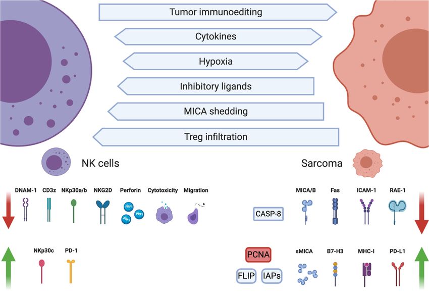
Figure 2. Interactions
Figure 2. Interactions between between
naturalnatural
killerkiller
(NK) (NK) cells,
cells, cancercells,
cancer cells,and
and tumor
tumor microenvironment
microenvironment (TME)
(TME) shape sarcomas’ immunoevasion mechanisms. Tumor immunoediting, cytokines, hypoxia,
shape sarcomas’ immunoevasion mechanisms. Tumor immunoediting, cytokines, hypoxia, and cells
and cells infiltrating the TME can change the sarcomas cell phenotype into an NK cell-resistant one,
infiltratingcharacterized
the TME can change the sarcomas cell phenotype into an NK cell-resistant one, characterized
by decreased expression of NK cell-activating ligands MHC class I-related chain A/B
by decreased expression
(MICA/B), retinoicofacid
NKearly
cell-activating ligandsintercellular
transcript 1 (RAE-1), MHC class I-related
adhesion chain1 A/B
molecule (MICA/B),
(ICAM-1), and retinoic
acid earlyproteins
transcript 1 (RAE-1),
necessary intercellular
for Fas ligand adhesion
(FasL)-mediated molecule
apoptosis 1 (ICAM-1),
(Fas, caspase-8). and expression
Conversely, proteins necessary
of inhibitory
for Fas ligand molecules such apoptosis
(FasL)-mediated as major histocompatibility
(Fas, caspase-8). complexConversely,
(MHC) class I,expression
programmed of cell inhibitory
death ligand 1 (PD-L1), B7-H3 (CD276), proliferating cell nuclear antigen (PCNA), and antiapoptotic
molecules such as major histocompatibility complex (MHC) class I, programmed cell death ligand 1
proteins cellular FLICE-inhibitory protein (c-FLIP), as well as inhibitors of apoptosis (IAPs), is
(PD-L1), B7-H3 (CD276),
increased. proliferating
NK cell phenotype cell nuclear
and function are alsoantigen
altered in(PCNA),
the sarcoma and
TME antiapoptotic proteins cellular
by cytokines, hypoxia,
FLICE-inhibitory protein
and inhibitory (c-FLIP),
ligands, resultingas
in well as inhibitors
a disturbed of apoptosis
balance between activating(IAPs), is increased.
and inhibitory receptor NK cell
phenotypeexpression and associated
and function are also cytotoxicity
altered impairment.
in the sarcoma CreatedTMEwith BioRender.com.
by cytokines, hypoxia, and inhibitory
ligands, resulting in a disturbed balance between activating and inhibitory receptor expression and
associated cytotoxicity impairment. Created with BioRender.com.Cancers 2020, 12, 3719 8 of 31
Cytotoxicity impairment of PB NK cells was reported in chemotherapy-naïve sarcoma patients,
in contrast to NK cells from renal cell carcinoma patients, which displayed normal cytolytic
activity [97,98]. The cytotoxic function of NK cells could be restored by IL-2 and Hsp-70-derived TKD
peptide. Additionally, NK cells in chemotherapy-naïve sarcoma patients had reduced proportions
of mature CD56dim population and slightly increased NKG2D expression compared to age-matched
controls. After disease progression or relapse, NK cell phenotypic alterations were more remarkable;
progressively reduced CD56dim proportions and decreased expression of NKG2D, CD3ζ, perforin,
together with reduced frequencies of differentiated CD57+ NK cells were all observed [98].
Suppression of the NK cell compartment increases at the tumor site. A significant decrease in
the NK cell proportions is observed in tumor-infiltrating lymphocytes (TILs) compared to matched
peripheral blood mononuclear cells (PBMCs). In contrast, no difference was observed between
tumor-infiltrating and PB CD3+ bulk T cells, CD4+ , and CD8+ T cells, indicating an impairment in
NK cell tumor homing or intratumoral persistence [99]. Profiling of TIL NK cells provided evidence
for decreased CD16, KIR2DL1, KIR2DL2/L3, and KIR3DL1 expression in the CD16+ KIR+ and CD16+
KIR− NK cell subsets, compared to NK cells in matched PBMCs. DNAM-1 and NKG2D expression on
TIL NK cells was also reduced in the vast majority of the patients compared to matched PBMCs [99].
Significantly, NKG2D and DNAM-1 downregulation might contribute to disease progression in these
patients as sarcoma cells are mostly recognized by NKG2D and DNAM-1 receptors [43,97]. In GIST,
the NKp30 receptor was preferentially downregulated on tumor-infiltrating NK cells. Interestingly,
PB NK cells in GIST patients expressed immunosuppressive NKp30c isoform more frequently with
proportionally less NKp30a and -b. The expression of NKp30c isoform was associated with an
unfavorable clinical outcome [64].
The co-culture experiments of NK and primary sarcoma cells provided further insights
into sarcoma-induced functional impairment. Sarcoma cells caused a decreased expression of
NKG2D, DNAM-1, and interfered with IL-15-induced expression of NKG2D, DNAM-1, and NKp30,
consecutively inhibiting the cytolytic activity of NK cells. The inhibition was contact-dependent,
and the cytotoxicity impairment was directly linked to the downregulation of the respective NK
cell-activating receptors. Five days of IL-15 pretreatment was able to increase NK cell resistance to
sarcoma suppression. In opposite to the above-mentioned changes in TIL NK cells, CD16 expression
and ADCC were not affected by the NK–sarcoma co-cultures [100].
Few reports show no differences in IFN-α signaling, NKG2D expression, and NK cytotoxic
properties between PB NK cells from sarcoma patients and healthy donors [43,101]. However,
the analyzed patient group was limited to freshly diagnosed patients with a most likely early-stage
disease, which indeed can be associated with mild or no functional impairment in PB NK cells [86,98].
3.1. Tumor-Infiltrating Immunosuppressive Cells
Other cells in the sarcoma microenvironment can contribute to creating a suppressive milieu.
Tumor stromal cells derived from fresh sarcoma samples display a potent antiproliferative effect
on PBMCs, decrease NK cell cytotoxicity and NKp44/46 expression, as demonstrated in co-culture
experiments [102]. Through rewiring chemokine and metabolic networks, sarcomas can induce
immunosuppressive CD4+ CD25+ Treg infiltration. Ghiringhelli et al. reported an inverse correlation
between NK cell activation and Treg abundance in GIST patients [103]. By expressing membrane-bound
transforming growth factor β (TGF-β), Tregs directly inhibit NK cell cytotoxicity, proliferation, and alter
NK cell phenotype by downregulating NKG2D receptor expression [103].
3.2. Cytokine-Dependent Inhibition
Cytokines present in the TME can also suppress NK cell function. Hafner et al. showed that TNF-α
suppresses NK cell cytotoxicity, consequently impairing NK cell antimetastatic function in murine
sarcoma model [86]. The molecular mechanism of TNF-α-mediated NK cell suppression has not been
fully elucidated, but new evidence indicates that TNF-α may contribute to NK cell exhaustion in aCancers 2020, 12, 3719 9 of 31
TIM-3-dependent manner [104]. The group has also provided insights into time-dependent changes in
NK cell cytotoxic function after sarcoma tumor inoculation. At first, the NK cell activity increases,
but after quickly reaching its peak it starts to decrease below the initial level [86]. On the other
hand, TNF-α was shown to increase human OS cells0 susceptibility to NK lysis by CD54 and CD58
upregulation, demonstrating a dual role of TNF-α in NK-sarcoma interactions [105,106]. Notably,
CD54 (ICAM-1) expression on cancer cells is essential for NK cell cytotoxicity, and its magnitude
directly correlates with OS susceptibility to NK cell lysis [106–108].
Interferons are proinflammatory peptides known for their antiviral properties. One of their
mechanisms of action is MHC class I upregulation, aiming to increase the presentation of viral peptides.
In murine MCA sarcoma, IFN-γ and IFN-α were shown to reduce expression of NKG2D ligand
H60. Downregulation occurred at the transcript level and was STAT1-dependent. IFN-γ-treated
MCA sarcomas with initially high levels of H60 were resistant to killing by IL-2-activated NK cells.
Resistance was not solely dependent on H60 downregulation but also on IFN-enhanced MHC class I
expression [82].
TGF-β is another vital player in sarcoma TME. It regulates the extracellular matrix (ECM) protein
composition and induces osteopontin synthesis in OS cells, increasing their malignant potential [109,110].
Additionally, Treg-derived TGF-β can inhibit NK cell effector functions such as cytotoxicity and tumor
homing [111–113]. Besides, Gao et al. provided evidence for TGF-β-induced transformation of NK cells
into intermediate type 1 innate lymphoid cells (intILC1) and ILC1 in the sarcoma microenvironment.
Importantly, intILC1s and ILC1s did not provide sufficient control of local tumor growth and metastasis,
whereas NK cells favored tumor immunosurveillance. ILC1-derived TNF-α was suggested to be
partially responsible for an escape from the innate immune system [114]. In soft tissue sarcoma patients,
high TGF-β1 intratumoral expression is associated with aggressive disease and shorter disease-specific
survival [115].
3.3. MHC-Dependent Inhibition
MHC class I molecules serve as ligands for inhibitory KIR and NKG2A receptors. Current evidence
shows that chemotherapy can increase classical and nonclassical MHC class I molecule expression
in OS cells, consequently inhibiting NK cell activity [43]. One of the critical NK-suppressive MHC
molecules is peptide-loaded HLA-E, which can be expressed in different tumor cell types, including
sarcomas. It is a potent inhibitor of NK cell activity, acting via NKG2A [116]. Moreover, it has been
shown that EWS treated with anti-GD2 chimeric antigen receptor (CAR)-NK cells developed resistance
to the treatment in an HLA-G-dependent manner, which was selectively upregulated on tumor cells
only in CAR-treated mice. NKG2A knockdown restored CAR-NK lytic function and allowed for
effective tumor eradication [117]. In OS patients, the MHC class I expression itself is associated with a
better prognosis, most likely due to T cell-mediated immune response [118].
Interestingly, the relationship between NK cell activation and MHC I expression appears to be
nonlinear. A moderate increase of MHC class I expression on EWS cells caused a highly NK-resistant
phenotype, whereas downregulation of MHC expression did not change the susceptibility, implicating
the existence of a threshold. That, in turn, would allow modest changes in the target cell surface
phenotype to significantly affect the susceptibility to NK cell-mediated lysis [119]. Not only the
surface expression but also the KIR-HLA mismatch degree between NK cells and OS determines their
susceptibility to NK cell lysis [120].
3.4. MICA Shedding
Shedding of NK cell-activating ligands can also contribute to sarcoma immunoevasion.
NKG2D ligand MICA is shed in a matrix metallopeptidase (MMP)-9-dependent manner in OS.
Soluble MICA (sMICA) was shown to cause NKG2D downregulation, impairing NK cell response [121].
High concentrations of sMICA were correlated with poor prognosis in multiple cancer types [122].
In sarcomas, sMICA concentration is increased in advanced disease, downmodulating NKG2DCancers 2020, 12, 3719 10 of 31
expression on NK cells. On the contrary, most of the early stage and well-differentiated sarcomas
were shown to express MICA on the cancer cell surface, indicating that MICA expression is lost
along the disease progression [41]. Therefore, MICA shedding might be an early event in sarcoma
immunoevasion, contributing to the disease progression [42,123]. Multiple studies have shown that
increased MICA and sMICA expression were associated with decreased NKG2D expression on NK cells
and correlated with advanced and metastatic disease [41,42]. Further, MMP-9 and MMP-2 expression
is associated with the presence of metastasis and poor survival in OS patients and could be potentially
used as a prognostic biomarker [124,125].
3.5. Apoptosis Resistance
Along with the tumor microevolution, neoplastic cells can acquire resistance to cell-mediated
cytotoxicity. Resistance to FasL-mediated apoptosis can be mediated through downregulated caspase-8
and increased expression of antiapoptotic proteins such as cellular FLICE-inhibitory protein (c-FLIP) or
Inhibitors of apoptosis (IAPs). Such mechanism has been demonstrated to play a role in immunoevasion
of rhabdomyosarcoma (RMS) and EWS cell lines, as well as primary EWS samples [46,47]. Notably,
FasL is constitutively expressed in the lung, implicating that Fas expressing cancer cells should be
eliminated by lung endothelium. However, metastatic OS acquire resistance to Fas-mediated death by
Fas downregulation, allowing for lung colonization [126]. Chemotherapeutic agents such as gemcitabine
upregulate Fas surface expression and may therefore be an important part of multimodal therapy
for OS lung metastases [127]. Cisplatin treatment can overcome FasL resistance by downregulating
c-FLIP-L, sensitizing OS cells to FasL mediated apoptosis [128]. Sensitization of OS cells to FasL can
also be induced by histone deacetylase inhibitor (HDACi) entinostat, which increases Fas transcription,
its localization in membrane lipid rafts and decreases the expression of antiapoptotic c-FLIP [129,130].
3.6. Immune Checkpoints
Immune checkpoints are molecules regulating the activity of immune system, which in
physiological setting play a protective role against autoimmunity and overactivation of lymphocytes.
Programmed cell death protein 1 (PD-1) is a well-known checkpoint molecule, functioning as a
“break” in the immune system [131]. Typically, only a small fraction of PB NK cells express PD-1,
but the proportion is increased on NK cells in cancer patients [132]. PB NK cells from KS patients
exhibit PD-1 expression in a CD56dim CD16+ population with otherwise normal surface phenotype.
However, despite the normal phenotype, PD-1+ NK cells demonstrated reduced cytotoxicity and
IFN-γ production ex vivo following the direct triggering of NKp30, NKp46, CD16, or short stimulation
with target cells, suggesting a role of PD-1 in KS-mediated NK cell exhaustion [133]. Moreover, PD-L1
expression in OS cell lines determined their susceptibility to NK cell lysis, shown by mAb blocking
experiments [134]. However, in OS patients, intratumoral PD-L1 expression positively correlated with
increased immune infiltration including NK cells and, surprisingly, event-free-survival [57].
B7 homolog 3 (B7-H3, CD276) is an immune checkpoint protein of the B7-CD28 family with a vital
role in T cell inhibition [135]. B7-H3 overexpression is observed in multiple cancers, including OS, RMS,
and EWS [135–138]. B7-H3 is expressed in 91.8% of OS tissues, where it promotes OS cell invasion
and is inversely correlated with TIL abundance [136]. High B7-H3 expression is also associated with
shorter survival and disease recurrence [136].
Interleukin-1 receptor 8 (IL-1R8) is a member of the IL-1 receptor family, acting as a negative
regulator of other IL-1 receptor and Toll-like receptor (TLR) signaling, that has been recently established
as a checkpoint molecule in NK cells. Knockout of IL-1R8 has been shown to restore NK cell
antitumor function in MCA-induced sarcomas, implicating a role of IL-1R8 in sarcoma-mediated NK
cell suppression [92]. Expression of other checkpoint molecules such as T-cell immunoglobulin and
mucin domain-3 (TIM-3) ligands and NKp44-inhibiting proliferating cell nuclear antigen (PCNA) was
also reported in sarcomas [45,139].Cancers 2020, 12, 3719 11 of 31
3.7. Altered Oxygen Metabolism
Rapid cell turnover and cancer growth are associated with decreased O2 gradient as tumors grow
beyond their vascular supply. Large murine sarcomas contain a severely hypoxic core, whereas smaller
tumors possess hypoxic gradients throughout the tumor mass. Evidence indicates that these hypoxic
gradients orchestrate sarcoma cell migration and ECM remodeling, increasing their metastatic potential.
Additionally, hypoxia-inducible factor 1-alpha (HIF-1α) increases CXCR4 expression on sarcoma cells,
contributing to metastasis development. Notably, in sarcoma patients, increased HIF-1α and CXCR4
expression are associated with advanced disease [140,141].
A hypoxic microenvironment is reported to alter the susceptibility of human OS cells to NK
cell-mediated lysis. Different OS cell lines expressed various NKG2D ligands such as MICA, MICB,
and ULBP1/2/3, with the MICA being most frequently expressed [44]. In a HIF-1α mediated way,
hypoxia decreased cell surface MICA expression without increasing the secretion of soluble MICA,
resulting in reduced susceptibility of the OS cells to the NK cell-mediated lysis [44]. Moreover,
by inducing HIF-1α, hypoxia impairs NK cell function by inhibiting their response to activating
cytokines as well as suppressing cell-mediated cytotoxicity capabilities, except for ADCC [142].
Significantly, in STS tumors, low oxygen content is associated with poor disease-specific and overall
survival [143]. Additionally, bone and soft-tissue sarcomas are characterized by increased oxidative
stress, which is known to inhibit NK cell effector functions [144,145].
3.8. HIV-KS-NK Cell Axis
NK cell-mediated immunity is significantly impaired in AIDS patients with progressing KS
compared to both HIV-negative patients with indolent classic KS and healthy blood donors. The highly
active antiretroviral therapy (HAART) is able to rescue impaired NK cell function in AIDS-KS patients,
inducing tumor regression and HHV8 clearance. However, AIDS-KS patients with more aggressive
disease and no response to therapy had persistent HHV8 viremia and reduced NK cell cytotoxicity.
These results suggest a crucial role of NK cells in the control of HHV8 infection and KS tumor, as well
as AIDS role in mediating NK cell suppression [74]. Additionally, NKG2C+ NKp46low NK cells were
discovered to form a novel, poorly functional subset present in AIDS-KS patients [146].
HIV-negative classical KS patients have significantly decreased NK cell cytotoxicity compared to
healthy controls, whereas healthy HHV8 carriers have phenotypically impaired NK cells with reduced
expression of NKp30, NKp46, and CD161 receptors [147]. Further, KS patients show downmodulation
of NKG2D, associated with impaired NK-cell lytic capacity, which could be restored upon KS
treatment [148]. Interestingly, KS cells exhibited high expression of NKG2D ligands confirmed in situ
by immunohistochemical (IHC) staining of KS biopsies. However, no tumor-infiltrating NK cells were
detected, suggesting a defect in NK cell homing or persistence in the KS microenvironment [148].
PGE2 was identified as a critical inhibitory mediator responsible for impairing NK cell response in KS,
acting by down-modulation of NKG2D expression on resting NK cells and impairing IL-15 induced
proliferation and phenotypic changes [148]. Other studies have demonstrated that PGE2 inhibits NK
cell migration properties and impairs their accumulation in TME, which could be the reason for the
lack of NK cells among the KS TILs [149,150]. Moreover, KSHV proteins K3 and K5 were shown to
drastically downregulate ICAM-1, MICA, MICB, and AICL (NKp80 ligand) expression on infected
cells, increasing their resistance to NK cells [151–153].
3.9. Iatrogenic NK Cell Suppression
NK cell functional impairment can be iatrogenic. Surgery was shown to transiently impair
NK cell cytotoxicity; however, the phenomenon’s significance is unclear [154]. While in other
cancers chemotherapy is reported to upregulate NK cell-activating ligands, a comparison of pre- and
post-chemotherapy OS tissue sections provided evidence for either unaltered or decreased expression
of MICA, CD112, and CD155 after chemotherapy [43]. Moreover, depending on the specific agentCancers 2020, 12, 3719 12 of 31
and the dose, chemotherapy can also directly suppress NK cell function [154–157]. Zoledronic acid
(ZA), tested as maintenance therapy in clinical trials in patients with bone sarcomas (OS and EWS),
acts by inhibiting bone resorption and inducing apoptosis in osteoclasts and tumor cells. Its effect on
NK cell activity is, however, unfavorable as ZA can impede in vitro NK cell expansion and cytolytic
responses to EWS, raising concerns against combining NK cell therapies with ZA in bone sarcoma
treatment [158].
4. NK Cell-Based Therapies in Sarcomas
Cancers 2020, 12, x 12 of 31
Numerous immunotherapeutic approaches have been tested in patients with sarcomas, and the
results have4. NKnot been asTherapies
Cell-Based impressive as in the treatment of other solid tumors. In theory, proper
in Sarcomas
management of immune checkpoint signaling
Numerous immunotherapeutic approaches by have
monoclonal
been testedantibodies
in patients withcansarcomas,
be one of and thethemodalities
to revive the functioning
results have not been of NK cells within
as impressive as in the tumor [159].
the treatment Immune
of other checkpoint
solid tumors. blockade
In theory, proper therapy
management
with antibodies of immune
blocking checkpoint
target signaling
cytotoxic by monoclonal antibodies canantigen
T lymphocyte-associated be one of the modalities and the
4 (CTLA-4)
to revive the functioning of NK cells within the tumor [159]. Immune checkpoint blockade therapy
PD-1/PD-L1 pathway leads to durable clinical responses in an increasing number of solid tumors.
with antibodies blocking target cytotoxic T lymphocyte-associated antigen 4 (CTLA-4) and the PD-
Unfortunately,
1/PD-L1responses
pathway leadsin patients
to durablewith sarcomas
clinical responses have
in an been observed
increasing number infrequently,
of solid tumors.except for
the TIL-rich undifferentiated
Unfortunately, responses pleomorphic sarcoma
in patients with sarcomas [160–162].
have been observedPreliminary
infrequently,clinical
except forevaluation
the of
TIL-rich undifferentiated pleomorphic sarcoma [160–162]. Preliminary
PD-1-PD-L1 axis inhibition in angiosarcoma and alveolar soft part sarcoma also shows promising clinical evaluation of PD-1-
PD-L1 axis inhibition in angiosarcoma and alveolar soft part sarcoma also shows promising results
results [163–165].
[163–165].
One of the Onereasons for the
of the reasons for inefficacy
the inefficacy ofofimmune checkpoint
immune checkpoint inhibition
inhibition therapytherapy
is the factisthat
the fact that
sarcomas sarcomas
have fewer have TILs
fewer per
TILsgram
per gramof of
tissue
tissueand lowerratios
and lower ratios of TIL
of TIL infiltration
infiltration when compared
when compared to to
other e.g.,
other cancers, cancers, e.g., melanoma
melanoma or renal
or renal cell
cell carcinoma [53].
carcinoma [53].However,
However, primary sarcomas
primary and sarcoma
sarcomas and sarcoma
cell lines were shown to be one of the most vulnerable tumor types to spontaneous NK cell
cell lines were shown to be one of the most vulnerable tumor types to spontaneous NK cell cytotoxicity,
cytotoxicity, making NK cell-based therapies novel and promising treatment alternative [30–32]. The
making NK NKcell-based therapies novelcan
cell-based immunotherapies andbe promising
divided into treatment alternative
two groups based on their[30–32].
principle The NK cell-based
of action:
immunotherapies can be divided
strategies augmenting into two
NK cell function andgroups based cancer
those sensitizing on theircells principle of action:
to NK cell-mediated lysis strategies
(Figure 3).
augmenting NK cell function and those sensitizing cancer cells to NK cell-mediated lysis (Figure 3).
Figure 3. Figure 3. A schematic illustration of selected prospective sarcoma treatment modalities based on:
A schematic illustration of selected prospective sarcoma treatment modalities based on:
modifying natural killer (NK) cell properties (left); or sensitizing sarcoma cells to NK cell cytotoxicity
modifying(right).
natural killer (NK) cell properties (left); or sensitizing sarcoma cells to NK cell cytotoxicity
Created with BioRender.com.
(right). Created with BioRender.com.Cancers 2020, 12, 3719 13 of 31
4.1. Hematopoietic Stem Cell Transplantation (HSCT)
HLA-mismatched HSCT combines the effects of chemotherapy and graft-versus-tumor (GvT)
phenomenon. HSCT with grafts from haploidentical donors was shown to be safe and beneficial
in pediatric solid tumors including sarcomas [166,167]. Already in 1984, a clinical trial in Moscow
demonstrated that allogeneic bone marrow transplant suppressed lung metastases development in a
group of OS patients after radical surgery. Importantly, all of the treated patients who did not develop
metastases had normal NK cytotoxicity levels, whereas, in the metastases group, the NK activity
was significantly lower, suggesting a critical role of NK cells in suppressing sarcoma metastases [94].
A clinical trial in pediatric cancer patients, including sarcomas, has shown that HSCT’s effects could
only be observed in patients with mismatched KIR-HLA [168]. However, the beneficial effect of
HLA-mismatched grafts was associated with a higher risk of toxicities [169]. A potential role for
NK cells in the graft-versus-tumor effect is supported by in vitro studies showing that the KIR-HLA
mismatch degree can predict OS cell lines susceptibility to NK cell lysis [120]. Another study
retrospectively investigated allo-HSCT in RMS patients and reported moderate results, prompting
further investigations and suggesting the potential use of allo-HSCT as a consolidation therapy [169].
A case report study has also shown haplo-HSCT to be effective in metastases control in two patients
with stage IV EWS [95]. Further clinical trials are being carried out to determine the clinical utility of
HSCT in high-risk sarcoma patients; however, there are currently no recommendations to use HSCT as
a sarcoma therapy irrespectively of the disease stage.
4.2. Cytokines
Because of their potent immunostimulatory properties, cytokines have always raised interest
as adjuvant therapies. The antitumor activity of the IFN-α-conjugated antibody against the OS cell
line was first reported in 1984 by Flannery et al. The treatment resulted in a modest increase in
NK cell cytotoxicity, attributed to IFN-α induced activation [170]. Multiple studies have shown the
efficacy of IFN-α, IL-2, IL-12, IL-15, IL-18, and IL-28 in augmenting NK cell functions by increasing
their cytotoxicity, rendering resistant to TME-mediated suppression, and altering sarcomas adhesion
molecule profile. Such an approach was proven useful through in vivo studies in multiple sarcoma
subtypes [43,78,97,171–179]. Because of synergistic effects between cytokines and chemotherapy,
they can be used together in sarcoma treatment. A combination of chemotherapy and NK cell-activating
cytokines has been shown to induce regression of both, sarcoma primary tumors and lung metastases
in murine models [78,175,176]. Importantly, the cytotoxic effect against EWS and RMS cells is almost
entirely dependent on NKG2D and DNAM-1 receptors in resting NK cells. However, IL-15-treatment
decreases the dependency on NKG2D and DNAM-1, reducing the probability of immune evasion by
downregulating their respective ligands [97,174].
Pegylated IFN-α-2b was tested as maintenance therapy in OS in phase III clinical trial NCT00134030.
The results show no benefit of IFN-α therapy and poor treatment tolerance due to associated
toxicities [180]. IL-2 treatment was also tested in pediatric OS patients [63]. Unfortunately,
systemic cytokine therapies are associated with severe side effects [181,182]. An exciting alternative is
direct intranasal therapy with either adenoviral or polyethyleneimine vector encoding IL-12, which can
locally increase NK cell antitumor potential and lead to the eradication of OS lung metastases [182–184].
A different way of localized cytokine treatment is based on isolated limb perfusion. After sarcoma
surgery, blood vessels of the treated limb are being reconnected to form a closed system with a pump
and treated with extremely high concentrations of melphalan, TNF-α, and mild hyperthermia [185].
Such an approach has proven to be useful by allowing a higher percentage of limb-sparing surgeries
and achieving greater response rates [185].You can also read