Anti-Flavivirus Vaccines: Review of the Present Situation and Perspectives of Subunit Vaccines Produced in Escherichia coli - MDPI
←
→
Page content transcription
If your browser does not render page correctly, please read the page content below
Review
Anti-Flavivirus Vaccines: Review of the Present
Situation and Perspectives of Subunit Vaccines
Produced in Escherichia coli
Sergio C. Araujo 1 , Lennon R. Pereira 2 , Rubens P. S. Alves 2 , Robert Andreata-Santos 2 ,
Alex I. Kanno 1 , Luis Carlos S. Ferreira 2, * and Viviane M. Gonçalves 1, *
 1    Laboratory of Vaccine Development, Instituto Butantan, São Paulo–SP 05503-900, Brazil;
      sergio.araujo@butantan.gov.br (S.C.A.); alex.kanno@butantan.gov.br (A.I.K.)
 2    Laboratory of Vaccine Development, Institute of Biomedical Sciences, Universidade de São Paulo,
      São Paulo–SP 05508-000, Brazil; lennon_rp@usp.br (L.R.P.); rpsa7@usp.br (R.P.S.A.);
      randreata@usp.br (R.A.-S.)
 *    Correspondence: lcsf@usp.br (L.C.S.F.); viviane.goncalves@butantan.gov.br (V.M.G.)

 Received: 23 July 2020; Accepted: 23 August 2020; Published: 31 August 2020

 Abstract: This article aims to review the present status of anti-flavivirus subunit vaccines, both those
 at the experimental stage and those already available for clinical use. Aspects regarding development
 of vaccines to Yellow Fever virus, (YFV), Dengue virus (DENV), West Nile virus (WNV), Zika virus
 (ZIKV), and Japanese encephalitis virus (JEV) are highlighted, with particular emphasis on purified
 recombinant proteins generated in bacterial cells. Currently licensed anti-flavivirus vaccines are
 based on inactivated, attenuated, or virus-vector vaccines. However, technological advances in
 the generation of recombinant antigens with preserved structural and immunological determinants
 reveal new possibilities for the development of recombinant protein-based vaccine formulations
 for clinical testing. Furthermore, novel proposals for multi-epitope vaccines and the discovery of
 new adjuvants and delivery systems that enhance and/or modulate immune responses can pave the
 way for the development of successful subunit vaccines. Nonetheless, advances in this field require
 high investments that will probably not raise interest from private pharmaceutical companies and,
 therefore, will require support by international philanthropic organizations and governments of the
 countries more severely stricken by these viruses.

 Keywords: flavivirus; mosquito-borne diseases; subunit vaccines

1. Introduction
     Flaviviruses are enveloped and positive-sense single-stranded RNA viruses of the Flavivirus genus
and Flaviviridae family. Most of them are transmitted to their hosts by hematophagous mosquitoes
or ticks. However, alternative transmission routes in humans, such as sexual and transplacental
transmission, have been described. The distinguishing characteristic of the Flavivirus genus is the
type I cap (m7 GpppAmp) at the 50 -end of the genome, which is not seen in viruses of the other
genera [1]. The yellow fever virus (YFV) is the prototype of the family, which encompasses other
species of viruses that cause important human diseases, such as Dengue virus (DENV), West Nile virus
(WNV), Zika virus (ZIKV), and Japanese encephalitis virus (JEV).
     Diseases caused by flaviviruses have great health and socioeconomic burden to countries mainly
located at tropical and subtropical regions. More than 3 billion people are at risk of JEV infection.
DENV infects approximately 390 million people annually. WNV is the most geographically widespread
flavivirus since it also affects countries in the Northern hemisphere. The last ZIKV outbreak alarmed
the world due to the congenital Zika syndrome, which includes microcephaly. Despite the existence of

Vaccines 2020, 8, 492; doi:10.3390/vaccines8030492                              www.mdpi.com/journal/vaccines
Vaccines2020,
Vaccines 2020,8,8,492
x 22 of
of 30
30
outbreak alarmed the world due to the congenital Zika syndrome, which includes microcephaly.
aDespite
potent vaccine, YFV has
the existence of are-emerged as a threat
potent vaccine, YFV tohaspublic health inasrecent
re-emerged years.
a threat Moreover,
to public factors
health such
in recent
as climate changes, increased human migration, and the spreading of mosquito vectors
years. Moreover, factors such as climate changes, increased human migration, and the spreading of have raised
concerns
mosquitoover the introduction
vectors of these viruses
have raised concerns over theinto new environments.
introduction Therefore,
of these viruses into the
newdevelopment
environments. of
new vaccines
Therefore, theand/or novel manufacturing
development of new vaccinestechniques
and/ortonovel
rapidly produce large techniques
manufacturing amounts of to flavivirus
rapidly
antigens
produce is urgent.
large This article
amounts is a review
of flavivirus of (1)isthe
antigens proposed
urgent. Thisantigens
article isfor use in the
a review development
of (1) the proposed of
subunit
antigensvaccines forthe
for use in DENV, ZIKV, WNV,
development YFV, and
of subunit JEV; (2)for
vaccines theDENV,
recombinant
ZIKV, platforms
WNV, YFV, thatand
have
JEV;been
(2)
used to produce these
the recombinant vaccines;
platforms thatand (3)been
have the potential
used to advantages
produce these andvaccines;
limitations
andof producing these
(3) the potential
antigens in Escherichia
advantages coli systems.
and limitations of producing these antigens in Escherichia coli systems.
2.
2. Flavivirus
Flavivirus Structure
Structure and
and Proteins
Proteins
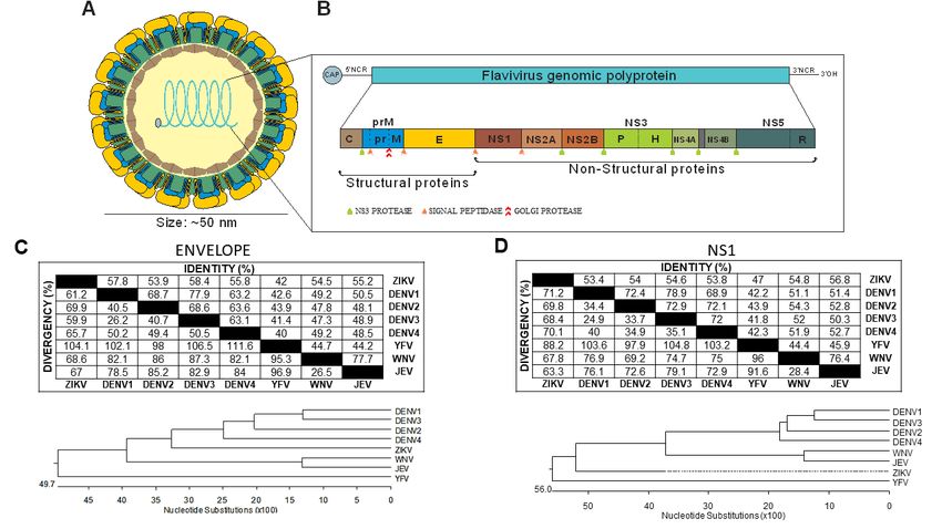
All
All flaviviruses
flaviviruses have
have the
the following
following three
three features
features in
in common:
common: (1)(1) identical genome organization,
identical genome organization,
(2) similar polyprotein
(2) similar polyproteinprocessing,
processing,andand(3)(3) tridimensional
tridimensional structure
structure (Figure
(Figure 1). contains
1). Each Each contains a
a single-
single-stranded positive RNA, which codifies a single polyprotein. Successive cleavages
stranded positive RNA, which codifies a single polyprotein. Successive cleavages of the single of the single
polyprotein,
polyprotein, byby both
both viral
viraland
andcellular
cellularproteases,
proteases, generate
generate three
three structural
structural proteins:
proteins: capsid
capsid (C),
(C), pre-
pre-membrane
membrane (prM), (prM), and envelope
and envelope (E) proteins.
(E) proteins. Seven nonstructural
Seven nonstructural (NS) proteins(NS) areproteins are also
also generated—
generated—NS1, NS2A, NS2B, NS3, NS4A, NS4B, and NS5. The virion structures of
NS1, NS2A, NS2B, NS3, NS4A, NS4B, and NS5. The virion structures of flaviviruses were determined flaviviruses were
determined mostly by cryo-electron
mostly by cryo-electron microscopymicroscopy
[2–6]. [2–6].
Figure 1.1. Genetic
Figure Genetic organization
organization and
and protein
protein homology
homology among
among different
differentflaviviruses.
flaviviruses. (A)
(A) Schematic
Schematic
representation of the virion structure. (B) Genome organization and polyprotein. (C,D)
representation of the virion structure. (B) Genome organization and polyprotein. (C,D) Homology Homology
profiles
profilesofofZika
Zikavirus (ZIKV),
virus Dengue
(ZIKV), virus
Dengue (DENV),
virus Yellow
(DENV), FeverFever
Yellow virus, virus,
(YFV), (YFV),
West Nile
West virus (WNV),
Nile virus
and
(WNV), and Japanese encephalitis virus (JEV) were determined using the reference sequences(E)
Japanese encephalitis virus (JEV) were determined using the reference sequences of envelope of
(C) or nonstructural
envelope 1 (NS1) (D) proteins.
(E) (C) or nonstructural 1 (NS1) (D) proteins.
The virion envelope shows an icosahedral symmetry in which envelope (E) protein dimers are
The virion envelope shows an icosahedral symmetry in which envelope (E) protein dimers are
arranged in a herringbone manner [1]. Immature virions become mature when prM proteins are
arranged in a herringbone manner [1]. Immature virions become mature when prM proteins are
processed in vivo, causing a conformational change on the virus surface, from a bumpy, looser surface
processed in vivo, causing a conformational change on the virus surface, from a bumpy, looser
to a smooth, compact surface, in which E proteins tightly interact with one another to form three sets of
surface to a smooth, compact surface, in which E proteins tightly interact with one another to form
dimers lying parallel to each other and forming a raft [2,3]. Recent work on ZIKV and DENV shows the
three sets of dimers lying parallel to each other and forming a raft [2,3]. Recent work on ZIKV and
induction of non-spherical, club-shaped, or caterpillar-shaped morphologies at temperatures ≥37 ◦ C.
DENV shows the induction of non-spherical, club-shaped, or caterpillar-shaped morphologies at
These morphologies were associated with the scape of the virus from the immune system [7].
temperatures ≥37 °C. These morphologies were associated with the scape of the virus from the
immune system [7].Vaccines 2020, 8, 492 3 of 30
The E protein of flaviviruses is responsible for the interaction with the host cell receptor,
which triggers virus internalization. When the virus enters the endosome, the low pH induces
conformational changes. These changes expose the fusion loop of E protein and lead to the fusion of the
viral protein with endosomal membranes. Next, the RNA of the virus is released and the translation of
the viral polyprotein starts. The polyprotein is cleaved to generate structural and NS proteins. The NS
proteins act in genome replication. Newly synthesized RNA and C protein are packaged by prM and E
to assemble immature virus particles. These particles bud into the endoplasmic reticulum and are
glycosylated. Finally, the immature virion is transported through the trans-Golgi network, where prM
is cleaved by the protease furin into pr peptide and M protein, and the trimeric prM-E are rearranged
to dimeric M-E heterodimers, thus forming the smooth mature virion particles, which are excreted to
infect other cells [1,8].
Each nonstructural flavivirus protein has a function in virus replication. The structure similarity
shared by these proteins from different flaviviruses was recently analyzed [8]. NS1 is a multifunctional
protein that has two forms: (1) a cell-associated form that acts in viral RNA replication and as
cofactor for virus infection and (2) a secreted form that regulates the innate immune response [9].
In some flaviviruses, a-1 ribosomal frameshift event produces also a NS10 protein, and mutations that
abolished NS10 production in JEV led to reduced viral neuroinvasiveness [10]. NS2A is small protein
reported to be involved in viral RNA replication [11,12], modulation of the host-antiviral interferon
response [13–16] and virus particle assembly/secretion [17–19]. NS2B acts as a cofactor to NS3 protease
domain, assisting its folding and catalytic activity [8,20]. NS3 has two domains: N-terminal protease
and C-terminal helicase. The NS3 protease domain is a chymotrypsin-type serine protease [21].
The NS3 helicase domain presents helicase and nucleoside 50 -triphosphatase activities [22]. NS4A and
NS4B have multiple functions involving viral replication and virus–host interactions. The reported
functions of NS4A involve endoplasmic reticulum membrane rearrangement [23], participation in
virus replication complexes formations [24], autophagy induction to prevent cell death and help viral
replication [25] and regulation of NS3 helicase ATPase activity [26]. NS4B is reported to interact
with the NS3 helicase domain and dissociate it from single-strand RNA [27]. NS4B can induce the
unfolded protein response in the host cells and inhibit interferon (IFN) signaling [16,28,29]. NS5 is the
largest NS protein directly involved in capping and RNA replication. It is also involved in interferon
suppression and has two domains: an N-terminal S-adenosylmethionine-dependent methyltransferase
domain, which adds the 50 -RNA cap to assist polyprotein translation and diminish activation of host
innate-immune responses, and a C-terminal RdRp region, which is involved in the RNA-replication
process [30–32].
The E protein is a natural candidate for subunit vaccines, since it is on the virus surface and plays
a direct role on host cell receptor binding and cell fusion. The ectodomain (soluble N-terminal region)
of E monomer has three domains: a beta-barrel domain I (EDI), a finger-like dimerization domain II
(EDII) that contains a fusion loop, and an immunoglobulin-like domain III (EDIII), which contains the
receptor-binding site and the major type-specific neutralization epitopes; consequently, the majority of
subunit vaccine candidates uses E protein or EDIII as antigen [33,34]. Nonetheless, cellular immune
response can also be protective for flavivirus and in some cases is required in order to generate robust
protection. For these reasons, there are some proposals for subunit vaccines that employ NS1, NS3,
and NS5 as vaccine antigens [35–41]. Since subunit vaccines are safer than virus attenuated vaccines
(but are less immunogenic), they could be the preferred antigen candidates for specific risk groups
such as young children, the elderly, and immunocompromised persons. Subunit vaccines could also
be employed as a safer strategy for prime-boost immunization regimens that combine live-attenuated
vaccines and subunit vaccines.
3. Yellow Fever Virus
Yellow fever (YF) is an acute disease that affects humans and non-human primates (NHPs) and
is caused by the Yellow Fever virus (YFV). Clinical manifestations of YF vary from asymptomaticVaccines 2020, 8, 492 4 of 30
individuals to a systemic viral sepsis with viremia; fever; prostration; liver, kidney and heart injury,
hemorrhage; and shock, which can result in death [42]. YFV remains endemic or enzootic in many
South American and African countries, recurrently causing outbreaks and epidemics [43,44]. In Brazil,
from 1967 to 1999, sylvatic human YF outbreaks were usually reported mostly in the Amazon Basin
in the north and mid-west regions, and fewer cases were reported in the Southeast region [45].
After 1999, however, most reported cases occurred outside of the Amazon Basin, mainly in southeast,
mid-west, and south regions of Brazil [45]. From 2016–2019, massive sylvatic YF outbreaks occurred in
states of Minas Gerais, São Paulo, Espirito Santo, Rio de Janeiro, and Bahia, affecting both humans
and NHPs [46–48]. During this period, the number of confirmed cases and deaths due YF was
respectively 2.82 and 1.57 times higher than the sum of all cases and deaths reported in the prior
36 years (1980–2015) [44]. This evidence highlights that even with licensed and efficient vaccine
available, government commitment in disease surveillance is essential to avoid outbreaks.
The YF live-attenuated vaccine (YFV-17D) was developed in 1936 using the 17D strain. All YF
vaccines produced today are derived from this strain. This vaccine is produced in embryonated chicken
eggs and the techniques applied to vaccine manufacture have changed little since its development in
1940s [49]. Even though the protection mechanisms are not yet fully elucidated and the manufacture
process is presumed to be outdated, the YFV-17D is an efficient and quite safe vaccine [50]. YFV-17D
had been considered the safest licensed vaccine, but this changed when some severe adverse events
related to vaccination were discovered in 1996. A few cases of YF-vaccine-associated viscerotropic
disease (YEL-AVD) and YF-vaccine-associated neurotropic disease (YEL-AND) have been reported
since then. These events have greater incidence in elderly people (over 60 years old). Fortunately,
these events are rare. For example, in the United States, these events were estimated at 0.4 cases
per 100,000 vaccinated subjects [45,51]. The adaptive immune responses to YFV-17D are fast, robust,
and durable (usually life-long). Studies show that neutralizing antibodies induced after vaccination
with YF-17D target a low number of conserved epitopes in the E protein, and antibody titers reach as
high as 30 times the needed value for protection after vaccination [52,53]. Despite antibodies being
considered the foremost mediator for YF protection, cellular immune response also plays an important
hole. After immunization, CD4+ and CD8+ T cells appear in the bloodstream, differentiate, and remain
there for the long term as memory T cells [45]. Another central feature of the YF-17D vaccine is its
capacity to induce robust innate immune responses including production of interferons, activation
of inflammasome, and activation of complement elements. These strong, fast, and integrated innate
response pathways are the reason for the robust and durable induced cellular and humoral immune
responses [54,55]. Because of these features, the YF-17D vaccine is recognized as one of the most
effective vaccine ever created [56].
Due to excellent immune properties, the YFV-17D vaccine has been used as a vector for expressing
epitopes of other flaviviruses, such as JEV [57], DENV [58], WNV [59], and ZIKV [60], and of
antigens from other pathogens, such as malaria [61] and HIV [62]. Despite their demonstrated efficacy,
YFV-17D-based vaccines are not recommended for elderly over 60 years old, infants younger than six
months, pregnant and breastfeeding women, people who are immunocompromised, and individuals
with egg-associated hypersensitivity [63–68]. Therefore, the development an alternative vaccine,
one that is safe for all people regardless of age and health conditions and capable of generating durable
and robust protection, continues to be a challenge.
Recombinant YFV proteins were mainly generated with sole purpose of structural characterization
(Table 1). Recombinant EDIII with an N-terminal His-tag was obtained as inclusion bodies in E. coli
using the pET-15b [34] or pET-20b vectors under control of the pelB signal sequence to be exported to
the periplasm [69]. Recombinant capsid protein lacking the C-terminal hydrophobic sequence was
produced in a soluble form by using E. coli strain BL21(DE3) RIL [70]. NS2A, NS2B, and NS4B were
obtained in recombinant E. coli strains in order to identify the cleavage sites [11], while recombinant
NS3 was produced to characterize the enzymatic activities of the protein [71,72]. Recombinant NS5
was produced in HEK293T cells for phosphorylation characterization [73,74]. Recombinant YFVVaccines 2020, 8, 492 5 of 30
NS1 was obtained in E. coli strain Lemo21 (DE3) using vector pBT7-N-His [75]. NS1 and E proteins
were also produced in insect and mammalian cells [76]. Except for two previous studies, none of
these recombinant proteins were tested as potential vaccine antigens under experimental conditions.
Immunizations performed with chimeric YFV NS1-β-galactosidase, produced in E. coli, induced
NS1-specific antibodies and conferred protection to mice [35]. In another study, a recombinant YFV E
protein produced in transgenic plants was used to immunize mice and monkeys. The immunization
elicited neutralizing antibodies, and most of the vaccinated mice were protected against lethal challenge
with the virus [77]. With the purpose of developing a safer and alternative YF vaccine, Tosta et al.
applied bioinformatics tools to design a multi-epitope YF vaccine based on the genome sequence of
137 YFV strains [78]. They proposed to reduce production costs by producing the recombinant protein
in E. coli. However, the efficacy of such a vaccine still awaits experimental validation.
Table 1. Recombinant YFV antigens produced in Escherichia coli and other systems.
Antigen System Purpose Soluble Ref.
EDIII E. coli, pET-15b vector Structural characterization No [34]
E. coli, pET-20b vector, pelB signal Thermodynamic stability and
EDIII No [69]
sequence molecular design
E. coli strain BL21(DE3) RIL, pET-30a
C protein Structural characterization Yes [70]
vector, lacking 20 C-terminal amino acids
NS2A, NS2B and Identification of the
E. coli strain C600, trpE fusion proteins No [11]
NS4B cleavage sites
E. coli strain W3110, cro full-length NS3 Enzymatic activity
NS3 No [71]
fusion and N-terminal truncated NS3 characterization
E. coli strain M15, pQE30 vector,
N-terminal His-tag, hydrophilic core Enzymatic activity
NS2B/NS3 Yes [72]
sequence of NS2B linked to NS3 via characterization
nonapeptide
HEK293T cells, pIRES/GFP bicistronic
Phosphorylation
NS5 mammalian expression vector, Yes [73,74]
characterization
His-tagged protein
E. coli strain Lemo21 (DE3),
NS1 Diagnostics No [75]
pBT7-N-His vector
E and NS1 proteins Vero and Spodoptera frugiperda cells Glycosylation characterization Yes [76]
E. coli strain BMH17-18, pUR vector, Mice immunization: ↑survival
NS1 No [35]
β-galactosidase fusion after i.c. challenge
Mice immunization: ↑
neutralizing Ab; ↑ IgG avidity;
Transgenic plant (Nicotiana benthamiana), protection after i.c. challenge
E protein Yes [77]
N-terminal His-tag Monkey immunization: ↓
Viremia; ↑ neutralizing Ab
titers; ↑ IFNy
↑ = increased and ↓ = decreased.
4. Japanese Encephalitis Virus
Japanese encephalitis (JE) is the most prevalent viral encephalitis in more than 20 countries
in South Asia and in the Western Pacific Region and is known as “Orient’s plague” [79]. In fact,
over 3 billion people (~43% of the human population) are at risk of infection. Annually, there are
approximately 68,000 cases each year and 20,000 deaths from JE. Most JE virus (JEV) infections are
asymptomatic or mild, with symptoms such as moderate headache and fever. However, 0.4% of the
cases evolve into a severe clinical form of JE characterized by high fever, headache, disorientation,
seizures, and coma. About 30% of severe infections are fatal, and up to 30% of those who survive a
severe infection will suffer permanent neurological sequelae [80].
The JE virus was isolated in 1924. It was described as a small icosahedral-enveloped arbovirus
transmitted by Culex mosquitoes [81]. Based on the nucleotide homology of E gene, JEV was categorized
into five JEV genotypes: GI, GII, GIII, GIV and GV, though all of these genotypes belong to the same
serotype. This is evidenced by the absence of secondary infections, in vitro seroneutralization tests ofVaccines 2020, 8, 492 6 of 30
heterologous genotypes, and the fact that vaccination has significantly decreased the burden of the
disease irrespective of the genotype [82–84].
The protection after JEV vaccination correlates with the level of neutralizing antibodies produced.
Several reports demonstrated that a protective status is reached when serum from vaccinated individuals
presents neutralization titers in plaque reduction neutralization test (PRNT50) of at least 10 [82,85,86],
a rather low PRNT50 titer value if compared with other viral diseases [87–89]. JEV vaccine-induced T
cell responses are observed in live attenuated vaccines only; and they preferentially target the NS3
protein. In addition, the generation of CD8+ T cell responses is better indicator of protective immunity
than that of CD4+ T cell responses.
Despite the efficacy of vaccination, the use of several JEV vaccine formulations approved
for humans has been discouraged or discontinued for a variety of reasons. Mouse brain-derived
inactivated (MBDI) vaccines were the first to be approved and implemented for large-scale human
immunization [90]. The Nakayama JEV strain was the first to be approved [91] and was later replaced
by the Beijing-1 JEV strain [92]. However, it was discontinued in 2005 due to its requirement of multiple
doses, limited immunogenicity, and adverse symptoms, especially in Caucasians. Cell-culture-derived
inactivated (CCDI) vaccines replaced MBDI vaccines because their production process is more easily
controlled. The development of several CCDI vaccines (especially using VERO cells) has been
reported [93–95]. Virus attenuation attempts led to the development of live-attenuated vaccines (LAVs),
with the subsequent approval of the SA-14-14-2 LAV formulation by several national governments [96].
The SA-14-14-2 LAV vaccine impaired vector transmission and showed greater immunogenicity than
inactivated vaccines. However, it fell into disuse due to concerns about its virulence rebound and the
threat that it posed to swine, which are frequently raised in JEV-endemic areas. Vector-based vaccines
have also been tested against JEV infections, but only the JE-CV construction, which is based on the
YFV-17D backbone, is licensed for human use. The NYVAC-JEV (attenuated vaccinia virus backbone)
and the ALVAC-JEV (canarypox virus backbone) vaccines showed lower immunogenicity or raised
safety concerns during clinical trials [97]. Moreover, in a comparison with the SA-14-14-2 LAV vaccine,
the JE-CV vaccine candidate showed equivalency when tested in a child cohort [98]. Subunit vaccines
were also evaluated in preclinical conditions, though they did not advance to clinical trials.
JEV subunit vaccines were produced for preclinical evaluations using the E. coli platform
(Table 2). Initial studies demonstrated that a minimum structural 95-mer antigenic domain within
the E glycoprotein was capable of binding neutralizing monoclonal antibodies (mAb). However,
immunization with this E fragment did not induce neutralizing immune responses in mice. Its inability
to induce neutralizing immune responses in mice was probably due to the loss of antigenic epitopes
after refolding of the protein from the insoluble fraction [99]. Two fragments of the JEV E glycoprotein,
Ea (N-terminal) and Eb (C-terminal), were produced in E. coli. Whereas the Eb fragment was
obtained in a soluble form, the Ea fragment was recovered from the insoluble fraction and refolded.
Both recombinant fragments were recognized by JEV neutralizing mAb and hyperimmune mouse serum,
but only mice immunized with two doses of Eb fragment administered with the Freund’s adjuvant
induced high neutralizing antibody titers and achieved partial protection. This evidence highlights
the importance of proper protein folding and generation of efficient neutralizing antibodies [100].
Later, the in vivo expression of Ea in mouse immunized with a DNA vaccine was also found to be
non-protective, unless it was preceded by the signal peptide of the gene of M protein, revealing the
importance of this peptide to Ea proper folding, immune activity, and induction of protection [101].Vaccines 2020, 8, 492 7 of 30
Table 2. Strategies for JEV subunit vaccines produced in E. coli for preclinical evaluation.
Target Strategy Immune Response Protection Refolding Ref.
Fragments of E protein
E protein capable of reacting with ↓Neutralizing Abs No significant protection Yes [99]
neutralizing mAbs
Two fragments of E Ea: ↓Neutralizing Abs Ea–No significant protection Ea–Yes
E protein [100]
protein Ea and Eb Eb: ↑ Neutralizing Abs Eb–Partial protection Eb–No
Freund’s adjuvant: ↑ Freund’s adjuvant:
Immunization with Freund’s
Neutralizing Abs 60% of protection
EDIII adjuvant or different charged No [102]
Cationic liposomes: ↑ Cationic liposomes:
liposomes in mice
Neutralizing Abs 80% of protection
Immunization with Freund’s
complete adjuvant
EDIII ↑ Neutralizing Abs 62.5% of protection No [36]
Boost with Immunization with
Freund’s incomplete adjuvant
Immunization with Freund’s
complete adjuvant
NS1 ↓Neutralizing Abs 87.5% of protection No [36]
Boost with Immunization with
Freund’s incomplete
↑ = increased and ↓ = decreased.
The receptor-binding domain III of E protein (EDIII) from attenuated JEV CH2195LA isolate was
fused with thioredoxin to produce the protein in a soluble form [102]. The immunization of mice
with this protein, which was associated with different adjuvants, induced neutralizing antibodies
and protective immunity [102]. In another study, the antigenicity, immunogenicity, and protective
immunity induced by JEV vaccines containing EDIII and NS1 were evaluated [36]. Mice immunized
with these recombinant proteins elicited antigen-specific antibody responses, but virus-neutralizing
antibodies were induced only in mice immunized with EDIII. Surprisingly, the NS1-containing vaccine
formulation induced high serum anti-NS1 IgG1 titers, which correlated with greater survival rates
(87.5% for NS1; 62.5% for EDIII) [100]. Although the generation of neutralizing antibodies to structural
virus proteins is considered the main protection correlate of JEV vaccines, these results demonstrated
that such a correlation is not complete. Taken together, these results emphasize the perspectives of
alternative JEV vaccines based on recombinant proteins produced in E. coli.
5. West Nile Virus
West Nile virus (WNV) is a neuroinvasive and neurotropic virus. While most of the infected
people are asymptomatic, around 20% will develop West Nile fever, a condition characterized by a
sudden onset of headache, fever, myalgia, fatigue, and vomiting. Patients can recover within a week
or they can remain in a debilitating state for months. Around one out of 150 patients infected with
WNV will develop neurological symptoms such as meningitis, encephalitis, acute flaccid paralysis,
and various ocular manifestations. West Nile fever is particularly serious in older adults, who usually
show more severe clinical manifestations [103–105].
WNV was first isolated in 1937 in the West Nile district in Uganda [106], but today, it can be found
in other parts of Africa, the Americas, Europe, and Oceania. It is the most widespread arbovirus in terms
of geographic distribution. While case reports are sparse, outbreaks are recurrent. One of the latest
outbreaks occurred in 2018 and involved over 2000 human cases across 15 countries in Europe [107].
Since its introduction into the US in 1999, almost 25,000 cases of neurological disease and 2300 deaths
were reported, making WNV the leading cause of an epidemic of mosquito-borne encephalitis [108].
WNV represents such a serious problem that, along with ZIKV, is the only mosquito-borne pathogen
that is tested in blood transfusions in the US [109].
Though there are veterinary vaccines licensed for use in susceptible animals, mainly horses,
no licensed human vaccine for WNV exists. The veterinary vaccines include inactivated whole-virus
vaccines (WEST-NILE INNOVATOR by Zoetis (Parsippany, NJ, USA) and Vetera WNV by Boehringer
Ingelheim Vetmedica (Duluth, GA, USA), a non-replicating vector based on live canarypox virus
(Recombitek Equine WNV by Boehringer Ingelheim Vetmedica (Duluth, GA, USA), and an inactivated
chimera flavivirus vaccine (Equi-Nile by Merck Animal Health (Madison, NJ, USA). These vaccinesVaccines 2020, 8, 492 8 of 30
confer immunity for one year only, so revaccination is recommended. Possibly, due to less regulatory
stringency, veterinary vaccines can move more quickly though clinical testing than those for human
use. Other WNV veterinary vaccines were licensed but are no longer available. These include the DNA
vaccine WEST NILE-INNOVATOR DNA and the live attenuated chimeric vaccine PreveNile [110].
Despite the potential threat of epidemic outbreaks, bringing a human WNV vaccine to phase III
clinical trials involves high costs and a rather reduced market value. A study published in 2006 indicated
that “universal vaccination against WNV disease would be unlikely to result in societal monetary
savings” [111]. Even 20 years after the introduction of WNV into the US, a recent study concluded that
it would be more cost-effective to invest in an age-based vaccination program as opposed to a universal
one [112]. Therefore, such vaccination programs do not interest large pharmaceutical companies.
The US National Institute of Allergy and Infectious Diseases (NIAID) supports the research and testing
of a variety of WNV vaccine candidates including inactivated virus, peptides, attenuated, and chimeric
virus vaccines. This research is mainly conducted by universities and biotech companies [113],
which usually face budgetary, academic bureaucracy, and marketing-related challenges.
The epidemiological profile of WNV presents another hurdle to the advancement of efficient
WNV vaccines to the clinical trial. The uncertainty of where and when WNV will appear and the
fact that it causes outbreaks at different scales makes it is hard to get approval for and set up phase
III clinical trials. The concomitant occurrence of other flaviviruses may have implications for the
approval and set-up of phase III clinical trials. Cross-reactivity of antibodies and the possibility of
the antibody-dependent enhancement (ADE) emergence are additional factors to be considered when
deciding to launch such trials. Additionally, the differences between the epitope-specific profile of
humans and mice may make the translation of mice results to clinical conditions difficult [114].
Human clinical trials of WNV vaccines have been done using DNA-based and vector-based
vaccines, inactivated virus vaccines, and subunit vaccines. Initial phase I/II trials of
formaldehyde-inactivated WNV, live attenuated YFV or DENV vectors expressing WNV prM/E
proteins, DNA plasmid carrying prM/E proteins, and recombinant E protein vaccine candidates
demonstrated promising results for the induction of neutralizing antibodies in most of the enrolled
participants [115,116]. In addition, no adverse effects were reported. Despite these promising results,
none of them progressed to phase III. The recombinant E protein fragment WN-80E is the only
subunit vaccine that entered clinical trials and was produced in Drosophila cells [117]. This vaccine
induced the generation of neutralizing antibodies after three doses in a dose-dependent manner [118].
A key aspect of this vaccine candidate is the exclusion of the transmembrane region of the antigen
(E protein). This deletion resulted in the expression of soluble protein secreted to the extracellular
medium. Moreover, unpublished data claim that the antigen is properly glycosylated and maintains
the native conformation of the viral protein [118].
Subunit vaccines based on recombinant proteins expressed in E. coli were developed and tested at
a preclinical level (Table 3), but they did not advance to clinical trials. Viral proteins are commonly
produced as inclusion bodies in E. coli. The recombinant ectodomain of WNV E protein was formulated
with the particulate saponin-based adjuvant Matrix-MTM. It induced neutralizing antibodies and 100%
protection in mice [119]. Likewise, the E protein domain III (EDIII), alone [120] or fused/conjugated to
other proteins [121,122] and formulated with adjuvants, induced protective immunity against lethal
challenge in mice. All these data demonstrate the potential for the development of WNV vaccines by
using formulations of recombinant proteins produced in E. coli.Vaccines 2020, 8, 492 9 of 30
Table 3. Strategies for WNV subunit vaccines in preclinical assays.
Target Strategy Immune Response Protection Refolding Ref.
Full length E and Mice: ↑IgG titers. Mice: 100% survival.
truncated Antibodies recognize Serum passive transfer No [123]
E protein
(80% N-terminal) WNV-infected cells 80% survival
Mice: ↑IgG and
E antigen in a DNA prime Mice: 100% survival. ↓
neutralizing Abs titers. ↑ Yes [124]
and protein boost viremia
CD8 + IFNg+ T cells
E ectodomain plus
Mice: ↑IgG and
E ectodomain Matrix-M (saponin) Mice: 100% survival Yes [119]
neutralizing Ab titers
adjuvant
Mice: ↑IgG, IgM and
IgA titers.
EDIII fused with
EDIII Complement-mediated ND No [125]
cholera toxin
killing of serum from
immunized mice
EDIII genetically fused
Mice: ↑IgG and
EDIII with flagelin Mice: 100% survival Yes [122]
neutralizing Ab titers
(TLR5 agonist)
EDIII fused with Horses: ↑IgG and
EDIII ND No [126]
CD40 ligand neutralizing Ab titers
EDIII conjugated with Mice: ↑IgG long-lasting
EDIII VLP of bacteriophage response (>1 year) and Mice: 100% survival Yes [121]
AP205 plus alum neutralizing Ab titers
Mice: ↑IgG titers. Abs Mice: ↑survival of mice
recognize WNV-infected receiving WNV premixed
EDIII EDIII plus CpG No [127]
cells. ↑T-cell proliferation. with antiserum from
↑cytokines (splenocytes) immunized mice
Mice: partially
neutralizing Abs. Different
Comparison EDIII and
EDIII immunogens induce ND No [128]
soluble full-length E
different potencies of
neutralizing Abs
EDIII plus CpG and Mice: ↑IgG and
EDIII Mice: 80% survival Yes [120]
boosted with oil neutralizing Ab titers
Mice: ↑ IgG and
EDIII boost after E
EDIII neutralizing Ab titers. Mice: 100% survival Yes [129]
ectodomain DNA prime
Moderate IFN-γ
EDIII plus KFE8 Mice: ↑ IgG and
EDIII Mice: 60% survival Yes [130]
peptide hydrogel neutralizing Ab titers
EDIII plus Mice: ↑IgG and
EDIII ND Yes [131]
Freund’s adjuvant neutralizing Ab titers
ND = not determined; ↑ = increased and ↓ = decreased.
Although it is difficult to establish a single explanation for their absence in clinical trials,
certain issues may have delayed the evolution of preclinical studies of WNV vaccine candidates
produced in E. coli. These include difficulties with achieving the correct folding of proteins from
inclusion bodies (IBs) and determining an appropriate adjuvant. Early studies showed that the
immunogenicity of WNV antigens is greatly affected by conformation. Also, disulfide bond patterns
differ when proteins are produced in insect cells or E. coli [132]. Several WNV vaccine candidates
employed proteins purified from IBs, and they were evaluated with different adjuvants to obtain
the best immune response (Table 3). Additionally, post-translational protein modifications, such as
glycosylation, may also affect the immunogenicity of WNV proteins. For example, only the glycosylated
WNV E protein can optimally bind to C-type lectin DC-SIGN(R), a receptor that can either enhance
the infection [133] or promote immune cell activation and antigen uptake [134]. Although bacterial
cells do not usually promote such post-translational modifications, novel genetically modified E. coli
strains that perform the glycosylation of recombinant proteins are good alternatives to deal with this
problem [135]. Culturing conditions, solubility tags, and different expression vectors and strains can
easily be evaluated in order to obtain a soluble antigen [136].
With the increasing number of options available, E. coli can become a suitable host for the
production of protein-based WNV vaccines for preclinical evaluations and, later, progress to clinical
trials. The incorporation of adjuvants and/or combinations with alternative immunization strategies,Vaccines 2020, 8, 492 10 of 30
such as prime-boost regimens may contribute to overcome the low immunogenicity of recombinant
proteins and improve immune responses.
6. Dengue Virus
Annually, dengue virus (DENV) infects approximately 390 million people worldwide. This results
in about 500,000 hospitalizations and a death toll of approximately 20,000 lives each year [137].
There are four serotypes of DENV capable of infecting humans. They are distributed among more
than 100 countries in tropical and subtropical regions around the world [138]. Most part of DENV
infections (about 75%) are asymptomatic and self-limited. However, symptomatic infections can
show different degrees of illness. The World Health Organization (WHO) classifies DENV infections
as DF for asymptomatic and/or dengue fever, DWS for a more severe dengue with warning signs,
and SD for severe dengue with clinical complications [139,140]. Patients with DWS can present fever
(37.5–38 ◦ C), headache, retro-orbital and abdominal pains, myalgia, arthralgia, a rash, flushing of the
face, anorexia, and nausea. In addition to these symptoms, SD patients can also have plasma leakage,
fluid accumulation, severe bleeding, and organ impairment that can be fatal [139]. Although the
determinants of the complications related to SD are not well understood, variables such as secondary
infections with different serotypes, immunity, age, and individual genetics can affect the risk of
developing the more severe forms of the disease [141–143].
There is not an effective therapy available for dengue. Current treatments address the symptoms of
the disease. This includes fluid replacement and blood transfusion, which require hospitalization [144].
There is, however, a prophylactic tetravalent live-attenuated virus vaccine, Dengvaxia® , also known
as CYD-TDV, that was developed by Sanofi Pasteur and is licensed in approximately 20 countries.
CYD-TDV consists of four chimeric viruses based on the 17D strain of YFV. The sequences encoding the
pre-membrane (prM) protein and the envelope glycoprotein (E) were replaced by those of each of the
four DENV serotypes. Despite being licensed, however, recent studies have shown that this vaccine
may present risks to individuals without prior immunity to DENV. This can result in an increase
in the number of severe cases after primary DENV infection and, consequently, in the number of
hospitalizations [140,145,146]. Additionally, according to WHO guidelines, CYD-TDV should not be
applied to the general population. Rather, it should only be used in those countries with demonstrated
seroprevalence (≥80%) and in individuals between nine and 45 years of age [143,147]. Thus, the search
for effective and safe DENV vaccines is still necessary.
During the past few years, another six DENV vaccine candidates advanced to clinical studies
(reviewed by Pinheiro-Michelsen, et al. [148]). Among them, two are in phase III trials. These are
LATV by NIAID (Bethesda, MD, USA) and the Butantan Institute (São Paulo, SP, Brazil) and TAK-003
by Takeda Pharmaceutical Company Limited (Tokyo, Japan). Both are based on the live-attenuated
DENV and are capable to induce neutralizing antibodies and cellular immune response to all DENV
serotypes [149–153]. Recently, TAK-003 demonstrated a promising efficacy of 80.2% in a phase III
study [154]. Although data related to the performance of LATV vaccine in phase III trial is not yet
available, this vaccine exhibited protective results in a human challenge model with the DENV 2
strain [5].
The main challenge to the development of an effective DENV vaccine is the need for a vaccine
that induces a safe, long-lasting, and balanced immune responses for all four DENV serotypes.
Such concerns are particularly relevant for formulations containing structural proteins, due to the
risk of they present of increasing of the severity of the disease by antibody-dependent enhancement
(ADE) [155]. This phenomenon occurs when the infectivity of the virus is increased in the cells that
express Fc receptors when the antibodies binding to DENV but do not have neutralizing capacity or
are present in sub-neutralization amounts [156,157]. Indeed, during CYD-TDV vaccine phase II and III
trials, variable serotype-specific protective responses were reported, with the lowest response reported
for DENV 2 (Serotype 1, 50–50.3%; Serotype 2, 9.2–35%; Serotype 3, 9.2–35%; Serotype 3, 74.0–78.4%;Vaccines 2020, 8, 492 11 of 30
Serotype 4, 75.3–77.7%). This is indicative of an unbalanced immune response [145,158], and may
represent a limitation of the CYD-TDV vaccine, especially among DENV-naive individuals [140].
Evidences indicate that induction of high titers of neutralizing antibodies does not signify a
complete protection correlation. In contrast, certain aspects of T cell–mediated immunity, CD8+ T cells
in particular, may be related to protection against DENV infection and disease [159–161]. Since the
non-structural (NS) proteins are the major targets of the anti-DENV CD8+ T cell responses [162],
vaccines that lack responses targeting NS proteins, such as the CYD-TDV vaccine, fall behind vaccines
that induce responses to all DENV proteins, such as the LATV vaccine. Despite its capacity to induce
cellular responses against the NS proteins, when administered in a tetravalent formulation, the NS
responses shift from a heterogeneous response between NS3 and NS5 proteins to a response mainly
targeting the NS5 protein. This is likely due to antigen accumulation, since the NS5 proteins are the
most conserved NS protein among DENV serotypes [162–164]. Similar difficulties also occur during
the development of humoral responses induced by attenuated vaccines. These difficulties may be
avoided by the use of recombinant antigens.
Over 30 DENV vaccine candidates based on recombinant antigens have been developed to date.
However, the majority of these vaccine candidates have not advanced to clinical validation despite
their promising preclinical results, as shown in Table 4. The tetravalent vaccine V180, developed
by Merck & Co. (Kenilworth, NJ, USA), is the only one that is presently under clinical testing.
The vaccine is based on truncated versions (DEN-80E) of the E protein from DENV1-4, produced
in Drosophila Schneider-2 (S2) cells, and uses the adjuvant ISCOMATRIXTM (CSL Behring, King of
Prussia, PA, USA) [165]. Preclinical tests using mice and NHPs showed the induction of Th1 biased
cellular immune responses, the presence of neutralizing antibodies and protective immunity against
lethal challenge. In humans, reports indicate that V180 induce neutralizing antibodies, with 85.7%
seroconversion [165–167]. On other hand, among the different DENV subunit vaccines tested until
now, those based on recombinant proteins produced in E. coli strains have been intensively evaluated
(Table 4). This begs the question, why haven’t DENV formulations based on this platform not advanced
to clinical trials?
Table 4. Strategies for DENV subunit vaccines produced in E. coli for preclinical evaluation.
Target Strategy Immune Response Protection Refolding Ref.
Mice: 50% survival
Fusion with P64k Mice and monkeys: ↑ (DENV2 i.c.)
E protein protein from N. IgG and neutralizing Monkeys: reduction of No [168–170]
meningitidis Ab titers after 4 doses viremia after challenge
(DENV2)
Mice: ↑ IgG titers; ↑
Chimeric protein with
CD8 and CD4 T cells;
E protein 11 peptides from ND Yes [171]
splenocytes (↑IFNy,
DENV1-4 E protein
IL-2,IL-4 and IL-17)
E protein DENV2 E protein Mice: ↑ IgG titers ND Yes [172]
Fragment of DENV1-4 Mice: 80% of survival
Mice and monkey: ↑
E proteins fused with (i.c. challenge);
E protein IgG and neutralizing No [173–176]
maltose binding Monkeys: no
Ab titers
protein (MBP) protection
Monkeys and mice: ↑
IgG and neutralizing Mice: ↑ survival
EDIII fused with C Ab titers, ↑ B cells (DENV1-4)
EDIII and C
proteins from antigen-specific; ↑ Monkeys: ↓ viremia Yes [177–183]
proteins
DENV1-4 IFNy (splenocytes or and ↑ survival against
PBMC); ↑ CD8 e CD8 T DENV1-4
cells (IFNy)
Mice: ↑ IgG and
neutralizing Ab titers
DENV1-4 EDIII
(heterologous Mice: ↑ survival
EDIII protein protein fused to fliC (S. No [184]
prime-boost with (DENV1-4)
typhimurium)
LATV vaccine);
↓ADE effectVaccines 2020, 8, 492 12 of 30
Table 4. Cont.
Target Strategy Immune Response Protection Refolding Ref.
DENV1-4 EDIII in Mice: ↑ IgG and Mice: ↑ survival
EDIII protein Yes [185]
tandem (B1234 protein) neutralizing Ab titers (DENV1-4)
DENV2 EDIII fused Mice: ↑ IgG and Mice: no protection; ↑
EDIII protein Yes [186]
with pIII coat protein neutralizing Ab titers ADE
DENV2 EDIII with
Mice: ↑ IgG and
EDIII protein different adjuvants ND Yes [187]
neutralizing Ab titers
(LT1, LTB and Alum)
Mice: ↑ IgG and Mice: ↑ survival
EDIII protein DENV2 EDIII Yes [188]
neutralizing Ab titers (DENV2)
Mice: ↑ IgG and
DENV1-4 EDIII fused
neutralizing Ab titers;
with lipid signal
EDIII protein ↑ IgG avidity; ↑ Mice: ↓ viremia Yes [189–192]
peptide of the
neutralization (PRNT);
lipoprotein Ag473
↓ ADE in vitro
DENV1 EDIII with Mice: ↑ IgG and
EDIII protein different adjuvants neutralizing Ab titers; ND No [193]
(PELC and CpG) ↑ IFNy (ELISPOT)
DENV1-2 EDIII or
Mice: ↑ IgG and Mice: ↑ survival
EDIII protein DENV3-4 EDIII in Yes [194,195]
neutralizing Ab titers (DENV1-4)
tandem
DENV2 EDIII with Mice: ↑ IgG and Mice: ↑ survival
EDIII protein Yes [196]
Freund’s adjuvant neutralizing Ab titers (DENV2)
Mice: ↑ IgG titers; ↑
DENV3 EDIII proliferation of
EDIII protein ND No [197]
consensus splenocytes; ↑ IFNy
and IL-4 (splenocytes)
Mice: ↑ IgG and
DENV1-4 EDIII
EDIII protein neutralizing ND No [198]
consensus
(DENV1-4) Ab titers
DENV (1,2) EDIII
EDIII protein Mice: ↑ IgG titers ND Yes [199]
protein
Mice: ↑ IgG and
neutralizing Ab titers;
↑ CD4 and CD8 T cells
EDIII protein DENV1-4 EDIII ND Yes [200]
(producing IFNy and
IL-2); ↑ IFNy, IL-2,
IL-12p40 (splenocytes)
DENV2 EDIII in
Mice: ↑ IgG and
EDIII protein Chimeric VLP ND Yes [201]
neutralizing Ab titers
(HBcAg-EDIII-2)
Fubc protein (peptides
from fusion and bc
EDII protein loop regions of EDII Mice: ↑ IgG titers ND Yes [202]
fused by link
sequence)
DENV2 NS1 with
Mice: ↑ survival
NS1 protein LTG33D, Alum or Mice: ↑ IgG titers Yes [37,203]
(DENV2)
Freund’s adjuvant
Mice: ↑ IgG titers; ↑
Mice: ↑ survival
NS5 protein DENV2 NS5 IFNy and TNFα No [38]
(DENV2)
(splenocytes)
Mice: ↑ IgG titers; ↑
NS3 protein DENV2 NS3 protein ND Yes [39]
IFNy (splenocytes)
Mice: ↓ viremia; ↓
Mice: ↑ IgG titers;↑ soluble NS1 levels; ↓
DJ NS1 chimera
CD4 and CD8 T cells; ↑ mouse tail bleeding
NS3/NS1 (DENV2 and JEV) and Yes [40]
CTL responses against time, and vascular
NS3
NS3 leakage at skin
injection sites
ND = not determined; ↑ = increased and ↓ = decreased.
The recombinant DENV antigens produced in E. coli cells include full length or fragments of
different proteins, fused or not with other proteins (chimeras), which were associated with different
adjuvants. Most part of the studies are based on the structural proteins (E and C) but non-structural
proteins, such as NS1, NS3, and NS5, were also investigated (Table 4). The most advanced strategies use
chimeric proteins to improve the immunogenicity of the DENV proteins. Tetra DIIIC vaccine, which isVaccines 2020, 8, 492 13 of 30
composed by chimeric forms of the E protein domain III (EDIII) fused to the capsid (C) protein from all
DENV serotypes, has been tested with different adjuvants and form protein aggregates when associated
with oligodeoxynucleotides. These formulations induced neutralizing antibodies, activation of B
and T cells, and protective immunity in mice and monkeys [177,179–181]. Additionally, the chimeric
protein also boosted the natural immune responses generated after DENV infection in monkeys [178]
or in a heterologous prime-boost regimen with the live-attenuated LATV vaccine [182]. Along these
lines, chimeric proteins using the E protein domain b (aa 298–400) from DENV 1–4 and fused with
maltose binding protein (MBP) induced neutralizing antibodies in mice and NHP. However, protective
immunity was registered only in mice [173–176]. Others monovalent or tetravalent formulations
based on full E protein or EDIII were also evaluated in mice, and some of them induced neutralizing
antibodies (Table 4).
Recently, the role played by recombinant DENV NS protein produced in E. coli has been evaluated.
Formulations using the NS1 protein associated with a derivative of the heat labile toxin (LTG33D) or
Freund adjuvants were capable to stimulate humoral and cellular immune responses in mice, inducing
partial protection to DENV2 [37,203]. NS5 protein was also protective to DENV2 strains, which was
accompanied of enhanced antigen-specific antibodies and expansion of IFNy/TNF-α producing T
cells, even in the absence of adjuvant [38]. Due to the presence of cytotoxic T cell epitopes in NS3,
this antigen was recently tested in combination with a chimeric DENV/JEV NS1. Mice immunized
with this vaccine showed activation of antigen-specific CD4+ and CD8+ T cells, elicited antibody
responses, NS3-specific cytotoxic T lymphocyte (CTL) activities, and protective effects on a DENV2
infection model [40]. Despite the fact that the protective role of NS3 has been demonstrated only in
vaccination platforms, such as DNA vaccines [41], the recombinant protein produced in E. coli proved
to be immunogenic in mice, leading to activation of antigen-specific antibody and cellular immune
responses [39].
Taken together, these studies demonstrate that recombinant DENV proteins produced in E. coli
are capable to induce protective immunity at experimental conditions and, thus, represent promising
candidates for testing in clinical conditions. Nonetheless, most of the DENV recombinant proteins
produced in E. coli showed reduced solubility and required the use of refolding techniques. This is
specifically critical for structural proteins, such as the E protein, for which conformational epitopes are
the main targets to neutralizing antibodies [204,205]. In addition, as usually observed with subunit
vaccines, DENV vaccines based on recombinant proteins require the use of adjuvants to improve
induction of antigen-specific immune responses. Such limitations may influence the development
pipeline and translation to clinical trials.
7. Zika Virus
The 2015–2016 Zika outbreak in the Americas infected up to 73% of the population in some
cities [206]. Before this, ZIKV infection was considered to be benign with trivial health consequences.
However, during the last epidemics, ZIKV infection was related to nervous system diseases,
including Guillain–Barré and congenital Zika syndromes, that, among other complications, resulted in
thousands of cases of microcephaly. The clinical manifestations of congenital Zika syndrome are not
yet fully elucidated. Many children born from infected mothers show no signs at birth, but they
developed abnormal neurodevelopmental outcomes later in life [207,208]. Although the number of
cases has decreased considerably since 2016, ZIKV is now endemic in tropical regions around the
world. Five years since the last epidemics, there are still no licensed vaccines available [209].
Antibody responses are considered the most important indicator for an effective flavivirus vaccine.
Indeed, monoclonal antibodies are capable of protecting mice from ZIKV infection [210], and T cell
depletion after immunization does not interfere in this protection [211]. Unlikely DENV infections,
in which CD4+ T cells target structural proteins and CD8+ T cells target mainly nonstructural proteins,
in ZIKV infection, both CD4+ and CD8+ T cells target structural proteins [212,213]. The depletion
of CD8+ T cells increased mortality in mice [214] and indicate that T cell responses also play anVaccines 2020, 8, 492 14 of 30
important role in protection. Even though neutralizing antibodies are considered the main contributor
to protective immunity, the aforementioned evidences indicate that an optimal ZIKV vaccine should
induce both cellular and humoral immune responses [181].
Several approaches have been taken in the pursuit of a ZIKV vaccine. Such approaches include
RNA-based, DNA-based, and viral-vector-based vaccines, as well as attenuated viruses, inactivated
viruses, and recombinant protein subunits. The formulations that have advanced to clinical trials use
attenuated viruses, inactivated viruses, RNA-based, DNA-based, or viral vector vaccines (Table 5).
To date, the only ZIKV vaccine that advanced to a phase II clinical trial is a DNA-based vaccine. It is
called RC-ZKADNA090-00-VP (ClinicalTrials.gov Identifier: NCT03110770), and it encodes the prM
and E proteins. During phase I, this vaccine did not show severe systemic reactions, but presented a
few mild to moderate adverse effects. Moreover, all the participants (14/14) developed neutralizing
antibodies. However efficacy, durability and protective effect in humans are not yet evaluated [215].
Results from a phase II trial are not yet available.
Table 5. ZIKV vaccines in clinical trials.
Inactivated Attenuated Viral
Clinical Trials Status DNA-Based RNA-Based Total
Virus Virus Vector-Based
NCT04033068
NCT03008122
Early phase I Ongoing - - NCT04064905 NCT04440774 6
NCT03343626
or phase I NCT04015648
NCT02937233
NCT02952833 NCT02840487
Completed NCT03611946 NCT03014089 NCT02996890 9
NCT02963909 NCT02996461
NCT03425149
Phase II Completed - - NCT03110770 - - 1
None of the ZIKV subunit vaccine advanced to clinical trials despite promising results in
pre-clinical conditions (Table 6). Ham and collaborators have demonstrated that a recombinant subunit
vaccine containing the N-terminus of the ZIKV envelope protein (E90) produced in E. coli induced
robust and specific humoral responses in mice. The passive transfer of sera collected from immunized
mice fully protected neonatal mice from a lethal virus challenge [216]. The same ZIKV vaccine was
tested in mouse models for in utero and neonatal infection. Considerable reduction of brain cells
infection was observed in fetus and suckling mice. Furthermore, this vaccine prevented the occurrence
of microcephaly compared to unvaccinated controls mice. In the same study, the authors demonstrated
that this vaccine protected mice even 140 days after immunization [217]. Despite its excellent results,
the E90 subunit vaccine did not advance to non-human primate testing nor to clinical trials.
Table 6. Strategies for ZIKV subunit vaccines produced in E. coli for preclinical evaluation.
Target Strategy Immune Response Protection Refolding Ref.
Neonatal mice: full
protection Pregnant
90% of the N-terminal whole E Mice: ↑IgG and mice: fetus and
E protein Yes [216]
protein + alum adjuvant neutralizing Ab titers offspring protection
from ZIKV-induced
microcephaly
Mice: ↑IgG and
80% of the N-terminal whole E
E protein neutralizing Ab titers; ↑ Mice: ↑survival Yes [218]
protein + alum adjuvant
ZIKV specific T cells; IFNy
Consensus E protein lacking
the whole stem and trans Mice: ↑IgG titer; ↑ ZIKV
E protein ND Yes [219]
membrane regions + poly (I:C) specific T cells; IFNy
or CpG ODN adjuvants
Mice: ↑IgG and
Full EDIII sequence + TMG or
EDIII protein neutralizing Ab titers; ND Yes [220]
alum adjuvants
↑IFNy, IL-4 and IL-6
ND = not determined and ↑ = increased.You can also read