WHOLE GENOME SEQUENCE ANALYSIS AND HOMOLOGY MODELLING OF A 3C LIKE PEPTIDASE AND A NON-STRUCTURAL PROTEIN 3 OF THE SARS-COV-2 SHOWS PROTEIN LIGAND ...
←
→
Page content transcription
If your browser does not render page correctly, please read the page content below

doi.org/10.26434/chemrxiv.11846943.v5

Whole Genome Sequence Analysis and Homology Modelling of a 3C
Like Peptidase and a Non-Structural Protein 3 of the SARS-CoV-2
Shows Protein Ligand Interaction with an Aza-Peptide and a
Noncovalent Lead Inhibitor with Possible Antiviral Properties
Arun Shanker, Anjani Alluri, Divya Bhanu

Submitted date: 09/03/2020 • Posted date: 10/03/2020
Licence: CC BY 4.0
Citation information: Shanker, Arun; Alluri, Anjani; Bhanu, Divya (2020): Whole Genome Sequence Analysis
and Homology Modelling of a 3C Like Peptidase and a Non-Structural Protein 3 of the SARS-CoV-2 Shows
Protein Ligand Interaction with an Aza-Peptide and a Noncovalent Lead Inhibitor with Possible Antiviral
Properties. ChemRxiv. Preprint. https://doi.org/10.26434/chemrxiv.11846943.v5

The family of viruses belonging to Coronaviridae mainly consist of virulent pathogens that have a zoonotic
property, Severe Acute Respiratory Syndrome (SARS-CoV) and Middle East Respiratory Syndrome
(MERS-CoV) of this family have emerged before and now the SARS-CoV-2 has emerged in China.
Characterization of spike glycoproteins, polyproteins and other viral proteins from viruses are important for
vaccine development. Homology modelling of these proteins with known templates offers the opportunity to
discover ligand binding sites and explore the possible antiviral properties of these protein ligand complexes.
Any information emerging from these protein models can be used for vaccine development. In this study we
did a complete bioinformatic analysis, sequence alignment, comparison of multiple sequences and homology
modelling of the SARS-CoV-2 whole genome sequences, the spike protein and the polyproteins for homology
with known proteins, we also analysed receptor binding sites in these models for possible binding with ligands
that exhibit antiviral properties. Our results showed that the tertiary structure of the polyprotein isolate
SARS-CoV-2_HKU-SZ-001_2020 had 98.94 percent identity with SARS-Coronavirus NSP12 bound to NSP7
and NSP8 co-factors. Our results indicate that a part of the viral genome (residues 3268 -3573 in Frame 2
with 306 amino acids) of the SARS-CoV-2 virus isolate Wuhan-Hu-1 (Genbank Accession Number
MN908947.3) when modelled with template 2a5i of the PDB database had 96 percent identity with a 3C like
peptidase of SARS-CoV which has ability to bind with Aza-Peptide Epoxide (APE) which is known for
irreversible inhibition of SARS-CoV main peptidase. The part of the genome (residues 1568-1882 in Frame 2
with 315 amino acids) when modelled with template 3e9s of the PDB database had 82 percent identity with a
papain-like protease/deubiquitinase which when complexed with ligand GRL0617 acts as inhibitor which can
block SARS-CoV replication. It is possible that these viral inhibiters can be used for vaccine development for
the SARS-CoV-2.

File list (18)

Coronavirus_MS_16.pdf (3.03 MiB)        view on ChemRxiv   download file

Figure 12.jpg (127.89 KiB)              view on ChemRxiv   download file

Coronavirus_MS_16.docx (1.49 MiB)       view on ChemRxiv   download file

Tables.docx (22.22 KiB)                 view on ChemRxiv   download file

Supplementary Tables.docx (35.94 KiB)   view on ChemRxiv   download file

Figure1.jpg (239.23 KiB)                view on ChemRxiv   download file

Figure 2.jpg (41.76 KiB)                view on ChemRxiv   download file

Figure 3.jpg (39.46 KiB)                view on ChemRxiv   download file

Figure 3.jpg (39.46 KiB)                view on ChemRxiv   download file

Figure 4.jpg (0.93 MiB)                 view on ChemRxiv   download file

Figure 5.jpg (695.68 KiB)               view on ChemRxiv   download file

Figure 6.jpg (52.36 KiB)                view on ChemRxiv   download file

Figure 7.jpg (53.73 KiB)                view on ChemRxiv   download file

Figure 8.jpg (82.26 KiB)                view on ChemRxiv   download file

Figure 9.jpg (14.38 KiB)                view on ChemRxiv   download file

Figure 10.jpg (79.40 KiB)               view on ChemRxiv   download file

Supplementaty Fig 1.pdf (157.41 KiB)    view on ChemRxiv   download file

Figure 11.JPG (159.45 KiB)              view on ChemRxiv   download file
1    Whole Genome Sequence Analysis and Homology Modelling of a 3C Like Peptidase and
2        a Non-Structural Protein 3 of the SARS-CoV-2 Shows Protein Ligand Interaction with
3         an Aza-Peptide and a Noncovalent Lead Inhibitor with Possible Antiviral Properties

4                                Arun K. Shanker1 *, Divya Bhanu1,2 and Anjani Alluri3

                            1
5                               ICAR - Central Research Institute for Dryland Agriculture

6                                       Santoshnagar, Hyderabad – 500059, India

7                                 Corresponding author email: arunshank@gmail.com

                2
8                   Centre for Plant Molecular Biology, Osmania University, Hyderabad, India

     3
9        Advanced Post Graduate Centre, Acharya N.G.Ranga Agricultural University, Guntur, India

10   Abstract

11   The family of viruses belonging to Coronaviridae mainly consist of virulent pathogens that have a zoonotic property, Severe
12   Acute Respiratory Syndrome (SARS-CoV) and Middle East Respiratory Syndrome (MERS-CoV) of this family have emerged
13   before and now the SARS-CoV-2 has emerged in China. Characterization of spike glycoproteins, polyproteins and other viral
14   proteins from viruses are important for vaccine development. Homology modelling of these proteins with known templates
15   offers the opportunity to discover ligand binding sites and explore the possible antiviral properties of these protein ligand
16   complexes. Any information emerging from these protein models can be used for vaccine development. In this study we did a
17   complete bioinformatic analysis, sequence alignment, comparison of multiple sequences and homology modelling of the
18   SARS-CoV-2 whole genome sequences, the spike protein and the polyproteins for homology with known proteins, we also
19   analysed receptor binding sites in these models for possible binding with ligands that exhibit antiviral properties. Our results
20   showed that the tertiary structure of the polyprotein isolate SARS-CoV-2_HKU-SZ-001_2020 had 98.94 percent identity with
21   SARS-Coronavirus NSP12 bound to NSP7 and NSP8 co-factors. Our results indicate that a part of the viral genome (residues
22   3268 -3573 in Frame 2 with 306 amino acids) of the SARS-CoV-2 virus isolate Wuhan-Hu-1 (Genbank Accession Number
23   MN908947.3) when modelled with template 2a5i of the PDB database had 96 percent identity with a 3C like peptidase of
24   SARS-CoV which has ability to bind with Aza-Peptide Epoxide (APE) which is known for irreversible inhibition of SARS-
25   CoV main peptidase. The part of the genome (residues 1568-1882 in Frame 2 with 315 amino acids) when modelled with
26   template 3e9s of the PDB database had 82 percent identity with a papain-like protease/deubiquitinase which when complexed
27   with ligand GRL0617 acts as inhibitor which can block SARS-CoV replication. It is possible that these viral inhibiters can be
28   used for vaccine development for the SARS-CoV-2.

29

30   Introduction

31   More than a decade has passed since the emergence human Coronavirus that caused Severe

32   Respiratory Syndrome (SARS-CoV) and it is about 7 years since the emergence of another

33   type of Coronavirus - Middle East Respiratory Syndrome (MERS-CoV) and now the SARS-

                                                                    1
34 CoV-2 has emerged in China. This repeated onslaught of these viruses goes to show that it can
35 assume pandemic proportions at any time and at any place.
36 The family of viruses belonging to Coronaviridae mainly consist of virulent pathogens that
37 have a zoonotic property and this large family of corona viruses, have been known to be
38 circulating in animals including camels, cats and bats. It has been seen in the past that Severe
39 Acute Respiratory Syndrome associated coronavirus (SARS-CoV) and Middle East
40 Respiratory Syndrome-associated coronavirus (MERS-CoV) belonging to this family of
41 viruses can be transmitted from animals to humans and can cause respiratory diseases. Human
42 to human transmission on this virus has been a concern and due to this search for antiviral
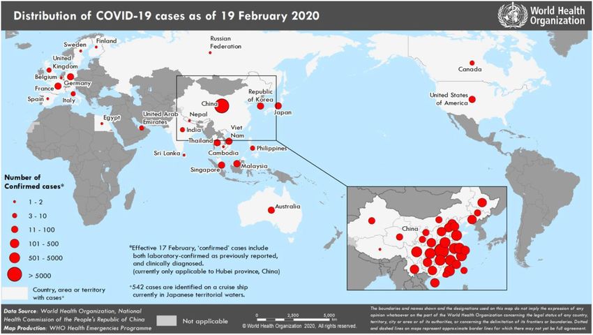
43 compounds and vaccine development for this family of virus becomes the need of the hour.
44 The SARS was first seen in 2002 in Guangdong province of China, and later spread globally
45 and has caused close to about 8096 cases (WHO 2004, de Vit et al., 2016). In 2012, a novel
46 betacoronavirus, designated Middle East respiratory syndrome coronavirus or MERS-CoV
47 associated with severe respiratory disease in humans, emerged in the Arabian Peninsula (de
48 Wit et al., 2013).
49 The World Health Organization (WHO), China Country Office was informed of cases of
50 pneumonia of unknown aetiology in Wuhan City, Hubei Province, on 31 December 2019
51 (WHO 2020). A novel coronavirus currently termed SARS-COV-2 was officially announced
52 as the causative agent by Chinese authorities on 7 January 2020. As on 20 Feb 2020 China’s
53 National Health Commission reported that there are 74,280 confirmed cases in China (Fig.1).
54 The World Health Organization reported 924 confirmed cases in 25 countries outside China
55 (WHO Situation Report 29 2020). This novel corona virus has been designated as SARS-CoV-
56 2.
57
2
58   Coronaviruses are RNA viruses and have large genomes structures and due to this they can

59   have high error in replication as compared to host genomes. It is also known that various CoVs

60   can do effective recombination of their genomes after infecting host cells (Luo et al 2018). This

61   recombination can be a factor for their evolution to novel types which may have new animals

62   as their intermediate hosts. These factors give the CoVs high adaptive ability and the capability

63   to jump across species and have a relatively large host range.

64   Characterization of Spike glycoproteins from viruses are important for vaccine development.

65   Any information coming from the protein model can be used for vaccine development. In Silico

66   Epitope, polyprotein and spike protein-based peptide vaccine designing for infectious viruses

67   is a way that can hasten the process of vaccine development. Spike (S) protein, polyprotein and

68   other viral proteins of the SARS-CoV-2 as a target for the development of vaccines and

69   therapeutics for the prevention and treatment of infection is an important approach. In the case

70   of SARS-CoV, these proteins can mediate binding of the virus with its receptor and promotes

71   the fusion between the viral and host cell membranes and virus entry into the host cell, hence

72   peptides, antibodies, organic compounds and short interfering RNAs that interact with the spike

73   protein can have a potential role in vaccine development (Du et al 2009).

74   There are multiple domain functions that are active in the replication of the coronavirus and

75   these domains are present in a protein designated as Non-structural protein 3 (nsp3) which is

76   the largest protein in the coronavirus genome (Chen et al 2015). 3C like protease (3CLpro) and

77   Papain like Protease (PLpro) are two important class of proteases that are involved in the

78   process of translation of the polypeptide from the genomic RNA to protein components that

79   are required structurally or non-structurally for replication and packaging of new generation

80   viruses (Liu et al 2020)

                                                    3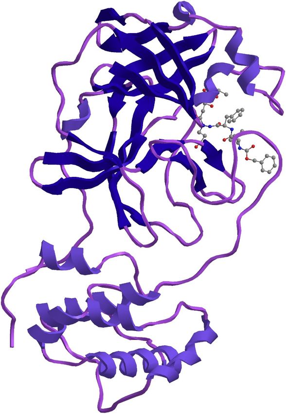
81   We hypothesised that there can be some proteins in the large chuck of proteins in the SARS-

 82   CoV-2 that could have homology with the Non-structural protein 3 (nsp3) SARS CoV and

 83   these proteins can possibly have binding sites with ligands that can bind with known ligand

 84   with antiviral properties.

 85   Here in this study we did a complete bioinformatic analysis, sequence alignment, comparison

 86   of multiple sequences of the SARS-CoV-2 whole genome sequences, the Spike protein and

 87   the polyproteins for homology with known spike proteins and also analysed receptor binding

 88   sites for possible vaccine development.

 89   Materials and Methods

 90   Six complete viral genome sequences, seven polyproteins (RdRp region) and seven

 91   glycoproteins available on NCBI portal on 4 Feb 2020 were taken for analysis. The sequence

 92   details and GenBank accession numbers are listed in Supplementary Table 1. Amongst the

 93   seven polyproteins, five are of Wuhan pneumonia virus isolate SARS-COV-2 and two

 94   sequences are of Wuhan pneumonia virus isolate SI200040-SP. The seven Glycoproteins are

 95   of the same isolate, Wuhan pneumonia virus isolate SARS-COV-2.

 96   The available polyproteins (RdRp region) and glycoprotein sequences were retrieved from

 97   Genbank, NCBI (Benson et al., 2000). These sequences were translated to amino acid

 98   sequences using sorted six frame translation with Bioedit (Hall et al., 2011). Multiple sequence

 99   alignment of the translated protein sequences was performed and phylogenetic tree was

100   constructed using Mega-X (Kumar et al., 2018). The alignment shows that amongst the seven

101   polyproteins, five sequences were identical being from the same isolate and two other

102   sequences of the other isolate are identical. Similar analysis of the seven glycoproteins was

103   done, all the seven glycoprotein sequences were found to be identical. Therefore, further

104   analysis was carried out for three sequences.

                                                      4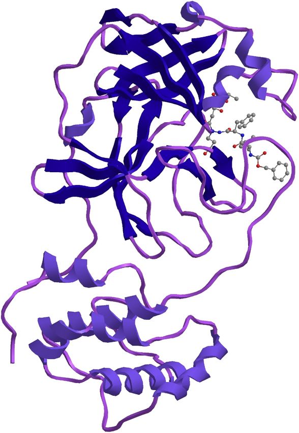
105   1.     MN938385.1 SARS-CoV-2 virus isolate SARS-COV-2 _HKU-SZ-001_2020 ORF1ab

106   polyprotein, RdRp region, (orf1ab) gene, partial cds: 0 to 284: Frame 3 95 aa

107   2.     MN970003.1 SARS-CoV-2 virus isolate SI200040-SP orf1ab polyprotein, RdRP

108   region, (orf1ab) gene, partial cds: 2 to 289: Frame 2 96 aa

109   3.     MN938387.1 SARS-CoV-2 virus isolate SARS-COV-2 _HKU-SZ-001_2020 surface

110   glycoprotein (S) gene, partial cds: 1 to 105: Frame 1 35 aa

111   Expasy proteomics server (Gasteiger et al., 2003) was used to study the protein sequence and

112   structural details. These peptides were studied for their physio-chemical properties using the

113   tool Protparam (Gasteiger et al., 2005). The secondary structure analysis was done using Chou

114   and Fasman algorithm with CFSSP (Kumar, 2013). To generate the 3D structure from the fasta

115   sequence, homology modelling was performed and the templates were identified. The model

116   was built using the template with highest identity. Swiss-model (Schwede et al., 2003) was

117   used to build and validate the 3D model, structural assessment was also performed to validate

118   the model built.

119   Complete genome sequence of the SARS-CoV-2 virus isolate Wuhan-Hu-1 (Genbank

120   Accession Number MN908947.3) which has 29903 bp ss-RNA           linear was translated sorted

121   6 frame with minimum ORF of 20 with any start codon and the resultant protein sequence was

122   used for homology modelling, homology models where done with large chunks of proteins

123   21503 to 25381 in Frame 2 with 1293 amino acids, 13450 to 21552 in Frame 1 with 2701

124   amino acids and 254 to 13480 in Frame 2 with 4409 amino acids.

125   SWISS-MODEL server was used for homology modelling (Waterhouse et al 2018) where

126   computation was on ProMod3 engine which is based on Open Structure (Biasini et al 2013).

127   Structural information is extracted from the template, sequence alignment is used to define

128   insertions and deletions.

                                                     5
129   Protein ligand interaction profile with hydrogen bonding, hydrophobic interactions, salt bridges

130   and π-Stacking was done with PLIP server (Salentin et al., 2015)

131   Results and Discussion

132   The physico- chemical properties and primary structure parameters of the 7 polyproteins RdRp

133   region of the SARS-CoV-2 virus isolate is given in Table 1. RdRP forms an important part of

134   the viral genome where in the RNA viruses its function is to catalyze the synthesis of the RNA

135   strand complementary to a given RNA template.

136   The isolates SI200040-SP orf1ab polyprotein and the isolate SI200121-SP orf1ab polyprotein

137   had 2 reading frames as compared to the rest of the isolates which had 3 reading frames. The

138   presence of multiple reading frames suggests the possibility of overlapping genes as seen in

139   many virus and prokaryotes and mitochondrial genomes. This could affect how the proteins

140   are made. The number of amino acid residues in all the polyproteins were the same expect one

141   isolate SI200040-SP which had one amino acid more than the other polyproteins. The

142   extinction coefficients of the two isolates SI200040-SP orf1ab polyprotein and the isolate

143   SI200121-SP orf1ab polyprotein was much higher compared to the rest of the polyproteins.

144   The extinction coefficient is important when studying protein-protein and protein-ligand

145   interactions. The instability index of these two isolates was also high when compared to the

146   others indicating the that these two isolates are instable. Regulation of gene expression by

147   polyprotein processing is known in viruses and this is seen in many viruses that are human

148   pathogens (Yost et al 2013).

149   The isolates here like many other viruses may be using replication strategy which could involve

150   the translation of a large polyprotein with subsequent cleavage by viral proteases. The two

151   isolates SI200040-SP orf1ab polyprotein and the isolate SI200121-SP orf1ab polyprotein also

152   showed shorter half-lives as compared to the other isolates indicating that they are susceptible

153   to enzymatic degradation.

                                                     6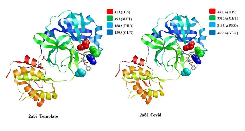
154   The tertiary structure analysis of the isolate SARS-CoV-2 _HKU-SZ-001_2020 ORF1ab

155   polyprotein is given in Table 2. It is seen that the polyprotein has a 98.94 percent identity with

156   PDB structure 6nur.1.A which is a hetero-1-2-1-mer. The polyprotein is an RNA directed RNA

157   polymerase. The protein is identical to the SARS-Coronavirus NSP12 bound to NSP7 and

158   NSP8 co-factors (Kirchdoerfer and Ward 2019). In SARS it is basically a nonstructural protein

159   with NSP12 being the RNA dependent RNA polymerase and the co factors NSP 7 and NSP 8

160   having the function of forming hexadecameric complexes and also act as processivity clamp

161   for RNA polymerase and primase (Fehr et al., 2016). This structure as in SARS CoV here in

162   SARS-CoV-2 may be involved in the machinery of core RNA synthesis and can be a template

163   for exploring antiviral properties.

164   The phylogenetic tree of the seven polyproteins is shown in Fig.2. It is seen that two

165   polyproteins were distinctly different from the rest. The Phylogenetic tree of the seven

166   glycoproteins of the SARS-CoV-2 virus isolate is shown in Fig.3, it is seen that the

167   glycoproteins are similar in all the isolates. Multiple alignment of the Polyproteins of the

168   SARS-CoV-2 is shown in Supplementary Fig.1.

169   This structure as in SARS CoV here in SARS-CoV-2 may be involved in the machinery of core

170   RNA synthesis and can be a template for exploring antiviral properties. Based on its functions

171   in the SARS CoV and its identity to the SARS-CoV-2, it is possible that it has the same

172   functions in SARS-CoV-2 an RNA polymerase which does de novo initiation and primer

173   extension with possible exonuclease activities, the activity itself being primer dependent useful

174   for understanding the mechanism of SARS-CoV-2 replication and can be used as an antiviral

175   target (Te Velthuis et al 2012; Te Velthuis et al 2010; Subissi et al 2014; Subissi et al 2014).

176

                                                      7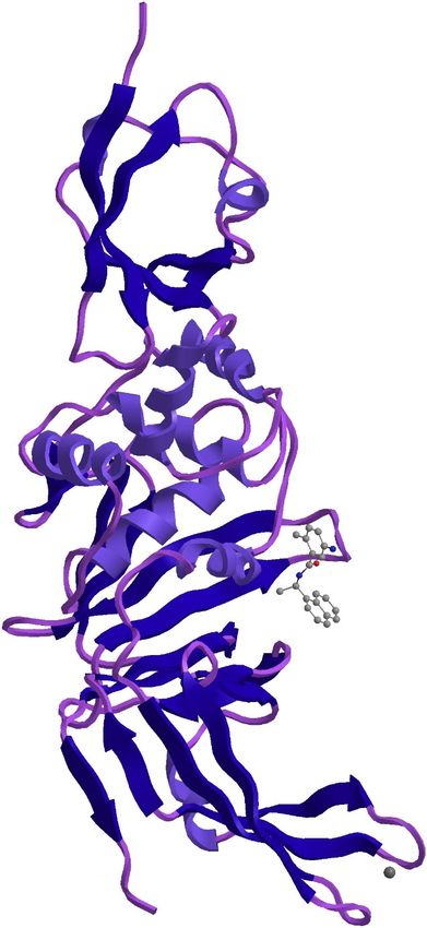
177   The polyprotein also has an identity of 19.74 percent with an ABC-type uncharacterized

178   transport system periplasmic component-like protein, this protein is known to be a substrate

179   binding protein and possible binding can be explored here (Bae et al 2019).

180   The homology model developed from the residues 254 to 13480 in Frame 2 with 4409 amino

181   acids from the Complete genome sequence of the SARS-CoV-2 virus isolate Wuhan-Hu-1

182   (Genbank Accession Number MN908947.3) which has 29903 bp with linear ss-RNA                 linear

183   showed interesting template alignments, in all the model aligned with 50 templates from the

184   PDB database with most of them being replicase polyprotein 1ab which is a SARS-CoV

185   papain-like protease (Daczkowski 2017). The maximum similarity of 97.3 percent was with

186   template structure of a Nsp9 protein from SARS-coronavirus indicating that this novel

187   coronavirus has high degree of similarity with the SARS-coronavirus and this can be used for

188   gaining insights into vaccine development. Nsp 9 is an RNA binding protein and has an

189   oligosaccharide/oligonucleotide fold-like fold, this protein can have an important function in

190   the replication machinery of the virus and can be important when designing antiviral for this

191   virus (Egloff et al 2004).

192   Two models were developed, one from residues 3268 -3573 in Frame 2 with 306 amino

193   acids and the other from the part of the genome residues 1568-1882 in Frame 2 with 315

194   amino acids of the SARS-CoV-2 virus isolate Wuhan-Hu-1 (Genbank Accession Number

195   MN908947.3). The models had similarity with the 3C like proteinase and a papain-like

196   protease/deubiquitinase protein which are known antiviral drug targets. Ligand binding with

197   these proteins and their action is on viral replication and inactivation can be useful in stopping

198   the viral replication (Baez-Santos et al 2015).

199

                                                        8
200 The homology models of the 4409 amino acid residues of the whole genome of the SARS-
201 CoV-2 virus isolate Wuhan-Hu-1 with the ligand association with templates 2a5i and 3e9s are
202 shown in Fig. 4 and Fig. 5 respectively.
203 The statistics of structural comparison with PDB templates is given in Table 5, it is seen that
204 the proteins from the SARS-CoV-2 are significantly close to the proteins of SARS CoV and
205 the amino acid alignment in the biding region is the same in both the viruses.
206 The alignment of the 305 residues from 3268-3573 aa of the Novel Coronavirus COVI-19 with
207 the template 2a5i is shown in Fig.6 and the alignment of the 315 residues from 1568-1882 aa
208 of the Novel Coronavirus COVI-19 with the template 3e9s is shown in Fig.7.
209 The important templates that aligned with this 4409 amino acid residues of the whole genome
210 of the SARS-CoV-2 virus isolate Wuhan-Hu-1were 2a5i of the PDB database which is a
211 crystal structure of SARS coronavirus main peptidase inhibited by an Aza-Peptide epoxide in
212 the space group C2 (Lee et al 2005) and 3e9s of the PDB database which is new class of
213 papain-like protease/deubiquitinase which when combined with ligand GRL0617 acts as
214 inhibitors blocking SARS virus replication (Ratia et al 2008). The model with template 2a5i of
215 the PDB database shows that Aza-Peptide Epoxide (APE; kinact/Ki=1900(±400) M−1 s−1)
216 which is a known anti SARS agent can be used to develop a molecular target with irreversible
217 inhibitor properties. The protein ligand interaction analysis of the Novel Coronavirus C3 like
218 peptidase and aza-peptide epoxide is shown in Fig.8. The substrate binding properties and
219 structural and chemical complementarity of this Aza-Peptide Epoxide can be explored as an
220 anti - Coronavirus SARS-COV-2 agent. The APE which is ethyl (2S)-4-[(3-amino-3-oxo-
221 propyl)-[[(2S)-2-[[(2S)-4-methyl-2-phenylmethoxycarbonylamino-pentanoyl]amino]-3-
222 phenyl-propanoyl]amino]amino]-2-hydroxy-4-oxo-butanoate structure is shown in Fig.9.
223
9224 The model with template 3e9s of the PDB database shows that the Coronavirus viral protein
225 can have a ligand which is a papain-like protease (PLpro) that is known to be a potent inhibitor
226 of viral replication in SARS (Ratia et al 2008).
227 The two parts of the Main protein from the whole genome of the SARS-CoV-2 aligned with
228 two SAR proteins and the ligand binding sites were similar, the alignment positions, number
229 of amino acids and ligand and the interacting residues is given in Table 3
230 The complete genome of MN908947.3 SARS-CoV-2 virus isolate Wuhan-Hu-1 encodes a
231 4409aa long protein along with the other glycoproteins and polyproteins. The homology
232 modelling of this protein showed sequence and structural alignment with two SARS proteases
233 with structural accession numbers 3e9s.1 and 2a5i.1 at positions 1568-1882 and 3268-3573
234 respectively. Reports suggests inhibition of virus replication by TTT ligand and an aza-peptide
235 epoxide inhibiting the main peptidase. The structural similarity of these templates are 83% and
236 96% respectively. The multiple sequence alignment shows complete conservation of the
237 sequence suggesting a high degree of homology. The protein ligand interaction analysis of the
238 Novel Coronavirus non structural protein and papain-like protease is shown in Fig. 10.
239 The Comparison of Hydrophobic interaction, hydrogen bonding, salt bridges of the
240 constructed model of the Novel Coronavirus protein from region 3268-3573 aa to ligand AZP
241 with Hydrophobic interaction, hydrogen bonding, salt bridges of the template 2a5i is given in
242 Suppl. Table 2, when comparing both it is seen that the binding properties are the same expect
243 for the presence of water bridge in the template 2a5i.
244
245 The Comparison of Hydrophobic interaction, hydrogen bonding, π-Stacking of the constructed
246 model of the Novel Coronavirus protein from region 1568-1882 aa to ligand Small molecule
247 Noncovalent Lead Inhibitor with the Hydrophobic interaction, hydrogen bonding, π-Stacking
10248 of the template 3e9s is given in Suppl. Table 3, when comparing both it is seen that the binding
249 properties are the same except or an additional π-Stacking at Tyr in the template 2a5i. This
250 shows that there is high possibility of binding of these antiviral compounds with the regions of
251 Novel Coronavirus protein that is in homology with the SARS protein.
252 Comparison of the hydrophobic interaction of the biding of the ligand AZP between the SARS-
253 CoV-2 protein and the template 2a5i of SARS CoV is shown in Fig.11 and the comparison of
254 the hydrophobic interaction of the biding of the ligand AZP between the SARS-CoV-2 protein
255 and the template 3e9s of SARS CoV is shown in Fig.12. It is seen that the interaction is the
256 same in both proteins with the same amino acids participating in the interaction indicating that
257 there is a possibility that these ligands with antiviral properties can bind to the new virus.
258 The similarity in the amin acids involved in the Hydrophobic interactions which are short range
259 interactions and have an important role in the affinities of the ligands and receptors shows that
260 the proteins of the SARS-CoV-2 may bind with the same affinity as seen in the SARS CoV
261 and this also shows a similar action of the ligand as seen in SARS CoV, indicating that these
262 ligands can be used as antivirals in the SARS-CoV-2.
263 The targeting of this part of the genome of the SARS-CoV-2 with the antiviral compounds
264 which have shown to bind in the similar region of the SARS virus can have implication in the
265 development of an effective antiviral compound against the SARS-CoV-2 . The SARS-CoV-2
266 shows homology with the SARS coronaviral proteases, papain-like protease (PLpro) and 3C-
267 like protease (3CLpro), these proteins have the function of processing the viral polyprotein and
268 also they perform the function of stripping ubiquitin and the ubiquitin-like interferon (IFN)-
269 stimulated gene 15 (ISG15) from the hosts to facilitate coronavirus replication and help in
270 evading immune response of the host, these inhibitors can also have a role in disrupting
271 signalling cascades in infected cells and protecting the uninfected cells.
11272 The chemical GRL0617 is 5-Amino-2-methyl-N-[(1R)-1-(1-naphthalenyl)ethyl]benzamide
273 and is known to inhibit the papainlike protease that is present in SARS CoV . This protease is
274 a potential target for antiviral compounds (Chaudhuri et al., 2011). We found the SARS-CoV-
275 2 has homology with this and the binding sites for this in the structural protein of the SARS-
276 CoV-2 is the same (Table 4). This compound inhibits the enzyme that is required for the
277 cleavage of the viral protein from the virus in SARS CoV, it also cleaves ubiquitin and has a
278 structural homology with the Deubiquitinases (DUBs) of the Ubiquitin-Specific Proteases
279 Compound GRL0617 binds in the S4 and S3 enzyme subsite that gets the C terminal tail of the
280 Ubiquitin (King and Finley 2014; Schauer et al., 2019). Our results show that Aza-Peptide
281 Epoxide an irreversible protease inhibitor and GRL0617 a viral replication inhibitor can be
282 used to develop inhibitors of the Novel Coronavirus SARS-COV-2.
283
284 References
285 Bae, J.E., Kim, I.J., Kim, K.J. and Nam, K.H., 2018. Crystal structure of a substrate-binding
286 protein from Rhodothermus marinus reveals a single α/β-domain. Biochemical and
287 Biophysical Research Communications, 497(1), pp.368-373.
288 Baez-Santos, Y.M., John, S.E.S. and Mesecar, A.D., 2015. The SARS-coronavirus papain-like
289 protease: structure, function and inhibition by designed antiviral compounds. Antiviral
290 Research, 115, pp.21-38.
291 Benson, D. A., Karsch-Mizrachi, I., Lipman, D. J., Ostell, J., Rapp, B. A., & Wheeler, D. L.
292 (2000). GenBank. Nucleic Acids Rresearch, 28(1), 15-18.
293 Biasini, M., Schmidt, T., Bienert, S., Mariani, V., Studer, G., Haas, J., Johner, N., Schenk,
294 A.D., Philippsen, A. and Schwede, T., 2013. OpenStructure: an integrated software
12295 framework for computational structural biology. Acta Crystallographica Section D:
296 Biological Crystallography, 69(5), pp.701-709.
297 Chaudhuri, R., Tang, S., Zhao, G., Lu, H., Case, D.A. and Johnson, M.E., 2011. Comparison
298 of SARS and NL63 papain-like protease binding sites and binding site dynamics:
299 inhibitor design implications. Journal of molecular biology, 414(2), pp.272-288.
300 Chen, Y., Savinov, S.N., Mielech, A.M., Cao, T., Baker, S.C. and Mesecar, A.D., 2015. X-ray
301 structural and functional studies of the three tandemly linked domains of non-structural
302 protein 3 (nsp3) from murine hepatitis virus reveal conserved functions. Journal of
303 Biological Chemistry, 290(42), pp.25293-25306.
304 Daczkowski, C.M., Dzimianski, J.V., Clasman, J.R., Goodwin, O., Mesecar, A.D. and Pegan,
305 S.D., 2017. Structural insights into the interaction of coronavirus papain-like proteases
306 and interferon-stimulated gene product 15 from different species. Journal of Molecular
307 Biology, 429(11), pp.1661-1683.
308 de Wit, E., Rasmussen, A.L., Falzarano, D., Bushmaker, T., Feldmann, F., Brining, D.L.,
309 Fischer, E.R., Martellaro, C., Okumura, A., Chang, J. and Scott, D., 2013. Middle East
310 respiratory syndrome coronavirus (MERS-CoV) causes transient lower respiratory tract
311 infection in rhesus macaques. Proceedings of the National Academy of Sciences,
312 110(41), pp.16598-16603.
313 de Wit, E., van Doremalen N., D. Falzarano, V. J. Munster, SARS and MERS: recent insights
314 into emerging coronaviruses. Nat Rev Microbiol 14, 523-534 (2016).
315 Du, L., He, Y., Zhou, Y., Liu, S., Zheng, B.J. and Jiang, S., 2009. The spike protein of SARS-
316 CoV—a target for vaccine and therapeutic development. Nature Reviews
317 Microbiology, 7(3), pp.226-236.
13318 Egloff, M.P., Ferron, F., Campanacci, V., Longhi, S., Rancurel, C., Dutartre, H., Snijder, E.J.,
319 Gorbalenya, A.E., Cambillau, C. and Canard, B., 2004. The severe acute respiratory
320 syndrome-coronavirus replicative protein nsp9 is a single-stranded RNA-binding
321 subunit unique in the RNA virus world. Proceedings of the National Academy of
322 Sciences, 101(11), pp.3792-3796.
323 Fehr, A.R. and Perlman, S., 2015. Coronaviruses: an overview of their replication and
324 pathogenesis. In Coronaviruses (pp. 1-23). Humana Press, New York, NY.
325 Gasteiger, E., Gattiker, A., Hoogland, C., Ivanyi, I., Appel, R. D., & Bairoch, A. (2003).
326 ExPASy: the proteomics server for in-depth protein knowledge and analysis. Nucleic
327 Acids Research, 31(13), 3784-3788.
328 Gasteiger, E., Hoogland, C., Gattiker, A., Wilkins, M. R., Appel, R. D., & Bairoch, A. (2005).
329 Protein identification and analysis tools on the ExPASy server. In The proteomics
330 protocols handbook (pp. 571-607). Humana press.
331 Hall, T., Biosciences, I., & Carlsbad, C. (2011). BioEdit: an important software for molecular
332 biology. GERF Bull Biosci, 2(1), 60-61.
333 King, R.W. and Finley, D., 2014. Sculpting the proteome with small molecules. Nature
334 chemical biology, 10(11), p.870.
335 Kirchdoerfer, R.N. and Ward, A.B., 2019. Structure of the SARS-CoV nsp12 polymerase
336 bound to nsp7 and nsp8 co-factors. Nature Communications, 10(1), pp.1-9.
337 Kumar, S., Stecher, G., Li, M., Knyaz, C., & Tamura, K. (2018). MEGA X: molecular
338 evolutionary genetics analysis across computing platforms. Molecular Biology and
339 Evolution, 35(6), 1547-1549.
14340 Kumar, T. A. (2013). CFSSP: Chou and Fasman secondary structure prediction server. Wide
341 Spectrum, 1(9), 15-19.
342 Lee, T.W., Cherney, M.M., Huitema, C., Liu, J., James, K.E., Powers, J.C., Eltis, L.D. and
343 James, M.N., 2005. Crystal structures of the main peptidase from the SARS coronavirus
344 inhibited by a substrate-like aza-peptide epoxide. Journal of Molecular Biology, 353(5),
345 pp.1137-1151.
346 Liu, W., Morse, J.S., Lalonde, T. and Xu, S., 2020. Learning from the Past: Possible Urgent
347 Prevention and Treatment Options for Severe Acute Respiratory Infections Caused by
348 2019‐nCoV. ChemBioChem.
349 Luo, C.M., Wang, N., Yang, X.L., Liu, H.Z., Zhang, W., Li, B., Hu, B., Peng, C., Geng, Q.B.,
350 Zhu, G.J. and Li, F., 2018. Discovery of novel bat coronaviruses in south China that
351 use the same receptor as Middle East respiratory syndrome coronavirus. Journal of
352 Virology, 92(13), pp.e00116-18.
353 Ratia, K., Pegan, S., Takayama, J., Sleeman K., Coughlin, M., Baliji, S., Chaudhuri, R., Fu,
354 W., Prabhakar, B.S., Johnson, M.E. and Baker, S.C., 2008. A noncovalent class of
355 papain-like protease/deubiquitinase inhibitors blocks SARS virus replication.
356 Proceedings of the National Academy of Sciences, 105(42), pp.16119-16124.
357 Salentin, S., Schreiber, S., Haupt, V.J., Adasme, M.F. and Schroeder, M., 2015. PLIP: fully
358 automated protein–ligand interaction profiler. Nucleic acids research, 43(W1),
359 pp.W443-W447
360 Schauer, N.J., Magin, R.S., Liu, X., Doherty, L.M. and Buhrlage, S.J., 2019. Advances in
361 Discovering Deubiquitinating Enzyme (DUB) Inhibitors. Journal of medicinal
362 chemistry.
15363 Schwede, T., Kopp, J., Guex, N., & Peitsch, M. C. (2003). SWISS-MODEL: an automated
364 protein homology-modeling server. Nucleic Acids Research, 31(13), 3381-3385.
365 Subissi, L., Imbert, I., Ferron, F., Collet, A., Coutard, B., Decroly, E. and Canard, B., 2014.
366 SARS-CoV ORF1b-encoded nonstructural proteins 12–16: replicative enzymes as
367 antiviral targets. Antiviral research, 101, pp.122-130.
368 Subissi, L., Posthuma, C.C., Collet, A., Zevenhoven-Dobbe, J.C., Gorbalenya, A.E., Decroly,
369 E., Snijder, E.J., Canard, B. and Imbert, I., 2014. One severe acute respiratory syndrome
370 coronavirus protein complex integrates processive RNA polymerase and exonuclease
371 activities. Proceedings of the National Academy of Sciences, 111(37), pp.E3900-
372 E3909.
373 Te Velthuis, A.J., Arnold, J.J., Cameron, C.E., van den Worm, S.H. and Snijder, E.J., 2010.
374 The RNA polymerase activity of SARS-coronavirus nsp12 is primer dependent.
375 Nucleic acids research, 38(1), pp.203-214.
376 Te Velthuis, A.J., van den Worm, S.H. and Snijder, E.J., 2012. The SARS-coronavirus nsp7+
377 nsp8 complex is a unique multimeric RNA polymerase capable of both de novo
378 initiation and primer extension. Nucleic acids research, 40(4), pp.1737-1747.
379 Waterhouse, A., Bertoni, M., Bienert, S., Studer, G., Tauriello, G., Gumienny, R., Heer, F.T.,
380 de Beer, T.A.P., Rempfer, C., Bordoli, L. and Lepore, R., 2018. SWISS-MODEL:
381 homology modelling of protein structures and complexes. Nucleic Acids Research,
382 46(W1), pp.W296-W303.
383 World Health Organization (WHO) 2004. [Accessed 11 Feb 2020]
384 https://www.who.int/csr/don/2004_05_18a/en/
16385 World Health Organization (WHO). Coronavirus. Geneva: WHO; 2020 [Accessed 4 Feb
386 2020]. Available from: https://www.who.int/health-topics/coronavirus
387 Yost, S.A. and Marcotrigiano, J., 2013. Viral precursor polyproteins: keys of regulation from
388 replication to maturation. Current Opinion in Virology, 3(2), pp.137-142.
389
390 Figure Captions
391
392 Fig.1 Countries, territories or areas with reported confirmed cases of SARS-COV-2 , 3
393 February 2020 Source WHO (https://www.who.int/docs/default-
394 ource/coronaviruse/situation-reports/20200219-sitrep-30-covid-
395 19.pdf?sfvrsn=6e50645_2)
17396
397 Fig.2 Phylogenetic tree of the seven polyproteins of Severe acute respiratory syndrome
398 coronavirus 2 isolate virus isolates
399
400 Fig.3 Phylogenetic tree of the seven glycoproteins of Severe acute respiratory syndrome
401 coronavirus 2 isolate virus isolates
402
18403
404
405
406 Fig. 4 Homology model with ligand binding of protein from amino acids 3268 -3573 in Frame
407 2 with 306 amino acids of the Complete genome sequence of the SARS-CoV-2 virus
408 isolate Wuhan-Hu-1 (Genbank Accession Number MN908947.3) which has 29903 bp
409 linear ss-RNA with 2a5i of the PDB database as template.
410
411
19412
413
414
415 Fig. 5 Homology model with ligand binding of protein from residues 1568-1882 in Frame 2
416 with 315 amino acids of the Complete genome sequence of the SARS-CoV-2 virus
417 isolate Wuhan-Hu-1 (Genbank Accession Number MN908947.3) which has 29903 bp
418 linear ss-RNA with 3e9s of the PDB database as template.
419
20420
421
422
423 Fig. 6 Alignment of the 305 residues from 3268-3573 aa of the Novel Coronavirus COVI-19
424 with the template 2a5i
425
426 Fig.7 the alignment of the 315 residues from 1568-1882 aa of the Novel Coronavirus COVI-
427 19 with the template 3e9s
21428
429 Fig.8 Protein Ligand interaction between the C3 like peptidase with aza-peptide epoxide of the
430 model with the template 2a5i
22431
432 Fig. 9 Structure of Aza-Peptide Epoxide (APE) ethyl (2S)-4-[(3-amino-3-oxo-propyl)-[[(2S)-
433 2-[[(2S)-4-methyl-2-phenylmethoxycarbonylamino-pentanoyl]amino]-3-phenyl-
434 propanoyl]amino]amino]-2-hydroxy-4-oxo-butanoate with possible anti Coronavirus
435 activity – (Source https://www.rcsb.org/ligand/AZP)
23436
437 Fig.10 Protein Ligand interaction between the Novel Coronavirus non structural protein and
438 papain-like protease of the model with the template 3e9s
439
440
441
442
443
444
445
446
447
24448
449
450
451 Fig.11 Comparison of the hydrophobic interaction of the biding of the ligand AZP between the
452 SARS-CoV-2 protein and the template 2a5i of SARS CoV
453
454
455
456
457
458
459
460
461
25462
463
464
465
466 Fig.12 comparison of the hydrophobic interaction of the biding of the ligand AZP between the
467 SARS-CoV-2 protein and the template 3e9s of SARS CoV
468
469
26470
471 Supplementary Fig.1 Multiple alignment of the Polyproteins of Severe acute respiratory
472 syndrome coronavirus 2 isolate virus isolates
473
474
475
27Coronavirus_MS_16.pdf (3.03 MiB) view on ChemRxiv download file
Figure 12.jpg (127.89 KiB) view on ChemRxiv download file
1 Whole Genome Sequence Analysis and Homology Modelling of a 3C Like Peptidase and
 2 a Non-Structural Protein 3 of the SARS-CoV-2 Shows Protein Ligand Interaction with
 3 an Aza-Peptide and a Noncovalent Lead Inhibitor with Possible Antiviral Properties

 4                            Arun K. Shanker1 *, Divya Bhanu1,2 and Anjani Alluri3

                          1
 5                         ICAR - Central Research Institute for Dryland Agriculture

 6                                    Santoshnagar, Hyderabad – 500059, India

 7                             Corresponding author email: arunshank@gmail.com

               2
 8                 Centre for Plant Molecular Biology, Osmania University, Hyderabad, India

 9 3 Advanced Post Graduate Centre, Acharya N.G.Ranga Agricultural University, Guntur, India

10Abstract

11The family of viruses belonging to Coronaviridae mainly consist of virulent pathogens that have a zoonotic property, Severe
12Acute Respiratory Syndrome (SARS-CoV) and Middle East Respiratory Syndrome (MERS-CoV) of this family have
13emerged before and now the SARS-CoV-2 has emerged in China. Characterization of spike glycoproteins, polyproteins and
14other viral proteins from viruses are important for vaccine development. Homology modelling of these proteins with known
15templates offers the opportunity to discover ligand binding sites and explore the possible antiviral properties of these protein
16ligand complexes. Any information emerging from these protein models can be used for vaccine development. In this study
17we did a complete bioinformatic analysis, sequence alignment, comparison of multiple sequences and homology modelling
18of the SARS-CoV-2 whole genome sequences, the spike protein and the polyproteins for homology with known proteins, we
19also analysed receptor binding sites in these models for possible binding with ligands that exhibit antiviral properties. Our
20results showed that the tertiary structure of the polyprotein isolate SARS-CoV-2_HKU-SZ-001_2020 had 98.94 percent
21identity with SARS-Coronavirus NSP12 bound to NSP7 and NSP8 co-factors. Our results indicate that a part of the viral
22genome (residues 3268 -3573 in Frame 2 with 306 amino acids) of the SARS-CoV-2 virus isolate Wuhan-Hu-1 (Genbank
23Accession Number MN908947.3) when modelled with template 2a5i of the PDB database had 96 percent identity with a
243C like peptidase of SARS-CoV which has ability to bind with Aza-Peptide Epoxide (APE) which is known for irreversible
25inhibition of SARS-CoV main peptidase. The part of the genome (residues 1568-1882 in Frame 2 with 315 amino acids)
26when modelled with template 3e9s of the PDB database had 82 percent identity with a papain-like protease/deubiquitinase
27which when complexed with ligand GRL0617 acts as inhibitor which can block SARS-CoV replication. It is possible that
28these viral inhibiters can be used for vaccine development for the SARS-CoV-2.

29

30Introduction

31More than a decade has passed since the emergence human Coronavirus that caused Severe

32Respiratory Syndrome (SARS-CoV) and it is about 7 years since the emergence of another

33type of Coronavirus - Middle East Respiratory Syndrome (MERS-CoV) and now the SARS-

                                                                 1
34CoV-2 has emerged in China. This repeated onslaught of these viruses goes to show that it
35can assume pandemic proportions at any time and at any place.
36The family of viruses belonging to Coronaviridae mainly consist of virulent pathogens that
37have a zoonotic property and this large family of corona viruses, have been known to be
38circulating in animals including camels, cats and bats. It has been seen in the past that Severe
39Acute Respiratory Syndrome associated coronavirus (SARS-CoV) and Middle East
40Respiratory Syndrome-associated coronavirus (MERS-CoV) belonging to this family of
41viruses can be transmitted from animals to humans and can cause respiratory diseases.
42Human to human transmission on this virus has been a concern and due to this search for
43antiviral compounds and vaccine development for this family of virus becomes the need of
44the hour.
45The SARS was first seen in 2002 in Guangdong province of China, and later spread globally
46and has caused close to about 8096 cases (WHO 2004, de Vit et al., 2016). In 2012, a novel
47betacoronavirus, designated Middle East respiratory syndrome coronavirus or MERS-CoV
48associated with severe respiratory disease in humans, emerged in the Arabian Peninsula (de
49Wit et al., 2013).
50The World Health Organization (WHO), China Country Office was informed of cases of
51pneumonia of unknown aetiology in Wuhan City, Hubei Province, on 31 December 2019
52(WHO 2020). A novel coronavirus currently termed SARS-COV-2 was officially announced
53as the causative agent by Chinese authorities on 7 January 2020. As on 20 Feb 2020 China’s
54National Health Commission reported that there are 74,280 confirmed cases in China (Fig.1).
55The World Health Organization reported 924 confirmed cases in 25 countries outside China
56(WHO Situation Report 29 2020). This novel corona virus has been designated as SARS-
57CoV-2.
258
59Coronaviruses are RNA viruses and have large genomes structures and due to this they can
60have high error in replication as compared to host genomes. It is also known that various
61CoVs can do effective recombination of their genomes after infecting host cells (Luo et al
622018). This recombination can be a factor for their evolution to novel types which may have
63new animals as their intermediate hosts. These factors give the CoVs high adaptive ability
64and the capability to jump across species and have a relatively large host range.
65Characterization of Spike glycoproteins from viruses are important for vaccine development.
66Any information coming from the protein model can be used for vaccine development. In
67Silico Epitope, polyprotein and spike protein-based peptide vaccine designing for infectious
68viruses is a way that can hasten the process of vaccine development. Spike (S) protein,
69polyprotein and other viral proteins of the SARS-CoV-2 as a target for the development of
70vaccines and therapeutics for the prevention and treatment of infection is an important
71approach. In the case of SARS-CoV, these proteins can mediate binding of the virus with its
72receptor and promotes the fusion between the viral and host cell membranes and virus entry
73into the host cell, hence peptides, antibodies, organic compounds and short interfering RNAs
74that interact with the spike protein can have a potential role in vaccine development (Du et al
752009).
76There are multiple domain functions that are active in the replication of the coronavirus and
77these domains are present in a protein designated as Non-structural protein 3 (nsp3) which is
78the largest protein in the coronavirus genome (Chen et al 2015). 3C like protease (3CLpro)
79and Papain like Protease (PLpro) are two important class of proteases that are involved in the
80process of translation of the polypeptide from the genomic RNA to protein components that
381are required structurally or non-structurally for replication and packaging of new generation
82viruses (Liu et al 2020)
83We hypothesised that there can be some proteins in the large chuck of proteins in the SARS-
84CoV-2 that could have homology with the Non-structural protein 3 (nsp3) SARS CoV and
85these proteins can possibly have binding sites with ligands that can bind with known ligand
86with antiviral properties.
87Here in this study we did a complete bioinformatic analysis, sequence alignment, comparison
88of multiple sequences of the SARS-CoV-2 whole genome sequences, the Spike protein and
89the polyproteins for homology with known spike proteins and also analysed receptor binding
90sites for possible vaccine development.
91Materials and Methods
92Six complete viral genome sequences, seven polyproteins (RdRp region) and seven
93glycoproteins available on NCBI portal on 4 Feb 2020 were taken for analysis. The sequence
94details and GenBank accession numbers are listed in Supplementary Table 1. Amongst the
95seven polyproteins, five are of Wuhan pneumonia virus isolate SARS-COV-2 and two
96sequences are of Wuhan pneumonia virus isolate SI200040-SP. The seven Glycoproteins are
97of the same isolate, Wuhan pneumonia virus isolate SARS-COV-2.
98The available polyproteins (RdRp region) and glycoprotein sequences were retrieved from
99Genbank, NCBI (Benson et al., 2000). These sequences were translated to amino acid
100sequences using sorted six frame translation with Bioedit (Hall et al., 2011). Multiple
101sequence alignment of the translated protein sequences was performed and phylogenetic tree
102was constructed using Mega-X (Kumar et al., 2018). The alignment shows that amongst the
103seven polyproteins, five sequences were identical being from the same isolate and two other
104sequences of the other isolate are identical. Similar analysis of the seven glycoproteins was
4105done, all the seven glycoprotein sequences were found to be identical. Therefore, further
106analysis was carried out for three sequences.
1071. MN938385.1 SARS-CoV-2 virus isolate SARS-COV-2 _HKU-SZ-001_2020 ORF1ab
108polyprotein, RdRp region, (orf1ab) gene, partial cds: 0 to 284: Frame 3 95 aa
1092. MN970003.1 SARS-CoV-2 virus isolate SI200040-SP orf1ab polyprotein, RdRP
110region, (orf1ab) gene, partial cds: 2 to 289: Frame 2 96 aa
1113. MN938387.1 SARS-CoV-2 virus isolate SARS-COV-2 _HKU-SZ-001_2020 surface
112glycoprotein (S) gene, partial cds: 1 to 105: Frame 1 35 aa
113Expasy proteomics server (Gasteiger et al., 2003) was used to study the protein sequence and
114structural details. These peptides were studied for their physio-chemical properties using the
115tool Protparam (Gasteiger et al., 2005). The secondary structure analysis was done using
116Chou and Fasman algorithm with CFSSP (Kumar, 2013). To generate the 3D structure from
117the fasta sequence, homology modelling was performed and the templates were identified.
118The model was built using the template with highest identity. Swiss-model (Schwede et al.,
1192003) was used to build and validate the 3D model, structural assessment was also performed
120to validate the model built.
121Complete genome sequence of the SARS-CoV-2 virus isolate Wuhan-Hu-1 (Genbank
122Accession Number MN908947.3) which has 29903 bp ss-RNA linear was translated
123sorted 6 frame with minimum ORF of 20 with any start codon and the resultant protein
124sequence was used for homology modelling, homology models where done with large chunks
125of proteins 21503 to 25381 in Frame 2 with 1293 amino acids, 13450 to 21552 in Frame 1
126with 2701 amino acids and 254 to 13480 in Frame 2 with 4409 amino acids.
127SWISS-MODEL server was used for homology modelling (Waterhouse et al 2018) where
128computation was on ProMod3 engine which is based on Open Structure (Biasini et al 2013).
5129Structural information is extracted from the template, sequence alignment is used to define
130insertions and deletions.
131Protein ligand interaction profile with hydrogen bonding, hydrophobic interactions, salt
132bridges and π-Stacking was done with PLIP server (Salentin et al., 2015)
133Results and Discussion
134The physico- chemical properties and primary structure parameters of the 7 polyproteins
135RdRp region of the SARS-CoV-2 virus isolate is given in Table 1. RdRP forms an important
136part of the viral genome where in the RNA viruses its function is to catalyze the synthesis of
137the RNA strand complementary to a given RNA template.
138The isolates SI200040-SP orf1ab polyprotein and the isolate SI200121-SP orf1ab polyprotein
139had 2 reading frames as compared to the rest of the isolates which had 3 reading frames. The
140presence of multiple reading frames suggests the possibility of overlapping genes as seen in
141many virus and prokaryotes and mitochondrial genomes. This could affect how the proteins
142are made. The number of amino acid residues in all the polyproteins were the same expect
143one isolate SI200040-SP which had one amino acid more than the other polyproteins. The
144extinction coefficients of the two isolates SI200040-SP orf1ab polyprotein and the isolate
145SI200121-SP orf1ab polyprotein was much higher compared to the rest of the polyproteins.
146The extinction coefficient is important when studying protein-protein and protein-ligand
147interactions. The instability index of these two isolates was also high when compared to the
148others indicating the that these two isolates are instable. Regulation of gene expression by
149polyprotein processing is known in viruses and this is seen in many viruses that are human
150pathogens (Yost et al 2013).
151The isolates here like many other viruses may be using replication strategy which could
152involve the translation of a large polyprotein with subsequent cleavage by viral proteases.
6153The two isolates SI200040-SP orf1ab polyprotein and the isolate SI200121-SP orf1ab
154polyprotein also showed shorter half-lives as compared to the other isolates indicating that
155they are susceptible to enzymatic degradation.
156The tertiary structure analysis of the isolate SARS-CoV-2 _HKU-SZ-001_2020 ORF1ab
157polyprotein is given in Table 2. It is seen that the polyprotein has a 98.94 percent identity
158with PDB structure 6nur.1.A which is a hetero-1-2-1-mer. The polyprotein is an RNA directed
159RNA polymerase. The protein is identical to the SARS-Coronavirus NSP12 bound to NSP7
160and NSP8 co-factors (Kirchdoerfer and Ward 2019). In SARS it is basically a nonstructural
161protein with NSP12 being the RNA dependent RNA polymerase and the co factors NSP 7 and
162NSP 8 having the function of forming hexadecameric complexes and also act as processivity
163clamp for RNA polymerase and primase (Fehr et al., 2016). This structure as in SARS CoV
164here in SARS-CoV-2 may be involved in the machinery of core RNA synthesis and can be a
165template for exploring antiviral properties.
166The phylogenetic tree of the seven polyproteins is shown in Fig.2. It is seen that two
167polyproteins were distinctly different from the rest. The Phylogenetic tree of the seven
168glycoproteins of the SARS-CoV-2 virus isolate is shown in Fig.3, it is seen that the
169glycoproteins are similar in all the isolates. Multiple alignment of the Polyproteins of the
170SARS-CoV-2 is shown in Supplementary Fig.1.
171This structure as in SARS CoV here in SARS-CoV-2 may be involved in the machinery of
172core RNA synthesis and can be a template for exploring antiviral properties. Based on its
173functions in the SARS CoV and its identity to the SARS-CoV-2, it is possible that it has the
174same functions in SARS-CoV-2 an RNA polymerase which does de novo initiation and
175primer extension with possible exonuclease activities, the activity itself being primer
176dependent useful for understanding the mechanism of SARS-CoV-2 replication and can be
7177used as an antiviral target (Te Velthuis et al 2012; Te Velthuis et al 2010; Subissi et al 2014;
178Subissi et al 2014).
179
180The polyprotein also has an identity of 19.74 percent with an ABC-type uncharacterized
181transport system periplasmic component-like protein, this protein is known to be a substrate
182binding protein and possible binding can be explored here (Bae et al 2019).
183The homology model developed from the residues 254 to 13480 in Frame 2 with 4409
184amino acids from the Complete genome sequence of the SARS-CoV-2 virus isolate Wuhan-
185Hu-1 (Genbank Accession Number MN908947.3) which has 29903 bp with linear ss-RNA
186linear showed interesting template alignments, in all the model aligned with 50 templates
187from the PDB database with most of them being replicase polyprotein 1ab which is a SARS-
188CoV papain-like protease (Daczkowski 2017). The maximum similarity of 97.3 percent was
189with template structure of a Nsp9 protein from SARS-coronavirus indicating that this novel
190coronavirus has high degree of similarity with the SARS-coronavirus and this can be used for
191gaining insights into vaccine development. Nsp 9 is an RNA binding protein and has an
192oligosaccharide/oligonucleotide fold-like fold, this protein can have an important function in
193the replication machinery of the virus and can be important when designing antiviral for this
194virus (Egloff et al 2004).
195Two models were developed, one from residues 3268 -3573 in Frame 2 with 306 amino
196acids and the other from the part of the genome residues 1568-1882 in Frame 2 with 315
197amino acids of the SARS-CoV-2 virus isolate Wuhan-Hu-1 (Genbank Accession Number
198MN908947.3). The models had similarity with the 3C like proteinase and a papain-like
199protease/deubiquitinase protein which are known antiviral drug targets. Ligand binding with
8200these proteins and their action is on viral replication and inactivation can be useful in
201stopping the viral replication (Baez-Santos et al 2015).
202
203The homology models of the 4409 amino acid residues of the whole genome of the SARS-
204CoV-2 virus isolate Wuhan-Hu-1 with the ligand association with templates 2a5i and 3e9s are
205shown in Fig. 4 and Fig. 5 respectively.
206The statistics of structural comparison with PDB templates is given in Table 5, it is seen that
207the proteins from the SARS-CoV-2 are significantly close to the proteins of SARS CoV and
208the amino acid alignment in the biding region is the same in both the viruses.
209The alignment of the 305 residues from 3268-3573 aa of the Novel Coronavirus COVI-19
210with the template 2a5i is shown in Fig.6 and the alignment of the 315 residues from 1568-
2111882 aa of the Novel Coronavirus COVI-19 with the template 3e9s is shown in Fig.7.
212The important templates that aligned with this 4409 amino acid residues of the whole genome
213of the SARS-CoV-2 virus isolate Wuhan-Hu-1were 2a5i of the PDB database which is a
214crystal structure of SARS coronavirus main peptidase inhibited by an Aza-Peptide epoxide in
215the space group C2 (Lee et al 2005) and 3e9s of the PDB database which is new class of
216papain-like protease/deubiquitinase which when combined with ligand GRL0617 acts as
217inhibitors blocking SARS virus replication (Ratia et al 2008). The model with template 2a5i
218of the PDB database shows that Aza-Peptide Epoxide (APE; kinact/Ki=1900(±400) M −1 s−1)
219which is a known anti SARS agent can be used to develop a molecular target with irreversible
220inhibitor properties. The protein ligand interaction analysis of the Novel Coronavirus C3 like
221peptidase and aza-peptide epoxide is shown in Fig.8. The substrate binding properties and
222structural and chemical complementarity of this Aza-Peptide Epoxide can be explored as an
223anti - Coronavirus SARS-COV-2 agent. The APE which is ethyl (2S)-4-[(3-amino-3-oxo-
9You can also read