IGM AND IGG IMMUNOREACTIVITY OF SARS-COV-2 RECOMBINANT M PROTEIN
←
→
Page content transcription
If your browser does not render page correctly, please read the page content below
International Journal of
               Molecular Sciences

Article
IgM and IgG Immunoreactivity of SARS-CoV-2 Recombinant
M Protein
Zorana Lopandić 1,† , Isidora Protić-Rosić 1,† , Aleksandra Todorović 2 , Sofija Glamočlija 2 , Marija Gnjatović 2 ,
Danica Ćujic 2 and Marija Gavrović-Jankulović 1, *

                                               1   Department of Biochemistry, Faculty of Chemistry, University of Belgrade, 11000 Belgrade, Serbia;
                                                   lopandic@chem.bg.ac.rs (Z.L.); proticrosic@chem.bg.ac.rs (I.P.-R.)
                                               2   Institute for the Application of Nuclear Energy, University of Belgrade, 11000 Beograde, Serbia;
                                                   aleksandra.todorovic@inep.co.rs (A.T.); sofija.glamoclija@inep.co.rs (S.G.); marijad@inep.co.rs (M.G.);
                                                   danicac@inep.co.rs (D.Ć.)
                                               *   Correspondence: mgavrov@chem.bg.ac.rs; Tel.: +381-11-333-6661
                                               †   These authors contributed equally to this work.

                                               Abstract: Diagnostic evaluation of specific antibodies against the SARS-CoV-2 virus is mainly based
                                               on spike (S) and nucleocapsid (N) proteins. Despite the critical functions in virus infection and
                                               contribution to the pattern of immunodominance in COVID-19, exploitation of the most abundant
                                               membrane (M) protein in the SARS-CoV-2 serology tests is minimal. This study investigated the
                                               recombinant M protein’s immunoreactivity with the sera from COVID-19 convalescents. In silico
                                               designed protein was created from the outer N-terminal part (19 aa) and internal C-terminal tail
                                               (101–222 aa) of the M protein (YP_009724393.1) and was recombinantly produced and purified.
                                     The designed M protein (16,498.74 Da, pI 8.79) revealed both IgM and IgG reactivity with serum

                                               samples from COVID-19 convalescents in Western blot. In ELISA, more than 93% (28/30) of COVID-
Citation: Lopandić, Z.; Protić-Rosić, I.;
                                               19 sera were positive for IgM detection, and more than 96% (29/30) were positive for specific IgG
Todorović, A.; Glamočlija, S.;
                                               detection to M protein. Based on the capacity to provoke an immune response and its strong antigenic
Gnjatović, M.; Ćujic, D.; Gavrović-
                                               properties, as shown here, and the fact that it is also involved in the virion entry into host cells, the M
Jankulović, M. IgM and IgG
Immunoreactivity of SARS-CoV-2
                                               protein of the SARS-CoV-2 virus as a good antigen has the potential in diagnostic purposes and
Recombinant M Protein. Int. J. Mol.            vaccine design.
Sci. 2021, 22, 4951. https://doi.org/
10.3390/ijms22094951                           Keywords: SARS-CoV-2 membrane protein; COVID-19; IgM reactivity; IgG reactivity; antigenicity; ELISA

Academic Editor: Yoshiro Kobayashi

Received: 26 March 2021                        1. Introduction
Accepted: 21 April 2021
                                                    An outbreak of coronavirus in Wuhan was reported by the World Health Organization
Published: 7 May 2021
                                               (WHO) country office in China in December 2019 [1]. The identified infectious agent,
                                               named the SARS-CoV-2 virus, rapidly spread worldwide and causing a global COVID-19
Publisher’s Note: MDPI stays neutral
                                               pandemic, with the severe acute respiratory syndrome (SARS) as the most severe clinical
with regard to jurisdictional claims in
                                               manifestation of the disease [2]. Soon after, the genome of the enveloped SARS-CoV-2 virus
published maps and institutional affil-
                                               was sequenced as a positive sense, single-stranded RNA, with the size of 29.8–30 kb [3].
iations.
                                               At the 50 terminal, two-thirds of the genome consists of two open reading frames (ORFs),
                                               ORF1 and ORF2, encoding the RNA polymerase and other non-structural proteins of the
                                               virus [4]. At the 30 terminal, four major structural proteins are encoded in the following
                                               order: the spike (S) glycoprotein (transcribed into 1273 aa), envelope (E) protein (76 aa),
Copyright: © 2021 by the authors.
                                               membrane (M) protein (222 aa), and nucleocapsid (N) protein (419 aa), all of which are
Licensee MDPI, Basel, Switzerland.
                                               involved in the production of the structurally complete viral particle [5].
This article is an open access article
                                                    By 9 March 2021 the number of infected persons reached more than 118,000,000 and
distributed under the terms and
                                               more than 2,600,000 death cases (https://coronavirus.jhu.edu/map.html, accessed on
conditions of the Creative Commons
                                               9 March 2021). The course of COVID-19 varies in clinical manifestations and disease
Attribution (CC BY) license (https://
                                               severity. The wide range of clinical manifestations is probably due to the virus infecting
creativecommons.org/licenses/by/
4.0/).
                                               almost every human cell [6]. Therefore, understanding viral structure and interaction

Int. J. Mol. Sci. 2021, 22, 4951. https://doi.org/10.3390/ijms22094951                                                  https://www.mdpi.com/journal/ijms
Int. J. Mol. Sci. 2021, 22, 4951                                                                                           2 of 11

                                   with host cells are important for developing strategies against the virus [7]. To decrease
                                   viral spreading and control pandemics, prophylactic, therapeutic, and diagnostic solutions
                                   are needed. So far, the most attention was given to the S and N proteins of the SARS-
                                   CoV-2 virus. This study focused on M protein, the most abundant structural protein
                                   of the SARS-CoV-2 which performs a variety of critical functions in the virus infection
                                   cycle. Due to its abundance, it may be one of the key components for virion assembly
                                   and morphogenesis [8]. It assists to assemble all other structural proteins (S, E, N) and
                                   participates in the budding process [9]. High-resolution structures of structural SARS-CoV-
                                   2 proteins have become available in the Protein Data Bank for S (6XEY), E (7K3G), and N
                                   (N-terminal domain 7CDZ, C-terminal domain 7CE0) protein; however, the structure of M
                                   protein has been only in silico predicted [10,11]. In addition, there is not much information
                                   about the immunoreactivity of SARS-CoV-2 M protein.
                                         To provide timely medical help to the COVID-19 patient, accurate diagnosis is of
                                   utmost importance. At the moment, there are two approaches in COVID-19 laboratory
                                   diagnostics: detection of virus presence or detection of antibody response to viral infection.
                                   PCR is assumed as a gold standard for COVID-19 confirmation, although the reliability of
                                   results is highly dependent on the time when the test is performed [12]. Antibodies specific
                                   for the important SARS-CoV-2 antigens, the S glycoprotein which binds to the human
                                   ACE2 receptor for viral entry, and the N protein necessary for viral replication, have been
                                   detected in actively infected and convalescent patients with the mild disease [13,14].
                                         Enormous efforts have been focused on developing effective and safe drugs and
                                   vaccines against SARS-CoV-2. Various therapeutic options, including herbal medicine,
                                   have been explored to fight against the SARS-CoV-2 virus [15,16]. Also, by 24 September
                                   2020, the SARS-CoV-2 vaccine landscape included 43 candidates based on inactivated
                                   vaccines, nucleic acid vaccines, vector vaccines, and protein vaccines which have been
                                   tested in clinical trials [16,17], with several that have already been authorized for vaccina-
                                   tion. By 18 April 2021 a guidance document, listing current COVID-19 vaccine candidates
                                   undergoing assessment for WHO Emergency Use Listing (EUL) and prequalification (PQ),
                                   is available on the following web address: https://www.who.int/publications/m/item/
                                   draft-landscape-of-covid-19-candidate-vaccines, accessed on 18 April 2021. Understanding
                                   the development of adaptive immunity to the SARS-CoV-2 virus is essential for vaccine de-
                                   velopment and setting respective pandemic control measures. Mapping the B-cell epitopes
                                   on the COVID-19 antigens, which recognize and bind specific antibodies, is valuable for the
                                   development of diagnostic tests, synthetic vaccines, and novel immunotherapeutics [18].
                                   Analysis of circulating SARS-CoV-2-specific CD8+ and CD4+ T cells in blood samples
                                   from COVID-19 patients revealed the immunodominance pattern for M, S, and N proteins,
                                   each recognized by 100% of COVID-19 cases studied. In the context of CD8+ cells reactivity
                                   in COVID-19 blood samples, M protein was identified as strong as the spike protein [19].
                                         Serology test detection of specific antibodies against the SARS-CoV-2 virus is mainly
                                   based on S and N proteins or a combination thereof [20], although other viral proteins also
                                   provoke an immune response. At this moment, data regarding the use of M protein in serol-
                                   ogy tests for COVID-19 are minimal. This study investigated the recombinant M protein’s
                                   immunoreactivity with IgM and IgG antibodies in sera of COVID-19 convalescents.

                                   2. Results
                                   2.1. Production and Purification of the Recombinant M Protein
                                         Analysis of the primary structure of the M protein SARS CoV-2 (NCBI YP_009724393.1)
                                   by bioinformatics tools (http://www.cbs.dtu.dk/services/TMHMM/, accessed on 25 April
                                   2021) revealed the N-terminal region (1–19 amino acids), three distinct transmembrane regions
                                   with helical structure: (TMI 20–39 amino acids, TMII 51–73 amino acids, TMIII 78–100 amino
                                   acids), and C-terminal region (101–222 amino acids) (Figure 1). Five continuous protein epi-
                                   topes were predicted in the program BEPITOP: 1 MADSNGTITVEE, 35 LQFAYANRNRFLYII,
                                   109 MWSFNPETNILLNV, 184 SQRVAGDSGFAAY, 202 GNYKLNTDHSSSSDNIALLV. There-

                                   fore, the cloning strategy employed the first 19 amino acids on the N-terminal and amino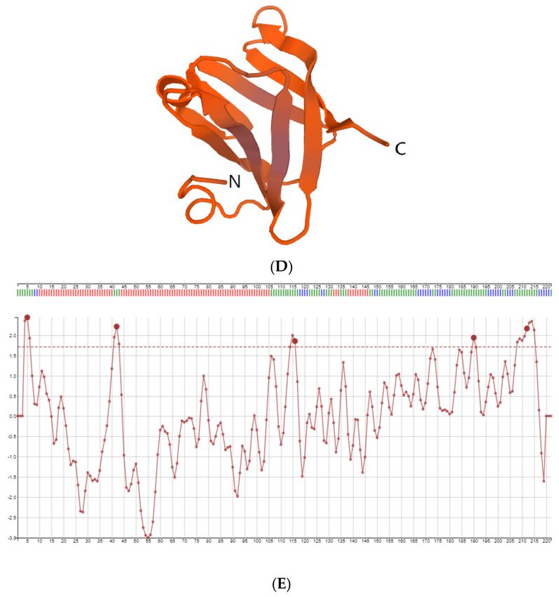
Int. J. Mol. Sci. 2021, 22, 4951                                                                                        3 of 11

                                   acids 104–222 at the C-terminal. The linker GGG was inserted between these two segments.
                                   Restriction site XhoI adds amino acids LE, and the addition of a 6His tag to the C terminus
                                   enhances protein purification. The 149 amino acids M protein’s theoretical molecular mass
                                   was 16,498.74 Da, and pI of 8.79. Designed M protein was produced in E. coli. SDS-PAGE
                                   analysis of purified M protein by the IMAC is shown in Figure 2. The gel stained with CBB
                                   revealed a protein band of about 17 kDa.

                                   Figure 1. Cont.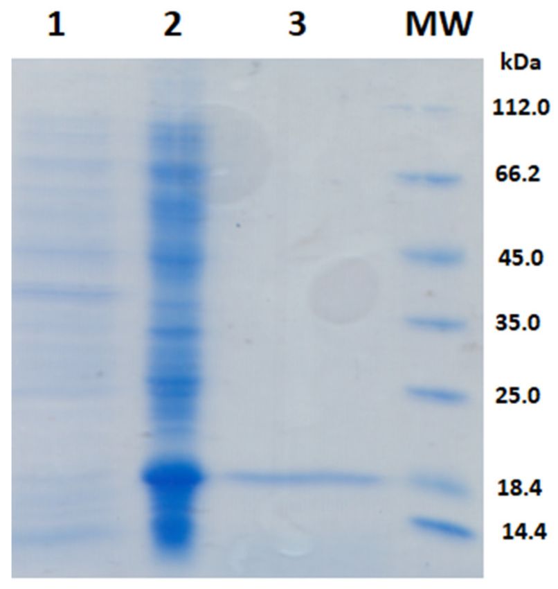
Int. J. Mol. Sci. 2021, 22, 4951                                                                                                 4 of 11

                                   Figure 1. Membrane (M) protein of SARS-CoV-2: (A) amino acid sequence NCBI YP_009724393.1;
                                   (B) Prediction of transmembrane regions in the primary structure of M protein in http://www.cbs.
                                   dtu.dk/services/TMHMM (accessed on 9 March 2021). (C) amino acid sequence of the recombinant
                                   M protein with 6His tag; Position of amino acids in the virion particle: amino acids exposed on the
                                   virion surface are shown in pink, amino acids inside the virion are shown in blue, amino acids in the
                                   transmembrane regions are shown red, and amino acids derived from the cloning strategy are shown
                                   in grey. (D) Structure of the M protein fragment obtained in SWISS-MODEL (25–121 aa). (E) Five B
                                   cell epitopes predicted in the SARS CoV-2 M protein in BEPITOP: 1MADSNGTITVEE, 35LQFAYAN-
                                   RNRFLYII, 109MWSFNPETNILLNV, 184SQRVAGDSGFAAY, 202GNYKLNTDHSSSSDNIALLV.

                                   2.2. Immunoreactivity of the Recombinant SARS-CoV-2 M Protein in Western Blot
                                         Immunoreactivity of the recombinant SARS-CoV-2 M protein was tested in Western
                                   blot (Figure 3). Samples from COVID-19 convalescents showed both IgM and IgG reactivity
                                   against M protein. A single band was detected, demonstrating good purity and specificity
                                   of produced M protein. In contrast, sera samples from non-COVID-19 subjects did not
                                   react with the recombinant M protein. Nonspecific binding of human IgM and IgG to M
                                   protein was not observed.

                                   2.3. Immunoreactivity of the SARS-CoV-2 M Protein in ELISA
                                        For optimization of the new assay based on SARS-CoV-2 M protein, 10 SARS-CoV-2
                                   positive and 10 negative sera were used for the ELISA SARS-CoV-2 IgG and IgM testing.
                                   The widest range in OD values for negative and positive sera for both classes of antibodies
                                   was observed when using M protein as the coating antigen at a final concentration of
Int. J. Mol. Sci. 2021, 22, 4951 5 of 11
2 µg/mL (OD 0.054−1.750 for IgG, OD 0.067−1.547 for IgM). The optimal dilution of the
test sera was 1:50. Using this optimal dilution of coating protein and sera, the optimal dilu-
tion of the HRP conjugated anti-human IgG was found to be 1:5000, and anti-human IgM
was 1:8000. The distribution of the OD values obtained from 40 SARS-CoV-2 negative and
30 SARS-CoV-2 positive human sera using IgG and IgM ELISA is presented in Figure 4A,B.
According to the receiver operating characteristic (ROC) curve analysis, the best cut-off
value for IgG detection was 0.220 OD (Figure 4C), and using this value, sensitivity was
96.7%, and specificity was 92.5%.
Figure 2. SDS-PAGE of the recombinant M protein: Samples were separated on 14% SAS-PAGE, after
which the gel was stained with CBB R 250. Lines: MW) molecular weight markers; (1) cell lysate
before the addition of IPTG, (2) cell lysate after 6 h of expression, (3) purified M protein after IMAC.
Figure 3. Representative Western blots showing the reaction of sera from COVID-19 subjects (samples 1−3) and healthy
individuals (samples 4−7); NSB–nonspecific binding of anti-human IgM and IgG antibodies.SARS-CoV-2 specific IgM antibodies in an assay based on M protein. The test perfor-
mance characteristics of IgM and IgG ELISA tests are summarized in Table 1, showing
that ELISA tests based on M protein could discriminate very well between COVID-19
subjects and healthy controls.
Int. J. Mol. Sci. 2021, 22, 4951 6 of 11
ELISA SARS-CoV-2 IgG ELISA SARS-CoV-2 IgM
1.8 1.8
1.6 1.6
1.4 1.4
OD (450 nm)
1.2
OD (450 nm)
1.2
1.0 1.0
0.8 0.8
0.6 0.6
0.4 0.4
>0.284
0.2 >0.22 0.2 Sens: 93.3
Sens: 96.7
Int. J. Mol. Sci.0.0
2021, 22, x FOR PEER REVIEW 0.0 Spec: 87.5 7 of 12
Spec: 92.5
0 1 0 1
Negative controls Positive controls Negative controls Positive controls
(A) (B)
ELISA SARS-CoV-2 IgG ELISA SARS-CoV-2 IgM
100 100
80 80
Sensitivity
Sensitivity
60 60
40 40
20 AUC = 0.985 20
AUC = 0.955
p < 0.001 p < 0.001
0 0
0 20 40 60 80 100 0 20 40 60 80 100
100-Specificity 100-Specificity
(C) (D)
Figure 4. 4.
Figure Distribution
Distributionofofthe
theOD
ODvalues
valuesobtained
obtainedfrom
from 40
40 SARS-CoV-2 negativeand
SARS-CoV-2 negative and30
30SARS-CoV-2
SARS-CoV-2 positive
positive human
human sera
sera
samples
samples using recombinant M protein-based SARS-CoV-2 IgG (A) and IgM (B) indirect ELISA. The ROC curve built forfor
using recombinant M protein-based SARS-CoV-2 IgG (A) and IgM (B) indirect ELISA. The ROC curve built
4040
SARS-CoV-2
SARS-CoV-2negative
negativeandand30
30SARS-CoV-2
SARS-CoV-2 positive
positive human sera analyzed
human sera analyzedby
bySARS-CoV-2
SARS-CoV-2IgGIgG(C)
(C)and
and IgM
IgM (D)
(D) ELISA
ELISA
based on M protein.
based on M protein.
Table 1. M Protein-Based
Based on theELISA IgM and IgG Tests
above-mentioned Performance
criteria, Characteristics.
37 samples from the 40 negative control
sera were negative, and 29 samples from the 30 positive control sera were positive (OD
ELISA IgM ELISA IgG
0.220−1.75) in the SARS-CoV-2 IgG ELISA. The area under the curve (AUC) was 0.985,
Sensitivity (%) 93.3 96.7
which suggests that defined cut-offs are suitable for interpreting ELISA results and attain-
Specificity (%) 87.5 92.5
ment of high precision in identifying the presence or absence of anti-SARS-CoV-2 specific
Negative predictive value (%) 91.1 97.4
IgG antibodies in human sera (the maximum AUC is 1, which corresponds to a perfect
Positive predictive value (%) 84.8 90.6
classifier, whereas large AUC values indicate better classifier performance).
The best cut-off value for IgM detection (Figure 4D) was 0.284, with a sensitivity of
3.93.3%
Discussion
and specificity of 87.5%. In detecting M protein-specific IgM antibodies, 35 sera
from themajority
The group ofofnegative control sera
the serological tests(n
for= the
40) detection
were negative in the antibodies
of specific assay, and 28
in sera
serum
from 30 SARS-CoV-2 positive samples were positive in the SARS-CoV-2 IgM
of COVID-19 patients on the market are based on the SARS-CoV-2 spike (S) glycoprotein ELISA assay.
Thethe
and AUC was 0.955, indicating
nucleocapsid an acceptable
(N) protein. degree of accuracy
The S glycoprotein protrudes infrom
detecting SARS-CoV-2
the virus envelope
specific
and IgM antibodies
consists in an assay
of the S1 subunit, basedcontains
which on M protein. The test performance
the receptor-binding characteristics
domain, and the S2
of IgM and
subunit, IgG is
which ELISA tests are
involved in summarized in Table 1,fusion.
host cell membrane showing that
The ELISA tests based
nucleocapsid on is
protein
M protein could discriminate very well between COVID-19 subjects and healthy
about three times smaller than S glycoprotein and lacks glycosylation sites. Both S and N controls.
proteins are immunogenic and are suitable for application in diagnostic assay [21]. Be-
sides diagnostic significance, immunoreactivity of viral proteins is also significant for
vaccine design. Currently, vaccine development is based either on attenuated
SARS-CoV-2 viral particle or S protein as dominant immunogen [22]. SARS-CoV M pro-
tein provoked both humoral and cellular immune responses [23]. Antibodies directed to
M protein were detected ten days post-onset in over 95% of the SARS-CoV convalescent
patients (n = 58) [24]. The primary structure of the SARS-CoV-2 M protein
(YP_009724393.1) has shown 91% (201/222) of sequence identities and 96% (214/222) of
sequence similarities to SARS-CoV M protein (UniProtKB-P59596) [25] and only 42%
(86/203) of sequence identity and 61% (124/203) of the sequence similarity with theInt. J. Mol. Sci. 2021, 22, 4951 7 of 11
Table 1. M Protein-Based ELISA IgM and IgG Tests Performance Characteristics.
ELISA IgM ELISA IgG
Sensitivity (%) 93.3 96.7
Specificity (%) 87.5 92.5
Negative predictive value (%) 91.1 97.4
Positive predictive value (%) 84.8 90.6
3. Discussion
The majority of the serological tests for the detection of specific antibodies in serum
of COVID-19 patients on the market are based on the SARS-CoV-2 spike (S) glycoprotein
and the nucleocapsid (N) protein. The S glycoprotein protrudes from the virus envelope
and consists of the S1 subunit, which contains the receptor-binding domain, and the S2
subunit, which is involved in host cell membrane fusion. The nucleocapsid protein is
about three times smaller than S glycoprotein and lacks glycosylation sites. Both S and
N proteins are immunogenic and are suitable for application in diagnostic assay [21].
Besides diagnostic significance, immunoreactivity of viral proteins is also significant for
vaccine design. Currently, vaccine development is based either on attenuated SARS-CoV-2
viral particle or S protein as dominant immunogen [22]. SARS-CoV M protein provoked
both humoral and cellular immune responses [23]. Antibodies directed to M protein
were detected ten days post-onset in over 95% of the SARS-CoV convalescent patients
(n = 58) [24]. The primary structure of the SARS-CoV-2 M protein (YP_009724393.1) has
shown 91% (201/222) of sequence identities and 96% (214/222) of sequence similarities to
SARS-CoV M protein (UniProtKB-P59596) [25] and only 42% (86/203) of sequence identity
and 61% (124/203) of the sequence similarity with the MERS-CoV M protein (UniProtKB-
T2BB40). In silico analysis of M protein’s primary structure revealed the transmembrane
helix topology, with the N- and C-terminal parts being exposed outside and inside the
virus particle, respectively. The predominant secondary structures of the long internal
part of the M protein seem to be β-strands with coiled coil. Thomas reported homology
of the M protein with the prokaryotic sugar transport protein SemiSWEET, which might
contribute to the rapid proliferation, replication, and immune evasion of the SARS-CoV-2
virus [11]. The three-dimensional model of the M protein fragment has been predicted in
SWISS-MODEL and it shared 15.6% of sequence identity with the Cryo-EM structure of
SARS-CoV-2 ORF3a protein (SMTL ID: 7kjr.1.A).
Grifoni et al. reported on the pattern of immunodominance in COVID-19 cases,
revealing M, S, and N proteins as clearly co-dominant, by each, recognized by 100% of
COVID-19 cases (n = 20) [19]. Our study aimed to investigate the immunological reactivity
of recombinant SARS-CoV-2 M protein. Humoral immune response to M protein was tested
using recombinant M protein as antigen for detection of specific IgM and IgG antibodies in
COVID-19 convalescent patients in ELISA and Western blot. The results demonstrated that
the M protein of SARS-CoV-2 has strong antigenic properties. M protein’s immunoreactivity
was very comparable to S and N protein’s immunoreactivity when measured in ELISA.
Immunodominant epitopes of the SARS-CoV virus are previously reported on both N-
and C-terminus of M protein [24,26]. In this study, we choose regions of M protein that
protrude outside and inside the virion, which bears four out of five in silico predicted
continuous epitopes and demonstrated their IgM and IgG reactivity. More than 96% of
COVID-19 sera were positive in the ELISA test for specific IgG detection, and more than
93% of COVID-19 sera were positive in the ELISA test for IgM detection.
Individual SARS-CoV-2 antigens revealed different performances to detect IgG and
IgM [27]. De Assis et al. also suggested that different purification tags may significantly affect
the antigen conformation and affect antibody binding. They indicated that combining addi-
tional antigens, such as the M protein, could contribute to the overall diagnostic performance.Int. J. Mol. Sci. 2021, 22, 4951 8 of 11
Viral infection provokes the immune system to produce neutralizing antibodies against
immunodominant virus epitopes. The main target for the induction of neutralizing anti-
bodies is S protein that protrudes from viral particles and interacts with host cells’ receptors.
Limited data are available regarding the functional properties of antibodies raised against
the SARS-CoV-2 M protein. Neutralizing antibodies directed to M protein of SARS-CoV
virus are described [28], while monoclonal antibodies to M protein also may demonstrate
blocking activity [29]. The engagement of multiple antibodies through different protective
mechanisms can effectively control virus infections [30]. Immunodominant epitopes on the
M protein of the SARS-CoV were identified on the N-terminal region (residues 1 to 31) and
the interior C-terminal region (residues 132 to 161), respectively [26]. Given the fact that
M protein possesses strong antigenic properties, as shown here and that can provoke an
immune response to the SARS-CoV-2 virus, as well as the fact that it is also involved in the
entry of virus in host cells, there are good reasons for further study potential of M protein
for diagnostic purposes either solely or in combination with other SARS-CoV-2 proteins.
The fact that M protein is conserved among coronaviruses and less prone to mutations
than S protein gives a justifiable reason for M protein to be a candidate for vaccine and
diagnostic assay development [31].
4. Materials and Methods
4.1. Design and Production of the Recombinant M Protein SARS-CoV−2
For a suitable 3D template the SWISS-MODEL was employed. We used amino acid
sequence of the M protein fragment and the 15.63% sequence identity was revealed by
SARS-CoV-2 Protein ORF3a (PDB ID: 6XDC), which looks like cytoplasmic part of pro-
tein. In silico prediction of immunoreactivity for the SARS-CoV-2 M protein was done
in the BEPITOPE program [32]. One continuous epitope was localized in the N-terminus
(1 MADSNGTITVEE) and three were distributed in the C terminus (109 MWSFNPETNILLNV,
184 SQRVAGDSGFAAY, 202 GNYKLNTDHSSSSDNIALLV, 109−221) therefore the cloning
strategy was to combine first 19 amino acids with amino acids 101−222 connected with
GGG linker. The gene for the M protein (Gene ID 43740571), from SARS-CoV-2 without
transmembrane regions was cloned in pET23b vector with restriction enzymes NdeI and
XhoI (ThermoFisher Scientific, Wathman, MA, USA). Expression of the M protein was
done in BL21-CodonPlus (DE3)-Ripl cells (Agilent Technologies Inc., La Jolla, CA, USA)
in Luria-Bertani medium (LB medium) supplemented with 100 µg/L ampicillin (Carl
Roth, Karlsruhe, Germany), 25 µg/L kanamycin (Carl Roth, Karlsruhe, Germany), 25 µg/L
chloramphenicol (Carl Roth, Karlsruhe, Germany). A single colony containing pET-23b-
M vector was inoculated in 5 mL of LB medium with antibiotics. The culture was left
overnight (ON) at 37 ◦ C in a shaking incubator (Bio San, Riga, Latvia). Five mL of the
ON culture was inoculated in 1 L of sterile LB medium with appropriate antibiotics at
37 ◦ C under constant shaking (250 rpm). When absorbance at 600 nm (OD600) reached 0.6,
the temperature of the medium was lowered to 22 ◦ C, and expression was induced using
1 mM isopropyl-β-D-1-thiogalactopyranoside (IPTG) (ThermoFisher Scientific, Waltham,
MA, USA). Cells were grown at 22 ◦ C ON under constant shaking (250 rpm). Cells were
harvested by centrifugation (3000× g, 25 min at 4 ◦ C, Eppendorf centrifuge 5430R, Ham-
burg, Germany), suspended in Lysis buffer (20 mM Tris-HCl (Merck KGaA, Darmstadt,
Germany), 150 mM NaCl (Beta Hem, Belgrade, Serbia), 1% Triton-X-100 (Merck KGaA,
Darmstadt, Germany), 10 mM dithiothreitol (Serva Electrophoresis GmbH, Heidelberg,
Germany), pH 7.8 and sonicated in an ice water bath (10 × 10 s at 30 W, Branson sonifier
150 (Branson Ultrasonic SA, Carouge, Switzerland). The M protein was solubilized from
inclusion bodies in 6 M urea (Serva Electrophoresis GmbH, Heidelberg, Germany), and ap-
plied onto a metal immobilized affinity chromatography (IMAC, Talon® Superflow™,
Uppsala, Sweden) according to the manufacturer’s instructions. The bound fraction was
eluted using 20 mM Tris-HCl, 150 mM NaCl, 6 M urea, 300 mM imidazole pH 7.8. After gel
electrophoresis, eluted fractions were pulled and dialyzed against 20 mM Tris-HCl, 150 mM
NaCl, 0.1% SDS (Serva Electrophoresis GmbH, Heidelberg, Germany) pH 7.8. The levelInt. J. Mol. Sci. 2021, 22, 4951 9 of 11
of protein purity was analyzed by staining SDS-PA gel with CBB (Serva, Electrophoresis
GmbH, Heidelberg, Germany).
4.2. Patient Serum Samples
The Medical Ethical Committee of the Institute for the Application of Nuclear Energy
(INEP, Belgrade, Serbia) approved this study. All study participants gave written informed
consent to use their serum samples for ELISA SARS-CoV-2 assay development. A total of
70 human serum samples were analyzed in ELISA. Blood samples for COVID-19 convales-
cents (n = 30) were taken at least four weeks after disease onset. COVID-19 infection was
confirmed with the quantitative RT-PCR, and anti-SARS-CoV-2 antibodies in these sera
were confirmed by ELISA SARS-CoV-2 IgG and IgM (INEP, Belgrade, Serbia). Sera samples
collected from healthy persons (n = 40) before the COVID-19 outbreak in Serbia taken from
the INEP sera sample bank were used as a negative control.
4.3. Detection of M Protein Specific IgM and IgG in Western Blot
Specific IgM and IgG for the SARS-CoV-2 membrane protein in COVID-19 patients
sera were tested in Western blot. M protein was resolved on (4/14%) SDS-PAGE under
reducing conditions (Mini Protean System, Bio-Rad, CA, USA). After semi-dry transfer to a
nitrocellulose membrane (ThermoFisher Scientific, Waltham, MA, USA), the membrane
was blocked in blocking buffer (1% BSA/10 mM PBS, 150 mM NaCl, 0.05% Tween-20,
pH 7.2) for 1 h at room temperature. Randomly selected sera samples from COVID-19
convalescents and control subjects, previously tested in ELISA as described above (diluted
1:50 in blocking buffer) were individually incubated with membrane overnight on 4 ◦ C.
Membranes incubated in blocking buffer, omitting serum, were used for assessment of
nonspecific binding. Incubation with sheep anti-human IgM/HRP or anti-human IgG/HRP
conjugate (INEP, Belgrade, Serbia), diluted 1:1000 in blocking buffer, was carried out one
hour at room temperature. Following each incubation step, membranes were washed
in PBST. DAB staining was used (EnVision Detection Systems Peroxidase/DAB, DAKO,
Santa Clara, CA, USA). Membranes were washed in water, dried, and scanned.
4.4. ELISA
Sera previously tested in the ELISA SARS-CoV-2 IgM and IgG INEP, Serbia (10 pos-
itive and 10 negative), were used to optimize the new ELISA based on recombinant M
protein. The plates were coated with the recombinant M protein in different concentration
(0.5 µg/mL, 1 µg/mL, 2 µg/mL, 3 µg/mL) in 0.1 M carbonate buffer pH 9.5 (100 µL/well).
The plates were incubated at +4 ◦ C for 18 h and washed three times with phosphate-
buffered saline pH 7.6 (PBS) containing 0.05% Tween 20 (PBST). The antigen-coated plates
were incubated with blocking solution (0.1 mg BSA in PBST, 100 µL/well) for 1 h at 37 ◦ C.
After washing, human sera diluted in blocking solution were added (1:50, 1:100, 1:200,
1:300, 1:400, 1:500; 100 µL/well) and plates were incubated for 1 h at 37 ◦ C. After washing,
the HRP-conjugated sheep anti-human IgM and anti-human IgG (INEP, Belgrade, Serbia)
were diluted in blocking solution and added to the wells (1:1000, 1:2000, 1:5000, 1:8000;
100 µL/well) and then incubated for 1 h at 37 ◦ C. After washing 4 times with washing
buffer (PBST), the color was allowed to develop for 10 min with the chromogenic solution
(3,3’,5,5’-tetramethylbenzidine (TMB) substrate, INEP, Serbia/hydrogen peroxide). After
stopping the reaction with Stop solution (INEP, Belgrade, Serbia), optical densities were
measured at 450 nm using an ELISA reader (Wallac Multilabel Counter 1420, Perkin Elmer,
Milano, Italy).
4.5. Statistical Analysis
In total, 70 sera samples (40 negative and 30 positive) were used for preliminary
ELISA assay validation. Statistical analyses were carried out using MedCalc version
10.4.0.0 (MedCalc Software, Ostend, Belgium) [33,34]. Sensitivity was defined as the
proportion of correctly identified COVID-19 positive patients who were initially positive byInt. J. Mol. Sci. 2021, 22, 4951 10 of 11
RT-PCR SARS-CoV-2 determination in respiratory samples. Specificity was defined as the
proportion of naive participants classified as positive as analyzed by ELISA SARS-CoV-2
M protein-specific antibodies.
Author Contributions: Conceptualization, M.G.-J. and D.Ć.; methodology, M.G.-J., D.Ć., M.G., Z.L.,
and I.P.-R.; software, M.G., Z.L.; validation, M.G.-J., D.Ć., and M.G.; investigation, Z.L., I.P.-R., A.T.,
and S.G.; resources, M.G.-J.; writing—original draft preparation, Z.L., I.P.-R., M.G.-J., D.Ć., and M.G.;
writing—review and editing, M.G.-J., D.Ć., and M.G.; visualization, Z.L., I.P.-R., M.G., and D.Ć.;
supervision, M.G.-J.; project administration, M.G.-J.; funding acquisition, M.G.-J. All authors have
read and agreed to the published version of the manuscript.
Funding: Ministry of Education, Science and Technological Development of Republic of Serbia
Contract number: 451-03-9/2021-14/200288 and 451-03-68/2020-14/200019.
Institutional Review Board Statement: The study was conducted according to the guidelines of
the Declaration of Helsinki, and approved by the Medical Ethical Committee of the Institute for
the Application of Nuclear Energy (INEP, Belgrade, Serbia) decision number No. 02–262/2 from
10 August 2020.
Informed Consent Statement: Informed consent was obtained from all subjects involved in the study.
Data Availability Statement: The data presented in this study are available on request from the
corresponding author.
Conflicts of Interest: The authors declare no conflict of interest.
References
1. WHO. WHO Statement Regarding Cluster of Pneumonia Cases in Wuhan, China. 9 January 2020. Available online: https://www.
who.int/china/news/detail/09-01-2020-who-statement-regarding-cluster-of-pneumonia-cases-in-wuhan-china (accessed on
1 October 2020).
2. Zhu, N.; Zhang, D.; Wang, W.; Li, X.; Yang, B.; Song, J.; Zhao, X.; Huang, B.; Shi, W.; Lu, R.; et al. A Novel Coronavirus from
Patients with Pneumonia in China, 2019. N. Engl. J. Med. 2020, 382, 727–733. [CrossRef] [PubMed]
3. Wu, J.; Yuan, X.; Wang, B.; Gu, R.; Li, W.; Xiang, X.; Tang, L.; Sun, H. Severe Acute Respiratory Syndrome Coronavirus 2:
From Gene Structure to Pathogenic Mechanisms and Potential Therapy. Front. Microbiol. 2020, 11, 1576. [CrossRef]
4. Shah, V.K.; Firmal, P.; Alam, A.; Ganguly, D.; Chattopadhyay, S. Overview of Immune Response During SARS-CoV-2 Infection:
Lessons From the Past. Front. Immunol. 2020, 11, 1–17. [CrossRef] [PubMed]
5. Klein, S.; Cortese, M.; Winter, S.L.; Wachsmuth-Melm, M.; Neufeldt, C.J.; Cerikan, B.; Stanifer, M.L.; Boulant, S.; Bartenschlager, R.;
Chlanda, P. SARS-CoV-2 structure and replication characterized by in situ cryo-electron tomography. Nat. Commun. 2020, 11, 5885.
[CrossRef] [PubMed]
6. Wang, Q.; Zhang, Y.; Wu, L.; Niu, S.; Song, C.; Zhang, Z.; Lu, G.; Qiao, C.; Hu, Y.; Yuen, K.-Y.; et al. Structural and functional basis
of SARS-CoV-2 entry by using human ACE2. Cell 2020, 181, 894–904. [CrossRef] [PubMed]
7. Uddin, M.; Mustafa, F.; Rizvi, T.A.; Loney, T.; Al Suwaidi, H.; Al-Marzouqi, A.H.H.; Kamal Eldin, A.; Alsabeeha, N.; Adrian, T.E.;
Stefanini, C.; et al. SARS-CoV-2/COVID-19: Viral Genomics, Epidemiology, Vaccines, and Therapeutic Interventions. Viruses
2020, 12, 526. [CrossRef] [PubMed]
8. Hu, Y.; Wen, J.; Tang, L.; Zhang, H.; Zhang, X.; Li, Y.; Wang, J.; Han, Y.; Li, G.; Shi, J.; et al. The M protein of SARS-CoV: Basic
structural and immunological properties. Genom. Proteom. Bioinform. 2003, 1, 118–130. [CrossRef]
9. Neuman, B.W.; Kiss, G.; Kunding, A.H.; Bhella, D.; Baksh, M.F.; Connelly, S.; Droese, B.; Klaus, J.P.; Makino, S.; Sawicki, S.G.; et al.
A structural analysis of M protein in coronavirus assembly and morphology. J. Struct. Biol. 2011, 174, 11–22. [CrossRef]
10. Bianchi, M.; Benvenuto, D.; Giovanetti, M.; Angeletti, S.; Ciccozzi, M.; Pascarella, S. Sars-CoV-2 Envelope and Membrane Proteins:
Structural Differences Linked to Virus Characteristics? BioMed Res. Int. 2020, 2020, 4389089. [CrossRef]
11. Thomas, S. The structure of the membrane protein of sars-cov-2 resembles the sugar transporter semisweet. Pathog. Immun. 2020,
5, 342–363. [CrossRef]
12. Dramé, M.; Teguo, M.T.; Proye, E.; Hequet, F.; Hentzien, M.; Kanagaratnam, L.; Godaert, L. Should RT-PCR be considered a gold
standard in the diagnosis of COVID-19? J. Med Virol. 2020, 92, 2312–2313. [CrossRef]
13. Amanat, F.; Stadlbauer, D.; Strohmeier, S.; Nguyen, T.H.O.; Chromikova, V.; McMahon, M.; Jiang, K.; Arunkumar, G.A.;
Jurczyszak, D.; Polanco, J.; et al. A serological assay to detect SARS-CoV-2 seroconversion in humans. Nat. Med. 2020,
26, 1033–1036. [CrossRef]
14. Weisberg, S.P.; Connors, T.J.; Zhu, Y.; Baldwin, M.R.; Lin, W.; Wontakal, S.; Szabo, P.A.; Wells, S.B.; Dogra, P.; Gray, J.; et al. Distinct
antibody responses to SARS-CoV-2 in children and adults across the COVID-19 clinical spectrum. Nat. Immunol. 2021, 22, 25–31.
[CrossRef]Int. J. Mol. Sci. 2021, 22, 4951 11 of 11
15. Borbone, N.; Piccialli, G.; Roviello, G.N.; Oliviero, G. Nucleoside Analogs and Nucleoside Precursors as Drugs in the Fight
against SARS-CoV-2 and Other Coronaviruses. Molecules 2021, 26, 986. [CrossRef]
16. Vicidomini, C.; Roviello, V.; Roviello, G.N. Molecular Basis of the Therapeutical Potential of Clove (Syzygium aromaticium L.) and
Clues to Its Anti-COVID-19 Utility. Molecules 2021, 26, 1880. [CrossRef]
17. Dong, Y.; Dai, T.; Wei, Y.; Zhang, L.; Zheng, M.; Zhou, F. A systematic review of SARS-CoV-2 vaccine candidates. Signal Transduct.
Target. Ther. 2020, 5, 237. [CrossRef]
18. Galanis, K.; Nastou, K.; Papandreou, N.; Petichakis, G.; Pigis, D.; Iconomidou, V. Linear B-Cell Epitope Prediction for In Silico
Vaccine Design: A Performance Review of Methods Available via Command-Line Interface. Int. J. Mol. Sci. 2021, 22, 3210.
[CrossRef]
19. Grifoni, A.; Weiskopf, D.; Ramirez, S.I.; Mateus, J.; Dan, J.M.; Moderbacher, C.R.; Rawlings, S.A.; Sutherland, A.; Premkumar, L.;
Jadi, R.S.; et al. Targets of T Cell Responses to SARS-CoV-2 Coronavirus in Humans with COVID-19 Disease and Unexposed
Individuals. Cell 2020, 181, 1489–1501.e15. [CrossRef]
20. Ainsworth, M.; Andersson, M.; Auckland, K.; Baillie, J.K.; Barnes, E.; Beer, S.; Beveridge, A.; Bibi, S.; Blackwell, L.; Borak, M.; et al.
Performance characteristics of five immunoassays for SARS-CoV-2: A head-to-head benchmark comparison. Lancet Infect. Dis.
2020, 20, 1390–1400. [CrossRef]
21. Meyer, M.A.M.B.; Drosten, C. Serological assays for emerging coronaviruses: Challenges and pitfalls. J. Adv. Res. 2014, 24, 91–98.
[CrossRef]
22. Corbett, K.S.; Edwards, D.K.; Leist, S.R.; Abiona, O.M.; Boyoglu-Barnum, S.; Gillespie, R.A.; Himansu, S.; Schäfer, A.;
Ziwawo, C.T.; DiPiazza, A.T.; et al. SARS-CoV-2 mRNA Vaccine Design Enabled by Prototype Pathogen Preparedness. Nature
2020, 586, 567–571. [CrossRef] [PubMed]
23. Liu, J.; Sun, Y.; Qi, J.; Chu, F.; Wu, H.; Gao, F.; Li, T.; Yan, J.; Gao, G.F. The Membrane Protein of Severe Acute Respiratory
Syndrome Coronavirus Acts as a Dominant Immunogen Revealed by a Clustering Region of Novel Functionally and Structurally
Defined Cytotoxic T-Lymphocyte Epitopes. J. Infect. Dis. 2010, 202, 1171–1180. [CrossRef] [PubMed]
24. Liu, C.; Li, X.; Zhang, C.; Xu, S.; Shao, Y.; Zhuang, H.; Che, X.; Qiu, Y.; Yin, H.; Li, D.; et al. Establishment of a reference panel for
the detection of anti-SARS-CoV antibodies. Biologicals 2007, 35, 203–210. [CrossRef] [PubMed]
25. Mahtarin, R.; Islam, S.; Islam, M.J.; Ullah, M.O.; Ali, M.A.; Halim, M.A. Structure and dynamics of membrane protein in
SARS-CoV-2. J. Biomol. Struct. Dyn. 2020. online ahead of print. [CrossRef]
26. He, Y.; Zhou, Y.; Siddiqui, P.; Niu, J.; Jiang, S. Identification of Immunodominant Epitopes on the Membrane Protein of the Severe
Acute Respiratory Syndrome-Associated Coronavirus. J. Clin. Microbiol. 2005, 43, 3718–3726. [CrossRef]
27. De Assis, R.R.; Jain, A.; Nakajima, R.; Jasinskas, A.; Felgner, J.; Obiero, J.M.; Norris, P.J.; Stone, M.; Simmons, G.; Bagri, A.; et al.
Analysis of SARS-CoV-2 antibodies in COVID-19 convalescent blood using a coronavirus antigen microarray. Nat. Commun. 2021,
12, 6. [CrossRef]
28. Pang, H.; Liu, Y.; Han, X.; Xu, Y.; Jiang, F.; Wu, D.; Kong, X.; Bartlam, M.; Rao, Z. Protective humoral responses to severe acute
respiratory syndrome-associated coronavirus: Implications for the design of an effective protein-based vaccine. J. Gen. Virol. 2004,
85, 3109–3113. [CrossRef]
29. Tripp, R.A.; Haynes, L.M.; Moore, D.; Anderson, B.; Tamin, A.; Harcourt, B.H.; Jones, L.P.; Yilla, M.; Babcock, G.J.;
Greenough, T.; et al. Monoclonal antibodies to SARS-associated coronavirus (SARS-CoV): Identification of neutralizing and
antibodies reactive to S, N, M and e viral proteins. J. Virol. Methods 2005, 128, 21–28. [CrossRef]
30. Howell, K.A.; Brannan, J.M.; Bryan, C.; McNeal, A.; Davidson, E.; Turner, H.L.; Vu, H.; Shulenin, S.; He, S.; Kuehne, A.; et al.
Cooperativity Enables Non-neutralizing Antibodies to Neutralize Ebolavirus. Cell Rep. 2017, 19, 413–424. [CrossRef]
31. Qamar, M.T.U.; Rehman, A.; Tusleem, K.; Ashfaq, U.A.; Qasim, M.; Zhu, X.; Fatima, I.; Shahid, F.; Chen, L.-L. Designing of a next
generation multiepitope based vaccine (MEV) against SARS-COV-2: Immunoinformatics and in silico approaches. PLoS ONE
2020, 15, e0244176. [CrossRef]
32. Odorico, M.; Pellequer, J.-L. BEPITOPE: Predicting the location of continuous epitopes and patterns in proteins. J. Mol. Recognit.
2003, 16, 20–22. [CrossRef]
33. Metz, C.E. Basic principles of ROC analysis. Semin. Nucl. Med. 1978, 8, 283–298. [CrossRef]
34. Zweig, M.H.; Campbell, G. Receiver-operating characteristic (ROC) plots: A fundamental evaluation tool in clinical medicine.
Clin. Chem. 1993, 39, 561–577. [CrossRef]You can also read