HOST DETERMINANTS OF MERS-COV TRANSMISSION AND PATHOGENESIS - MDPI
←
→
Page content transcription
If your browser does not render page correctly, please read the page content below
Review
Host Determinants of MERS-CoV Transmission and
Pathogenesis
W. Widagdo, Syriam Sooksawasdi Na Ayudhya, Gadissa B. Hundie and Bart L. Haagmans *
 Department of Viroscience, Erasmus Medical Center, Rotterdam, The Netherlands;
 w.widagdo@erasmusmc.nl (W.W.); s.sooksawasdi@erasmusmc.nl (S.S.N.A.);
 g.hundie@erasmusmc.nl (G.B.H.)
 * Correspondence: b.haagmans@erasmusmc.nl

 Received: 1 March 2019; Accepted: 13 March 2019; Published: 19 March 2019

 Abstract: Middle East respiratory syndrome coronavirus (MERS-CoV) is a zoonotic pathogen that
 causes respiratory infection in humans, ranging from asymptomatic to severe pneumonia. In
 dromedary camels, the virus only causes a mild infection but it spreads efficiently between
 animals. Differences in the behavior of the virus observed between individuals, as well as between
 humans and dromedary camels, highlight the role of host factors in MERS-CoV pathogenesis and
 transmission. One of these host factors, the MERS-CoV receptor dipeptidyl peptidase-4 (DPP4),
 may be a critical determinant because it is variably expressed in MERS-CoV-susceptible species as
 well as in humans. This could partially explain inter- and intraspecies differences in the tropism,
 pathogenesis, and transmissibility of MERS-CoV. In this review, we explore the role of DPP4 and
 other host factors in MERS-CoV transmission and pathogenesis—such as sialic acids, host
 proteases, and interferons. Further characterization of these host determinants may potentially
 offer novel insights to develop intervention strategies to tackle ongoing outbreaks.

 Keywords: MERS-CoV; transmission; pathogenesis; host factors; DPP4

1. Introduction
      Middle East respiratory syndrome coronavirus (MERS-CoV) is a novel pathogen that was
isolated in late 2012 [1]. Since then, the virus has caused multiple outbreaks and infected more than
2000 individuals, [2] who then develop a respiratory infection ranging in severity from
asymptomatic to fatal [3,4]. Severe-to-fatal MERS-CoV patients have a higher chance of transmitting
this virus since they shed a higher amount of virus progeny in comparison to the
asymptomatic-to-mild ones [5–8]. Identifying and quarantining these patients in healthcare facilities
where outbreaks have occurred, together with implementing proper infection control, has been
effective in reducing transmission and containing these outbreaks [9,10]. However, new MERS-CoV
cases are still being reported, especially in the Arabian Peninsula [2,11]. This is partly due to the
continuous zoonotic introduction of this virus to the human population in this region by
dromedaries [12]. The dromedary camel is the only animal species that has been reported to transmit
this virus to humans [13–16]. MERS-CoV infection in these animals merely causes mild upper
respiratory tract infection [17,18], but seroepidemiological studies showed that this virus has been
circulating in dromedary camels for decades, suggesting the efficient transmission of MERS-CoV in
this species [19–22].
      Although the clinical manifestations, as well as transmission, are remarkably different in
MERS-CoV-infected humans and dromedary camels, the viruses isolated from these two species are
highly similar, if not indistinguishable [12,16]. This indicates that host factors play a significant role
in MERS-CoV pathogenesis and transmission. However, the identity of these host factors and how
they affect the pathogenesis and transmission of MERS-CoV are generally not well understood.

Viruses 2019, 11, 280; doi:10.3390/v11030280                                  www.mdpi.com/journal/viruses
Viruses 2019, 11, 280 2 of 14
Dipeptidyl peptidase-4 (DPP4)—the MERS-CoV receptor, sialic acids, proteases, and interferons are
all examples of potentially critical host factors that have been shown to affect MERS-CoV infection in
vitro [23–26]. This review highlights the role of some MERS-CoV-interacting host factors—especially
DPP4—in MERS-CoV pathogenesis and transmission.
2. MERS-CoV-Interacting Host Factors
MERS-CoV infection of a target cell is initiated by the virus attachment to the cell surface
[23,27]. MERS-CoV uses the N-terminal part of its spike (S)—the so called S1 protein (Figure 1A)—to
bind to two host cell surface molecules, dipeptidyl peptidase-4 (DPP4) and α2,3-sialic acids [23,24].
DPP4 is the functional receptor of MERS-CoV; its absence renders cells resistant to this virus, while
its transient expression in non-susceptible cells permits viral replication [23]. DPP4 is a serine
exopeptidase, which is either expressed at the cell surface or shed in a soluble form. It has the
capacity to cleave-off dipeptides from polypeptides with either L-proline or L-alanine at the
penultimate position. Accordingly, DPP4 is capable of cutting various substrates, such as hormones,
cytokines, chemokines, and neuropeptides, allowing it to be involved in multiple physiological
functions as well as pathophysiological conditions [28]. This enzymatic activity is mediated by the
α/β hydrolase domain of DPP4, while MERS-CoV infection is mediated by the binding of S1 protein
to the β-propeller domain of this exopeptidase (Figure 1B) [28–31]. There are 11 critical residues
within the β-propeller domain that directly interact with the S1 protein [29–31]. These residues are
quite conserved in camelids, primates, and rabbits—species shown to be susceptible to MERS-CoV
[17,31–33]. In contrast, ferrets, rats, and mice resist MERS-CoV infection due to differences in some
critical DPP4 residues [31,34–36]. These data illustrate that DPP4 has the capacity to determine the
host range of MERS-CoV.
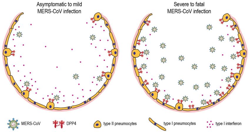
Figure 1. Schematic figure depicting four structural proteins of Middle East respiratory syndrome
coronavirus (MERS-CoV), i.e., S, E, M, and N proteins (A); a cartoon representation of MERS-CoV S1
protein binding to DPP4 (PDB code 4L72) (B). The S protein consists of the S1 and S2 subunits. The
α/β hydrolase domain of DPP4 is indicated in red, β-propeller domain in green, while part of the
MERS-CoV S1 protein is shown in blue.
Other MERS-CoV-interacting host factors besides DPP4 are less extensively studied and have
mostly been investigated in vitro. Glycotopes of α2,3-sialic acids coupled with 5-N-acetylated
neuraminic acid are recognized by the S1 protein of MERS-CoV during attachment [24]. In the
absence of these glycotopes, MERS-CoV entry is reduced but not abolished, indicating their function
as an attachment factor rather than a receptor [24]. Besides α2,3-sialic acids, CEACAM5 and GRP78
have also been suggested to be attachment factors for MERS-CoV, but their roles in vivo during
MERS-CoV infection are not clear at this moment [37,38]. Post attachment, MERS-CoV uses the
C-terminal part of its S protein—known as S2 (Figure 1A)—to interact with host proteases, such as
furin, TMPRSS2, and cathepsins [39–42]. These proteases cleave the S protein and induce
conformational changes, allowing fusion between viral and host cellular membranes, resulting in the
release of viral RNA into the cell cytoplasm [27]. TMPRSS2 and DPP4 are held in one complex at the
Viruses 2019, 11, 280                                                                            3 of 14

cell surface by a scaffolding protein, the tetraspanin CD9, leading to a rapid and efficient entry of
MERS-CoV into the susceptible cells [43]. Once fusion with host cell membranes has occurred,
MERS-CoV subsequently replicates its genetic material and produces viral proteins in the cell
cytoplasm to generate new virus progeny. During this stage, MERS-CoV uses its nsp3–4
polyproteins to build its replication organelles as well as its accessory proteins such as the 4a and 4b
proteins to inhibit host anti-viral defense mechanisms [44–54]. However, the capacity of MERS-CoV
accessory proteins to impede several pathways of host immune response in the lungs may be
limited. MERS-CoV inoculation of macaques and genetically modified mice generally results in
limited clinical manifestations; thus, adapting this virus through serial passaging or defecting the
type I interferon pathway may be needed to enhance viral replication and pathogenesis in these
animals [32,55–58]. These observations, together with studies showing type I interferon capacity to
inhibit MERS-CoV infection in vitro [25,59], highlight the importance of the innate immune response,
especially type I interferon, as an inhibiting factor for MERS-CoV.

3. Host Factors in MERS-CoV Transmission
      So far MERS-CoV has been isolated from dromedary camels and humans [1,60]. Both species
are not only susceptible to MERS-CoV infection, but also capable of transmitting this virus [7,12–
18,22]. However, current data indicate that virus spread is more efficient in dromedary camels than
in humans [5,7,19–21,61]. This difference in transmissibility could be partially due to the different
tropism of MERS-CoV in these two species. In dromedaries, MERS-CoV has been shown to replicate
in the nasal epithelium upon experimental in vivo infection [17], while in humans, MERS-CoV
mainly replicates in the lower respiratory tract, particularly in the bronchiolar and alveolar epithelia
[23,62–65]. Higher viral RNA levels in the sputum and lavage samples of MERS-CoV patients
compared to nasal and throat swabs are consistent with the tropism of MERS-CoV in humans [66–
68]. This different MERS-CoV tropism in dromedary camels and humans is in line with the
localization of DPP4 in the respiratory tract tissues of these two species. In humans, DPP4 is absent
in the nasal epithelium but present in the lower respiratory tract epithelium, mainly in type II
pneumocytes [69,70]. In contrast, DPP4 is expressed in the nasal epithelium of dromedary camels
[69]. This difference in DPP4 localization between humans and dromedary camels therefore explains
MERS-CoV tropism in these two species and highlights DPP4 as an essential determinant of
MERS-CoV tropism.
      DPP4 localization has also been investigated in many other MERS-CoV-susceptible species. In
Gambian and Egyptian fruit bats, DPP4 is expressed in the respiratory tract and intestinal
epithelium, suggesting that MERS-CoV can target both tissues [71]. In line with this finding,
MERS-CoV inoculation via intranasal and intraperitoneal routes in the Jamaican fruit bat led to viral
RNA shedding both in the respiratory tract and the intestinal tract [72]. In contrast to frugivorous
bats, DPP4 is limitedly expressed in the respiratory tract epithelium of two insectivorous bats, i.e.,
common pipistrelle and common serotine bats, but abundant in their intestinal epithelium [71].
Accordingly, sequences of MERS-like-CoVs were mainly obtained from rectal swabs and fecal
samples of insectivorous bats [73–80]. These findings not only support insectivorous bats as the
origin host of MERS-CoV [73–80], but also indicate the importance of intestinal tropism and fecal–
oral transmission of MERS-like-CoV in these insectivorous bats.
      Besides bats, humans, and dromedary camels, other animal species have also been proposed as
potential hosts of MERS-CoV. Remarkably, DPP4 of horses, llamas, alpacas, pigs, bovines, goats,
sheep, and rabbits has been demonstrated to recognize the S protein of MERS-CoV [81,82]. In most
of these species, there is a preferential upper respiratory tract expression of DPP4 observed. Rabbits
express DPP4 in the upper and lower respiratory tract epithelium, and thus may allow MERS-CoV
to replicate in both compartments [33,83]. Horses, llamas, and pigs mainly express DPP4 in the
upper respiratory tract—particularly the nasal epithelium [84]. Upon intranasal MERS-CoV
inoculation, llamas, alpacas, and pigs developed upper respiratory tract infection, while horses did
not seroconvert and only shed infectious virus in a limited amount [84–88]. The reason why horses
seem to be less permissive to MERS-CoV remains to be investigated, but a chronic co-infection in the
Viruses 2019, 11, 280 4 of 14
guttural pouch, a common disease among horses, might be one of the explanations. This guttural
pouch infection results in excessive mucus production that might hinder MERS-CoV from attaching
and entering the nasal epithelium [84,89,90]. Sheep, on the other hand, did not seem to express
significant levels of DPP4 in their respiratory tract, and thus did not seroconvert nor shed infectious
virus upon experimental MERS-CoV inoculation [84,88]. Comparable to sheep, goats limitedly shed
infectious virus upon experimental infection and did not transmit this virus to other naïve goats
upon direct contact [88]. The results of experimental MERS-CoV infection in livestock animals are in
line with data from epidemiological studies. MERS-CoV seropositive llamas and alpacas are present
in the field, while horses, goats, and sheep are generally found to be seronegative [22,86,87,91–98].
Given the fact that experimental in vivo infection studies and DPP4 expression analysis in
different animal species revealed that dromedary camels are not the only animals in which
MERS-CoV has an upper respiratory tract tropism [17,18,83,84], it is then relevant to question
whether other animals can potentially spread MERS-CoV as well. New World camelids, i.e., alpacas
and llamas, are able to transmit the virus to respective naïve animals upon contact [86]. Pigs and
rabbits, on the other hand, hardly transmit the virus—neither by contact nor airborne routes [83,99].
Most likely, this is caused by the fact that pigs and rabbits, unlike dromedary camels, shed low levels
of infectious virus upon MERS-CoV inoculation (Figure 2). This difference indicates that other host
factors besides DPP4 could cause interspecies variation in MERS-CoV infection. Indeed, several
glycotopes of α2,3-sialic acids that function as attachment factors of MERS-CoV are present in the
nasal epithelium of dromedary camels but absent in that of rabbits and pigs (Figure 3) [24,100]. The
lack of these glycotopes in pigs and rabbits might limit the susceptibility and transmission of
MERS-CoV in these animals. Although the role of these glycotopes in MERS-CoV transmission still
requires further investigation, it remains plausible that an efficient transmission of this virus might
require the presence of both DPP4 and MERS-CoV-recognized glycotopes of α2,3-sialic acids (Figure
3).
Figure 2. Schematic overview of viral RNA and infectious virus shedding of MERS-CoV-inoculated
dromedary camels, pigs, and rabbits. Each data point represents the average data from previous
experiments [17,33,84]. Viral RNA is measured in TCID50/mL genome equivalents, while infectious
virus is expressed in TCID50/mL.
Besides entry and attachment receptors, MERS-CoV has been demonstrated to use both cell
surface and lysosomal proteases to enter its target cells [39,40,43,101]. The preference of MERS-CoV
to use certain host proteases is influenced by the type of target cell and the cleavage stage of their S
protein prior to infection [40]. It has also been reported that the lysosomal proteases from bat cells
support coronavirus spike-mediated virus entry more efficiently than their counterparts from
human cells [39]. These observations suggest that host proteases from different host species may
determine the species and tissue tropism of MERS-CoV.
Because MERS-CoV has been circulating in dromedary camels for decades before emerging in
the human population [19–22], it is plausible that this virus inhibits the immune response of
dromedary camels more efficiently than that of other species, including pigs and rabbits. The
difference in immune response among MERS-CoV-susceptible species is therefore another factor
that might yield interspecies variation in permissiveness to MERS-CoV. Characterizing theViruses 2019, 11, 280 5 of 14
difference in host proteases and immune responses among MERS-CoV-susceptible species, as
performed for DPP4 and MERS-CoV-recognized α2,3-sialic acid glycotopes (Figure 3), has not yet
been investigated. These data, however, may further explain interspecies variation in MERS-CoV
infection and transmission.
Figure 3. Schematic representation of DPP4 expression and MERS-CoV-recognized α2,3-sialic acid
glycotopes in the respiratory tract of dromedary camel, pig, rabbit, human, and sheep.
4. Host Factors in MERS-CoV Pathogenesis
MERS-CoV causes respiratory infection in humans ranging from asymptomatic to severe
pneumonia [3,4]. However, it is currently unclear what causes this intraspecies variation.
Epidemiology data indicate that individuals with certain risk factors are at higher risk of developing
severe MERS-CoV infection [4,102]. This implies that some host factors may dictate the outcome of
MERS-CoV infection, thus rendering intraspecies variation. Two of the risk factors, i.e., smoking and
chronic obstructive pulmonary disease (COPD), have been shown to upregulate DPP4 expression in
the lungs [70,102–104], suggesting DPP4 as a possible reason for intraspecies variation observed
among MERS-CoV patients. In healthy human lungs, DPP4 is almost exclusively expressed in type II
pneumocytes [69,70]. Type II pneumocytes are small cuboidal cells that can regenerate alveolar
epithelium upon injury, and roughly cover 2% of the alveolar surface area. Meanwhile, around 95%
of the surface area of the alveolus is occupied by type I pneumocytes that are morphologically flat
and responsible for gas exchange [105,106]. In the lungs of smokers and COPD patients, unlike in
healthy human lungs, DPP4 is prominently expressed in both type I and II pneumocytes, indicating
upregulated expression on type I pneumocytes [104]. Autopsy reports from fatal MERS-CoV
patients showed that both type I and II pneumocytes expressed DPP4 and became infected by
MERS-CoV, proposing a role of DPP4-expressing type I pneumocytes in MERS-CoV pathogenesis
[64,107]. Damage to type I cells in the lung alveoli during viral infection may lead to diffuse alveolar
damage [108]. In line with observations made in human MERS cases, common marmosets that
express DPP4 in both type I and II pneumocytes have been reported to produce more infectious
virus upon experimental MERS-CoV infection, compared to rhesus and cynomolgus macaques that
merely expressed DPP4 in type II pneumocytes [58,109–112]. Accordingly, these common
marmosets developed moderate-to-severe infection, while macaques generally developed mild
transient pneumonia [32,58,109–112]. Similarly, in genetically modified mice that displayed
MERS-CoV tropism for type II pneumocytes, only mild clinical manifestations were observed upon
MERS-CoV infection [56,113]. Adapting MERS-CoV through serial passaging or upregulating DPP4
expression throughout the airway epithelium in mice, however, will induce severe clinical disease
[55,56]. These data altogether support the role of DPP4-expressing type I pneumocytes in the
pathogenesis of severe MERS-CoV infection.Viruses 2019, 11, 280 6 of 14
The differential expression of host factors that limits the infection should also be taken into
account. DPP4 in soluble form has been demonstrated to protect against MERS-CoV infection in vitro
and in a mouse model [23,114]; however, its presence in the lungs and role in MERS-CoV
pathogenesis remain to be investigated. The host immune response also has the capacity to inhibit
MERS-CoV infection. MERS-CoV has been shown to replicate to higher levels in
immunocompromised rhesus macaques [115], consistent with the observation that
immunocompromised individuals have difficulties clearing MERS-CoV upon infection [68,107,116].
The survivors of MERS-CoV infection have been shown to develop virus-specific CD4+ and CD8+ T
cell responses, implying the role of T cells in virus clearance [117]. However, the depletion of T cells
in mice can either lead to failure in MERS-CoV clearance or improvement in clinical outcome,
depending on the type of mouse model used [57,118]. Therefore, the role of adaptive immune
response in MERS-CoV pathogenesis is currently unclear. On the other hand, one of the main
components of the host innate immune response, type I interferon, inhibits MERS-CoV replication in
susceptible cells, partly by inhibiting double membrane vesicles (DMV) formation [25,57,59,119,120].
The absence of type I interferon signaling in mice also resulted in more severe clinical manifestations
and histopathological lesions upon MERS-CoV infection [57]. Advance age, which can cause delayed
type I interferon response upon viral infection, is a well-known risk factor for fatal MERS-CoV
infection [4,102,121–123]. Collectively, these data highlight the role of host innate immune response
as a potent inhibitor for MERS-CoV infection.
It is indubitable that severe MERS-CoV infection is not solely driven by the pathogen.
Additional underlying conditions increase MERS-CoV replication and induce severe-to-fatal clinical
manifestations [4,11,103,124,125]. It is plausible that more than one underlying condition is needed
to yield a fatal outcome . DPP4 upregulation in type I pneumocytes and insufficient type I interferon
response might be crucial determinants for severe MERS-CoV infection (Figure 4). Further
investigation of the host determinants of MERS-CoV pathogenesis may offer insights for developing
novel therapeutic measures.
Figure 4. MERS-CoV infection in the lungs of asymptomatic-to-mild (left panel) and severe-to-fatal
cases (right panel). Shown is a hypothetical model with two critical host determinants, DPP4 and
interferon, differentially expressed in asymptomatic-to-mild and severe-to-fatal MERS-CoV
infection.
5. Concluding Remarks and Future Perspectives
Although MERS-CoV has been reported to undergo some genotypic changes since it emerged
in the human population [12,126–129], this has not resulted in distinct phenotypic changes so far
[63,126]. Therefore, host factors remain the most significant determinant in explaining inter- andViruses 2019, 11, 280 7 of 14
intraspecies variations observed in MERS-CoV pathogenesis and transmission. DPP4 and
MERS-CoV-recognized α2,3-sialic acids might partially explain these variations, since their
localization has been demonstrated to be variable between MERS-CoV-susceptible species
[69,71,84,100]. DPP4 expression in human lungs has also been shown to vary due to certain
comorbidities [70,96,104]. Nevertheless, it is undoubtable that the inter- and intraspecies variation in
MERS-CoV pathogenesis and transmission is a complex phenomenon influenced by more than one
host factor. Current data suggest proteases and interferons as other critical host factors, but how
they instigate inter- and intraspecies variations, as well as their role in MERS-CoV pathogenesis and
transmission, still remain to be further elucidated. Characterization of the host determinants of
MERS-CoV pathogenesis and transmission could potentially offer insight into this virus
epidemiology and guide novel therapeutic development. It may also help to identify the most
vulnerable individuals to protect against MERS-CoV infection—for example, by using vaccination.
Author Contributions: All authors contributed to the writing of the manuscript and carefully evaluated the
manuscript before submission.
Funding: Our work is supported by Zoonotic Anticipation and Preparedness Initiative (Innovative Medicines
Initiative grant 115760), with assistance and financial support from Innovative Medicines Initiative and the
European Commission, and contributions from European Federation of Pharmaceutical Industries and
Associations partners. Syriam Sooksawasdi Na Ayudhya received the Royal Thai Government Scholarship
supported by the Ministry of Science and Technology of Thailand to perform her doctoral study.
Conflicts of Interest: The authors declare no conflict of interest.
References
1. Zaki, A.M.; van Boheemen, S.; Bestebroer, T.M.; Osterhaus, A.D.; Fouchier, R.A. Isolation of a novel
coronavirus from a man with pneumonia in Saudi Arabia. N. Engl. J. Med. 2012, 367, 1814–1820.
2. World Health Organization MERS Situation Update, March 2018. Available online:
http://www.emro.who.int/pandemic-epidemic-diseases/mers-cov/mers-situation-update-march-2018.htm
l (accessed on 1 March 2019).
3. Widagdo, W.; Okba, N.M.A.; Stalin Raj, V.; Haagmans, B.L. MERS-coronavirus: From discovery to
intervention. One Health 2017, 3, 11–16.
4. The WHO Mers-CoV Research Group. State of Knowledge and Data Gaps of Middle East Respiratory
Syndrome Coronavirus (MERS-CoV) in Humans. PLoS Curr. 2013, 5,
doi:10.1371/currents.outbreaks.0bf719e352e7478f8ad85fa30127ddb8.
5. Moon, S.Y.; Son, J.S. Infectivity of an Asymptomatic Patient With Middle East Respiratory Syndrome
Coronavirus Infection. Clin. Infect. Dis. 2017, 64, 1457–1458.
6. Memish, Z.A.; Al-Tawfiq, J.A.; Makhdoom, H.Q.; Al-Rabeeah, A.A.; Assiri, A.; Alhakeem, R.F.; AlRabiah,
F.A.; Al Hajjar, S.; Albarrak, A.; Flemban, H.; et al. Screening for Middle East respiratory syndrome
coronavirus infection in hospital patients and their healthcare worker and family contacts: A prospective
descriptive study. Clin. Microbiol. Infect. 2014, 20, 469–474.
7. Drosten, C.; Meyer, B.; Muller, M.A.; Corman, V.M.; Al-Masri, M.; Hossain, R.; Madani, H.; Sieberg, A.;
Bosch, B.J.; Lattwein, E.; et al. Transmission of MERS-coronavirus in household contacts. N. Engl. J. Med.
2014, 371, 828–835.
8. Kim, S.W.; Park, J.W.; Jung, H.D.; Yang, J.S.; Park, Y.S.; Lee, C.; Kim, K.M.; Lee, K.J.; Kwon, D.; Hur, Y.J.; et
al. Risk factors for transmission of Middle East respiratory syndrome coronavirus infection during the
2015 outbreak in South Korea. Clin. Infect. Dis. 2017, 64, 551–557.
9. Normile, D. South Korea finally MERS-free. Science 2015, doi:10.1126/science.aae0157.
10. World Health Organization Middle East Respiratory Syndrome Coronavirus (MERS-CoV), Republic of
Korea—Disease Outbreak News 25 October 2015. Available online:
http://www.who.int/csr/don/25-october-2015-mers-korea/en/ ( 1 March 2019).
11. World Health Organization MERS-CoV Global Summary and Assessment of Risk. July 2017. Available
online: http://www.who.int/emergencies/mers-cov/risk-assessment-july-2017.pdf?ua=1 (1 March 2019).
12. Dudas, G.; Carvalho, L.M.; Rambaut, A.; Bedford, T. MERS-CoV spillover at the camel-human interface.
Elife 2018, 7, eLife.31257.Viruses 2019, 11, 280 8 of 14
13. Haagmans, B.L.; Al Dhahiry, S.H.; Reusken, C.B.; Raj, V.S.; Galiano, M.; Myers, R.; Godeke, G.J.; Jonges,
M.; Farag, E.; Diab, A.; et al. Middle East respiratory syndrome coronavirus in dromedary camels: An
outbreak investigation. Lancet Infect. Dis. 2014, 14, 140–145.
14. Memish, Z.A.; Cotten, M.; Meyer, B.; Watson, S.J.; Alsahafi, A.J.; Al Rabeeah, A.A.; Corman, V.M.; Sieberg,
A.; Makhdoom, H.Q.; Assiri, A.; et al. Human infection with MERS coronavirus after exposure to infected
camels, Saudi Arabia, 2013. Emerg. Infect. Dis. 2014, 20, 1012–1015.
15. Paden, C.R.; Yusof, M.; Al Hammadi, Z.M.; Queen, K.; Tao, Y.; Eltahir, Y.M.; Elsayed, E.A.; Marzoug, B.A.;
Bensalah, O.K.A.; Khalafalla, A.I.; et al. Zoonotic origin and transmission of Middle East respiratory
syndrome coronavirus in the UAE. Zoonoses. Public Health 2018, 65, 322–333.
16. Briese, T.; Mishra, N.; Jain, K.; Zalmout, I.S.; Jabado, O.J.; Karesh, W.B.; Daszak, P.; Mohammed, O.B.;
Alagaili, A.N.; Lipkin, W.I. Middle East respiratory syndrome coronavirus quasispecies that include
homologues of human isolates revealed through whole-genome analysis and virus cultured from
dromedary camels in Saudi Arabia. MBio 2014, 5, e01146-14.
17. Haagmans, B.L.; van den Brand, J.M.; Raj, V.S.; Volz, A.; Wohlsein, P.; Smits, S.L.; Schipper, D.; Bestebroer,
T.M.; Okba, N.; Fux, R.; et al. An orthopoxvirus-based vaccine reduces virus excretion after MERS-CoV
infection in dromedary camels. Science 2016, 351, 77–81.
18. Adney, D.R.; van Doremalen, N.; Brown, V.R.; Bushmaker, T.; Scott, D.; de Wit, E.; Bowen, R.A.; Munster,
V.J. Replication and shedding of MERS-CoV in upper respiratory tract of inoculated dromedary camels.
Emerg. Infect. Dis. 2014, 20, 1999–2005.
19. Meyer, B.; Muller, M.A.; Corman, V.M.; Reusken, C.B.; Ritz, D.; Godeke, G.J.; Lattwein, E.; Kallies, S.;
Siemens, A.; van Beek, J.; et al. Antibodies against MERS coronavirus in dromedary camels, United Arab
Emirates, 2003 and 2013. Emerg. Infect. Dis. 2014, 20, 552–559.
20. Muller, M.A.; Corman, V.M.; Jores, J.; Meyer, B.; Younan, M.; Liljander, A.; Bosch, B.J.; Lattwein, E.; Hilali,
M.; Musa, B.E.; et al. MERS coronavirus neutralizing antibodies in camels, Eastern Africa, 1983-1997.
Emerg. Infect. Dis. 2014, 20, 2093–2095.
21. Reusken, C.B.; Messadi, L.; Feyisa, A.; Ularamu, H.; Godeke, G.J.; Danmarwa, A.; Dawo, F.; Jemli, M.;
Melaku, S.; Shamaki, D.; et al. Geographic distribution of MERS coronavirus among dromedary camels,
Africa. Emerg. Infect. Dis. 2014, 20, 1370–1374.
22. Alagaili, A.N.; Briese, T.; Mishra, N.; Kapoor, V.; Sameroff, S.C.; Burbelo, P.D.; de Wit, E.; Munster, V.J.;
Hensley, L.E.; Zalmout, I.S.; et al. Middle East respiratory syndrome coronavirus infection in dromedary
camels in Saudi Arabia. MBio 2014, 5, e00884-14.
23. Raj, V.S.; Mou, H.; Smits, S.L.; Dekkers, D.H.; Muller, M.A.; Dijkman, R.; Muth, D.; Demmers, J.A.; Zaki,
A.; Fouchier, R.A.; et al. Dipeptidyl peptidase 4 is a functional receptor for the emerging human
coronavirus-EMC. Nature 2013, 495, 251–254.
24. Li, W.; Hulswit, R.J.G.; Widjaja, I.; Raj, V.S.; McBride, R.; Peng, W.; Widagdo, W.; Tortorici, M.A.; van
Dieren, B.; Lang, Y.; et al. Identification of sialic acid-binding function for the Middle East respiratory
syndrome coronavirus spike glycoprotein. Proc. Natl. Acad. Sci. USA 2017, 114, E8508–E8517.
25. de Wilde, A.H.; Raj, V.S.; Oudshoorn, D.; Bestebroer, T.M.; van Nieuwkoop, S.; Limpens, R.W.; Posthuma,
C.C.; van der Meer, Y.; Barcena, M.; Haagmans, B.L.; et al. MERS-coronavirus replication induces severe in
vitro cytopathology and is strongly inhibited by cyclosporin A or interferon-alpha treatment. J. Gen. Virol.
2013, 94 Pt 8, 1749–1760.
26. Iwata-Yoshikawa, N.; Okamura, T.; Shimizu, Y.; Hasegawa, H.; Takeda, M.; Nagata, N. TMPRSS2
contributes to virus spread and immunopathology in the airways of murine models after coronavirus
infection. J. Virol. 2019, doi:10.1128/JVI.01815-18.
27. Fehr, A.R.; Perlman, S. Coronaviruses: An overview of their replication and pathogenesis. Methods. Mol.
Biol. 2015, 1282, 1–23.
28. Boonacker, E.; Van Noorden, C.J. The multifunctional or moonlighting protein CD26/DPPIV. Eur. J. Cell.
Biol. 2003, 82, 53–73.
29. Wang, N.; Shi, X.; Jiang, L.; Zhang, S.; Wang, D.; Tong, P.; Guo, D.; Fu, L.; Cui, Y.; Liu, X.; et al. Structure of
MERS-CoV spike receptor-binding domain complexed with human receptor DPP4. Cell Res. 2013, 23, 986–
993.
30. Lu, G.; Hu, Y.; Wang, Q.; Qi, J.; Gao, F.; Li, Y.; Zhang, Y.; Zhang, W.; Yuan, Y.; Bao, J.; et al. Molecular basis
of binding between novel human coronavirus MERS-CoV and its receptor CD26. Nature 2013, 500, 227–
231.Viruses 2019, 11, 280 9 of 14
31. Bosch, B.J.; Raj, V.S.; Haagmans, B.L. Spiking the MERS-coronavirus receptor. Cell Res. 2013, 23, 1069–1070.
32. de Wit, E.; Rasmussen, A.L.; Falzarano, D.; Bushmaker, T.; Feldmann, F.; Brining, D.L.; Fischer, E.R.;
Martellaro, C.; Okumura, A.; Chang, J.; et al. Middle East respiratory syndrome coronavirus (MERS-CoV)
causes transient lower respiratory tract infection in rhesus macaques. Proc. Natl. Acad. Sci. USA 2013, 110,
16598–16603.
33. Haagmans, B.L.; van den Brand, J.M.; Provacia, L.B.; Raj, V.S.; Stittelaar, K.J.; Getu, S.; de Waal, L.;
Bestebroer, T.M.; van Amerongen, G.; Verjans, G.M.; et al. Asymptomatic Middle East respiratory
syndrome coronavirus infection in rabbits. J. Virol. 2015, 89, 6131–6135.
34. Raj, V.S.; Smits, S.L.; Provacia, L.B.; van den Brand, J.M.; Wiersma, L.; Ouwendijk, W.J.; Bestebroer, T.M.;
Spronken, M.I.; van Amerongen, G.; Rottier, P.J.; et al. Adenosine deaminase acts as a natural antagonist
for dipeptidyl peptidase 4-mediated entry of the Middle East respiratory syndrome coronavirus. J. Virol.
2014, 88, 1834–1838.
35. Coleman, C.M.; Matthews, K.L.; Goicochea, L.; Frieman, M.B. Wild-type and innate immune-deficient
mice are not susceptible to the Middle East respiratory syndrome coronavirus. J. Gen. Virol. 2014, 95 Pt 2,
408–412.
36. Iwata-Yoshikawa, N.; Fukushi, S.; Fukuma, A.; Suzuki, T.; Takeda, M.; Tashiro, M.; Hasegawa, H.; Nagata,
N. Non Susceptibility of Neonatal and Adult Rats against the Middle East Respiratory Syndrome
Coronavirus. Jpn. J. Infect. Dis. 2016, 69, 510–516.
37. Chu, H.; Chan, C.M.; Zhang, X.; Wang, Y.; Yuan, S.; Zhou, J.; Au-Yeung, R.K.; Sze, K.H.; Yang, D.; Shuai,
H.; et al. Middle East respiratory syndrome coronavirus and bat coronavirus HKU9 both can utilize
GRP78 for attachment onto host cells. J. Biol. Chem. 2018, 293, 11709–11726.
38. Chan, C.M.; Chu, H.; Wang, Y.; Wong, B.H.; Zhao, X.; Zhou, J.; Yang, D.; Leung, S.P.; Chan, J.F.; Yeung,
M.L.; et al. Carcinoembryonic Antigen-Related Cell Adhesion Molecule 5 Is an Important Surface
Attachment Factor That Facilitates Entry of Middle East Respiratory Syndrome Coronavirus. J. Virol. 2016,
90, 9114–9127.
39. Zheng, Y.; Shang, J.; Yang, Y.; Liu, C.; Wan, Y.; Geng, Q.; Wang, M.; Baric, R.; Li, F. Lysosomal proteases
are a determinant of coronavirus tropism. J. Virol. 2018, 92, e01504.
40. Park, J.E.; Li, K.; Barlan, A.; Fehr, A.R.; Perlman, S.; McCray, P.B. Jr.; Gallagher, T. Proteolytic processing of
Middle East respiratory syndrome coronavirus spikes expands virus tropism. Proc. Natl. Acad. Sci. USA
2016, 113, 12262–12267.
41. Shirato, K.; Kawase, M.; Matsuyama, S. Middle East respiratory syndrome coronavirus infection mediated
by the transmembrane serine protease TMPRSS2. J. Virol. 2013, 87, 12552–12561.
42. Millet, J.K.; Whittaker, G.R. Host cell entry of Middle East respiratory syndrome coronavirus after
two-step, furin-mediated activation of the spike protein. Proc. Natl. Acad. Sci. USA2014, 111, 15214–15219.
43. Earnest, J.T.; Hantak, M.P.; Li, K.; McCray, P.B. Jr.; Perlman, S.; Gallagher, T. The tetraspanin CD9
facilitates MERS-coronavirus entry by scaffolding host cell receptors and proteases. PLoS Pathog. 2017, 13,
e1006546.
44. Muller, C.; Hardt, M.; Schwudke, D.; Neuman, B.W.; Pleschka, S.; Ziebuhr, J. Inhibition of Cytosolic
Phospholipase A2alpha Impairs an Early Step of Coronavirus Replication in Cell Culture. J. Virol. 2018, 92,
e01463-17.
45. Belov, G.A.; van Kuppeveld, F.J. (+)RNA viruses rewire cellular pathways to build replication organelles.
Curr. Opin. Virol. 2012, 2, 740–747.
46. Belov, G.A.; Nair, V.; Hansen, B.T.; Hoyt, F.H.; Fischer, E.R.; Ehrenfeld, E. Complex dynamic development
of poliovirus membranous replication complexes. J. Virol. 2012, 86, 302–312.
47. Rabouw, H.H.; Langereis, M.A.; Knaap, R.C.; Dalebout, T.J.; Canton, J.; Sola, I.; Enjuanes, L.; Bredenbeek,
P.J.; Kikkert, M.; de Groot, R.J.; et al. Middle East Respiratory Coronavirus Accessory Protein 4a Inhibits
PKR-Mediated Antiviral Stress Responses. PLoS Pathog. 2016, 12, e1005982.
48. Canton, J.; Fehr, A.R.; Fernandez-Delgado, R.; Gutierrez-Alvarez, F.J.; Sanchez-Aparicio, M.T.;
Garcia-Sastre, A.; Perlman, S.; Enjuanes, L.; Sola, I. MERS-CoV 4b protein interferes with the
NF-kappaB-dependent innate immune response during infection. PLoS Pathog. 2018, 14, e1006838.
49. Siu, K.L.; Yeung, M.L.; Kok, K.H.; Yuen, K.S.; Kew, C.; Lui, P.Y.; Chan, C.P.; Tse, H.; Woo, P.C.; Yuen, K.Y.;
et al. Middle east respiratory syndrome coronavirus 4a protein is a double-stranded RNA-binding protein
that suppresses PACT-induced activation of RIG-I and MDA5 in the innate antiviral response. J. Virol.
2014, 88, 4866–4876.Viruses 2019, 11, 280 10 of 14
50. Nakagawa, K.; Narayanan, K.; Wada, M.; Popov, V.L.; Cajimat, M.; Baric, R.S.; Makino, S. The
endonucleolytic RNA cleavage function of nsp1 of Middle East respiratory syndrome coronavirus
promotes the production of infectious virus particles in specific human cell lines. J. Virol. 2018, 92,
e01157-18.
51. Knoops, K.; Kikkert, M.; Worm, S.H.; Zevenhoven-Dobbe, J.C.; van der Meer, Y.; Koster, A.J.; Mommaas,
A.M.; Snijder, E.J. SARS-coronavirus replication is supported by a reticulovesicular network of modified
endoplasmic reticulum. PLoS Biol. 2008, 6, e226.
52. Thornbrough, J.M.; Jha, B.K.; Yount, B.; Goldstein, S.A.; Li, Y.; Elliott, R.; Sims, A.C.; Baric, R.S.; Silverman,
R.H.; Weiss, S.R. Middle East Respiratory Syndrome Coronavirus NS4b Protein Inhibits Host RNase L
Activation. MBio 2016, 7, e00258-16.
53. Menachery, V.D.; Mitchell, H.D.; Cockrell, A.S.; Gralinski, L.E.; Yount, B.L. Jr.; Graham, R.L.; McAnarney,
E.T.; Douglas, M.G.; Scobey, T.; Beall, A.; et al. MERS-CoV Accessory ORFs Play Key Role for Infection and
Pathogenesis. MBio 2017, 8, e00665–17.
54. Oudshoorn, D.; Rijs, K.; Limpens, R.; Groen, K.; Koster, A.J.; Snijder, E.J.; Kikkert, M.; Barcena, M.
Expression and Cleavage of Middle East Respiratory Syndrome Coronavirus nsp3-4 Polyprotein Induce
the Formation of Double-Membrane Vesicles That Mimic Those Associated with Coronaviral RNA
Replication. MBio 2017, 8, e01658–17.
55. Li, K.; Wohlford-Lenane, C.L.; Channappanavar, R.; Park, J.E.; Earnest, J.T.; Bair, T.B.; Bates, A.M.;
Brogden, K.A.; Flaherty, H.A.; Gallagher, T.; et al. Mouse-adapted MERS coronavirus causes lethal lung
disease in human DPP4 knockin mice. Proc. Natl. Acad. Sci. USA 2017, 114, E3119–E3128.
56. Cockrell, A.S.; Yount, B.L.; Scobey, T.; Jensen, K.; Douglas, M.; Beall, A.; Tang, X.C.; Marasco, W.A.; Heise,
M.T.; Baric, R.S. A mouse model for MERS coronavirus-induced acute respiratory distress syndrome. Nat.
Microbiol. 2016, 2, 16226.
57. Zhao, J.; Li, K.; Wohlford-Lenane, C.; Agnihothram, S.S.; Fett, C.; Zhao, J.; Gale, M.J. Jr.; Baric, R.S.;
Enjuanes, L.; Gallagher, T.; et al. Rapid generation of a mouse model for Middle East respiratory
syndrome. Proc. Natl. Acad. Sci. USA 2014, 111, 4970–4975.
58. Yao, Y.; Bao, L.; Deng, W.; Xu, L.; Li, F.; Lv, Q.; Yu, P.; Chen, T.; Xu, Y.; Zhu, H.; et al. An animal model of
MERS produced by infection of rhesus macaques with MERS coronavirus. J. Infect. Dis. 2014, 209, 236–242.
59. Falzarano, D.; de Wit, E.; Martellaro, C.; Callison, J.; Munster, V.J.; Feldmann, H. Inhibition of novel beta
coronavirus replication by a combination of interferon-alpha2b and ribavirin. Sci. Rep. 2013, 3, 1686.
60. Raj, V.S.; Farag, E.A.; Reusken, C.B.; Lamers, M.M.; Pas, S.D.; Voermans, J.; Smits, S.L.; Osterhaus, A.D.;
Al-Mawlawi, N.; Al-Romaihi, H.E.; et al. Isolation of MERS coronavirus from a dromedary camel, Qatar,
2014. Emerg. Infect. Dis. 2014, 20, 1339–1342.
61. Cho, S.Y.; Kang, J.M.; Ha, Y.E.; Park, G.E.; Lee, J.Y.; Ko, J.H.; Lee, J.Y.; Kim, J.M.; Kang, C.I.; Jo, I.J.; et al.
MERS-CoV outbreak following a single patient exposure in an emergency room in South Korea: An
epidemiological outbreak study. Lancet. 2016, 388, 994–1001.
62. Chan, R.W.; Chan, M.C.; Agnihothram, S.; Chan, L.L.; Kuok, D.I.; Fong, J.H.; Guan, Y.; Poon, L.L.; Baric,
R.S.; Nicholls, J.M.; et al. Tropism of and innate immune responses to the novel human betacoronavirus
lineage C virus in human ex vivo respiratory organ cultures. J. Virol. 2013, 87, 6604–6614.
63. Chan, R.W.; Hemida, M.G.; Kayali, G.; Chu, D.K.; Poon, L.L.; Alnaeem, A.; Ali, M.A.; Tao, K.P.; Ng, H.Y.;
Chan, M.C.; et al. Tropism and replication of Middle East respiratory syndrome coronavirus from
dromedary camels in the human respiratory tract: An in-vitro and ex-vivo study. Lancet Respir. Med. 2014,
2, 813–822.
64. Ng, D.L.; Al Hosani, F.; Keating, M.K.; Gerber, S.I.; Jones, T.L.; Metcalfe, M.G.; Tong, S.; Tao, Y.; Alami,
N.N.; Haynes, L.M.; et al. Clinicopathologic, Immunohistochemical, and Ultrastructural Findings of a
Fatal Case of Middle East Respiratory Syndrome Coronavirus Infection in United Arab Emirates, April
2014. Am. J. Pathol. 2016, 186, 652–658.
65. Hocke, A.C.; Becher, A.; Knepper, J.; Peter, A.; Holland, G.; Tonnies, M.; Bauer, T.T.; Schneider, P.;
Neudecker, J.; Muth, D.; et al. Emerging human middle East respiratory syndrome coronavirus causes
widespread infection and alveolar damage in human lungs. Am. J. Respir. Crit. Care Med. 2013, 188, 882–
886.
66. Bermingham, A.; Chand, M.A.; Brown, C.S.; Aarons, E.; Tong, C.; Langrish, C.; Hoschler, K.; Brown, K.;
Galiano, M.; Myers, R.; et al. Severe respiratory illness caused by a novel coronavirus, in a patient
transferred to the United Kingdom from the Middle East, September 2012. Eurosurveillance 2012, 17, 20290.Viruses 2019, 11, 280 11 of 14
67. Corman, V.M.; Albarrak, A.M.; Omrani, A.S.; Albarrak, M.M.; Farah, M.E.; Almasri, M.; Muth, D.; Sieberg,
A.; Meyer, B.; Assiri, A.M.; et al. Viral Shedding and Antibody Response in 37 Patients With Middle East
Respiratory Syndrome Coronavirus Infection. Clin. Infect. Dis. 2016, 62, 477–483.
68. Drosten, C.; Seilmaier, M.; Corman, V.M.; Hartmann, W.; Scheible, G.; Sack, S.; Guggemos, W.; Kallies, R.;
Muth, D.; Junglen, S.; et al. Clinical features and virological analysis of a case of Middle East respiratory
syndrome coronavirus infection. Lancet Infect. Dis. 2013, 13, 745–751.
69. Widagdo, W.; Raj, V.S.; Schipper, D.; Kolijn, K.; van Leenders, G.J.; Bosch, B.J.; Bensaid, A.; Segales, J.;
Baumgartner, W.; Osterhaus, A.D.; et al. Differential Expression of the Middle East Respiratory Syndrome
Coronavirus Receptor in the Upper Respiratory Tracts of Humans and Dromedary Camels. J. Virol. 2016,
90, 4838–4842.
70. Meyerholz, D.K.; Lambertz, A.M.; McCray, P.B. Jr. Dipeptidyl Peptidase 4 Distribution in the Human
Respiratory Tract: Implications for the Middle East Respiratory Syndrome. Am. J. Pathol. 2016, 186, 78–86.
71. Widagdo, W.; Begeman, L.; Schipper, D.; Run, P.R.V.; Cunningham, A.A.; Kley, N.; Reusken, C.B.;
Haagmans, B.L.; van den Brand, J.M.A. Tissue Distribution of the MERS-Coronavirus Receptor in Bats. Sci.
Rep. 2017, 7, 1193.
72. Munster, V.J.; Adney, D.R.; van Doremalen, N.; Brown, V.R.; Miazgowicz, K.L.; Milne-Price, S.;
Bushmaker, T.; Rosenke, R.; Scott, D.; Hawkinson, A.; et al. Replication and shedding of MERS-CoV in
Jamaican fruit bats (Artibeus jamaicensis). Sci. Rep. 2016, 6, 21878.
73. Annan, A.; Baldwin, H.J.; Corman, V.M.; Klose, S.M.; Owusu, M.; Nkrumah, E.E.; Badu, E.K.; Anti, P.;
Agbenyega, O.; Meyer, B.; et al. Human betacoronavirus 2c EMC/2012-related viruses in bats, Ghana and
Europe. Emerg. Infect. Dis. 2013, 19, 456–459.
74. Yang, L.; Wu, Z.; Ren, X.; Yang, F.; Zhang, J.; He, G.; Dong, J.; Sun, L.; Zhu, Y.; Zhang, S.; et al.
MERS-related betacoronavirus in Vespertilio superans bats, China. Emerg. Infect. Dis. 2014, 20, 1260–1262.
75. Wacharapluesadee, S.; Sintunawa, C.; Kaewpom, T.; Khongnomnan, K.; Olival, K.J.; Epstein, J.H.; Rodpan,
A.; Sangsri, P.; Intarut, N.; Chindamporn, A.; et al. Group C betacoronavirus in bat guano fertilizer,
Thailand. Emerg. Infect. Dis. 2013, 19, 1349–1351.
76. Kim, H.K.; Yoon, S.W.; Kim, D.J.; Koo, B.S.; Noh, J.Y.; Kim, J.H.; Choi, Y.G.; Na, W.; Chang, K.T.; Song, D.;
et al. Detection of Severe Acute Respiratory Syndrome-Like, Middle East Respiratory Syndrome-Like Bat
Coronaviruses and Group H Rotavirus in Faeces of Korean Bats. Transbound. Emerg. Dis. 2016, 63, 365–372.
77. Corman, V.M.; Ithete, N.L.; Richards, L.R.; Schoeman, M.C.; Preiser, W.; Drosten, C.; Drexler, J.F. Rooting
the phylogenetic tree of middle East respiratory syndrome coronavirus by characterization of a conspecific
virus from an African bat. J. Virol. 2014, 88, 11297–11303.
78. Ithete, N.L.; Stoffberg, S.; Corman, V.M.; Cottontail, V.M.; Richards, L.R.; Schoeman, M.C.; Drosten, C.;
Drexler, J.F.; Preiser, W. Close relative of human Middle East respiratory syndrome coronavirus in bat,
South Africa. Emerg. Infect. Dis. 2013, 19, 1697–1699.
79. Anthony, S.J.; Gilardi, K.; Menachery, V.D.; Goldstein, T.; Ssebide, B.; Mbabazi, R.; Navarrete-Macias, I.;
Liang, E.; Wells, H.; Hicks, A.; et al. Further Evidence for Bats as the Evolutionary Source of Middle East
Respiratory Syndrome Coronavirus. MBio 2017, 8, e00373-17.
80. Luo, C.M.; Wang, N.; Yang, X.L.; Liu, H.Z.; Zhang, W.; Li, B.; Hu, B.; Peng, C.; Geng, Q.B.; Zhu, G.J.; et al.
Discovery of Novel Bat Coronaviruses in South China That Use the Same Receptor as Middle East
Respiratory Syndrome Coronavirus. J. Virol. 2018, 92, e00116-18.
81. Barlan, A.; Zhao, J.; Sarkar, M.K.; Li, K.; McCray, P.B. Jr.; Perlman, S.; Gallagher, T. Receptor variation and
susceptibility to Middle East respiratory syndrome coronavirus infection. J. Virol. 2014, 88, 4953–4961.
82. van Doremalen, N.; Miazgowicz, K.L.; Milne-Price, S.; Bushmaker, T.; Robertson, S.; Scott, D.; Kinne, J.;
McLellan, J.S.; Zhu, J.; Munster, V.J. Host species restriction of Middle East respiratory syndrome
coronavirus through its receptor, dipeptidyl peptidase 4. J. Virol. 2014, 88, 9220–9232.
83. Widagdo, W.; Okba, N.M.A.; Richard, M.; de Muelder, D.; Bestebroer, T.M.; Lexmond, P.; J.M.A. v. d. B.;
Haagmans, B.L.; Herfst, S. Middle East respiratory syndrome coronavirus transmission in rabbits.
Unpublished work, 2019.
84. Vergara-Alert, J.; van den Brand, J.M.; Widagdo, W.; Munoz, M. t.; Raj, S.; Schipper, D.; Solanes, D.;
Cordon, I.; Bensaid, A.; Haagmans, B.L.; et al. Livestock Susceptibility to Infection with Middle East
Respiratory Syndrome Coronavirus. Emerg. Infect. Dis. 2017, 23, 232–240.Viruses 2019, 11, 280 12 of 14
85. de Wit, E.; Feldmann, F.; Horne, E.; Martellaro, C.; Haddock, E.; Bushmaker, T.; Rosenke, K.; Okumura, A.;
Rosenke, R.; Saturday, G.; et al. Domestic Pig Unlikely Reservoir for MERS-CoV. Emerg. Infect. Dis. 2017,
23, 985–988.
86. Adney, D.R.; Bielefeldt-Ohmann, H.; Hartwig, A.E.; Bowen, R.A. Infection, Replication, and Transmission
of Middle East Respiratory Syndrome Coronavirus in Alpacas. Emerg. Infect. Dis. 2016, 22, 1031–1037.
87. Crameri, G.; Durr, P.A.; Klein, R.; Foord, A.; Yu, M.; Riddell, S.; Haining, J.; Johnson, D.; Hemida, M.G.;
Barr, J.; et al. Experimental Infection and Response to Rechallenge of Alpacas with Middle East
Respiratory Syndrome Coronavirus. Emerg. Infect. Dis. 2016, 22, 1071–1074.
88. Adney, D.R.; Brown, V.R.; Porter, S.M.; Bielefeldt-Ohmann, H.; Hartwig, A.E.; Bowen, R.A. Inoculation of
Goats, Sheep, and Horses with MERS-CoV Does Not Result in Productive Viral Shedding. Viruses 2016, 8,
230.
89. Harris, S.R.; Robinson, C.; Steward, K.F.; Webb, K.S.; Paillot, R.; Parkhill, J.; Holden, M.T.; Waller, A.S.
Genome specialization and decay of the strangles pathogen, Streptococcus equi, is driven by persistent
infection. Genome Res. 2015, 25, 1360–1371.
90. Waller, A.S. Strangles: A pathogenic legacy of the war horse. Vet. Rec. 2016, 178, 91–92.
91. Reusken, C.B.; Haagmans, B.L.; Muller, M.A.; Gutierrez, C.; Godeke, G.J.; Meyer, B.; Muth, D.; Raj, V.S.;
Smits-De Vries, L.; Corman, V.M.; et al. Middle East respiratory syndrome coronavirus neutralising serum
antibodies in dromedary camels: A comparative serological study. Lancet Infect. Dis. 2013, 13, 859–866.
92. Hemida, M.G.; Perera, R.A.; Wang, P.; Alhammadi, M.A.; Siu, L.Y.; Li, M.; Poon, L.L.; Saif, L.; Alnaeem, A.;
Peiris, M. Middle East Respiratory Syndrome (MERS) coronavirus seroprevalence in domestic livestock in
Saudi Arabia, 2010 to 2013. Eurosurveillance 2013, 18, 20659.
93. van Doremalen, N.; Hijazeen, Z.S.; Holloway, P.; Al Omari, B.; McDowell, C.; Adney, D.; Talafha, H.A.;
Guitian, J.; Steel, J.; Amarin, N.; et al. High Prevalence of Middle East Respiratory Coronavirus in Young
Dromedary Camels in Jordan. Vector Borne Zoonotic Dis. 2017, 17, 155–159.
94. Meyer, B.; Garcia-Bocanegra, I.; Wernery, U.; Wernery, R.; Sieberg, A.; Muller, M.A.; Drexler, J.F.; Drosten,
C.; Eckerle, I. Serologic assessment of possibility for MERS-CoV infection in equids. Emerg. Infect. Dis. 2015,
21, 181–182.
95. Hemida, M.G.; Chu, D.K.W.; Perera, R.; Ko, R.L.W.; So, R.T.Y.; Ng, B.C.Y.; Chan, S.M.S.; Chu, S.; Alnaeem,
A.A.; Alhammadi, M.A.; et al. Coronavirus infections in horses in Saudi Arabia and Oman. Transbound.
Emerg. Dis. 2017, 64, 2093–2103.
96. Ali, M.; El-Shesheny, R.; Kandeil, A.; Shehata, M.; Elsokary, B.; Gomaa, M.; Hassan, N.; El Sayed, A.;
El-Taweel, A.; Sobhy, H.; et al. Cross-sectional surveillance of Middle East respiratory syndrome
coronavirus (MERS-CoV) in dromedary camels and other mammals in Egypt, August 2015 to January
2016. Eurosurveillance 2017, 22, 30487.
97. David, D.; Rotenberg, D.; Khinich, E.; Erster, O.; Bardenstein, S.; van Straten, M.; Okba, N.M.A.; Raj, S.V.;
Haagmans, B.L.; Miculitzki, M.; et al. Middle East respiratory syndrome coronavirus specific antibodies in
naturally exposed Israeli llamas, alpacas and camels. One Health 2018, 5, 65–68.
98. Reusken, C.B.; Schilp, C.; Raj, V.S.; De Bruin, E.; Kohl, R.H.; Farag, E.A.; Haagmans, B.L.; Al-Romaihi, H.;
Le Grange, F.; Bosch, B.J.; et al. MERS-CoV Infection of Alpaca in a Region Where MERS-CoV is Endemic.
Emerg. Infect. Dis. 2016, 22, 1129.
99. Vergara-Alert, J.; Raj, V.S.; Munoz, M.; Abad, F.X.; Cordon, I.; Haagmans, B.L.; Bensaid, A.; Segales, J.
Middle East respiratory syndrome coronavirus experimental transmission using a pig model. Transbound.
Emerg. Dis. 2017, 64, 1342–1345.
100. Widagdo, W.; Okba, N.M.A.; Li, W.; de Jong, A.; de Swart, R.; Begeman, L.; Cunningham, A.A.; van Riel,
D.; van den Brand, J.M.A.; Segales, J.; et al. Species specific binding of the MERS-coronavirus S1A protein.
Unpublished work, 2019.
101. Yang, Y.; Du, L.; Liu, C.; Wang, L.; Ma, C.; Tang, J.; Baric, R.S.; Jiang, S.; Li, F. Receptor usage and cell entry
of bat coronavirus HKU4 provide insight into bat-to-human transmission of MERS coronavirus. Proc. Natl.
Acad. Sci. USA 2014, 111, 12516–12521.
102. Nam, H.S.; Park, J.W.; Ki, M.; Yeon, M.Y.; Kim, J.; Kim, S.W. High fatality rates and associated factors in
two hospital outbreaks of MERS in Daejeon, the Republic of Korea. Int. J. Infect. Dis. 2017, 58, 37–42.
103. Alraddadi, B.M.; Watson, J.T.; Almarashi, A.; Abedi, G.R.; Turkistani, A.; Sadran, M.; Housa, A.;
Almazroa, M.A.; Alraihan, N.; Banjar, A.; et al. Risk Factors for Primary Middle East Respiratory
Syndrome Coronavirus Illness in Humans, Saudi Arabia, 2014. Emerg. Infect. Dis. 2016, 22, 49–55.Viruses 2019, 11, 280 13 of 14
104. Seys, L.J.M.; Widagdo, W.; Verhamme, F.M.; Kleinjan, A.; Janssens, W.; Joos, G.F.; Bracke, K.R.;
Haagmans, B.L.; Brusselle, G.G. DPP4, the Middle East Respiratory Syndrome Coronavirus Receptor, is
Upregulated in Lungs of Smokers and Chronic Obstructive Pulmonary Disease Patients. Clin. Infect. Dis.
2018, 66, 45–53.
105. Dahlin, K.; Mager, E.M.; Allen, L.; Tigue, Z.; Goodglick, L.; Wadehra, M.; Dobbs, L. Identification of genes
differentially expressed in rat alveolar type I cells. Am. J. Respir. Cell. Mol. Biol. 2004, 31, 309–316.
106. Evans, M.J.; Cabral, L.J.; Stephens, R.J.; Freeman, G. Renewal of alveolar epithelium in the rat following
exposure to NO2. Am. J. Pathol. 1973, 70, 175–198.
107. Alsaad, K.O.; Hajeer, A.H.; Al Balwi, M.; Al Moaiqel, M.; Al Oudah, N.; Al Ajlan, A.; AlJohani, S.;
Alsolamy, S.; Gmati, G.E.; Balkhy, H.; et al. Histopathology of Middle East respiratory syndrome
coronovirus (MERS-CoV) infection-clinicopathological and ultrastructural study. Histopathology. 2018, 72,
516–524.
108. Haagmans, B.L.; Kuiken, T.; Martina, B.E.; Fouchier, R.A.; Rimmelzwaan, G.F.; van Amerongen, G.; van
Riel, D.; de Jong, T.; Itamura, S.; Chan, K.H.; et al. Pegylated interferon-alpha protects type 1 pneumocytes
against SARS coronavirus infection in macaques. Nat. Med. 2004, 10, 290–293.
109. Chen, Z.; Bao, L.; Chen, C.; Zou, T.; Xue, Y.; Li, F.; Lv, Q.; Gu, S.; Gao, X.; Cui, S.; et al. Human Neutralizing
Monoclonal Antibody Inhibition of Middle East Respiratory Syndrome Coronavirus Replication in the
Common Marmoset. J. Infect. Dis. 2017, 215, 1807–1815.
110. Chan, J.F.; Yao, Y.; Yeung, M.L.; Deng, W.; Bao, L.; Jia, L.; Li, F.; Xiao, C.; Gao, H.; Yu, P.; et al. Treatment
With Lopinavir/Ritonavir or Interferon-beta1b Improves Outcome of MERS-CoV Infection in a
Nonhuman Primate Model of Common Marmoset. J. Infect. Dis. 2015, 212, 1904–1913.
111. Falzarano, D.; de Wit, E.; Feldmann, F.; Rasmussen, A.L.; Okumura, A.; Peng, X.; Thomas, M.J.; van
Doremalen, N.; Haddock, E.; Nagy, L.; et al. Infection with MERS-CoV causes lethal pneumonia in the
common marmoset. PLoS. Pathog. 2014, 10, e1004250.
112. Widagdo, W.; Wiersma, L.C.M.; Smits, S.L.; de Vries, R.D.; Schipper, D.; Raj, V.S.; van den Ham, H.J.;
Brown, R.; Zambon, M.; Kondova, I.; et al. DPP4-expressing type I pneumocytes in a fatal human
MERS-coronavirus case. Unpublished work, 2019.
113. Li, K.; Wohlford-Lenane, C.; Perlman, S.; Zhao, J.; Jewell, A.K.; Reznikov, L.R.; Gibson-Corley, K.N.;
Meyerholz, D.K.; McCray, P.B. Jr. Middle East Respiratory Syndrome Coronavirus Causes Multiple Organ
Damage and Lethal Disease in Mice Transgenic for Human Dipeptidyl Peptidase 4. J. Infect. Dis. 2016, 213,
712–722.
114. Algaissi, A.; Agrawal, A.S.; Han, S.; Peng, B.H.; Luo, C.; Li, F.; Chan, T.S.; Couch, R.B.; Tseng, C.K.
Elevated Human Dipeptidyl Peptidase 4 Expression Reduces the Susceptibility of hDPP4 Transgenic Mice
to Middle East Respiratory Syndrome Coronavirus Infection and Disease. J. Infect. Dis. 2018, 219, 829–835.
115. Prescott, J.; Falzarano, D.; de Wit, E.; Hardcastle, K.; Feldmann, F.; Haddock, E.; Scott, D.; Feldmann, H.;
Munster, V.J. Pathogenicity and Viral Shedding of MERS-CoV in Immunocompromised Rhesus
Macaques. Front. Immunol. 2018, 9, 205.
116. Kim, S.H.; Ko, J.H.; Park, G.E.; Cho, S.Y.; Ha, Y.E.; Kang, J.M.; Kim, Y.J.; Huh, H.J.; Ki, C.S.; Jeong, B.H.; et
al. Atypical presentations of MERS-CoV infection in immunocompromised hosts. J. Infect. Chemother. 2017,
23, 769–773.
117. Zhao, J.; Alshukairi, A.N.; Baharoon, S.A.; Ahmed, W.A.; Bokhari, A.A.; Nehdi, A.M.; Layqah, L.A.;
Alghamdi, M.G.; Al Gethamy, M.M.; Dada, A.M.; et al. Recovery from the Middle East respiratory
syndrome is associated with antibody and T-cell responses. Sci. Immunol. 2017, 2, eaan5393.
118. Coleman, C.M.; Sisk, J.M.; Halasz, G.; Zhong, J.; Beck, S.E.; Matthews, K.L.; Venkataraman, T.;
Rajagopalan, S.; Kyratsous, C.A.; Frieman, M.B. CD8+ T Cells and Macrophages Regulate Pathogenesis in
a Mouse Model of Middle East Respiratory Syndrome. J. Virol. 2017, 91, e01825-16.
119. Hart, B.J.; Dyall, J.; Postnikova, E.; Zhou, H.; Kindrachuk, J.; Johnson, R.F.; Olinger, G.G. Jr.; Frieman, M.B.;
Holbrook, M.R.; Jahrling, P.B.; et al. Interferon-beta and mycophenolic acid are potent inhibitors of Middle
East respiratory syndrome coronavirus in cell-based assays. J. Gen. Virol. 2014, 95 Pt 3, 571–577.
120. Oudshoorn, D.; van der Hoeven, B.; Limpens, R.W.; Beugeling, C.; Snijder, E.J.; Barcena, M.; Kikkert, M.
Antiviral Innate Immune Response Interferes with the Formation of Replication-Associated Membrane
Structures Induced by a Positive-Strand RNA Virus. MBio 2016, 7, e01991-16.
121. Uno, K.; Yagi, K.; Yoshimori, M.; Tanigawa, M.; Yoshikawa, T.; Fujita, S. IFN production ability and
healthy ageing: Mixed model analysis of a 24 year longitudinal study in Japan. BMJ Open 2013, 3, e002113.Viruses 2019, 11, 280 14 of 14
122. Li, G.; Ju, J.; Weyand, C.M.; Goronzy, J.J. Age-Associated Failure To Adjust Type I IFN Receptor Signaling
Thresholds after T Cell Activation. J. Immunol. 2015, 195, 865–874.
123. Channappanavar, R.; Fehr, A.R.; Vijay, R.; Mack, M.; Zhao, J.; Meyerholz, D.K.; Perlman, S. Dysregulated
Type I Interferon and Inflammatory Monocyte-Macrophage Responses Cause Lethal Pneumonia in
SARS-CoV-Infected Mice. Cell Host Microbe 2016, 19, 181–193.
124. Zumla, A.; Hui, D.S.; Perlman, S. Middle East respiratory syndrome. Lancet 2015, 386, 995–1007.
125. Alfaraj, S.H.; Al-Tawfiq, J.A.; Alzahrani, N.A.; Altwaijri, T.A.; Memish, Z.A. The impact of co-infection of
influenza A virus on the severity of Middle East Respiratory Syndrome Coronavirus. J. Infect. 2017, 74,
521–523.
126. Chu, D.K.W.; Hui, K.P.Y.; Perera, R.; Miguel, E.; Niemeyer, D.; Zhao, J.; Channappanavar, R.; Dudas, G.;
Oladipo, J.O.; Traore, A.; et al. MERS coronaviruses from camels in Africa exhibit region-dependent
genetic diversity. Proc. Natl. Acad. Sci. USA 2018, 115, 3144–3149.
127. Assiri, A.M.; Biggs, H.M.; Abedi, G.R.; Lu, X.; Bin Saeed, A.; Abdalla, O.; Mohammed, M.; Al-Abdely,
H.M.; Algarni, H.S.; Alhakeem, R.F.; et al. Increase in Middle East Respiratory Syndrome-Coronavirus
Cases in Saudi Arabia Linked to Hospital Outbreak With Continued Circulation of Recombinant Virus,
July 1-August 31, 2015. Open Forum Infect. Dis. 2016, 3, ofw165.
128. Payne, D.C.; Biggs, H.M.; Al-Abdallat, M.M.; Alqasrawi, S.; Lu, X.; Abedi, G.R.; Haddadin, A.; Iblan, I.;
Alsanouri, T.; Al Nsour, M.; et al. Multihospital Outbreak of a Middle East Respiratory Syndrome
Coronavirus Deletion Variant, Jordan: A Molecular, Serologic, and Epidemiologic Investigation. Open
Forum Infect. Dis. 2018, 5, ofy095.
129. Lamers, M.M.; Raj, V.S.; Shafei, M.; Ali, S.S.; Abdallh, S.M.; Gazo, M.; Nofal, S.; Lu, X.; Erdman, D.D.;
Koopmans, M.P.; et al. Deletion Variants of Middle East Respiratory Syndrome Coronavirus from
Humans, Jordan, 2015. Emerg. Infect. Dis. 2016, 22, 716–719.
© 2019 by the authors. Licensee MDPI, Basel, Switzerland. This article is an open access
article distributed under the terms and conditions of the Creative Commons Attribution
(CC BY) license (http://creativecommons.org/licenses/by/4.0/).You can also read