A Phenome-Wide Association Study of the Effects of Fusarium graminearum Transcription Factors on Fusarium Graminearum Virus 1 Infection - Frontiers
←
→
Page content transcription
If your browser does not render page correctly, please read the page content below
ORIGINAL RESEARCH
                                                                                                                                              published: 11 February 2021
                                                                                                                                         doi: 10.3389/fmicb.2021.622261

                                             A Phenome-Wide Association Study
                                             of the Effects of Fusarium
                                             graminearum Transcription Factors
                                             on Fusarium Graminearum Virus 1
                                             Infection
                                             Jisuk Yu 1 and Kook-Hyung Kim 1,2,3*
                                             1
                                               Plant Genomics and Breeding Institute, Seoul National University, Seoul, South Korea, 2 Department of Agricultural
                                             Biotechnology, College of Agriculture and Life Sciences, Seoul, South Korea, 3 Research Institute of Agriculture and Life
                                             Sciences, Seoul National University, Seoul, South Korea

                           Edited by:        The Fusarium graminearum virus 1 (FgV1) causes noticeable phenotypic changes
               Eeva Johanna Vainio,          such as reduced mycelial growth, increase pigmentation, and reduced pathogenicity
   Natural Resources Institute Finland
                       (Luke), Finland
                                             in its host fungi, Fusarium graminearum. Previous study showed that the numerous
                        Reviewed by:
                                             F. graminearum genes including regulatory factors were differentially expressed upon
                        Hideki Kondo,        FgV1 infection, however, we have limited knowledge on the effect(s) of specific
          Okayama University, Japan          transcription factor (TF) during FgV1 infection in host fungus. Using gene-deletion
                 Shin-Yi Lee Marzano,
        Agricultural Research Service,       mutant library of 657 putative TFs in F. graminearum, we transferred FgV1 by
            United States Department         hyphal anastomosis to screen transcription factors that might be associated with viral
         of Agriculture, United States
                                             replication or symptom induction. FgV1-infected TF deletion mutants were divided
                  *Correspondence:
                     Kook-Hyung Kim
                                             into three groups according to the mycelial growth phenotype compare to the FgV1-
                   kookkim@snu.ac.kr         infected wild-type strain (WT-VI). The FgV1-infected TF deletion mutants in Group
                                             1 exhibited slow or weak mycelial growth compare to that of WT-VI on complete
                   Specialty section:
         This article was submitted to
                                             medium at 5 dpi. In contrast, Group 3 consists of virus-infected TF deletion mutants
   Microbe and Virus Interactions with       showing faster mycelial growth and mild symptom compared to that of WT-VI. The
                                Plants,
                                             hyphal growth of FgV1-infected TF deletion mutants in Group 2 was not significantly
               a section of the journal
             Frontiers in Microbiology       different from that of WT-VI. We speculated that differences of mycelial growth among
         Received: 29 October 2020           the FgV1-infected TF deletion mutant groups might be related with the level of FgV1
         Accepted: 07 January 2021           RNA accumulations in infected host fungi. By conducting real-time quantitative reverse
        Published: 11 February 2021
                                             transcription polymerase chain reaction, we observed close association between FgV1
                              Citation:
          Yu J and Kim K-H (2021) A
                                             RNA accumulation and phenotypic differences of FgV1-infected TF deletion mutants in
   Phenome-Wide Association Study            each group, i.e., increased and decreased dsRNA accumulation in Group 1 and Group
            of the Effects of Fusarium
                                             3, respectively. Taken together, our analysis provides an opportunity to identify host’s
graminearum Transcription Factors on
      Fusarium Graminearum Virus 1           regulator(s) of FgV1-triggered signaling and antiviral responses and helps to understand
                              Infection.     complex regulatory networks between FgV1 and F. graminearum interaction.
         Front. Microbiol. 12:622261.
    doi: 10.3389/fmicb.2021.622261           Keywords: Fusarium graminearum, transcription factor, mycovirus, FgV1, phenome

Frontiers in Microbiology | www.frontiersin.org                                      1                                         February 2021 | Volume 12 | Article 622261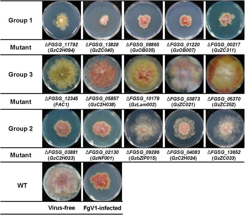
Yu and Kim                                                                                                        TF Functions on FgV1 Infection

INTRODUCTION                                                           summary of fungal host proteins that might be associated
                                                                       with FgV1 accumulation, mycovirus transmission, and symptom
Virus divert many cellular resources to produce virus-specific         development in F. graminearum (Yu and Kim, 2020). FgHex1
components and counteract to host defense responses                    that functions in the maintenance of cellular integrity enhances
during virus infection (Carrera and Elena, 2012). This                 the FgV1 RNA synthesis by binding to the FgV1 genomic
virus-host interaction often leads to the expression of                RNA (Son et al., 2016b). FgHal2 is required for the vegetative
disease symptoms in the host by triggering physiological               growth and methionine biosynthesis of F. graminearum and
alteration and modifying cytoskeleton or membrane structures           FgHal2 gene deletion reduces FgV1 RNA accumulation and
(Osterbaan and Fuchs, 2019).                                           vertical transmission of virus (Yu et al., 2015; Yun et al.,
   Transcription factors (TFs) are DNA-binding proteins                2015). Transcriptional reduction of FgSWI6, encoding possible
responsible for modulating gene regulatory systems by                  transcription cofactor in F. gramimearum, following FgV1
cooperating with a range of proteins, including other upstream         infection might be related to the changes of fungal host colony
or downstream TFs, transcription initiation complex, and               morphology caused by FgV1 infection (Son et al., 2016c).
epigenetic regulators (Spitz and Furlong, 2012; Shelest, 2017;         However, these studies focused on biological functions of
Mitsis et al., 2020). During virus-host interaction, TFs are           individual components, and specific biological processes and
directly or indirectly regulate defense response by activation         pathways remains elusive. Using transcriptomic analysis, we
or repression of downstream signaling pathways (Alves                  previously demonstrated that numerous F. graminearum genes
et al., 2014). In plant, members of TF families belonging to           including transcription factors were differentially expressed upon
WRKY family, myeloblastosis related proteins (MYB), basic              FgV1 infection (Lee et al., 2014). Recent study reported that
leucine zipper (bZIP), APETELA2/Ethylene-Responsive Factor             FgV1 protein pORF2 could inhibit transcriptional induction
(AP2/ERF) family, and NAC transcription factors have been              of FgDICER and FgAGO genes to counteract host’s antiviral
shown to be associated with defense response against plant             RNA silencing response (Yu et al., 2020). This previous study
viruses as well as abiotic stress responses (Alves et al., 2014;       proposed that FgV1 might be able to affect gene regulatory
Ng et al., 2018).                                                      networks directly or indirectly, which lead to pleiotropic
   A filamentous fungus Fusarium graminearum causes                    phenotypes on the presence of significant amount of viral
Fusarium head blight of major cereal crops, such as wheat,             RNA in fungal host.
barley, and rice (Dweba et al., 2017). Fusarium species also              Here, to gain new insights on the role(s) of host transcription
produce mycotoxins such as deoxynivalenol (DON), nivalenol,            factors that might be associated with viral RNA replication or
and zearalenone that are considered threat to the animals              symptom development following FgV1 infection, we transferred
and human health (Ferrigo et al., 2016). Since the report of           FgV1 to gene-deletion mutant library of 657 putative TFs in
the complete genome sequence of F. graminearum, many                   F. graminearum. Based on this library, we analyzed phenotype
researchers have attempt to characterize function(s) of TFs and        of virus-infected mutants and the relationship between FgV1
their target genes in gene regulatory network using diverse            RNA accumulation and phenotypic differences of FgV1-
computational and experimental approaches (Son et al., 2011;           infected TF deletion mutants. To our knowledge, this is
Liu et al., 2019; Guo et al., 2020). Systematic loss-of function       the first description of phenome-based association study in
studies and transcriptomic studies under comparable condition          characterizing effects of Fusarium graminearum transcription
provide new insights into the role of TFs in complex regulatory        factors on FgV1 infection.
networks for mycotoxin biosynthesis, sexual development, and
virulence in F. graminearum (Son et al., 2011; Kim et al., 2015;
Kazan and Gardiner, 2018; Chen Y. et al., 2019; Guo et al.,            MATERIALS AND METHODS
2020). Their interconnection and specific roles on signaling
pathways, however, are largely unknown. In previous study, to          Fungal Strains and Growth Condition
determine the functions and interconnectedness of individual           Fusarium graminearum GZ03639 WT strain and 657 TF deletion
TFs, the gene-disruption library of 657 potential TF genes in          mutant library were provided by the Center for Fungal Genetic
F. graminearum was constructed and analyzed (Son et al., 2011).        Resources (Seoul, South Korea). All fungal isolates were stored
Each of TF deletion mutant was categorized by phenotypic               in 20% (v/v) glycerol at −80◦ C and TF deletion mutants were
characteristics, such as mycelial growth, sexual and asexual           reactivated at 25◦ C on potato dextrose agar (PDA) with geneticin
developments, virulence, toxin production, and stress responses        (50 µg/ml). TF deletion mutants were subcultured on complete
(Son et al., 2011).                                                    medium (CM) agar containing geneticin for further experiment.
   Fusarium graminearum virus 1 (FgV1) is a single-stranded            Fungal colonies incubated on CM agar at 25◦ C for 120 h were
RNA (ssRNA) virus and closely related to the proposed                  photographed. Fungal cultures used for extraction of RNA were
family “Fusariviridae” (Kwon et al., 2007; Honda et al.,               prepared as previously described (Lee et al., 2014). Briefly, freshly
2020). FgV1 infection causes remarkable phenotypic change              grown mycelia was inoculated into CM broth, and the cultures
such as reduced growth rate, increased pigmentation, reduced           were incubated at 25◦ C for 120 h. Hyphae were collected by
mycotoxin synthesis, reduced pathogenicity, and defects in             filtering through 3 MM paper followed by washing with distilled
sexual development in F. graminearum (Chu et al., 2002;                water, dried by blotting mycelia between paper towels, and frozen
Lee et al., 2014). Previous literature review provides a               at −80◦ C.

Frontiers in Microbiology | www.frontiersin.org                    2                                   February 2021 | Volume 12 | Article 622261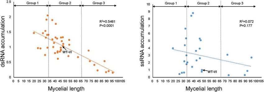
Yu and Kim                                                                                                        TF Functions on FgV1 Infection

Virus Transmission                                                      factor 1α (EF1α, locus FGSG_08811), were used as internal
FgV1-infected F. graminearum GZ03639 (WT-VI) was generated              controls to normalize qRT-PCR results. Data were analyzed using
by using protoplast fusion method (Lee et al., 2011). We                the Bio-Rad CFX Manager V1.6.541.1028 software (Bio-Rad).
confirmed FgV1 infection by total RNA extraction and reverse            RNA samples were extracted from at least two independent,
transcription polymerase chain reaction (RT-PCR) using virus            biologically replicated experiments, and each PCR product was
specific primer pair and selected WT-VI as positive control             evaluated in at least three independent experiments, including
for further experiments. FgV1 was introduced into TF deletion           three technical replicates. All primer sets used in this study are
mutant by hyphal anastomosis between WT-VI and TF deletion              listed in Supplementary Table 2.
mutant library. An agar block of WT-VI and individual TF
deletion mutant strain was placed on CM agar media and                  Viral dsRNA Confirmation and
incubated at 25◦ C for 4 days. Overlapped region of two fungal          Semi-Quantification
strains were isolated, transferred to CM agar contained geneticin       Three micrograms of DNaseI-treated total RNAs from all virus-
(50 µg/ml), and subcultured twice to eliminate unstable virus-          infected TF deletion mutants were treated by 30 units of S1
infected colony. Multiple replicates of all virus-infected mutant       Nuclease (Takara Bio). Samples were loaded into 1% agarose
strains have obtained. After virus transmission has failed during       gel for analysis of viral double-stranded (dsRNA) accumulation.
at least three times repetition, we determined these TF deletion        After separation on agarose gel, dsRNA was visualized in a UV
mutant strains as non-transmissible via hyphal anastomosis.             transilluminator. To measure relative accumulation of FgV1 viral
                                                                        dsRNA in TF deletion mutants, 3 µg of total RNA from all
Measurement of Mycelial Growth                                          virus-infected mutants were loaded and separated on 1% agarose
For phenotype analysis, virus-infected TF deletion mutants              gel. Ethidium bromide-stained gels were visualized in a UV
were photographed after 5 days of cultivation (Supplementary            transilluminator. Band intensity were measured using ImageJ
Figure 1). Radial growth of mycelia from the inoculum was               software (Schneider et al., 2012). The relative amount of viral
measured using ImageJ software (Schneider et al., 2012). The TF         genomic dsRNA was estimated by measuring the amount of FgV1
deletion mutants that showed reduced mycelial growth after gene         RNA relative to 18S rRNA.
deletion was also assessed (Supplementary Table 1).

Preparation of Total RNA Samples and                                    RESULTS
cDNA Synthesis
For nucleic acid extraction, frozen mycelia were pulverized using       Phenotype Analysis of FgV1-Infected TF
liquid nitrogen and a mortar and pestle. Total RNAs were                Gene-Deletion Mutants
extracted with RNAiso Plus reagent (Takara Bio, Shiga, Japan)           To investigate the effect of TF genes on FgV1 infection in
followed by treatment with DNaseI (Takara Bio) to remove                F. graminearum, we transferred FgV1 to putative 657 TF gene
genomic DNA according to the manufacturer’s instructions. As            deletion mutants. Among 709 TF genes, 657 TF genes were
described previously, 4 M LiCl was added to total RNA extract           successfully disrupted and other 52 TF genes were excluded due
to a final concentration of 2 M to isolate ssRNA fraction (Yu           to lethality or technical problem of generation of homologous
et al., 2018). Samples were then incubated at −20◦ C for 2 h,           recombination construct (Son et al., 2011). FgV1 could effectively
ssRNA pellets were washed in 75% ethanol and suspended in               transmitted by hyphal anastomosis between FgV1-infected strain
RNase-free water. Next, 3 µg of ssRNA of each sample was                GZ03639 (WT-VI) strain and virus-free TF deletion mutant
used to synthesize first-strand cDNA with an oligo (dT)18 primer        strains. Among total of 657 TF deletion mutants, we could not
and GoScriptTM reverse transcriptase (Promega, Madison, WI,             transmit FgV1 onto a 17 TF deletion mutants despite repeated
United States) according to the manufacturer’s protocols. All           trials (Table 1 and Supplementary Table 1). Representative
synthesized cDNAs were diluted to 20 ng of mixture with                 image of colony morphologies for each FgV1-infected TF
nuclease-free water.                                                    deletion mutants (total 640) were shown in Supplementary
                                                                        Figure 1. Typically, colony morphology of WT-VI (a FgV1-
Real-Time RT-PCR Analysis                                               infected strain) includes irregular colony shape, no aerial
Real-time quantitative RT-PCR (qRT-PCR) was performed with              mycelium, and dense mycelia with deep red or brown color. Most
a Bio-Rad CFX384TM Real-time PCR system using gene-specific             of FgV1-infected TF deletion mutants showed similar colony
internal primers as described previously with slight modification       morphologies compared to that of WT-VI, but several virus
(Yu et al., 2018). Each reaction mix (10 µl) consisted of               infected fungal colonies showed abnormal colony morphology
20 ng of cDNA, 5 µl of 2 X iQTM SYBR Green Supermix
                                                  R
                                                                        (e.g., slower or faster mycelial growth, low density and scarce
(Bio-Rad, Hercules, CA, United States), and 10 pmoles of                hyphal growth, curly mycelia, aerial mycelia development, and
each primer. The thermal profile was as follows: 3 min at               change of pigment production).
95◦ C and 40 cycles of 10 s at 95◦ C, 30 s at 59◦ C, and                    FgV1-infected TF deletion mutants were classified into three
melting curve data obtained by increasing the temperature from          groups according to the mycelial growth phenotype compared
55 to 95◦ C. Two endogenous reference genes, i.e., ubiquitin            to that of WT-VI or virus-free TF deletion mutant (Figure 1
C-terminal hydrolase (UBH, locus FGSG_01231) and elongation             and Table 1). For this, the mycelial length of WT-VF was set

Frontiers in Microbiology | www.frontiersin.org                     3                                  February 2021 | Volume 12 | Article 622261
Yu and Kim TF Functions on FgV1 Infection
TABLE 1 | Analysis of FgV1-infected transcription factor deletion mutants. candidates belong to the groups of bHLH (basic–helix–loop–
helix) motif and heteromeric CCAAT-binding factor showed
TF Classification TF 1TF TF-FgV1 Groupa
relatively high phenotypic variation following FgV1 infection
1 2 3 compared to that of other TF families (Table 1).
bHLH 16 15 15 0 11 4
TF Factors That Might Be Involved in
bZIP 22 22 22 0 21 1
C2H2 zinc finger 98 94 94 5 84 5
FgV1-Derived Host Symptom
Heteromeric CCAAT factors 8 8 8 3 5 0
Previous study analyzed phenotypes of putative 657 TF deletion
HMG 37 34 34 3 30 1
mutants and divided them based upon their major phenotypic
Homeodomain-like 14 7 7 1 6 0
categories such as mycelial growth, sexual development, conidia
Nucleic acid-binding, OB-fold 47 40 40 4 35 1
production, toxin production, and stress responses (Son
Winged helix repressor DNA-binding 27 26 26 0 23 3
et al., 2011). This phenome-based analysis demonstrated that
Helix-turn-helix, AraC type 8 7 7 0 6 1
fungal virulence, growth, and DON production were highly
GATA type zinc finger 8 7 7 0 7 0
correlated with sexual development (Son et al., 2011). Because
Zinc finger, CCHC-type 12 12 12 0 12 0 FgV1 infection causes multiple phenotypic alterations, this
Zn2Cys6 zinc finger 316 296 286b 16 254 16 simultaneous and multiple FgV1-derived symptom might also be
Myb 19 17 17 1 13 3 consequences of interaction between virus and host factor that
Others 77 72 65b 2 52 11 have pivotal roles in gene regulatory network. In this regard,
Total 709 657 640 35 559 46 we selected 35 TF deletion mutants that exhibit multiple defects
a Groups
in mycelial growth, virulence, sexual development, and toxin
1–3 were determined by mycelial growth on complete media (CM).
Average radial growth on CM of FgV1-infected TF deletion mutants compared to
production (Figure 2 and Supplementary Table 3; Son et al.,
virus-free strain or wild-type (WT) strain (set to a value of 100) was divided as 2011). Ten out of 35 selected TF deletion mutants that were not
groups (Group 1, less than 33; Group 2, 33–62.9; Group 3, 63–100 of WT strain. related with environmental stress responses or DNA damages
b Some of TF deletion mutants did not obtain FgV1 through hyphal anastomosis.
were shown in Figure 2A, except FgNHP6A (FGSG_00385)
deletion mutant that showed pH 4-resistance. Among these 35
TF deletion mutants, most of gene deletion mutants showed
to a value of 100 (±11). Mycelial length of WT-VI decreased decreased mycelial growth compared to WT. Colony morphology
to 40–55 (average 47.5 ± 7) compared to that of WT-VF. In of virus-free deletion mutants including FgSWI6 (FGSG_04220),
case of TF deletion mutant showing growth retardation, this FgNOT3 (FGSG_13746), GzC2H090 (FGSG_10517), GzWing019
virus-free TF deletion mutant was used as standard to compare (FGSG_08572), and FgCrz1A (FGSG_13711) showed similar
FgV1-infected TF mutant. Comparing with each virus-free TF phenotypes to those of WT-VI such as reduced aerial mycelia,
deletion strain or wild-type (WT) strain, we classified Group reduced mycelial growth, and increased pigmentation (Figure 2A
1 and 3 of FgV1-infected TF deletion mutants determined by and Supplementary Table 1, compare to WT-VI in Figure 1).
growth under 33 (0–32.9) or over 63 (63–100), respectively. When we confirmed gene expression level of these five TFs
The FgV1-infected TF deletion mutants in Group 1 indicated after FgV1 infection by qRT-PCR, those of all five TFs showed
slow or weak mycelial growth compared to that of WT-VI. decreased level compared to WT-VF (Supplementary Table 4).
Among FgV1-infected TF deletion mutants in Group 2 (mycelial Comparing phenotype changes upon FgV1 infection in Group
growth, 33–62.9), we could divide them into two subgroups. One 1, GzC2H003 (FGSG_00477) and FgNHP6A (FGSG_00385) TF
showed similar colony morphology of WT-VI while the other deletion mutants showed slow mycelial growth after FgV1
subgroup did not follow typical phenotype WT-VI and showed infection compared to WT-VI (Figure 2A, compare to WT-VI
fluffy but low density of mycelia phenotype. In contrast, Group in Figure 1). On the contrary, mycelial growth in virus-infected
3 consists of virus-infected TF deletion mutants showing mild TF deletion mutants in Group 3, FgCrz1A (FGSG_13711),
symptoms, such as faster mycelial growth and partially restored GzZC108 (FGSG_08769), GzMADS003 (FGSG_09339), and
aerial mycelia formation. FgStuA (FGSG_10129) showed little reduction of mycelial
Approximately 88% of FgV1-infected TF deletion mutants growth compared to WT-VI. Six out of those 35 TF deletion
(Group 2) showed similar phenotype and mycelial growth mutants including GzAPSES004 (FGSG_10384) and FgFSR1
regardless of phenotype of virus-free TF gene deletion mutants. (FGSG_01665) mutants were not able to uptake FgV1 via hyphal
Among FgV1-infected TF deletion mutants in Group 3, which anastomosis method, this result might be explained as growth
growth reduction rate was lower than 10%, colony morphology defect in those deletion mutants (Supplementary Figure 1).
of most mutants in this group showed recovery phenotype
and viral accumulation level was as low as 10% of WT-VI Comparison of Phenotypes Produced in
although FgV1 infection might still affect colony morphology.
In this regard, comparisons of colony morphology including
Response to Deletion and FgV1 Infection
mycelial growth of TF deletion mutants following hypovirulence- of the TFs Associated With
associated FgV1 infection help screening host gene(s) that Environmental Stress Response
might attribute or affect development of virus-derived symptom TF phenome analysis described phenotypic change under diverse
and FgV1 replication. We observed that F. graminearum TF abiotic stresses, including osmotic stress, reactive oxygen species
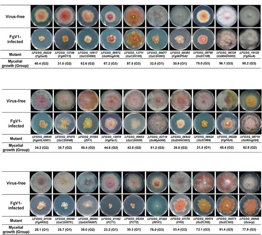
Frontiers in Microbiology | www.frontiersin.org 4 February 2021 | Volume 12 | Article 622261Yu and Kim TF Functions on FgV1 Infection FIGURE 1 | Representative FgV1-infected colony morphology according to mycelial growth. Representative examples of colony morphology of FgV1-infected TF deletion mutants are shown. All cultures were photographed after incubating 5 days on complete media (CM). WT, Fusarium graminearum wild-type (WT) strain GZ03639. The virus-free TF deletion mutants that correspond to FgV1-infected TF deletion mutants in this figure did not changed mycelial growth compared to virus-free WT. (ROS) stress, fungicide, cell wall stress, and acidic (pH = 4) or (Group 3) showed much faster mycelial growth and formed basic (pH = 11) conditions (Son et al., 2011). We compared rhombus-shape of red line at the outside region of colony. In TF phenotypes in response to those stress factors, considering case of GzDHHC003 (FGSG_06542) and GzZC086 (FGSG_8924), FgV1 infection as biotic stress in F. graminearum, whether TF related with oxidative stress response, showed normal growth gene disruptions that showed different response to abiotic stress but with relatively reduced mycelia growth compared to WT- might also relate to response against to the FgV1 infection VI. FgFlbA (FGSG_06228) showed increased transcript level (Figure 2B). In results, among pH 11-sensitive or pH 11-resistant following FgV1 infection and deletion mutant showed resistance TF deletion mutants, GzbHLH001 (FGSG_00545), GzC2H048 phenotype in all stress factors except for pH 11 stress response. (FGSG_07075), and ZIF1 (FGSG_01555) deletion mutants The virus-infected 1FgFlbA (Group 2) colony grow normally showed more damaged virus-infected phenotype compared to but contain clustering region around fungal colony. In contrast, WT-VI though they were belong to the Group 2. The other pH 11 while 1GzWing020 (FGSG_08719, Group 3) showed sensitive responsive gene FgPac1 (FGSG_12970) deletion mutant showed responses to all stress responsive factors in phenome data, FgV1- similar phenotype to WT-VI. 1GzC2H048 and 1ZIF1 also infected colony showed increased aerial mycelia production and showed osmotic stress response. Between two pH 4-resistance TF hyphal growth compared to WT-VI. Obtained results indicated deletion mutants, 1GzC2H005 (FGSG_00653) and 1GzMyb008 that some of F. graminearum TF candidates that showed sensitive (FGSG_02719), they showed different phenotype. FgV1-infected response in pH, fungicide or ROS stress alone also involved in 1GzC2H005 deletion mutant showed very weak and low density response to FgV1 infection as well as TF candidates that response of colony morphology. In contrast, FgV1-infected 1GzMyb008 in broad range of environmental stress factors. Frontiers in Microbiology | www.frontiersin.org 5 February 2021 | Volume 12 | Article 622261
Yu and Kim                                                                                                                            TF Functions on FgV1 Infection

  FIGURE 2 | Selected colony morphology of FgV1-infected TF deletion mutants. Colony morphology of virus-free and of FgV1-infected TF gene-deletion mutant
  strains. (A) TF gene-deletion mutants that showed multiple defect phenotypes after single gene deletion. (B) TF gene-deletion mutants that related to sensitive
  response against abiotic stress factor. (C) TF gene-deletion mutants that related to DNA damage response. All cultures were photographed after 5-day incubation
  on CM.

The Relationship Between FgV1 Infection                                               showed retarded growth compared to WT-VI or to the
and TFs Involved in DNA Damage                                                        typical colony morphologies of FgV1-infected TF deletion
                                                                                      mutants belonging to Group 2 (Figure 2C, compare to WT-
Response                                                                              VI in Figure 1). GzC2H075 (FGSG_09368) and GzGATA007
Previous study identified 16 putative TFs involved in DNA                             (FGSG_09565) deletion mutants, which showed apparent
damage responses (DDRs) (Son et al., 2016a). In this study, we                        reduction in mycelial growth following FgV1 infection, exhibited
found that these DDR TF gene deletion group included relatively                       sensitive response only to DNA damage reaction in phenome
high portion of FgV1-infected TF deletion mutants that belong                         data. Individual mycelial growth value of RFX1 (FGSG_07420;
to Groups 1 and 3. Among 13 FgV1-infected TF, four FgV1-                              Group 3) deletion mutant was slightly higher than that of WT-
infected TF deletion mutants including FgARS2 (FGSG_01106),                           VI, however, virus-infected mutant showed strong inhibition of
GzC2H075 (FGSG_09368), FCT1 (FGSG_01182), and FCT2                                    mycelial growth phenotype so we are unable to process further
(FGSG_05304) were divided in Group 1. FgV1-infected                                   experiment. FHS (FGSG_01176; Group 2) deletion mutant
1GzGATA007 (FGSG_09565) belong to Group 2, however,                                   that showed oxidative stress and ROS response along with

Frontiers in Microbiology | www.frontiersin.org                                   6                                       February 2021 | Volume 12 | Article 622261
Yu and Kim TF Functions on FgV1 Infection
DDR did not show significant change of colony morphology the expression levels of some putative TF genes, however,
following FgV1 infection. The colony morphology of GzZC302 all of these changes might not be directly related with
(FGSG_00574; Group 3) and GzZC303 (FGSG_00573; Group FgV1 accumulation or FgV1-mediated colony morphology in
3) deletion mutants showed similar reduced aerial mycelia, F. graminearum.
increased pigmentation, and responded to multiple stress factors
include oxidative, ROS and pH, however, their virus-infected
phenotypes were not significantly different compared to WT-VI. TFs That Might Be Involved in FgV1 RNA
1Gzscp (FGSG_06948; Group 3), which exhibit multiple defects Accumulation
along with DDR, showed little reduction of mycelial growth To isolate TFs that might be associated with viral replication,
compared to WT-VI. we confirmed dsRNA and viral ssRNA accumulation levels
Although all DDR-related putative TF genes exhibited in FgV1-infected TF deletion mutants (Table 2). We selected
different sensitivity to DNA damaging agent include methyl several FgV1-infected TF deletion mutants belong to Groups 1,
methanesulfonate, hydroxyurea, bleomycin, and camptothecin 2, and 3. Selected isolates include FgV1-infected mutants with
(Son et al., 2016a), we could not correlate a common DNA phenotypic changes such as defect in sexual development, TF
damaging agent that links to displayed phenotype among FgV1- genes responsive to stress or DNA damage, and significantly up-
infected TF deletion mutants belong to Group 1. or down-regulated TF genes upon FgV1 infection from RNA-
Seq analysis. In results, increased viral ssRNA accumulation
level was observed in FgNHP6A (FGSG_00385), GzZC040
Comparisons With RNA-Seq Data and (FGSG_13828), GzZC086 (FGSG_8924), TRI15 (FGSG_03881),
Phenome Data FgFlbB (FGSG_03597), GzDHHC003 (FGSG_06542), and
Previous study demonstrated that 24 TF genes were up- or GzZC267 (FGSG_01669) deletion mutants. Among them,
down-regulated following FgV1 infection using transcriptomics- dsRNA accumulation level was also significantly increased
based analysis (Lee et al., 2014). We validated these RNA- in FgV1-infected GzZC086 deletion mutants. 1GzZC021
Seq data with selected TF genes in this study (Supplementary (FGSG_03873), GzZC252 (FGSG_05370), and 1GzZC303
Table 5). Among those 24 TF genes, only two TF genes (FGSG_00573) in Group 3 showed significant decrease in
including GzZC252 (FGSG_05370) and GzZC311 (FGSG_00217) viral dsRNA accumulation level. FgV1-infected 1GzZC252
were grouped into 1 and 3, respectively, following FgV1 showed significant reduction in dsRNA accumulation but
infection (Figure 1). Interestingly, expression of both GzZC252 not in viral ssRNA accumulation level compared to that of
(FGSG_05370) and GzZC311 (FGSG_00217) genes were up- WT-VI. In addition, we observed dsRNA patterns of these TF
regulated upon FgV1 infection (Lee et al., 2014; Supplementary deletion mutants (Figure 3). TF deletion mutants classified
Table 5). 1GzZC311 did not show significant phenotypic change into Group 3 including 1GzZC021 (FGSG_03873), 1GzZC252
in mycelial growth compared to WT. Phenotype of FgV1- (FGSG_05370), and 1GzZC197 (FGSG_03892) showed
infected 1GzZC311 showed slow growth of mycelia compared decreased dsRNA accumulation compared to those of WT-VI
to that of WT-VI. In contrast, 1GzZC252-VF showed flat and other mutants classified into Groups 1 and 2 (Figure 3A).
colony morphology with scarce growth of aerial mycelia but Defective RNAs (D-RNAs, approximately 2–3 kbp long) were
regular growth of mycelial growth in length. FgV1-infected often observed in TF gene deletion mutants that showed multiple
1GzZC252 did not decrease mycelial growth but caused color phenotypic changes and related with stress or DNA damage
change from pale yellow to dark yellow in overall area of responses (Figure 3B). Among TF deletion mutants in Group 2,
culture plate. The other 21 TF gene deletion mutants showed FgV1-infected mutants including 1GzbZIP015 (FGSG_09286),
similar colony morphology compared to WT-VI. Although 1GzC2H024 (FGSG_04083), and 1GzZC033 (FGSG_13652)
RNA-Seq analysis also identified GzbHLH006 (FGSG_02516), produced fluffy but low density of aerial mycelia (Figure 1) and
GzbHLH007 (FGSG_02814) GzC2H006 (FGSG_00764), TRI15 also accumulated D-RNAs during FgV1 replication (Figure 3C,
(FGSG_03881), and GzGATA003 (FGSG_04626) that showed right panel). These results indicated that deletion of single TF
significant changes of gene expression levels upon FgV1, gene affects FgV1 replication at different step(s) and generation
Fusarium graminearum virus 2 (FgV2; a Chrysovirus), FgV3 of D-RNAs. In Figure 1, we simplify FgV1-infected TF deletion
(a Fusagravirus), and Fusarium graminearum hypovirus 1 mutants by grouping based upon mycelial growth rate as the
infections (Lee et al., 2014; Wang et al., 2016), those deletion first step. We postulated that mycelial growth and viral RNA
mutants did not show significant change of colony morphology accumulation might inversely correlated in FgV1-infected
following FgV1 infection. In addition, we selected several TF fungal strains if mycelial growth of mutant did not changed by
genes in Groups 1 and 3 for confirmation of change of gene target gene deletion. To examine relationship between mycelial
expressions following FgV1 infection (Supplementary Table 4). growth and viral RNA accumulation, we plot dsRNA or ssRNA
In Group 1, FGSG_08865 and FGSG_13828 genes showed accumulation (y) against mycelial length (x) using selected
significantly increased expression levels following FgV1 infection FgV1-infected TF deletion mutants (Figure 4). In general, FgV1-
among 5 genes. In Group 3, expression levels of FGSG_09339, infected TF deletion mutants that grew slower than WT-VI
FGSG_08455, and FGSG_03873 genes were decreased while accumulated higher level of viral dsRNA compared to WT-VI.
expression level of FGSG_12742 was increased compared to In contrast, FgV1-infected TF deletion mutants that grow faster
that of WT-VI. These results showed FgV1 infection affects than WT-VI accumulated lower level of viral dsRNA compared
Frontiers in Microbiology | www.frontiersin.org 7 February 2021 | Volume 12 | Article 622261Yu and Kim TF Functions on FgV1 Infection
to WT-VI. Altogether, this result indicates that the relative TABLE 2 | Comparisons of relative ratio of mycelial growth, dsRNA accumulation,
and ssRNA accumulation of FgV1-infected TF deletion mutants.
levels of viral dsRNA accumulation in fungal colonies negatively
correlate with mycelial growth of FgV1-infected TF mutant. Mycelial dsRNAb ssRNAc Noted
lengtha
WT-VI 47.5 1.04 ± 0.1 0.98 ± 0.1
DISCUSSION Group 1
1FGSG_00477 32.5 0.98 ± 0.2 3.01 ± 0.9 MD
Identifying host factors involved in FgV1-derived symptom
1FGSG_01106 28.1 1.46 ± 0.7 0.62 ± 0.02 MD, DDR
induction and viral RNA accumulation is a key aspect
1FGSG_09368 25.7 0.96 ± 0.2 0.45 ± 0.1 DDR
of understanding the molecular mechanism during
1FGSG_00385 30.9 1.32 ± 0.1 4.41 ± 1.4* MD, pH4(R)
F. graminearum-FgV1 interactions. Previous studies suggested
1FGSG_08865 30.4 1.54 ± 0.3 2.19 ± 0.7 N
that viral components interfere with host cell signaling pathways
1FGSG_13828 19.4 1.47 ± 0.8 3.98 ± 1.8* N
and progressively cause alteration in physiological and
1FGSG_08924 31.5 1.91 ± 0.6* 6.68 ± 3.0* Fung, virus
developmental processes, which culminate in visible virus- response
induced symptoms (Urbanowski et al., 2008; Pesti et al., 2019). 1FGSG_00217 32.2 1.41 ± 0.4 5.34 ± 2.5* Virus response
In this respect, we used genome-wide TF deletion mutant 1FGSG_00324 32.6 2.05 ± 1.3 1.79 ± 1.0 MD, os
library for F. graminearum to find host transcription factors Group 2
and host-cell signaling pathways that might be associated with 1FGSG_09286 46.3 1.20 ± 0.2 5.09 ± 2.4 Virus response
pleiotropic effects of FgV1 infection on fungal host and to 1FGSG_03881 41.7 1.28 ± 0.3 3.62 ± 1.7* Virus response
identify novel host factor which might be involved in FgV1 RNA 1FGSG_04083 47.3 1.33 ± 0.1 0.78 ± 0.2 Virus response
accumulation in host cell. 1FGSG_08617 42.2 0.86 ± 0.3 2.32 ± 0.4 Virus response
We observed different phenotype change in fungal colony 1FGSG_08893 37.2 1.26 ± 0.1 8.44 ± 0.7* N
color which turns yellow after virus infection without greater 1FGSG_06110 45.5 1.32 ± 0.01 2.61 ± 0.6* Virus response
reduction of mycelial growth in some TF deletion mutants. 1FGSG_02615 33.4 1.41 ± 0.2 2.05 ± 0.5 Virus response
Those genes were not listed in phenome data as pH sensitive 1FGSG_03597 45.3 0.98 ± 0.1 3.94 ± 1.1* N
responsive mutants that showed reduced mycelial growth at pH 1FGSG_06542 38.8 1.14 ± 0.5 8.81 ± 5.2* MD, ROS
4 or pH 11 (Son et al., 2011). However, it is worth noting 1FGSG_11686 38.0 1.00 ± 0.1 1.43 ± 0.2 N
that pH also impacts on pigment production and mycelium 1FGSG_01669 45.3 0.89 ± 0.3 5.31 ± 0.5* Virus response
color. For example, the red pigment of F. graminearum is pH Group 3
sensitive and changes color from red to yellow as the pH drop 1FGSG_03873 88.9 0.17 ± 0.2** 0.31 ± 0.1** N
(Leslie and Summerell, 2008). Because pH affects wide range of 1FGSG_13625 73.3 0.54 ± 0.2 0.29 ± 0.1** N
fungal physiological processes and gene expression in fungal cells 1FGSG_05370 95.3 0.15 ± 0.1** 2.71 ± 0.2* Virus response
(Bousset et al., 2019), change of colony morphology of those TFs 1FGSG_00574 72.1 0.89 ± 0.1 3.10 ± 0.5* MD, DDR,
deletion mutants might be related with pH stability following Os,ROS,Fung,pH11
virus infection. In addition, dsRNA accumulation was decreased 1FGSG_00573 91.4 0.20 ± 0.1** 0.87 ± 0.3 DDR,
in 1GzZC197 (FGSG_03892) as shown in Figure 3, pH stability Os,ROS,Fung,pH11
might also affect replication of viral RNA. Since whether pH a Radial growth was measured after a 5-day incubation on CM. Average radial
impacts virus-host interactions is not clear in F. graminearum, growth on CM of FgV1-infected TF deletion mutants compared to virus-free strain
further studies are needed. or WT strain (set to a value of 100) was divided as groups (Group 1, less than 33;
Group 2, 33–62.9; Group 3, 63–100 of WT strain).
In TF deletion mutant library, we were interested in TF b Relative accumulation of FgV1 viral dsRNA in TF deletion mutants was measured
deletion mutants showing pleiotropic phenotype similar with using 3 µg total RNA samples at 120 h post-inoculation (hpi). Values were
FgV1-infection derived phenotype in F. graminearum. We normalized to the FgV1 RNA in WT-VI (set to a value of 1), with the standard
deviation based on at least two independent RNA preparations. Different number
expected it would provide information for identifying host
of asterisks indicate data are significantly differed (P < 0.05) from each other and
factor(s) or characterizing signaling pathway that related with mean for FgV1-infected GZ03639 WT strain based on LSD test.
hypovirulence-associated traits of FgV1 regardless of phenotype c Quantification of FgV1 viral ssRNA at 120 hpi using real-time RT-PCR. cDNAs
observation of FgV1-infected TF deletion mutant. As mentioned were generated from ssRNA samples obtained after 120 h of incubation. EF1α and
UBH gene transcripts were used as internal controls. Values are means (+ SD) of
above, several TF deletion mutants showed WT-VI like colony two biological replicates with at least two experimental replication.Different number
morphology. Some of these TFs might play central roles of asterisks indicate data are significantly differed (P < 0.05) from each other and
in reprogramming transcriptional network or function as an mean for FgV1-infected GZ03639 WT strain based on LSD test.
d MD, multiple defects after TF gene deletion; DDR, TF deletion mutant related
important downstream regulator, which results in pleiotropic
with DNA damage responses; os, sensitive in osmotic stress; ROS, sensitive in
effects by gene deletion. For example, FgSWI6 and GzAPSES004 ROS stress; Fung, sensitive in fungicide response; pH4(R), resistance response
were suggested as hub regulators of virulence, mycotoxin against pH 4 stress; N, TF deletion mutant not showing any significant change;
synthesis, and sexual reproduction-associated networks (Guo virus response, level of gene expression was changed after virus infection.
et al., 2020). Previous study reported that decreased expression
level of FgSWI6 following FgV1 infection seems to be attenuate symptom expression by FgV1 infection although it
related with FgV1-derived phenotypic alteration (Son et al., contained increased FgV1 accumulation level compared to
2016c). Because constitutive overexpression FgSWI6 moderately WT (Son et al., 2016c). FgCrz1A is reported as a possible
Frontiers in Microbiology | www.frontiersin.org 8 February 2021 | Volume 12 | Article 622261Yu and Kim TF Functions on FgV1 Infection
FIGURE 3 | Accumulation of FgV1 viral double-stranded RNA in virus-infected TF deletion mutant strains of F. graminearum. (A) dsRNA accumulation of TF
gene-deletion mutants that belong to Groups 1 to 3. (B) TF gene-deletion mutants that showed multiple defect phenotypes after single gene deletion, TF
gene-deletion mutants related to sensitive response against abiotic stress factor and DNA damage response. (C) TF gene-deletion mutants showing significant
changes of gene expression levels upon FgV1 (left) and TF gene-deletion mutants showing abnormal colony morphology in Group 2 (right). Number in parenthesis
represents group of each sample. A 3 µg quantity of total RNAs per sample was treated with DNaseI and S1 Nuclease and separated in 1% agarose gel. The largest
band in the each sample represents the full-length FgV1 dsRNA (6.6 kb); smaller bands indicate internally deleted forms of viral dsRNA. The lane marked M
correspond to lambda DNA digested with HindIII. WT-VI, FgV1-infected WT GZ03639 strain.
ortholog of Saccharomyces cerevisiae Crz1 that has crucial 1GzZC302 (FGSG_00574; Figure 2). However, ss and dsRNA
role in regulating calcineurin- and Ca2+ /calmodulin-dependent accumulations of those mutants were similar or slightly decreased
signaling (Chen L. et al., 2019). 1FgCrz1A displayed multiple compared to WT-VI (Table 2). These results indicated DNA
abnormalities in phenotypes including increased sensitivity to damage might be induced by FgV1 infection and those DDR-
metal cations Ca2+ , Mg2+ , Mn2+ , and Li+ (Chen L. et al., related gene deletion caused significant change in mycelial
2019). We observed small decrease in mycelial growth in FgV1- growth even they contain relatively low or similar amount of
infected 1FgCrz1A compared to WT-VI. However, including viral RNA compared to WT-VI. Accordingly, genetic instability
FgCrz1A, we are not sure whether these TF function in facilitating plays a considerable role in pathogenicity of FgV1 and is likely
FgV1 replication or in regulating defense pathways of fungal to be a key factor of FgV1-associated symptom development. In
host. The attempts to establish relationships between FgV1 and addition, we often found FgV1 infection resulted in significant
phenotype-associated cellular signaling pathways are required accumulation of DI RNAs as were in case of 1FgARS2,
in further study. 1GzC2H075, 1GzZC303, and 1GzZC302 mutants. It might
Recent study demonstrated that FgARS2 physically interacts suggest possible role of DDR in supporting virus replication.
with the cap-binding complex to form a stable tertiary complex Like other DNA viruses, some RNA viruses also have ability
(Bui et al., 2019), however, key components and regulation to trigger DDR signaling to assist host cellular conditions
processes of DDR in F. graminearum are largely unknown. In that are beneficial for viral replication (Ryan et al., 2016).
this study, we found FgV1 infection significantly impacts on Although it has not been determined whether FgV1 replication
mycelial growth of several DDR-related TF deletion mutants, i.e., and symptom development are closely related with DDR in
1FgARS2 (FGSG_01106; Bui et al., 2019), 1FCT1 (FGSG_01182; present study, this phenotype-based analysis would lead to
Kim et al., 2020), 1FCT2 (FGSG_05304; Kim et al., 2020), investigate the interactions between FgV1 and the DDR in
1GzC2H075 (FGSG_09368), 1GzZC303 (FGSG_00573), and F. graminearum in further.
Frontiers in Microbiology | www.frontiersin.org 9 February 2021 | Volume 12 | Article 622261Yu and Kim TF Functions on FgV1 Infection Previous transcriptome study described that 24 TF genes showed increased gene expression level following FgV1 infection were differentially expressed upon FgV1 infection, however, 114 but result of FgV1 transmission into each gene deletion of 709 TF genes were not detected (Lee et al., 2014). Because mutant showed different colony morphology. GzZC311 and expression levels of many TF genes vary through different GzZC252 encoded hypothetical protein contain fungal-specific phase of fungal development and environmental condition, it regulatory protein domain, however, their cellular functions might has limitation in finding crucial TF genes that play have not been identified yet. 1GzZC252 negatively affects FgV1 crucial role(s) during FgV1 infection from transcriptome profiles dsRNA accumulation but not for ssRNA accumulation. Further obtained at a particular time-point measurement under a certain investigation with complementation or overexpression mutant is condition. Several studies analyzed transcription profiles of required to confirm whether this gene is required for FgV1 RNA F. graminearum in response to different mycovirus infections accumulation. Among Group 2, FgV1 infections in 1GzbZIP015 (Lee et al., 2014; Wang et al., 2016; Bormann et al., 2018). (FGSG_09286) and 1GzZC050 (FGSG_12597) mutants showed In addition, other transcription profiles has revealed subsets similar colony morphology like WT-VI (Figure 1). GzbZIP015 of transcriptionally regulated genes using mutant strains that encoded protein which has similarity with cross-pathway control involved in mycotoxin synthesis, asexual or sexual development, protein 1, the ortholog of GCN4 in the yeast S. cerevisiae, is RNAi process, abiotic stress response, and post-translational a main regulator of protein synthesis and might have role in modification (Brauer et al., 2020). For examples, GzZC196 longevity and stress response in Neurospora crassa (Hinnebusch, (FGSG_03912; Group 1) and GzZC197 (FGSG_03892; Group 3) 2005). Given that increased expression of GzbZIP015 gene upon showed reduced gene expression level in FgDICERs or FgAGOs FgV1 infection (Lee et al., 2014) and increased accumulation of double knockout mutant strains compared to WT (Son et al., FgV1 ssRNA in FgV1-infected 1GzbZIP015 mutant, GzbZIP015 2017; Supplementary Table 1). GzbHLH011 (FGSG_06262) and might serve as an antiviral host factor following virus GzHOMEL018 (FGSG_07243) showed reduced gene expression infection (Table 2). level in both 1FgGCN5 and 1FgSAS3 that are putative histone We confirmed ss and dsRNA accumulation using several acetyltransferase (HATs) in F. graminearum (Kong et al., 2018). TF genes deletion mutants belong to Group 2 but it showed Both 1GzbHLH011 and 1GzHOMEL018 belong to Group 3 differential gene expression upon FgV1 infection (Table 2). FgV1 (Supplementary Table 1). In case of GzZC086 (FGSG_08924), ssRNA accumulation levels were increased compared to WT-VI, involved in oxidative stress in F. graminearum (Lee et al., however, gene deletion did not seem to directly affect dsRNA 2018), FgV1-infected 1GzZC086 (Group 1) showed significantly accumulation. This result suggested that dsRNA accumulation increased viral RNA accumulation level (Table 2). Therefore, level could determine FgV1-derived symptom rather than ssRNA combined phenome data from this study and transcriptome data accumulation (Figure 4). obtained from diverse conditions will help in understanding Viruses need host factors not only to assist their replication common and unique roles of TFs and signaling pathways that but also to face the host antiviral defense response. Mycovirus might be associated with host response against virus infection. infection in host cell boosts host antiviral response such In this study, among FgV1-infected TF deletion mutants that as RNA interference (RNAi). Interestingly, Cryphonectria belonged to Groups 1 and 3, we found overlapped result with hypovirus 1 (CHV1) and FgV1 exhibit suppression activity transcriptome analysis. GzZC311 (FGSG_00217) and GzZC252 against host antiviral response through suppression of RNAi (FGSG_05370) that were grouped into 1 and 3, respectively, component-related gene transcription (Sun et al., 2009; FIGURE 4 | Relationship between mycelial growth and the viral RNA accumulation in TF deletion mutants. Relationship between radial growth of mycelia and FgV1 RNA replication was estimated by calculating the viral dsRNA (A) and ssRNA (B) accumulations in FgV1-infected TF deletion mutants. Dotted and dashed lines indicate the linear regression between the mycelial growth and viral RNA accumulation. Frontiers in Microbiology | www.frontiersin.org 10 February 2021 | Volume 12 | Article 622261
Yu and Kim                                                                                                                                     TF Functions on FgV1 Infection

Yu et al., 2020). Previous studies demonstrated that the Spt–                             pathways promote viral replication and activate antiviral
Ada–Gcn5 acetyltransferase (SAGA) transcriptional activator                               response (Dunn and Connor, 2012). Since systematic
regulates the induction of the essential antiviral RNA-silencing                          characterization of the kinome and phosphatome has been
components, dicer-like 2 (dcl2) and argonaute-like 2 (agl2) in                            reported in F. graminearum previously (Wang et al., 2011; Yun
Cryphonectria parasitica (Andika et al., 2017). We attempted                              et al., 2015), applying FgV1 into kinome and phosphatome in
to identify TFs that involved in transcriptional regulation of                            F. graminearum will provide a valuable resource to understand
FgDICERs or FgAGOs genes using TF deletion mutant library,                                fungal host cell signaling pathway involved in antiviral or
however, FgV1 was not suitable for screening candidate genes                              proviral functions. Although the TF phenome data illustrated
because of the presence of pORF2, suppressor of RNAi (Yu                                  characteristics of phenotype of all TF deletion mutants in
et al., 2020). Further investigations are in progress to identify                         previous research, it has limitation in expecting possible
TFs that play roles in regulating gene expressions of FgDICERs or                         functions of TF genes that do not show distinct phenotypic
FgAGOs combined with present research and other FgV-infected                              change. In this regard, FgV1-infected TF deletion mutant
TF deletion mutants.                                                                      library obtained in present study would provide chance to
    As mentioned earlier, we had failed to transmit FgV1 via                              better characterize function(s) of novel TF genes that showed
hyphal anastomosis into several TF deletion mutants including                             distinguishable phenotypes following FgV1-infection. Further
1GzZC030 (FGSG_06380), 1GzZC032 (FGSG_00153),                                             study will explore the roles of these TF genes and their putative
1GzZC044 (FGSG_12094), 1GzZC060 (FGSG_08808),                                             target genes during FgV1 infection.
1GzZC232 (FGSG_07067), 1FgArt1 (FGSG_02083), 1GzZC301
(FGSG_00404), and 1GzZC316 (FGSG_00125). Most of those
gene deletion mutants did not show specific alteration in mycelial                        DATA AVAILABILITY STATEMENT
growth. Among them, we confirmed gene expression levels of
9 TFs by qRT-PCR (Supplementary Table 4). Seven out of                                    The raw data supporting the conclusions of this article will be
nine genes showed significant changes of gene expression levels                           made available by the authors, without undue reservation.
following FgV1 infection. Some of these genes might be involved
in cell-to-cell interaction regulation that has been proposed as
a defense mechanism of host fungi to limit the transmission                               AUTHOR CONTRIBUTIONS
of mycoviruses (Nuss, 2011). For example, GzZC232 encoded
protein shares 59% sequence identity with Epichloë festucae ProA,                         JY and K-HK designed the experiments, analyzed the data,
which is similar to N. crassa ADV-1 and Sordaria macrospora                               and wrote the manuscript. JY performed the experimental
Pro1 (Tanaka et al., 2013). ProA deletion mutant is defective                             work. Both authors contributed to the article and approved the
in hyphal fusion under nutrient limitation condition (Tanaka                              submitted version.
et al., 2013). In case of FgArt1, it is associated with biosynthesis
of trichothecene and fumonisin by regulating genes involved in
starch hydrolysis, however, it remains unclear if FgArt1 plays a                          FUNDING
role in cell fusion or related biological processes (Oh et al., 2016).
                                                                                          This research was supported in part by grants from the National
Many genes and molecular signaling networks are involved
                                                                                          Research Foundation of Korea funded by the Ministry of Science
during hyphal fusion in N. crassa including MAPKinase cascades,
                                                                                          and ICT (NRF-2020R1C1C1011779) and Agenda Program
a STRIPAK complex, transcription factors, a NADPH-oxidases
                                                                                          (No. PJ01488703), the Rural Development Administration
complex, ROS systems, and Ca2+ -binding regulators (Fischer
                                                                                          (RDA), South Korea.
and Glass, 2019). Further detailed study is required to explain
this inability of hyphal fusion in some TF deletion mutants in
F. graminearum.
                                                                                          SUPPLEMENTARY MATERIAL
    Kinases and phosphatases also contribute to the regulation
of gene expression by interacting with transcription factors                              The Supplementary Material for this article can be found
(Ariño et al., 2019; González-Rubio et al., 2019). Both                                   online at: https://www.frontiersin.org/articles/10.3389/fmicb.
phosphatidylinositol-3-kinase (PI3K) and Akt signaling                                    2021.622261/full#supplementary-material

REFERENCES                                                                                Ariño, J., Velázquez, D., and Casamayor, A. (2019). Ser/Thr protein phosphatases
                                                                                             in fungi: structure, regulation and function. Microb. Cell 6, 217–256. doi:
Alves, M. S., Dadalto, S. P., Gonçalves, A. B., De Souza, G. B., Barros, V. A.,              10.15698/mic2019.05.677
   and Fietto, L. G. (2014). Transcription factor functional protein-protein              Bormann, J., Heinze, C., Blum, C., Mentges, M., Brockmann, A., Alder, A., et al.
   interactions in plant defense responses. Proteomes 2, 85–106. doi: 10.3390/               (2018). Expression of a structural protein of the mycovirus FgV-ch9 negatively
   proteomes2010085                                                                          affects the transcript level of a novel symptom alleviation factor and causes virus
Andika, I. B., Jamal, A., Kondo, H., and Suzuki, N. (2017). SAGA complex mediates            infection-like symptoms in Fusarium graminearum. J. Virol. 92, e00326-18.
   the transcriptional up-regulation of antiviral RNA silencing. Proc. Natl. Acad.        Bousset, L., Ermel, M., Soglonou, B., and Husson, O. (2019). A method to measure
   Sci. U.S.A. 114, E3499–E3506.                                                             redox potential (Eh) and pH in agar media and plants shows that fungal growth

Frontiers in Microbiology | www.frontiersin.org                                      11                                           February 2021 | Volume 12 | Article 622261
Yu and Kim TF Functions on FgV1 Infection is affected by and affects pH and Eh. Fungal Biol. 123, 117–124. doi: 10.1016/j. Lee, K.-M., Cho, W. K., Yu, J., Son, M., Choi, H., Min, K., et al. (2014). A funbio.2018.11.008 comparison of transcriptional patterns and mycological phenotypes following Brauer, E. K., Subramaniam, R., and Harris, L. J. (2020). Regulation and infection of Fusarium graminearum by four mycoviruses. PLoS One 9:e100989. dynamics of gene expression during the life cycle of Fusarium graminearum. doi: 10.1371/journal.pone.0100989 Phytopathology 110, 1368–1374. doi: 10.1094/phyto-03-20-0080-ia Lee, K.-M., Yu, J., Son, M., Lee, Y.-W., and Kim, K.-H. (2011). Transmission of Bui, D.-C., Kim, J.-E., Shin, J., Lim, J. Y., Choi, G. J., Lee, Y.-W., et al. (2019). Fusarium boothii mycovirus via protoplast fusion causes hypovirulence in other ARS2 plays diverse roles in DNA damage response, fungal development, and phytopathogenic fungi. PLoS One 6:e21629. doi: 10.1371/journal.pone.0021629 pathogenesis in the plant pathogenic fungus Fusarium graminearum. Front. Lee, Y., Son, H., Shin, J. Y., Choi, G. J., and Lee, Y. W. (2018). Genome-wide Microbiol. 10:2326. doi: 10.3389/fmicb.2019.02326 functional characterization of putative peroxidases in the head blight fungus Carrera, J., and Elena, S. F. (2012). Computational design of host transcription- Fusarium graminearum. Mol. Plant Pathol. 19, 715–730. doi: 10.1111/mpp. factors sets whose misregulation mimics the transcriptomic effect of viral 12557 infections. Sci. Rep. 2:1006. Leslie, J. F., and Summerell, B. A. (2008). The Fusarium Laboratory Manual. Chen, L., Tong, Q., Zhang, C., and Ding, K. (2019). The transcription factor Hoboken, NJ: John Wiley & Sons. FgCrz1A is essential for fungal development, virulence, deoxynivalenol Liu, Z., Jian, Y., Chen, Y., Kistler, H. C., He, P., Ma, Z., et al. (2019). A biosynthesis and stress responses in Fusarium graminearum. Curr. Genet. 65, phosphorylated transcription factor regulates sterol biosynthesis in Fusarium 153–166. doi: 10.1007/s00294-018-0853-5 graminearum. Nat. Commun. 10, 1–17. Chen, Y., Kistler, H. C., and Ma, Z. (2019). Fusarium graminearum trichothecene Mitsis, T., Efthimiadou, A., Bacopoulou, F., Vlachakis, D., Chrousos, G. P., and mycotoxins: biosynthesis, regulation, and management. Annu. Rev. Eliopoulos, E. (2020). Transcription factors and evolution: an integral part of Phytopathol. 57, 15–39. gene expression. World Acad. Sci. 2, 3–8. Chu, Y.-M., Jeon, J.-J., Yea, S.-J., Kim, Y.-H., Yun, S.-H., Lee, Y.-W., et al. (2002). Ng, D. W., Abeysinghe, J. K., and Kamali, M. (2018). Regulating the regulators: Double-stranded RNA mycovirus from Fusarium graminearum. Appl. Environ. the control of transcription factors in plant defense signaling. Int. J. Mol. Sci. Microbiol. 68, 2529–2534. doi: 10.1128/aem.68.5.2529-2534.2002 19:3737. doi: 10.3390/ijms19123737 Dunn, E. F., and Connor, J. H. (2012). HijAkt: the PI3K/Akt pathway in virus Nuss, D. L. (2011). Mycoviruses, RNA silencing, and viral RNA recombination. replication and pathogenesis. Prog. Mol. Biol. Transl. Sci. 106, 223–250. Adv. Virus Res. 80, 25–48. doi: 10.1016/b978-0-12-385987-7.00002-6 Dweba, C., Figlan, S., Shimelis, H., Motaung, T., Sydenham, S., Mwadzingeni, L., Oh, M., Son, H., Choi, G. J., Lee, C., Kim, J. C., Kim, H., et al. (2016). Transcription et al. (2017). Fusarium head blight of wheat: pathogenesis and control strategies. factor ART 1 mediates starch hydrolysis and mycotoxin production in Fusarium J. Crop Prot. 91, 114–122. doi: 10.1016/j.cropro.2016.10.002 graminearum and F. verticillioides. Mol. Plant Pathol. 17, 755–768. doi: 10.1111/ Ferrigo, D., Raiola, A., and Causin, R. (2016). Fusarium toxins in cereals: mpp.12328 occurrence, legislation, factors promoting the appearance and their Osterbaan, L. J., and Fuchs, M. (2019). Dynamic interactions between plant viruses management. Molecules 21:627. doi: 10.3390/molecules21050627 and their hosts for symptom development. Plant Pathol. J. 101, 885–895. doi: Fischer, M. S., and Glass, N. L. (2019). Communicate and fuse: how filamentous 10.1007/s42161-019-00323-5 fungi establish and maintain an interconnected mycelial network. Front. Pesti, R., Kontra, L., Paul, K., Vass, I., Csorba, T., Havelda, Z., et al. (2019). Microbiol. 10:619. doi: 10.3389/fmicb.2019.00619 Differential gene expression and physiological changes during acute or González-Rubio, G., Fernández-Acero, T., Martín, H., and Molina, M. (2019). persistent plant virus interactions may contribute to viral symptom differences. Mitogen-activated protein kinase phosphatases (MKPs) in fungal signaling: PLoS One 14:e0216618. doi: 10.1371/journal.pone.0216618 conservation, function, and regulation. Int. J. Mol. Sci. 20:1709. doi: 10.3390/ Ryan, E. L., Hollingworth, R., and Grand, R. J. (2016). Activation of the DNA ijms20071709 damage response by RNA viruses. Biomolecules 6:2. doi: 10.3390/biom6010002 Guo, L., Ji, M., and Ye, K. (2020). Dynamic network inference and association Schneider, C. A., Rasband, W. S., and Eliceiri, K. W. (2012). NIH Image to ImageJ: computation discover gene modules regulating virulence, mycotoxin and sexual 25 years of image analysis. Nat. Methods 9, 671–675. doi: 10.1038/nmeth.2089 reproduction in Fusarium graminearum. BMC Genomics 21:179. doi: 10.1186/ Shelest, E. (2017). Transcription factors in fungi: TFome dynamics, three major s12864-020-6596-y families, and dual-specificity TFs. Front. Genet. 8:53. doi: 10.3389/fgene.2017. Hinnebusch, A. G. (2005). Translational regulation of GCN4 and the general amino 00053 acid control of yeast. Annu. Rev. Microbiol. 59, 407–450. doi: 10.1146/annurev. Son, H., Fu, M., Lee, Y., Lim, J. Y., Min, K., Kim, J.-C., et al. (2016a). A novel micro.59.031805.133833 transcription factor gene FHS1 is involved in the DNA damage response in Honda, S., Eusebio-Cope, A., Miyashita, S., Yokoyama, A., Aulia, A., Shahi, S., Fusarium graminearum. Sci. Rep. 6, 1–12. et al. (2020). Establishment of Neurospora crassa as a model organism for fungal Son, M., Choi, H., and Kim, K.-H. (2016b). Specific binding of Fusarium virology. Nat. Commun. 11, 1–13. graminearum Hex1 protein to untranslated regions of the genomic RNA of Kazan, K., and Gardiner, D. M. (2018). Transcriptomics of cereal–Fusarium Fusarium graminearum virus 1 correlates with increased accumulation of both graminearum interactions: what we have learned so far. Mol. Plant Pathol. 19, strands of viral RNA. Virology 489, 202–211. doi: 10.1016/j.virol.2015.12.013 764–778. doi: 10.1111/mpp.12561 Son, M., Lee, Y., and Kim, K.-H. (2016c). The transcription cofactor Swi6 of the Kim, H.-K., Jo, S.-M., Kim, G.-Y., Kim, D.-W., Kim, Y.-K., and Yun, S.-H. Fusarium graminearum is involved in fusarium graminearum virus 1 infection- (2015). A large-scale functional analysis of putative target genes of mating- induced phenotypic alterations. Plant Pathol. J. 32, 281–289. doi: 10.5423/ppj. type loci provides insight into the regulation of sexual development of the oa.12.2015.0267 cereal pathogen Fusarium graminearum. PLoS Genet. 11:e1005486. doi: 10. Son, H., Park, A. R., Lim, J. Y., Shin, C., and Lee, Y.-W. (2017). Genome- 1371/journal.pgen.1005486 wide exonic small interference RNA-mediated gene silencing regulates sexual Kim, J.-E., Nam, H., Park, J., Choi, G. J., Lee, Y.-W., and Son, H. (2020). reproduction in the homothallic fungus Fusarium graminearum. PLoS Genet. Characterization of the ccAAt-binding transcription factor complex in the plant 13:e1006595. doi: 10.1371/journal.pgen.1006595 pathogenic fungus Fusarium graminearum. Sci. Rep. 10, 1–11. Son, H., Seo, Y.-S., Min, K., Park, A. R., Lee, J., Jin, J.-M., et al. (2011). A phenome- Kong, X., van Diepeningen, A. D., van der Lee, T. A., Waalwijk, C., Xu, J., Xu, based functional analysis of transcription factors in the cereal head blight J., et al. (2018). The Fusarium graminearum histone acetyltransferases are fungus. Fusarium graminearum. PLoS Pathog. 7:e1002310. doi: 10.1371/journal. important for morphogenesis. DON biosynthesis, and pathogenicity. Front. ppat.1002310 Microbiol. 9:654. doi: 10.3389/fmicb.2018.00654 Spitz, F., and Furlong, E. E. (2012). Transcription factors: from enhancer binding Kwon, S.-J., Lim, W.-S., Park, S.-H., Park, M.-R., and Kim, K.-H. (2007). ). to developmental control. Nat. Rev. Genet. 13, 613–626. doi: 10.1038/nrg3207 Molecular characterization of a dsRNA mycovirus, Fusarium graminearum Sun, Q., Choi, G. H., and Nuss, D. L. (2009). A single argonaute gene is required virus-DK21, which is phylogenetically related to hypoviruses but has a genome for induction of RNA silencing antiviral defense and promotes viral RNA organization and gene expression strategy resembling those of plant potex-like recombination. Proc. Natl. Acad. Sci. U.S.A. 106, 17927–17932. doi: 10.1073/ viruses. Mol. Cells 23, 304–315. pnas.0907552106 Frontiers in Microbiology | www.frontiersin.org 12 February 2021 | Volume 12 | Article 622261
You can also read