Innate Immune Components That Regulate the Pathogenesis and Resolution of hRSV and hMPV Infections
←
→
Page content transcription
If your browser does not render page correctly, please read the page content below
viruses
Review
Innate Immune Components That Regulate the
Pathogenesis and Resolution of hRSV and
hMPV Infections
Catalina A. Andrade 1 , Gaspar A. Pacheco 1 , Nicolas M. S. Gálvez 1 , Jorge A. Soto 1 ,
Susan M. Bueno 1 and Alexis M. Kalergis 1,2, *
 1    Millennium Institute of Immunology and Immunotherapy, Departamento de Genética Molecular y
      Microbiología, Facultad de Ciencias Biológicas, Pontificia Universidad Católica de Chile, Santiago 8320000,
      Chile; cnandrade@uc.cl (C.A.A.); grpacheco@uc.cl (G.A.P.); nrgalvez@uc.cl (N.M.S.G.); jasoto6@uc.cl (J.A.S.);
      sbueno@bio.puc.cl (S.M.B.)
 2    Departamento de Endocrinología, Facultad de Medicina, Pontificia Universidad Católica de Chile,
      Santiago 8320000, Chile
 *    Correspondence: akalergis@bio.puc.cl; Tel.: +56-2-686-2842; Fax: +56-2-222-5515

 Received: 1 May 2020; Accepted: 9 June 2020; Published: 12 June 2020

 Abstract: The human respiratory syncytial virus (hRSV) and human Metapneumovirus (hMPV) are
 two of the leading etiological agents of acute lower respiratory tract infections, which constitute
 the main cause of mortality in infants. However, there are currently approved vaccines for neither
 hRSV nor hMPV. Moreover, despite the similarity between the pathology caused by both viruses,
 the immune response elicited by the host is different in each case. In this review, we discuss how
 dendritic cells, alveolar macrophages, neutrophils, eosinophils, natural killer cells, innate lymphoid
 cells, and the complement system regulate both pathogenesis and the resolution of hRSV and hMPV
 infections. The roles that these cells play during infections by either of these viruses will help us to
 better understand the illnesses they cause. We also discuss several controversial findings, relative
 to some of these innate immune components. To better understand the inflammation in the lungs,
 the role of the respiratory epithelium in the recruitment of innate immune cells is briefly discussed.
 Finally, we review the main prophylactic strategies and current vaccine candidates against both hRSV
 and hMPV.

 Keywords: hRSV; hMPV; innate immune response; vaccines; immunotherapy

1. Introduction
     The human respiratory syncytial virus (hRSV), recently renamed human orthopneumovirus, is
the leading viral agent that causes acute lower respiratory tract infections (ALRTIs), making this virus
the main etiological viral agent by which infants under the age of five die [1–3]. It has been reported
that hRSV-caused ALRTIs in infants can reach nearly 33 million cases per year, of which 3.2 million
require hospitalization, and 59,600 of the hospitalized cases, unfortunately, resulted in the deaths of
the infected infants [4]. The economic cost associated with hospitalizations can reach USD 394 million
per year, making this virus a serious health care matter [5].
     Infection by hRSV affects young children more severely, as well as the immunocompromised and
the elderly [6,7]. Infections by this virus usually take place during late fall, winter, and early spring,
and can produce symptoms, such as coughing, dyspnea, feeding difficulties, hypoxemia, and wheezing,
and this infection can lead to a more severe pathology involving bronchiolitis and pneumonia [8,9].
Additionally, it has been observed that hRSV RNA or proteins can be detected in the central nervous
system (CNS), which has been associated with neuro-pathologies, such as esotropia, seizures, and

Viruses 2020, 12, 637; doi:10.3390/v12060637                                         www.mdpi.com/journal/viruses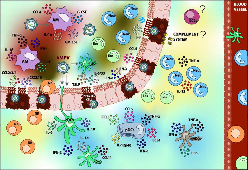
Viruses 2020, 12, 637                                                                                2 of 46

encephalopathies [10–12]. Reinfections of this virus occur frequently throughout the lives of the infants,
due to the inefficient development of immunological memory produced by the first infection with
hRSV. Even though a less severe disease with fewer clinical complications is caused by the reinfection
with this virus, cases with lower respiratory tract disease have been reported [13,14].
      Even though hRSV was first identified over 60 years ago and has been widely studied ever since
its discovery, no safe and effective vaccine has been approved to prevent the disease caused by this
virus [15,16].
      The human Metapneumovirus (hMPV) is another major viral agent that causes ALRTIs, making
it an important pathogen that contributes significantly to the mortality of infants under the age of
five [1,2]. A report made in the United States (US) showed that the numbers of outpatient visits are
55/1000, and the cases of children infected by hMPV that needed hospitalization represents 1/1000
children, lower than the 3/1000 cases that hRSV is responsible for, estimating that 20,000 children are
hospitalized per year in the US due to hMPV infection [17]. The hospitalization costs associated with
this virus are nearly USD 277 million per year, making hMPV an important health care problem as
well [18].
      hMPV infections cause more complications to young children, the immunocompromised,
and elderly patients [19]. hMPV infection is characterized by symptoms, such as cough, rhinitis, fever,
wheezing, and hyperventilation, and can lead to a more severe pathology involving bronchiolitis
and pneumonia [9]. Additionally, it has been reported that hMPV can produce alterations in the
CNS, resulting in seizures and encephalopathies [11,20]. Infection by this virus has been observed to
take place during the entire year, but there is generally a peak during the winter or spring seasons.
Furthermore, it can cause reinfections throughout the lives of the infants [8,21].
      hMPV was discovered more recently than hRSV in 2001, and similar to the latter, no vaccine
against hMPV has been approved yet [22,23].
      Even though both of these viruses are closely related and share similar symptoms, infection with
hRSV causes more cases of bronchiolitis and fewer cases of pneumonia as compared to hMPV [24].
Moreover, immune cell types involved in the resolution of each infection also vary—the immune
response elicited in the host by these viruses is responsible for some differences in the symptoms [24,25].
Because of this, along with the fact that the immune responses produced against these viruses are not
the same, it is important to characterize the specific innate immune responses for each of these viruses,
and their contributions to pathogenesis.
      In this article, we will discuss the general characteristics of these viruses along with the innate
immune response that these pathogens induce in the host. This work will be focused on dendritic
cells (DCs), alveolar macrophages (AMs), neutrophils, eosinophils, natural killer (NK) cells, innate
lymphoid cells (ILCs), and the complement system. Additionally, we will discuss how this response can
contribute to pathogenesis and/or disease resolution. How the pulmonary epithelium can contribute
to both pathogenesis and the recruitment of these immune cells will also be addressed. Current and
prospective prophylactic approaches, such as vaccines and monoclonal antibodies that can help to
control the infection caused by these viruses, will also be considered. Lastly, we will discuss the
similarities and the differences in the immune responses elicited by hRSV and hMPV infections.

2. General Features of These Two Respiratory Viruses
     hRSV and hMPV belong to the same taxonomical family of viruses, but due to individual
characteristics in their genomes, they are classified in different genera within this family. In the
following section, the general characteristics of these viruses regarding their genetics and life cycles
will be discussed.
Viruses 2020, 12, 637 3 of 46
2.1. Human Respiratory Syncytial Virus (hRSV)
2.1.1. Viral Genes and Structure
hRSV is an enveloped, single-stranded, non-segmented, negative-sensed RNA virus that belongs
to the Pneumoviridae family, Orthopneumovirus genus [3]. Its genome is 15.2 kb in length and contains
10 genes that code for 11 proteins in the following order: 30 -NS1-NS2-N-P-M-SH-F-G-M2-L-50 [3]. It is
noteworthy that the proteins M2-1 and M2-2 are two distinct proteins, a product of the transcription of
two different open reading frames (ORFs) of the m2 gene [3].
The envelope of hRSV contains three proteins on the surface: the glycoprotein (G), the fusion
protein (F), and the short hydrophobic protein (SH). The G protein is a heavily glycosylated glycoprotein
involved in the attachment of the virus to the target cell via the binding of heparin and/or annexin II on
the cell surface [26,27]. As for the F protein, most of the evidence suggests that it binds to the receptor
nucleolin [28,29]. This binding mediates the fusion between the viral envelope and the cell membrane,
as well as cell–cell fusion, leading to syncytia formation. Similar to other fusion proteins, the F protein
exists in two distinct conformational states (pre-fusion and post-fusion) [30,31], which are relevant for
the humoral response elicited against this viral antigen, and the exposure of the epitopes that these
antibodies recognize [31]. Such transition is presumably triggered by the interaction between F and its
receptor nucleolin and is required to bring the viral envelope and the cell membrane closer together
to induce the fusion of both [32]. Lastly, the SH protein is a small protein that is expressed on the
membranes of infected cells, and is not essential for virus attachment or fusion [33], but rather acts as a
viroporin on the surface of infected cells [34,35].
The genome of hRSV is associated with the nucleoprotein (N), the phosphoprotein (P), and the
viral RNA-dependent RNA polymerase (L) to form the ribonucleoprotein complexes (RNPs). The main
functions of the N protein are to coat the viral RNA in a left-handed helical nucleocapsid to protect
it from mechanical, chemical, and physical damage [36,37], and to participate in the replication of
the viral genome [38,39]. The P protein is an essential factor for the replication and transcription
of the viral genome and is also implicated in the packaging in the nucleocapsid [40,41]. The L
protein is responsible for the synthesis of a positive-sensed antigenome that serves as a template for
replication, and the transcription of the viral genome into mono- and polycistronic mRNAs [42,43].
The efficient transcription of long polycistronic mRNAs requires the M2-1 protein, since it serves as an
anti-termination factor [44] and the M2-2 protein is used as a cofactor necessary for the fine-tuning of
gene expression [45].
Matrix proteins M and M2-1 are also present in the virion as structural components [46,47]. The M
protein in particular is a bridge between the RNPs and the lipid bilayer envelope. It also serves as
an inhibitor of virus transcription in the late stages of infection and facilitates virion assembly and
budding by coating the RNPs [48] and modifying the actin cytoskeleton [49].
Lastly, hRSV possesses two non-structural proteins, NS1 and NS2, which are expressed in the
early stages of replication. These proteins are considered to be major virulence factors of hRSV since
they play an important role in the inhibition of type I IFN expression, thus promoting viral replication
and spread to neighboring cells [50–52].
2.1.2. Infectious Cycle
hRSV is able to infect bronchial respiratory epithelia. Interestingly, it has been shown that it
can also infect neurons in vitro [12,53], as well as DCs inhibiting their capacity to activate T cells
by preventing immunological synapse assembly [54,55]. To infect a target cell, hRSV must trigger a
two-step entry process involving the electrostatic attachment of the viral particle to the cell membrane
through the G protein and the subsequent fusion of both the viral envelope and the cell membrane
through the F protein. The G protein is not completely essential for infection to occur, but it facilitates
viral entry [33,56]. After viral and host membranes have been fused, the viral contents of hRSV are
released into the cell cytoplasm. The uncoating of the genome takes place and the replication andViruses 2020, 12, 637 4 of 46
transcription of the viral genome begin. The N, P, L, M2-1, and M2-2 proteins participate in these
processes. The M2-2 protein also acts as a regulatory element in the transcription and replication of the
viral genome [57]. Finally, the matrix protein (M) is key for understanding the formation of inclusion
bodies, virion assembly, and budding, although it has also been observed that it contributes to cell
cycle arrest in infected lung epithelial cells [58].
Interestingly, SH protein has been shown to act as a viroporin, assembling into pentameric
pores in infected cells [57,58], thus altering the permeability of the membrane to cations [34,35].
Additionally, this protein can inhibit both apoptosis and TNF-α signaling [59], and may be implicated
in downregulating the IL-1β response [60], suggesting a role as an important virulence factor. Besides,
the SH protein could contribute to the replication and infection process [34].
Lastly, non-structural proteins 1 and 2 (NS1 and NS2) are encoded by the first two genes of hRSV
and play crucial roles as virulence factors in early time points of infection [61]. Both of these proteins
are able to partly inhibit hRSV-mediated and TNF-α-mediated cellular apoptosis in the early stages of
infection [62]. Moreover, they interfere with key viral recognition signaling pathways, downregulating
IRF3 and STAT2 expression, thereby limiting the type I interferon (IFN-I) response [50,51].
2.2. Human Metapneumovirus (hMPV)
2.2.1. Viral Genes and Structure
hMPV is an enveloped, non-segmented, single-stranded, and negative-sensed RNA virus, which
belongs to the genus Metapneumovirus of the family Pneumoviridae [63,64]. The genome of this virus is
13.3 kb of length and is constituted by eight genes that encode for nine proteins, whose genomic order
is the following: 30 -N-P-M-F-M2-SH-G-L-50 . Each of these genes encodes a single protein, except for
the m2 gene, which codes for two different proteins. M2-1 and M2-2 are the result of the overlapping of
two ORFs located in the mRNA from the m2 gene [64]. The genome of hMPV differs from the genome
of hRSV in the lack of the NS1 and NS2 proteins.
The hMPV virion consists of an envelope that has three glycoproteins on the surface: the small
hydrophobic protein (SH), the attachment protein (G) and the fusion protein (F). The SH protein is a
transmembrane protein of type II, which is capable of inhibiting the transcriptional activity of NF-κB,
resulting in a decrease in the transcription of genes that use this pathway [65,66]. It has been reported
that the SH protein can play an important role in the cycle of the virus through the modulation of the
permeability of the membrane [65]. The G protein is a type II transmembrane protein that can bind to
the cellular glycosaminoglycans (GAGs), which are found in the membranes of the cells, promoting
the attachment of the virus to the cell and playing a role in the infection capability of hMPV [67]. The F
protein is a type I fusion protein that is synthesized in the form of an inactive precursor (F0), and,
in order to be biologically activated, a cleavage is needed between the subunits F1 and F2, which are
connected through disulphide bonds [68]. This protein mediates the attachment and the fusion of
the viral envelope of hMPV with the cell membrane to promote the entry of the virus into the cell to
be infected.
These glycoproteins found on the surface are associated with the matrix protein (M), which is also
connected with the inner membrane from the surface. The M protein interacts with various components
from the virus and the cells, promoting the assembly and budding of the virus [69]. Additionally,
the M protein can inhibit the transcription of the virus before it is packaged [70].
hMPV RNA is united with the nucleoprotein (N), the phosphoprotein (P), the large polymerase
protein (L), the putative transcription factor (M2-1), and the RNA synthesis regulatory factor (M2-2),
forming the nucleocapsid. The association between two domains from the N protein and the virus RNA
causes the helical nucleocapsid form and protects the genome from any damage [71,72]. The N protein
interacts with the P protein, inducing the recruitment of the L protein along with the M2-1. The P
protein makes the assembly of the ribonucleoprotein (RNP) complex possible, due to the stabilization
of the L protein. Both the P and L proteins conform to the RNA-dependent RNA polymerase (RdRp),
and position themselves in the 30 end of the viral genome to begin the transcription process [72,73].Viruses 2020, 12, 637 5 of 46
2.2.2. Infectious Cycle
Ciliated epithelial cells are the primary target of the hMPV infection, and then the virus spreads
to all the cells of the respiratory tract where viral replication takes place [74]. The replication process
begins in the upper respiratory tract and can spread to the lower respiratory tract, where it can reach
the bronchioles and the alveoli. In these structures, the virus has shown higher replication capacities,
as compared to the upper respiratory tract [74]. Within the immune cells that can get infected by hMPV,
DCs are one of the most relevant, although viral replication is not efficient in these cells [75].
Once the virus arrives at the airway epithelia, the F and G proteins promote the attachment
to the airway epithelial cell (AEC), where there is an interaction between the F protein and αvβ1
integrin, along with an interaction between the G protein and GAGs [67,76,77]. When the fusion of the
membranes is accomplished, a fusion pore is formed and the hMPV RNP complex can enter into the
cytoplasm of the cell to be infected. In the cytoplasm, the N, P, and L proteins detach from the viral
RNA and arrange themselves to create the polymerase complex [76]. Additionally, proteins N and
P are the only proteins needed to generate inclusion bodies that appear as a result of the replication
process [78]. Importantly, the M2-2 protein plays a role in the evasion of the immune response elicited
against hMPV infection, inhibiting the expression of genes associated with the MyD88 pathway [79].
This contributes to the spreading of the infection.
hMPV exhibits two phases of replication, characterized by their kinetics of infection: an acute
phase of infection with a peak of viral titer 7 days post-infection (d.p.i.) and a second phase of infection
with a peak of viral titer at 14 or 28 d.p.i., depending on the virus strain. Of note, the genetic material
of hMPV can be found up to 180 d.p.i. within the lungs of BALB/c mice [80,81].
3. Differential Regulation of the Innate Immune Response by Each Virus
The innate immune response is the first line of defense against virtually all kinds of pathogens,
including those of viral natures. Within this complex response, innate immunity plays a key role in the
control of infections given its fast and antigen non-specific responses against pathogens. The most
relevant innate immune response components in the contexts of hRSV and hMPV infections are
neutrophils, eosinophils, macrophages, dendritic cells, natural killer cells, and the complement system.
How each of these cells are activated and the immune pathology, or contribution for disease resolution
that results from their recruitment, is discussed below.
3.1. Dendritic Cells
Dendritic cells (DCs) are considered part of the first line of defense against viruses and,
most importantly, are considered to orchestrate both the innate and the adaptive immune response [82].
DCs can be found in the lung epithelium and are capable of responding quickly during pathogen-caused
inflammation [83]. Moreover, DCs are classified into two conventional subtypes (cDC1 and cDC2) and
a plasmacytoid one (pDC), of mixed lymphoid and myeloid ontogeny [84–86].
During a hRSV infection, all DCs subtypes have been shown to migrate to the lung as early as
2 d.p.i. [87,88], and peak migration occurs at 6 d.p.i. [88]. Importantly, DCs can serve as an important
IFN-I sources upon encounter with hRSV [89], although there is one controversial report that found
no IFN-I secretion in either DC subset in the lungs of infected mice [90]. This last study showed
that only pDCs—among all classically defined DC subsets—contributed minimally to IFN-I secretion.
The authors used an Ifna6gfp/+ mouse strain to define which immune cells contributed to IFN-I secretion
and validated their results through the quantitation of Ifna5 and Ifnb mRNA expression by quantitative
RT-PCR on FACS-sorted populations of immune cells. Considering that neither of the approaches of
these authors considered direct IFN-α protein recognition and that mice possessed 14 genes coding for
IFN-α [91], DCs may not have been identified as major producers of IFN-I, given that they focused
only on two of the genes responsible for IFN-α secretion, which could explain their results.
DCs are susceptible to hRSV infection [92–95]. However, DCs are poorly permissive to the virus
as the infection is abortive and leads to poor production of new viral particles [54,93,96]. Human DCs
subsets can be infected in different degrees by hRSV, as pDCs are less permissive than cDCs, and bothViruses 2020, 12,
Viruses 2020, 12,637
x FOR PEER REVIEW 66 of
of 46
45
DCs subsets can be infected in different degrees by hRSV, as pDCs are less permissive than cDCs,
cDC1 along with cDC2 are equally permissive to hRSV infection [97]. Interestingly, DC infection
and both cDC1 along with cDC2 are equally permissive to hRSV infection [97]. Interestingly, DC
is enhanced by the presence of infected macrophages as in vitro models of airway epithelium have
infection is enhanced by the presence of infected macrophages as in vitro models of airway
shown [98]. It can also occur through the internalization of antibody-coated hRSV through FcγRIIb and
epithelium have shown [98]. It can also occur through the internalization of antibody-coated hRSV
FcγRIII [96]. Moreover, FcγRIII knockout mice also show less severe hRSV symptoms, suggesting that
through FcγRIIb and FcγRIII [96]. Moreover, FcγRIII knockout mice also show less severe hRSV
this receptor contributes to hRSV pathogenesis, possibly by enhancing DC infection [96]. hRSV-infected
symptoms, suggesting that this receptor contributes to hRSV pathogenesis, possibly by enhancing
DCs can promote airway obstruction, enhance disease, and promote more severe allergic responses in
DC infection [96]. hRSV-infected DCs can promote airway obstruction, enhance disease, and
recipient mice [99].
promote more severe allergic responses in recipient mice [99].
hRSV infection induces DC maturation, as observed by the upregulation of markers, such as CD40,
hRSV infection induces DC maturation, as observed by the upregulation of markers, such as
B7.1 (CD80), CD83, B7.2 (CD86), MHC-I, and MHC-II [54,94,100], although the upregulation of the
CD40, B7.1 (CD80), CD83, B7.2 (CD86), MHC-I, and MHC-II [54,94,100], although the upregulation
latter is controversial. Moreover, the secretion of TH 2-polarizing and inflammatory cytokines, including
of the latter is controversial. Moreover, the secretion of TH2-polarizing and inflammatory cytokines,
TNF-α, IL-1β, IL-6, and low quantities of IL-10 has been observed, as shown in Figure 1 [54,94,95].
including TNF-α, IL-1β, IL-6, and low quantities of IL-10 has been observed, as shown in Figure 1
The constitutive and TLR-induced secretion of IFN-I is impaired in infected DCs, but is still considered
[54,94,95]. The constitutive and TLR-induced secretion of IFN-I is impaired in infected DCs, but is
relevant [88,92,95,97,101–103]. Interestingly, the quantities and relative amounts of type I IFNs (α and β)
still considered relevant [88,92,95,97,101–103]. Interestingly, the quantities and relative amounts of
and type III IFNs (λ) secreted by hRSV-infected DCs depend on the specific strain that infects the cell [102].
type I IFNs (α and β) and type III IFNs (λ) secreted by hRSV-infected DCs depend on the specific
Finally, infected DCs exhibit a diminished capacity to activate CD4+ T cells [54,100]. Surprisingly, this
strain that infects the cell [102]. Finally, infected DCs exhibit a diminished capacity to activate CD4+ T
phenomenon is not mediated by soluble factors—it is the expression of the nucleoprotein of hRSV on
cells [54,100]. Surprisingly, this phenomenon is not mediated by soluble factors—it is the expression
the surface of infected DCs which impairs the establishment of a proper immune synapse between
of the nucleoprotein of hRSV on the surface of infected DCs which impairs the establishment of a
both cell types [54,55].
proper immune synapse between both cell types [54,55].
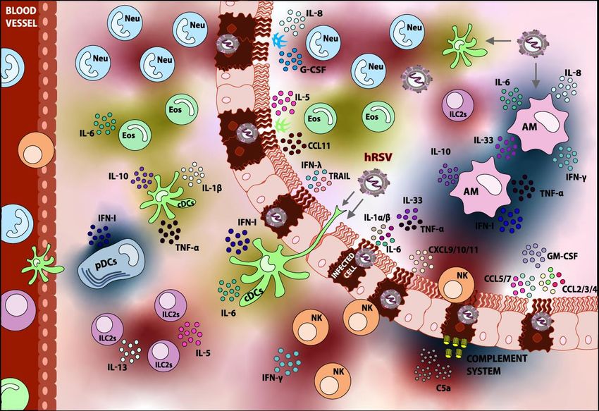
Figure 1.
Figure 1. Innate
Innate immune
immune response
response elicited
elicited upon
upon hRSV
hRSV infection.
infection. Following
Following thethe detection
detection ofof hRSV,
hRSV,
various types of immune cells are recruited to the lung. Within the innate immune cells,
various types of immune cells are recruited to the lung. Within the innate immune cells, Dendritic cells Dendritic
cells (DCs),
(DCs), Alveolar
Alveolar Macrophages
Macrophages (AMs),(AMs), Neutrophils
Neutrophils (Neu),(Neu), Eosinophils
Eosinophils (Eos), (Eos), Natural
Natural Killer Killer cells
cells (NK),
(NK),
and and Group
Group 2 Innate2 Lymphoid
Innate Lymphoid Cells can
Cells (ILC2s) (ILC2s) can beAccordingly,
be found. found. Accordingly,
the fixationthe
andfixation and
activation
activation of the Complement System is detected. Most of these components play
of the Complement System is detected. Most of these components play a role in the pathology of the a role in the
pathology of the infection. The role of the respiratory epithelium in the recruitment
infection. The role of the respiratory epithelium in the recruitment of innate immune cells can also of innate
immune
be cells can
appreciated. also be appreciated.
Components shaded in Components shaded
blue are elements thatincontribute
blue are elements that contribute to
to hRSV disease-resolving,
hRSV disease-resolving,
whereas components shaded whereas components
in red are consideredshaded in red
detrimental andare considered
contribute detrimental
to hRSV pathogenesisand
contribute
overall. to hRSVdepicted
Elements pathogenesis overall.
in yellow Elements
are those depicted
whose in yellow
contributions toare
hRSVthose whose contributions
pathology are not well
to hRSVor
studied pathology are not well
are controversial. Thestudied
arrows or are controversial.
indicate the possibleThe arrows
targets indicate the possible targets
of infection.
of infection.Viruses 2020, 12, 637 7 of 46
During hRSV infection, poor or inadequate activation of T cells by DCs is not uncommon [54,100].
TH 2 polarization is key for hRSV pathogenesis and is largely influenced by DCs. It has been shown
that the cDC2 subset is capable of polarizing T cell responses in an IL-4Rα-dependent manner, which
induces poor DC maturation and TH 2 bias [104]. Moreover, the TLR3- and TLR7-mediated secretion of
IL-33 by DCs could induce the generation of a TH 2-biased adaptive response [105], and the secretion
of TSLP by hRSV-infected epithelial cells can induce the maturation of cDCs, almost certainly to a
TH 2-polarizing profile [106].
As previously mentioned, cDCs are of myeloid origin and both subtypes are present in the lungs
during hRSV infection [87], and in the BAL of hRSV-infected children [107]. Studies are controversial in
determining their relative abundance in the peripheral blood of infected children [107,108]. However,
it has been observed that the ratio between cDC1 and cDC2 is low in infected mice and could contribute
to pathogenesis [88], since the cDC2 subset is more prone to polarize CD4+ T cells to a TH 2 profile [104].
On the other hand, the cDC1 subset has vast antiviral capacities, given its high cross-presenting
activity, which activates CD8+ T cells [109–114], further explaining why a low cDC1:cDC2 ratio is
detrimental. Both cDC subtypes are equally susceptible to infection and are more permissive to hRSV
than pDCs [94,97]. The infection with hRSV induces the upregulation of costimulatory markers B7.1
(CD80) and B7.2 (CD86) [94,97], and limits CD4+ T cell activation [88,94], as previously mentioned.
Additionally, hRSV infection lowers IFN-α secretion in the cDC1 compartment and lowers IFN-β
secretion in the cDC2 compartment [88,97].
On the other hand, pDCs are of mixed ontogeny and are characterized by their plasmacytoid
appearance and vast secretion of IFN-I. These cells contribute greatly to disease resolution, lowering
viral titers, and ameliorating pulmonary inflammation and airway obstruction [87,115,116].
Even though there is one report stating the opposite [90], the consensus is that pDCs are one of the
foremost producers of type I interferons (IFN-I) in the lungs during hRSV infection [92,95,117].
Moreover, it has been observed that IFN-I secretion is TLR7/MyD88- and IFNαR1-dependent,
but MAVS-independent, ruling out the involvement of RIG-like receptors (RLRs) in the triggering of
IFN-I secretion [117]. Immunoglobulin-complexed hRSV (IC-hRSV) has been shown to induce TLR
internalization and endosomal signaling in pDCs, which results in IFN-I secretion and can partly
explain the observed TLR7/MyD88 dependency [118]. Additionally, the presence of IFN-I-producing
infected monocytes or epithelial cells can further stimulate IFN-I secretion by pDCs, thus explaining
the observed IFNαR1 dependency [118], without ruling out the possibility of autocrine IFN-I signaling.
Although pDCs do not interfere with CD4+ T cell responses [117,119], they do potentiate
antigen-specific, IFN-γ-producing CD8+ T cell antiviral responses [116,117,119]. Despite their vast
antiviral potential, hRSV-ALRTI children possess fewer pDCs in their peripheral blood than healthy
controls [107,108], which also correlates with hRSV-induced asthma development in the future [120].
pDCs are susceptible to hRSV infection, albeit abortive and very scarce [97]. Infection readily
induces the upregulation of CD40 and B7.2 (CD86) at even higher levels than cDCs [97]. Even though
pDCs still produce IFN-I when infected [95], they do so at lower levels [88,101] and are less responsive
to TLR-induced IFN-I secretion [88,92,95,103] and TLR-induced cytokine secretion [88].
Studies using neonate models of hRSV infection are extremely relevant, considering that newborns
and children less than two years of age are an important high-risk population. Murine neonate models
of infection have revealed that neonate pDCs are capable of processing and presenting antigens,
but possess an intrinsic insufficient IFN-I response in comparison to mature adult pDCs [101]. Similar
conclusions have recently been drawn for neonate cDCs [121]. Other studies regarding the role of cDCs
in neonate models of infection reveal that neonate cDCs do not mature correctly in response to hRSV,
since they do not upregulate costimulatory markers or CCR7, and maintain the expression of CCR5,
which restrains migration to lymph nodes and retains DCs in the site of inflammation without proper
maturation [122]. Another interesting observation is that the cDC2 compartment is less abundant in
neonates, possibly leading to a weaker DC response against hRSV [110]. Interestingly, non-specific DC
expansion in neonates leads to a better antiviral response based on the IFN-γ-secreting CD8+ T cell,Viruses 2020, 12, 637 8 of 46
which limits airway inflammation [116]. Furthermore, the cDC1 compartment is functionally limited
in neonates when compared to adult cDC1s—neonate DCs internalize and process less antigen and
upregulate B7.1 and B7.2 to a lesser extent [110].
Interestingly, lung migratory cDC1s from newborns establish different antigen-specific CD8+ T
cell responses to adult cDC1s, giving rise to an immature-like immunodominant hierarchy among
CD8+ T cells [110]. Briefly, these data suggest that there are differences between adults and neonates
regarding the relative abundance of CD8+ T cell populations that recognize particular epitopes derived
from hRSV antigens. The pattern of epitope dominance is relevant in the context of infection since it
shapes the adaptive immune response and defines the epitopes from an antigen that will induce a
more potent immune response [123–125]. Interestingly, neonate CD103high cDC1s—but not CD103low
cDC1s—can establish the same immunodominant CD8+ T cell hierarchy that is observed in adult
cDC1-mediated CD8+ T cell responses [111]. This suggests that CD103 upregulation in the cDC1
subset of neonate DCs is indicative of proper cDC1 development towards an adult-like phenotype.
Lastly, the TLR4 and TLR9 stimulation of DCs in neonates leads to the cDC1 and cDC2 expansion and
upregulation of B7.1 (CD80) and B7.2 (CD86), which also leads to an adult-like immunodominant
hierarchy among CD8+ T cells [109].
To date, there are no clinical reports that describe the role of DCs in the pathology induced by
the infection with hMPV [126]. However, in vitro studies, along with animal models, are able to
demonstrate the role and function of DCs during this infection.
During the acute phase of infection with hMPV, DCs comprise the main immune cell population
that senses this virus, along with macrophages and AECs [127,128]. As previously stated, DCs are
susceptible to infection by hMPV, but the replication in these cells is not as efficient as in epithelial
cells [75]. hMPV infection leads to an increase in the amounts of pDCs and cDCs in the lungs, reaching
their peak at 8 and 10 d.p.i., respectively, and cDCs are found more abundantly and persist until 18 d.p.i.
However, the CD103+ cDC1 subset decreases during the first weeks after the infection, but 8 weeks
after the infection it reaches its normal amount [88]. The hMPV-infected DCs upregulate the maturation
marker B7.1 (CD80) compared to non-infected cells, but additional markers, such as CD83 and B7.2
(CD86) do not exhibit significant differences. The infection is not able to cause the maturation of cDCs,
but unlike hRSV, hMPV-infection has no cytopathic effects [129].
In vitro studies demonstrated that monocyte-derived DCs (moDCs) were capable of being infected
by hMPV, but it was an unproductive infection [95]. The effect of the G and SH proteins during
the infection with hMPV demonstrate a mechanism that hMPV might use to evade the immune
system. Both of these proteins were not necessary to infect moDCs, and in their absence the rate of
infection increased within moDCs [130]. The G and SH proteins of hMPV minimize the capability of
internalizing the virus in moDC through a pathway similar to macropinocytosis, and by doing this,
the virus decreases CD4+ T cell activation and, as a consequence, it reduces the TH 1 response [130].
This correlates with previous findings, indicating that the infection of DCs with hMPV prevents the
activation of naïve T cells [75,131].
When moDCs are infected with hMPV they can secrete low concentrations of IL-6 and TNF-α,
and when these cytokines levels are compared to non-infected cells, the difference is not significant.
However, moDCs secrete significantly higher IFN-α levels compared to non-infected cells [95]. On the
other hand, hMPV-infected pDCs secrete the cytokines IFN-α, TNF-α, IL-12p40, CCL3, CCL4, and CCL5
upon infection, while in cDCs infected with hMPV, the cytokines IFN-β, IL-1α, IL-6, IL-10, and CCL11
(eotaxin) are secreted, as shown in Figure 2 [88]. IL-12p40 is a pro-inflammatory cytokine that is
secreted in response to viral infection, and in the case of a hMPV infection, it plays an important role
in the control of the inflammatory response and damage in the lung tissue. The absence of IL-12p40
increases the inflammation in the lungs and modifies the cytokine response, leading to deteriorated
functionality of the lungs [132]. This observation suggests that the DCs play a protective role in the
immune response against hMPV infection.Viruses 2020, 12, 637 9 of 46
Viruses 2020, 12, x FOR PEER REVIEW 9 of 45
Figure 2.
Figure 2. Innate
Innateimmune
immuneresponse
response elicited
elicited uponupon
hMPV hMPV infection.
infection. AfterAfter the detection
the detection of hMPV, of various
hMPV,
various types of immune cells are recruited to the lung. Within the innate immune
types of immune cells are recruited to the lung. Within the innate immune cells, Dendritic cells (DCs), cells, Dendritic
cells (DCs),
Alveolar Alveolar Macrophages
Macrophages (AMs), (Neu),
(AMs), Neutrophils Neutrophils (Neu),(Eos),
Eosinophils Eosinophils (Eos),Killer
and Natural and Natural
cells (NK)Killer
can
cells (NK) can be found. Most of these cells play a role in the pathology of the
be found. Most of these cells play a role in the pathology of the infection. However, Group 2 Innateinfection. However,
Group 2 Innate
Lymphoid Cells 2Lymphoid
(ILC2s) andCells 2 (ILC2s)
the role and the roleSystem
of the Complement of the during
Complement System
the infection during
have the
not been
infection yet,
reported haveandnotthis
been reported
lack yet, andisthis
of information lack of information
illustrated with “?” in theis illustrated
figure. Thewith “?”the
role of in respiratory
the figure.
The role ofinthe
epithelium the respiratory
recruitment of epithelium in thecells
innate immune recruitment
can also be ofappreciated.
innate immune cells canshaded
Components also be
in
appreciated. Components shaded in blue are elements that contribute to hMPV
blue are elements that contribute to hMPV disease resolving, whereas components shaded in red are disease resolving,
whereas components
considered detrimental and shaded in red
contribute are considered
to hMPV pathogenesis detrimental and contribute
overall. Elements to hMPV
shaded in yellow are
pathogenesis
those overall. Elements
whose contributions to hMPVshaded in yellow
pathology areare
notthose
wellwhose
studiedcontributions to hMPVThe
or are controversial. pathology
arrows
are not well
indicate studied or
the possible are controversial.
targets of infection. The arrows indicate the possible targets of infection.
As mentioned before, pDCs are an important source of IFNs that are crucial for the appropriate
antiviral response. A recent report demonstrated that the SH protein of hMPV suppresses the
TLR7/MyD88 pathway in pDCs, leading to the the inhibition
inhibition of of the
the IFNs
IFNs expression
expression [133].
[133]. The M2-2
protein of hMPV can impair the TLR7/9 pathway as well, thus thus inhibiting
inhibiting the
the secretion
secretion of
of IFN-I
IFN-I [134].
[134].
This indicates
indicatesthat
thatSHSH
andand
M2-2 proteins
M2-2 have have
proteins a role ainrole
the evasion of the immune
in the evasion of theresponse,
immune interfering
response,
with the proper
interfering with response
the proper ofresponse
pDCs. of pDCs.
IFNs are secreted via sensing the viral RNA. However, IFNs can be secreted through the sensing
of IFN by the alpha interferon receptor (IFNAR) in the cells as well. The number of DCs decline when
this receptor is absent during the infection with hMPV, hMPV, causing
causing the
the debilitated
debilitated response
response of CD8++ TT cell,
of CD8 cell,
since IFNAR enhances
enhances thethe increase
increasein inDCs
DCsininthe
thefirst
firststage
stageofofreplication
replication[135].
[135].
In vitro studies have proven that the M protein can be released from the cell during the infection
and can bind with DCs, internalizing this protein in a fast way. way. The M protein has the ability to induce
the maturation of DCs and, as a consequence, the the secretion
secretion of of cytokines
cytokines and
and chemokines,
chemokines, such
such as
as TNF,
TNF,
IL-1β, IL-6, IL-12p70, IL-8, and IL-10, that promote the inflammatory response [70]. Some reports have
indicated that
that the
theinfection
infectionwith
withhMPV
hMPV provokes
provokeslow levels
low of IL-10,
levels and and
of IL-10, that that
the low
the secretion of IL-10
low secretion of
might play aplay
IL-10 might negative role in
a negative theinseverity
role of hMPV-infection
the severity of hMPV-infection within preterm
within infants
preterm [136,137].
infants [136,137].
In conclusion, DCs play varied roles during hRSV infection. Quite importantly, they are largely
responsible for the TH2 polarization and poor CD4+ T cell responses against hRSV, especially when
DCs themselves are infected. On the other hand, pDCs are an especially important and highlyViruses 2020, 12, 637 10 of 46
In conclusion, DCs play varied roles during hRSV infection. Quite importantly, they are largely
responsible for the TH 2 polarization and poor CD4+ T cell responses against hRSV, especially when DCs
themselves are infected. On the other hand, pDCs are an especially important and highly regulated
source of IFN-I and are critical for the establishment of an antiviral state in the lung epithelium, as well as
for the establishment of a CD8+ T cell-mediated antiviral response. Lastly, cDC1 responses also appear
to be vital in the establishment of a proper CD8+ T cell response, whose immunodominant hierarchy is
immature in neonates. As for DCs during hMPV-infection, it seems they play a protective role against
the infection of this virus. The secretion of the cytokine IL-12p40 enhances the pro-inflammatory
responses, and without it the inflammation causes intense damage in the lung tissue. The infection of
DCs with hMPV impairs the proper priming of naïve T cells and can inhibit the signaling of TLR7/9,
reducing the secretion of IFNs in pDCs. However, IFN levels can still be secreted by the different types
of DCs. A comparison of the role of DCs between hRSV and hMPV infections can be found in Table 1.
Table 1. Role of DCs in the pathology caused by hRSV and hMPV.
Study Model hRSV hMPV
# FcγRIII receptor contributes to hRSV
pathogenesis, possibly by enhancing
DC infection. # The secretion of IL-12p40 of
# Infected DCs can promote airway obstruction, infected pDCs might contribute
In vivo model
enhance disease, and promote more severe to the protection of the lungs.
allergic responses.
# A low cDC1:cDC2 ratio correlates with
enhanced disease severity.
# One of the main sources of IFN-I. # Main immune cell population to
# Secretion of TH 2-polarizing (IL-6, IL-10, IL-33) sense this virus.
and proinflammatory cytokines (IL-1β, TNF-α), # hMPV decreases the CD4+ T cell
and poor CD4+ T cell activation. activation and, as a consequence,
In vitro model
# Neonate DC subsets secrete less IFN-I, possess it reduces the TH 1 response.
an impaired capacity to activate CD4+ T cells, # DCs are capable of secreting
and elicit different epitope-specific CD8+ T cell IFNs, promoting the
responses during the infection. antiviral response.
3.2. Macrophages
Macrophages are within the first cells that protect the host against the viral infection, and there
are two types in the lungs: the alveolar macrophages (AMs) and the interstitial macrophages
(IMs) [138]. AMs are a type of macrophage, resident to the alveoli, that represent the first line
of defense against respiratory pathogens [139]. Similarly to other tissue-resident macrophages,
AMs are excellent phagocytes, possess a wide array of antimicrobial enzymes, can secrete ROS,
can act as antigen-presenting cells, and mediate responses against both intracellular and extracellular
pathogens [140]. They are also involved in the repairing of tissues after the infection has been resolved,
through the secretion of anti-inflammatory cytokines as well as mitogenic factors [140].
In the case of hRSV, AMs are crucial for the early control of infection. During a hRSV infection,
they represent one of the foremost and only sources of IFN-I, contributing to the establishment of an
antiviral state in neighboring cells [90]. IFNs are secreted through RNA sensing by RLRs and their
subsequent interaction with the mitochondrial antiviral signaling protein (MAVS) [90], although the
possibility of TLR involvement should not be discarded, given that MyD88 is also necessary for proper
IFN secretion [89].
Moreover, the depletion of AMs leads to a more severe disease caused by hRSV [141]. For example,
it leads to airway obstruction [142], weight loss, and higher viral loads in the lungs [143] in murine
models. Moreover, the activation of AMs with IFN-γ promotes the clearing of the viral infection [143].
Additionally, the expansion of the AM subset in mice recently recovered from allergically inducedViruses 2020, 12, 637 11 of 46
eosinophilia leads to a reduced hRSV immunopathology [144], supporting the notion that AMs play a
protective role in hRSV infection.
The protective role of AMs can be at least partly explained through the previously mentioned
secretion of type I IFNs, but also through the balanced and controlled secretion of pro-inflammatory
cytokines, such as TNF-α, IL-6, and IL-8, upon the encounter with the live or inactivated virus, or upon
AMs infected with hRSV, as shown in Figure 1 [141,145–149]. It is noteworthy that TNF-α has been
shown to have antiviral properties in the context of their respiratory viruses, such as influenza [150].
Even though TNF-α is a pro-inflammatory cytokine in nature, which could enhance pulmonary
inflammation and airway obstruction, the local and balanced secretion of TNF-α by AMs proves to
be beneficial during hRSV infection, probably because of the promotion of an antiviral local state.
Interestingly, AMs have been observed to promote the recruitment and activation of Natural Killer
(NK) cells to the site of infection, further promoting a protective innate immune response [149].
AMs can be infected acutely by hRSV, as indicated above [151,152]. Interestingly, infection can
occur through the internalization of IC-hRSV via the Fc receptors [153]. A report indicated that
a persistently infected murine macrophage cell line for more than 87 passages can upregulate the
expression of FcγRIIB and FcγRIII, which could render AMs more susceptible to hRSV infection [152].
Moreover, infected macrophages have shown to enhance DCs infection by hRSV, which can in turn lead
to the development of a TH 2 type adaptive immune response that is not efficient for virus clearance [98].
However, AM infection by hRSV is abortive and restricted, leading to the replication of only a few viral
particles [145]. Interestingly, infection is also controlled in AMs lacking MAVS or IFNAR1, although
virus and lysosome colocalization is less pronounced [145]. IL-6, TNF-α, IFN-α/β, and IFN-γ secretion
is somewhat lower in infected AMs, but still relevant altogether [145,147,154]. On the other hand,
phagocytosis and ROS production are also partially impaired in infected AMs [147]. Still, AMs are
capable of playing a protective role, even when infected.
Nonetheless, some concerns have arisen regarding the possible role AMs could play in adaptive
response polarization. Importantly, they can secrete IL-33 [105], a powerful driver of TH 2 responses and
harmful in the context of hRSV infection. IL-33 secretion was found to be induced by TLR7 agonists,
suggesting a possible mechanism for IL-33 secretion during a hRSV infection [105]. Additionally,
and even though a moderate amount of IL-6 promotes local inflammation and limits viral spreading,
the uncontrolled secretion of IL-6 could result in harmful TH 2 polarization [155].
Another controversial point is the finding that AMs can secrete considerable amounts of IL-10 at
early time points of hRSV infection [156]. Concerns arise given the suppressive role that IL-10 can
exert, which may result in the incomplete control of early infection.
In sharp contrast to hRSV infection, AMs seem to contribute to the pathogenesis caused by hMPV,
rather than protect against the infection. In the bronchoalveolar lavage (BAL) of patients infected by
hMPV, macrophages are found. Along this line, a biopsy of the lungs from these patients indicated
chronic inflammation and the presence of alveolar macrophages in the tissue [157]. The characteristics
of these macrophages found in the pulmonary tissue suggested the development of bronchiolitis in
these patients.
During the acute phase of infection with hMPV, macrophages are among the main immune cell
population to sense hMPV [127,128]. Studies performed in mice have demonstrated that macrophages
reach the lungs 3 d.p.i., and are present until 7 d.p.i. [158]. The macrophages are recruited to the place
of the infection due to the chemokine CCL2 (MCP-1), as a consequence of its high expression 1–5 d.p.i.
This chemokine not only acts as a chemoattractant for macrophages but also has a role in the regulation
of TH 1/TH 2 responses, especially since it promotes the secretion of IL-4, thereby stimulating the TH 2
response even more [158].
AMs promote the spreading of hMPV though a macrophage infection-dependent mechanism,
resulting in viral dissemination and the infection of the cells of the airway epithelium. Moreover,
when AMs were depleted by the administration of a liposome-encapsulated clodronate suspension,
hMPV infection caused a lesser decrease in the weights of infected mice, compared to non-depletedViruses 2020, 12, 637 12 of 46
mice—the pathology score, along with the obstruction in the airways, decreased as well [141].
Additionally, the absence of AMs during the infection with hMPV leads to the decreased recruitment
of neutrophils, but no significant difference in the recruitment of cDC2s [141]. The data indicate that
AMs play an important role in the pathogenesis of the hMPV infection.
AMs are one of the main sources of IL-6, TNF-α, IFN-α/β, CCL4, GM-CSF, and G-CSF in the lungs,
as shown in Figure 2. Furthermore, this study suggests that AMs are the main cells secreting IFN-I
during a hMPV infection, similar to what happens during a hRSV infection [141]. A study performed
in IFNAR−/− mice demonstrated that AMs can function properly independently of IFNAR signaling,
unlike IMs [135]. Additionally, the absence of this receptor does not cause AMs to be less recruited to
the site of infection.
The detrimental role that AMs possess might be explained by the secretion of IL-1, since this
cytokine is an important enhancer of inflammation in the lungs. The secretion of IL-1β is able to
generate an inflammatory response by itself and promotes the secretion of more pro-inflammatory
cytokines [159]. Moreover, it is known that the secretion of IL-1α/β can produce an inflammatory
pathological response in the lungs in infectious contexts with other viruses, such as influenza, while
playing no role in the clearance of the virus [160].
Generally speaking, the evidence points out that the activation of AMs in the lungs is a desirable
response against hRSV. Their resilience against infection, their major contribution of type I IFNs,
the secretion of adequate amounts of TNF-α and IL-6, and the recruitment of NK cells to the lungs
are key for the early control of infection. Their possible role as TH 2-polarizing agents would not be
unexpected, but their involvement in the development of adaptive immune response seems to be
minimal [149]. Hence, we highlight the positive role of AMs in the early control of hRSV infection in
the lungs. On the other hand, the activation of AMs in the lungs does not seem to be an appropriate
response against hMPV. The studies indicate that the presence of these cells does more damage than
protection in response to this virus. It is worth mentioning that IL-1 has a pro-inflammatory effect
that is linked with pulmonary damage. Moreover, these cytokines are not secreted by AMs during
the hRSV infection, but they are during the hMPV infection. Additionally, studies comparing the
amount of AMs recruited to the lungs by both viruses demonstrated that hRSV-infected mice are
able to recruit more macrophages to the site of infection by day 7 p.i. compared to hMPV-infected
mice [161]. The difference in cytokine secretion and the intensity of macrophage recruitment is where
the difference between protection and damage might lie. A comparison of the role of AMs between
hRSV and hMPV infections can be found in Table 2.
Table 2. Role of AMs in the pathology caused by hRSV and hMPV.
Study Model hRSV hMPV
# Contribute to disease resolution and
# Contributes to the pathogenesis of
ameliorate symptoms.
the infection.
In vivo model # Controversial secretion of TH 2-polarizing
# Promotes the spreading of the
IL-33 and anti-inflammatory IL-10 at early
virus in the airway epithelium.
time-points of infection.
# One of the main sources of IFN-I upon
# One of the main sources of IFN-I.
RNA sensing.
In vitro model # The secretion of IL-1α/β can
# Balanced early secretion of
generate an inflammatory
proinflammatory cytokines (IL-6, IL-8,
pathological response in the lungs.
and TNF-α) Abortive infection can occur.
3.3. Neutrophils
Neutrophils are the most abundant type of immune cells in the bloodstream and possess
important antimicrobial capacities. They are the first line of defense against many pathogens, given
their notable phagocytic capacities, the release of proteolytic enzymes and other antimicrobial peptidesViruses 2020, 12, 637 13 of 46
via degranulation, and the formation of web-like structures specialized in the capture and elimination
of pathogens, called neutrophil extracellular traps (NETs) [162].
During the infection with hRSV, the excessive infiltration of neutrophils and other granulocytes are
a hallmark symptom of severe disease in children [163]. In vitro studies have shown that the secretion
of IL-17 by TH 17 CD4+ T cells along with the production of IL-8 (CXCL8) by epithelial alveolar cells
are involved in the recruitment of neutrophils to the lungs, as shown in Figure 1 [164]. Moreover,
hRSV infection and local IL-17A secretion have been shown to potentiate IL-8 secretion and enhance
neutrophil recruitment [164–166].
Despite their antimicrobial capacities, a recent study shows that neutrophils are neither implicated
in the elimination of hRSV in the lungs nor in the recruitment of effector or memory T cells to
the lungs [167]. Moreover, the authors show that neutrophils do not contribute to disease severity
since there was no pronounced weight loss or secretion of pro-inflammatory cytokines in the lungs
(such as TNF-α, IL-1β, or IL-6) when the neutrophils were recruited to the lung via intranasal CXCL1
administration [167].
However, there is evidence that neutrophils can contribute to pathogenesis, causing airway
inflammation and tissue damage. For instance, it has been shown that hRSV is able to induce NETosis
in human neutrophils in vitro and in the BAL from children with hRSV-ALRTI [168,169], and that the
F protein alone can induce NETosis in vitro through TLR4 activation [170]. Even though these NETs
are able to trap hRSV and prevent further infection in vitro [168,169], their protective capacity in vivo
needs to be evaluated. However, it has been observed that NET formation can contribute to airway
obstruction in calves infected with bovine RSV (bRSV), a species closely related to human hRSV [169].
This would indicate that, although NETosis may contribute to limit viral replication, this occurs at
the cost of developing one of the hallmark symptoms associated with ALRTIs—airway obstruction
that might lead to bronchiolitis. Interestingly, the BALs of children with severe hRSV bronchiolitis
contain neutrophils expressing the same levels of TLR4 protein on the cell surface as healthy controls
but contain less TLR4 overall, which indicates that they possess lower levels of intracellular endosomal
TLR4 [171]. Although this may not affect NETosis through the interaction of the cell surface TLR4
with the F protein of hRSV, it may influence the balance between the cell surface and endosomal
TLR4 signaling upon activation. There may be a bias in signaling towards the activation of NF-κB
and the subsequent secretion of pro-inflammatory cytokines, rather than the activation of IRF3 or
IRF7. However, as already noted, Kirsebom and colleagues found no implication of neutrophils on the
generation of the pro-inflammatory milieu observed in the lungs of infected mice [167].
Another harmful effect of neutrophils in the context of hRSV infection is their ability to release
great amounts of reactive oxygen species (ROS) in a process called oxidative burst, non-specifically
oxidizing biomolecules in the surroundings of the cell. Although quite effective for controlling
pathogens, it can be very harmful to the tissues of the host. Interestingly, it has been observed that the
pro-inflammatory milieu elicited by hRSV is capable of promoting an oxidative burst in neutrophils
in vitro [172]. Moreover, hRSV is capable of downregulating the antioxidant enzymes needed for
withstanding the oxidative stress, both in vitro and in the BALs of hRSV-infected children [173,174],
leading to epithelial damage and lung injury.
In vitro studies have also shown that the hRSV infection of an epithelial alveolar cell monolayer
promotes neutrophil transmigration and further supports the notion of excessive neutrophil recruitment
to the site of infection [175]. More importantly, in a hRSV infection context, migrating neutrophils express
higher levels of CD11b and myeloperoxidase, and induce considerable damage to the epithelial cell
monolayer, as evaluated through monolayer integrity, cell count, and soluble LDH [175]. Additionally,
it has been shown that the hRSV F protein is able to induce the neutrophil-mediated overexpression
of mucin, one of the main components of mucus [176]. Thus, neutrophils may contribute to the
production of excessive mucus that occurs in a hRSV infection and collapses the airways.
Regarding the infection with hMPV, during a study performed in infected patients aged between
1 and 16 years, numerous neutrophils were found in their BALF. Additionally, during a biopsy ofYou can also read