Keratin Based Biopolymer Films As Substrate For Ocular Surface Reconstruction In Vitro
←
→
Page content transcription
If your browser does not render page correctly, please read the page content below
Aus der Augenklink

          der Heinrich-Heine-Universität Düsseldorf
             Direktor: Univ.-Prof. Dr. med. G. Geerling

              Keratin Based Biopolymer Films

As Substrate For Ocular Surface Reconstruction In Vitro

                           Dissertation

        zur Erlangung des Grades eines Doktors der Medizin

der Medizinischen Fakultät der Heinrich-Heine-Universität Düsseldorf

                           vorgelegt von

                           Yaqing Feng

                               2012
Als Inauguraldissertation gedruckt mit der Genehmigung der

Medizinischen Fakultät der Heinrich-Heine-Universität Düsseldorf

gez.:
Dekanin/Dekan:
Referentin/Referent:

Koreferentin/Koreferent:
SUMMARY
BACKGROUND : Severe ocular surface disease can result in blindness intractable for
surgical cure by means of corneal transplantation. Limbal stem cell transplantation

which uses human amniotic membrane (AM) as cell carrier is the most widelyused
therapeutic modality to reconstruct the corneal or conjunctival surface. However, AM
has downsides including potential disease transmission, variable quality and limited
transparency. Hence it is necessary to find standardized alternative substrates for ocular
surface reconstruction.

MATERIALS AND METHODS: My study investigated the cell growth behavior of
human corneal epithelial cell line (HCE-T) on a new, transparent keratin film (KF)
based on human hair and compared this with human denuded AM and plastic as control
in vitro. The cultivation was carried out in 24-well culture plates with 5×10³ or 1×10⁴

cells/well seeded onto KF, AM and plastic for 72 h to detect cell proliferation by an
MTT-assay. In addition, cell migration was observed by a scratch-wound healing assay
for 48 h and cell attachment behavior was investigated for 30 min with an adhesion
assay. Cells cultured on KF and AM at an air- liquid interface for 2 weeks were stained
with hematoxylin and eosin for histology.

RESULTS: The group seeded with 5×10³ cells/well indicated that there was no
difference between AM and KF after proliferating 24 h and 72 h (p = 0.582 and p =
0.066, respectively) while proliferation was higher on AM than KF after 48 h (p =
0.005). Cells cultivated on plastic showed significantly superior proliferation behavior

than on AM and KF (P < 0.05) after 72 h. Another group seeded with 1×10⁴ cells/well
demonstrated superior proliferation on AM than KF after 48 h and 72 h (p = 0.001 and p
= 0.003, respectively). After 24 h proliferation on AM was similar to KF (p = 0.252).
However, proliferation was significantly better on plastic than on AM and KF (p =
0.002 and p = 0.001, respectively) after 24 h and still better compared to on AM and KF

(p = 0.002 and p = 0.000, respectively) after 48 h, while similar proliferation was
                                            I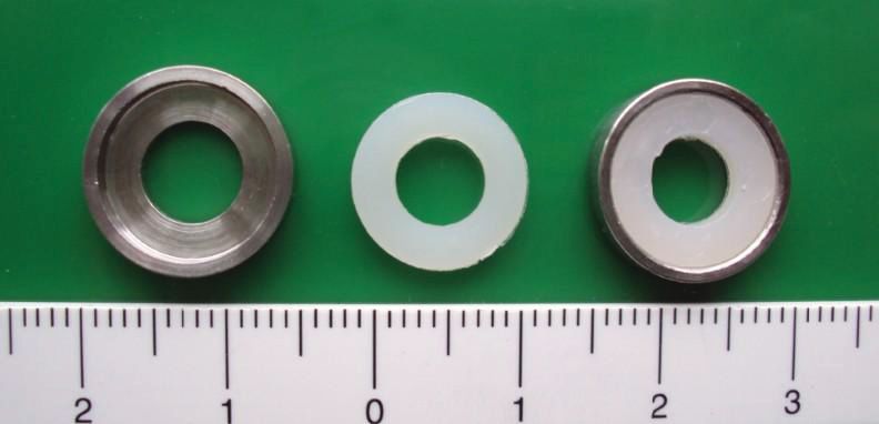
observed on plastic and AM after 72 h (p = 0.145); The results of the scratch-wound
healing assay demonstrated a significant superior cell migration on KF than on AM over
48 h (p < 0.05). However cell migrated on plastic still better than on KF and AM (p <
0.05). Moreover, there were significantly more cells attached to AM compared to plastic
and KF (p = 0.032 and p = 0.001, respectively) and no significant difference was
observed between KF and plastic (p = 0.147) after 30 min. In summary, cell
proliferation and migration was best on plastic, proliferation better on AM compared to
KF and migration faster on KF than AM. While adhesion was best on AM and it was
similar on KF and plastic (see table). Histology demonstrated HCE-T cells cultured on
KF and AM form a multilayered epithelium at an air- liqiud interface which is similar to

the normal human corneal epithelium.

                                  Proliferation                   Migration                     Adhesion
            KF                            +                           ++                             +

          AM                             ++                            +                           +++

         Plastic                        +++                           +++                            +
“+ ~ +++” indicate progressive increase in cell growth behavior on the substrates. (KF: keratin film – AM : amniotic
membrane)

CONCLUSION : Transparent biopolymer keratin films based on human hair support
cell proliferation, migration, adhesion and differentiation of HCE- T cells in vitro
Therefore, it could be a promising candidate as a cell substrate for ocular surface
reconstruction.

                                                        II
ZUSAMMENFASSUNG
HINTERGRUND: Eine schwere Augenoberflächenerkrankung kann zu einer
chirurgisch   therapieresistenten    Erblindung    führen.   Die   Transplantation   von

Limbus-Stammzellen auf Amnion-Membran (AM) als Zellträger ist die am häufigsten
verwendete therapeutische Modalität zur Rekonstruktion der Augenoberfläche. Aber die
AM hat einige Nachteile, einschließlich der mögliche Übertragung von Infektionen,
eine variable Qualität und eine geringe Transparenz. Es ist daher notwendig,
standardisierte alternative Substrate zu finden.

MATERIAL UND METHODEN : Das Zellwachstumsverhalten einer menschlichen
kornealen Epithelzelllinie (HCE- T) wurde auf einem neuen, transparenten Keratin-Film
(KF) auf Basis von menschlichen Haaren untersucht und mit AM und Kunststoff in
vitro verglichen. 5×10³ oder 1×10⁴ Zellen/Loch wurden auf KF, AM und Kunststoff für
72 h ausgesät, um die Zellproliferation durch MTT-Assay zu bestimmen. Die
Zellmigration wurde mit einem Kratz-Wundheilungsassay für 48 h beobachtet und die
Zellanheftung nach 30 min durch einen Adhäsionstest untersucht. Für die Histologie
wurden Zellen auf KF und AM an einer Luft-Flüssigkeitsgrenzfläche für 2 Wochen
kultiviert und mit Hämatoxylin und Eosin gefärbt.

ERGEBNISSE: Proliferation und Migration waren am besten auf Kunststoff. Eine
bessere Proliferation fand sich auf AM als auf KF und eine schnellere Migration auf KF
gegenüber AM. Die Adhäsion war am besten auf AM und schlechter auf KF und
Kunststoff (siehe Tabelle). HCE- T-Zellen kultiviert auf KF und AM an einer
Luft-Flüssigkeitsgrenzschicht zeigten ein mehrschichtiges Epithel ähnlich der normalen
menschlichen Hornhaut.

                                            III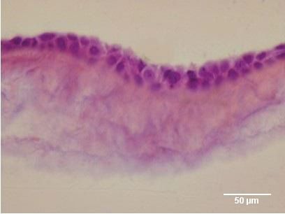
Proliferation                Migration                   Adhäsion

          KF                           +                          ++                          +

         AM                           ++                           +                        +++

     Kunststoff                      +++                          +++                         +
“+ ~ +++” zeigen progressive Zunahme des Zellwachstumsverhalten auf den Substraten. (KF: Keratin-Film – AM :
Amnion-M embran)

SCHLUSSFOLGERUNG:                     Transparente       Biopolymer-Keratin-Filme            auf     Basis
menschlichen       Haars      unterstützen      Zellproliferation,      Migration,      Adhäsion       und
Differenzierung       von     Hornhaut-Epithelzellen         in    vitro.   Daher könnte es ein
vielversprechender Kandidat als Substrat für eine Augenoberflächenrekonstruktion sein.

                                                    IV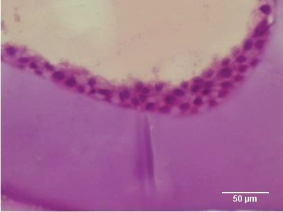
ABBREVIATIONS

°C      degree Celsius                       LESC   limbal epithelial stem cell
%       percent                              LSCD   limbal stem cell deficiency
μL      microliter                           M      mole
μm      micrometer                           min    minute
μM      micromole                            mL     milliliter
AM      amniotic membrane                    mM     millimole
ANOVA   analysis of variance                 MTT    3-(4,5-dimethylthiazol-2-y
BCVA    best corrected visual                       l) -2,5-diphenyltetrazolium

        acuity                                      bromide
bFGF    basic fibroblast growth factor       NaCl   sodium chloride
BSS     balanced salt solution               NaOH   sodium hydroxide
CO2     carboxic oxide                       OS     ocular surface
Da      dalton                               PBS    phosphate-buffered saline

DMEM    Dulbecco’s modified                  PCL    Poly (ε-caprolactone)
        Eagle’s medium                       PET    polyethylene terephthalate
DMSO    dimethyl sulfoxide                   PH     potential of hydrogen
EDTA    ethylene diamine tetraacetic         SCID   severe combined immuno-
        acid                                        deficiency disease

EGF     epidermal growth factor              SD     standard deviation
FBS     fetal bovine serum                   SDS    sodium dodecyl sulfate
g       gram                                 SJS    Stevens-Johnson syndrome
GC      gelatin-chitosan                     SPSS   statistical product and
h       hour                                        service solutions

HCE-T   human corneal epithelial cell line TGF      transforming growth factor
KF      keratin film                         VEGF   vascular endothelial
KGF     keratinocyte growth factor                  growth factor

                                         V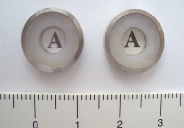
TABLE OF CONTENTS

1 INTRODUCTION....................................................................................1
    1.1 THE OCULAR SURFACE...................................................................................1
        1.1.1 Anatomy of the ocular surface……………………………………………..1
        1.1.2 Ocular surface diseases…………………………………………………….3
    1.2 LIMBAL STEM CELL DEFICIENCY…………………………………………4
        1.2.1 Introduction of the limbal stem cell deficiency……………………………4
        1.2.2 Conventional therapy for ocular surface reconstruction……………….5

        1.2.3 Tissue engineering for ocular surface reconstruction…….……………6
        1.2.4 Keratin films as cell carriers for ocular surface reconstruction……….….18
    1.3 PURPOSE OF THE INVESTIGATION……………………………………….19

2 MATERIALS AND METHODS……………………………...............20
    2.1 MATERIALS…………………………………………………………………..20
    2.2 METHODS……………………………………………………………………..21
        2.2.1 Keratin film fabrication….......………………………………………........21

        2.2.2 Human amniotic membrane preparation……………………………….23
        2.2.3 Cell culture………………………………………….……………………..24
        2.2.4 Cell proliferation: MTT-assay….………………………………………….27
        2.2.5 Cell migration: scratch-wound healing assay.……………………………..28
        2.2.6 Cell attachment: adhesion assay.…………………………………………..29

        2.2.7 Histology (H&E stain)…………………………………………………......29
        2.2.8 Statistical analysis.………………………………………………………....30

                                                    VI
3 RESULTS………………………………………………………………31
3.1 CELL PROLIFERATION IN VITRO…………………………...…………….31
3.2 CELL MIGRATION IN VITRO……………………………………………….33
3.3 CELL ADHESION IN VITRO………………………………………………...36
3.4 HISTOLOGY (H&E STAIN)…………………………………………………..37
4 DISCUSSION…..………………………………………………………38
4.1AMNIOTIC MEMBRANE AS CELL CARRIER FOR OCULAR
SURFACE RECONSTRUCTION………………………………………….....38
4.2 ALTERNATIVE CELL CARRIERS FOR OCULAR SURFACE
RECONSTRUCTION………………………………………………………....41
4.2.1 Contact lens as cell carrier for ocular surface reconstruction…………....42
4.2.2 Fibrin gel as cell carrier for ocular surface reconstruction……………....42
4.3 NEW CELL CARRIER – KERATIN FILM – FOR OCULAR SURFACE
RECONSTRUCTION………………………………………………………....43
4.3.1 Cell proliferation in vitro………………………………………………...45
4.3.2 Cell migration in vitro…………………………………………………...46
4.3.3 Cell adhesion in vitro and histology……………………………………..47
4.4 LIMITATIONS OF MY STUDY……………………………………………..48
5 CONCLUSION….……………………………………………………..50
6 REFERENCES…………………………………………………….......51
7 APPENDIX……….…………………………………………………….69
VIIIntroduction
1 INTRODUCTION
1.1 THE OCULAR SURFACE
1.1.1 Anatomy of the ocular surface
The ocular surface (OS) is the outermost layer of the eye and is composed of cornea and
conjunctiva, which includes bulbar, palpebral and forniceal conjunctiva. The normal
anterior surface is covered by stratified epithelia which can protect the ocular surface
from pathogens and avoid infection by producing antimicrobial peptides (McDermott,
2004).
The cornea – a main refractive element of the eye – consists of five layers which are
epithelium, Bowman’s membrane, stroma, Descemet’s membrane and endothelium.
Due to the uniform structure, avascularity and deturgescence, the cornea is transparent
which is important for maintaining clear vision. Aging or damaged corneal epithelial
cells can be replaced by the limbal epithelial stem cells (LESC) – which reside in the
peripheral corneal limbus andhave superior proliferative capacity compared to the
central corneal epithelial cells. LESC can migrate from the limbus into the center of the
cornea and differentiate into mature corneal epithelial cells to regenerate corneal
epithelium (Sun and Green, 1977; Schermer, Galvin et al. 1986; Pellegrini, Golisano et
al. 1999; Dua and Azuara-Blanco, 2000; Sun and Lavker, 2004). (Fig. 1).
-1-Introduction
Figure 1. Location of the limbus, which i s the transition zone between peripheral cornea
and conjunctiva. *From O'Callaghan and Daniels, 2011.
The conjunctiva, a thin and transparent mucous membrane covering the sclera, can
secrete mucus and tears to help lubricate the eye. It has three parts – bulbar, forniceal
and palpebral conjunctiva, and it is composed of a stratified epithelium and vascularized
stroma histologically. The conjunctival epithelium includes goblet cells, which are
responsible for producing the conjunctival mucin component of the tear film. The
conjunctival stroma includes the accessory lacrimal glands of Krause and Wolfring,
which can secrete the baseline tear. When the LESC are seriously injured, the
conjunctival epithelium can migrate onto the corneal surface and result in corneal
neovascularisation and opacity(Kinoshita, Friend et al. 1983; Danjo, Friend et al.
1987).
The corneal and conjunctival epithelia express different cytokeratins (CK), which exist
in almost all the invertebrates epithelial cells. There are two subfamilies of cytokeratins,
which are type І (acidic, including CK9 to CK20) and type Ċ (neutral/basic, including
CK1 to CK8). The type of cytokeratin expression can be used to evaluate the phenotype
of the ocular surface epithelial cells, because different epithelial cells have different
patterns of cytokeratins. It has been shown that keratins of the corneal epithelium are
composed of a major cytokeratin pair, basic keratin CK3 and acid keratin CK12, and
they have been used as a corneal type differentiation marker (Moll, Franke et al. 1982;
Meller and Tseng, 1999). In the normal human conjunctival epithelium, the cytokeratin
-2-Introduction
pair – CK4 and CK13 – have been used as conjunctival differentiation marker (Krenzer
and Freddo, 1997). Therefore, evaluating the cytokeratin pattern is useful for the
epithelial cells culture in vitro to investigate if the cells maintain the phenotype, which
potentially would be important for ensuring a good epithelial graft for transplantation in
vivo.
1.1.2 Ocular surface diseases
Etiologically, ocular surface diseases can be classified as inflammatory, infectious,
congenital, hereditary, traumatic, toxic, nutrient, iatrogenic,immunological as well as
degenerative and they can lead to discomfort, superficial punctate keratopathy and loss
of corneal transparency. Advanced disorders of the ocular surface, Stevens-Johnson
syndrome (SJS), severe keratitis, chemical or thermal burns, ocular cicatricial
pemphigoid and aniridia, can result in severe discomfort, ulceration, severe impairment
of vision and even blindness (Shapiro, Friend et al. 1981; Dua and Forrester, 1990).
Cornea-related disease is the second main blindness cause all over the world reported by
the World Health Organization. There are about 50 million people who suffer from
bilateral blindness and at least 150 million people who have impaired vision in both
eyes mainly as a result of corneal disease. Moreover, patients with corneal blindness are
increasing by 1.5 million to 2 million every year worldwide (Whitcher, Srinivasan et al.
2001; Foster, 2003). Therefore, the treatment of ocular surface diseases is a great
challenge for the ophthalmologists to reduce global blindness.
-3-Introduction
1.2 LIMBAL STEM CELL DEFICIENCY
1.2.1 Introduction of the limbal stem cell deficiency
Corneal epithelial stem cells are located at the limbus which is the transition zone
between the conjunctiva and the peripheral cornea. The limbal epithelial stem cells are
the source of the corneal epithelial renewal and they reconstruct the aging or the
damaged corneal epithelium to maintain the corneal integrity (Sun and Green, 1977).
During homeostasis or after injury of the corneal epithelium, the limbal stem cells
migrate into the center of the cornea from the limbus and produce transient amplifying
cells to replace the lost corneal epithelial cells and transform them into normal
multilayer corneal epithelium gradually (Cotsarelis, Cheng et al. 1989˗Lavker, Dong
et al. 1991˗Pellegrini, Golisano et al. 1999) (Fig. 2). However, this can not occur if the
limbal stem cells are depleted.
Figure 2. Schematic diagram of the limbal stem cells transform into central corneal
epithelial cells. Limbal stem cells (S C) which locate at the limbus migrate to the center of
the cornea, differentiate into transient amplifying cells (TAC) and finally transform into
terminally differentiated cells (TDC) to replace the lost corneal epithelium and become
mature normal corneal epithelial cells. *From Holland, 1996.
-4-Introduction
According to the causes, limbal stem cell deficiency (LSCD) can be classified as
primary and secondary. The primary LSCD is related to an insufficient stromal
microenvironment which can not support limbal stem cell function, such as aniridia,
neurotrophic keratopathy and chronic limbitis. However, secondary LSCD ismore
common and associated with external factors that damage the limbal stem cells, such as
thermal or chemical burn, multiple surgeries, Stevens-Johnson syndrome, ocular
cicatricial pemphigoid, severe microbial infection and contact lens- induced keratopathy.
In LSCD patients, the neighbouring conjunctival epithelial cells migrate across the
limbus onto the cornea surface. This leads to corneal opacity and loss of vision.
Meanwhile, these patients suffer severe discomfort accompanied by chronic
inflammation, neovascularization, persistent corneal epithelial defects, ulceration and
stromal scar (Shapiro, Friend et al. 1981; Puangsricharern and Tseng, 1995; Daniels,
Dart et al. 2001; O'Callaghan and Daniels, 2011).
1.2.2 Conventional therapy for ocular surface reconstruction
Because medical therapy often fails, surgical treatments such as penetrating or lamellar
corneal transplantation are necessary to restore vision. The most widely applied
treatment is full-thickness replacement of damaged cornea which is penetrating
keratoplasty. If the corneal endothelium is normal, lamellar keratoplasty is used for
ocular surface reconstruction which maintains the recipient’s endothelium (Geerling and
Seitz, 2005). However, patients who suffer from limbal stem cell deficiency are poor
candidates for conventional corneal transplantation, as grafting the central cornea does
not restore limbal stem cell population. Therefore, the vision progression of these
patients is limited (Williams, Roder et al. 1992; Kuckelkorn, Keller et al. 2001). In
addition, long-term healing of the grafted epithelial surface is plagued with recurrent
and persistent epithelial defects which often lead to reopacification, revascularization of
the graft as well as persistent photophobia of the patients. For treating persistent corneal
epithelial defects, human amniotic membrane transplantation has been widely used to
-5-Introduction
support cell repair and has also been used as cell substrate for expansion of cell sheets
(Dua, Gomes et al. 2004).
At present, LESC transplantation is being performed with cadaveric, living
related-donor or autologous limbus from the contralateral healthy eye for treating
patients who suffer from severe limbal stem cell deficiency. However, living- related or
autologous LESC transplantation requires to take a large limbal graft from a healthy eye
and thus carries the risk of inducing ocular surface disease of the allogeneic donor eye
or the contralateral healthy donor eye (Chen and Tseng, 1991; Holland, 1996; Pellegrini,
Traverso et al. 1997; Dua and Azuara-Blanco, 2000). Transplantation from allogeneic
donors may induce rejection and requires the use of long-term systemic
immunosuppressant therapy which can have significant side effects (Daya, Bell et al.
2000; Kim, Tchah et al. 2003).
1.2.3 Tissue engineering for ocular surface reconstruction
In recent years, methods of tissue engineering for OS reconstruction have received
increased interest. Epithelial cell sheets have been expanded in vitro from small
autologous limbal biopsies, which can reduce the risk of inducing ocular surface disease
of the donor. To date, different cell carriers for cell sheets expansion have been
proposed for ocular surface reconstruction (summarized in Table 1).
-6-Introduction
Table 1. Cell substrates for ocular surface reconstruction
Substrates References
In vitro experiments In vivo experiments
(cultured cells in vitro)
Animals Humans (n eyes)
Amniotic membrane M eller et al.1999 Koizumi et al.2000 Tsai et al.2000(n=6)
(Rabbit conjunctival epithelial cells) (Rabbits) Shimazaki et al.2002(n=13)
Nakamura et al.2003(n=13)
Koizumi et al.2000 Wan et al.2011 Nakamura et al.2004(n=6)
(Rabbit corneal epithelial cells) (Rabbits) Shortt et al.2008(n=10)
Basu et al.2012(n=50)
Nakamura et al.2004
(Human limbal epithelial cells)
Collagen gels Geggel et al.1985 Shimmura et al.2003 –—
(Rabbit corneal epithelial cells) (Rabbits)
He et al.1991 M errett et al.2008
(Human limbal epithelial cells) (Minipigs)
Dravida et al.2008 Liu et al.2008
(Human limbal epithelial cells) (Minipigs)
M cIntosh et al.2009 Lagali et al.2008
(Human limbal epithelial cells and (Minipigs)
bovine keratocytes)
M i et al.2010
(Bovine limbal epithelial cells)
Levis et al.2010
(Human limbal epithelial cells)
Fibrin gels Han et al.2002 Talbot et al.2006 Rama et al.2001(n=18)
(Human limbal epithelial cells) (Rabbits) Rama et al.2010(n=113)
Silicone hydrogel Di Girolamo et al.2007 –— Di Girolamo et al.2009
Contact lenses (Human limbal epithelial cells) (n=3)
-7-Introduction
Substrates References
In vitro experiments In vivo experiments
(cultured cells in vitro)
Animals Humans (n eyes)
Poly(ε-caprolactone) Ang et al.2006 Ang et al.2006 –—
membranes (Rabbit conjunctival epithelial cells) (Mice)
Sharma et al.2011
(Human corneal epithelial cell line:
HCE-T and human limbal epithelial
cells)
Gelatin-chitosan Zhu et al.2006 –— –—
membranes (Rabbit conjunctival epithelial cells)
Silk fibroin films Higa et al.2008 –— –—
(Murine and human limbal epithelial
cells)
Chirila et al.2008
(Human limbal epithelial cells)
Higa et al.2010
(Rabbit limbal epithelial cells)
Bray et al.2011
(Human limbal epithelial cells)
Human anterior Galal et al.2007 –— –—
lens capsules (Human limbal epithelial cells)
Keratin films Reichl et al.2011 –— –—
(Human corneal epithelial cell line:
HCE-T)
“–—” indicates no animal experiment in vivo or clinical result is available.
-8-Introduction
Human Amniotic Membranes
Human amniotic membrane (AM), the innermost layer of the placenta, consists of three
layers – epithelium, basement membrane and avascular stroma – and has been
successfully used as carrier for expansion of cell sheets in vitro and further transplanted
to the ocular surface in vivo.
Several studies have shown that human AM can support proliferation, migration,
differentiation and adhesion of epithelial cells in vitro (Meller and Tseng, 1999; Schwab,
1999; Koizumi, Inatomi et al. 2000) and it has also been found to encourage healing of
epithelial wounds and as carrier for transplantation of limbal stem cells, oral mucosal
epithelial cells and conjunctival epithelial cells in vivo. Expansion of autologous
epithelial cells on a carrier for transplantation clearly reduces the risk of donor site
morbidity and avoids the use of long-term systemic immunosuppression (Kenyon and
Tseng, 1989; Tsai and Tseng, 1994; Tseng, Prabhasawat et al. 1998; Nakamura, Inatomi
et al. 2004; Ang, Tan et al. 2005).
In this emerging clinical field and in the absence of any statistics, no standard technique
for ocular surface reconstruction with bioengineered tissue has been defined, although it
maybe likely that based on the numbers of clinical reports today and because AM
stimulates cell growth and has antiinflammatory and antiangiogenic properties, human
AM is probably the most commonly used cell carrier for tissue engineering, particularly
in OS reconstruction (Tsai, Li et al. 2000; Nakamura, Koizumi et al. 2003; Nakamura,
Inatomi et al. 2004; Kruse and Cursiefen, 2008; Wan, Wang et al. 2011; Basu, Ali et al.
2012). On the downsides, human AM has variable quality (differences in protein
expression which is dependent on donor age and length of pregnancy influence clinical
results after transplantation) (Hopkinson, McIntosh et al. 2006; Gicquel, Dua et al. 2009;
López-Valladares, Teresa Rodríguez-Ares et al. 2010), carries the risk of disease
transmission from donor to recipient, induces potential immunological reactions and has
-9-Introduction
a limited transparency (Conno, Doutch et al. 2010; Dua, Rahman et al. 2010). Therefore,
it is not surprising that clinical results also vary, that vision remains limited and that
AM grafts are frequently lost in the early postoperative period (Ijiri, Kobayashi et al.
2007˗Shortt, Secker et al. 2008). To date, a number of alternative carriers have been
used as well as potential carriers proposed for ocular surface reconstruction.
Collagen Gels
Since the human corneal major component is collagen, the choice of collagen as
substrate for corneal epithelial cell expansion appears obvious. Collagen has a low
immunogenicity, is naturally biocompatible and relatively not expensive to isolate. In
addition, collagen gels could easily be prepared from pepsin solubilized bovine dermal
collagen (Elsdale and Bard, 1972; Fagerholm, Lagali et al. 2009).
Some investigations revealed that collagen gels support the growth of human and rabbit
corneal epithelial cells in vitro (Geggel, Frienid et al. 1985; He and McCulley, 1991;
McIntosh Ambrose, Salahuddin et al. 2009) and of rabbits in vivo (Shimmura, Doillon
et al. 2003). However, conventional collagen hydrogels are inherently weak because of
their high water content (Bell, Ivarsson et al. 1979; Freed, Guilak et al. 2006). Their
mechanical properties can be improved by chemical crosslinking or plastic
compression.
Cross-linked collagen gels can be fabricated by mixing collagen with 1-ethyl-3-
(3-dimethyl aminopropyl) carbodiimide (EDC) and N-hydroxysuccinimide (NHS) and
cast to produce a recombinant cross- linked collagen scaffold. These cross- linked gels
supported primary human limbal epithelium growth in vitro (Dravida, Gaddipati et al.
2008) and the findings were confirmed by transplanting the constructs with expanded
corneal epithelial cells into corneas of mini-pigs in vivo (Lagali, Griffith et al. 2008˗Liu,
Merrett et al. 2008˗Merrett, Fagerholm et al. 2008). Moreover, these cross- linked
- 10 -Introduction
collagen grafts could be used to substitute corneal grafts in patients with keratoconus or
corneal central scar (Fagerholm, Lagali et al. 2010).
Plastic compressed collagen gels can be produced by sandwiching the collagen
hydrogels between layers of nylon and metal mesh for 5 min in order to increase the
mechanical strength by reducing the water content. Human limbal epithelial stem cells
cultured on these gels can form multilayered epithelium which were very similar to the
human central corneal epithelium in vitro (Levis, Brown et al. 2010˗Mi, Chen et al.
2010). There is no report about plastic compressed collagen gels for OS reconstruction
in vivo so far.
Fibrin Gels
Fibrin gels can be prepared by mixing fibrinogen with thrombin to induce the last step
of the natural coagulation cascade. Both components – fibrinogen and thrombin – can
be harvested from human plasma. Hence, fibrin gels can be produced from a totally
autologous material.
The fibrin gels have successfully been used as substrate for epithelial cell expansion and
differentiation in vitro and to repair corneal ulcers in humans (Lagoutte, Gauthier et al.
1989˗Duchesne, Hassan et al. 2001˗Han, Schwab et al. 2002; Talbot, Carrier et al.
2006). In 2001, Rama et al. cultured autologous limbal stem cells on the fibrin gels and
grafted them to 18 eyes of 18 patients who suffered from unilateral limbal stem cell
deficiency. After a follow-up of 12 – 27 months, 14 eyes were considered successful as
evaluated by symptoms, best corrected visual acuity (BCVA), corneal transparency and
impression cytology (Rama, Bonini et al. 2001). Recently, the same group reported
result of more patients (113 eyes of 112 patients who had severe limbal stem cells
deficiency) showing that 76.6 % of these grafts were successful by evaluating
symptoms, BCVA, corneal transparency and impression cytology after a follow- up of 1
- 11 -Introduction
– 10 years (Rama, Matuska et al. 2010). Therefore, the fibrin gel is a clinically viable
alternative to human AM. However, the components used for fibrin gels fabrication are
harvested from plasma. This could carry the risk of disease transmission and infection.
Table 2. Fibrin gels used as cell carrier for human eyes in vivo. (LP: light perception – CF:
counting fingers – HM: hand movement)
Author Year of Indications N Follow-up Success BCVA
publication (eyes) improvement
Rama et al 2001 LSCD 18 12-27 months 77.8 % eyes from LP or CF
to 0.1– 0.8
Rama et al 2010 LSCD 113 1-10 years 76.6 % eyes from HM or CF
to 0.3-0.9
Silicone Hydrogel Contact Lenses
Human limbal epithelial stem cells expanded on silicone hydrogel contact lenses
showed that contact lenses can sustain proliferation and migration from human limbal
tissue to confluency with a corneal phenotype which was confirmed by
immunohistochemistry in vitro (Di Girolamo, Chui et al. 2007). So far, these silicone
hydrogel contact lenses have been used for human ocular surface reconstruction in three
human eyes with limbal stem cell deficiency. With a follow-up of 8 – 13 months,
transparent corneal epithelium, significant improvement of symptoms and best
corrected visual acuity and no corneal conjunctivalization or neovascularization were
observed (Di Girolamo, Bosch et al. 2009). Further investigations with more patients
are necessary in order to confirm if they are suitable alternative carriers to human AM
for ocular surface reconstruction.
Table 3. Silicone hydrogel contact lense s used as cell carrier for human eyes in vivo.
Author Year of Indications N Follow- Morphological BCVA
publication (eyes) up outcome improvement
Di Girolamo 2009 - Aniridia 3 8-13 No From CF / 0.03
et al -Conjunctival months conjunctivalization to 0.1- 0.5
melanoma or vascularization.
- 12 -Introduction
Poly (ε-Caprolactone) Membranes
Poly (ε-caprolactone) (PCL) is a material for pharmaceutical products and wound
dressings, and can be prepared into highly flexible and strong membranes by dissolving
PCL solution in trifluoroethanol via an electrospinning process.
Rabbit conjunctival epithelial cells which were cultured on the PCL membranes
maintained their conjunctival phenotype, showed cell proliferation and formed 8 to 10
epithelial cell layers after xenografting o nto dorsal subcutaneous tissue of severe
combined immunedeficient (SCID) mice (Ang, Cheng et al. 2006). Moreover, human
corneal epithelial cells line (HCE-T) expanded on the PCL films retained a normal
corneal phenotype and limbal epithelial stem cells grown on PCL films showed similar
morphology compared with glass coverslips and human AM in vitro (Sharma, Mohanty
et al. 2011). However, reports of clinical applications for ocular surface reconstruction
in humans are still missing.
Gelatin-chitosan Membranes
Chitosan is a major constituent of cornea and dermis, and as such known to play a
critical role in wound healing in vitro and in vivo. It can be fabricated into gels by
dissolving deacetylated chitosan into acetic acid solution and cast onto tissue culture
plates until it is dry (Kratz, Arnander et al. 1997; Sechriest, Miao et al. 2000˗Suh and
Matthew, 2000).
Pure chitosan membranes are too stiff for application to the curved ocular surface.
Integration with gelatin – a soft, elastic natural material – can reduce the membrane’s
stiffness while further improving its mechanical strength by increasing molecular
interactions (Berger, Reist et al. 2004). Rabbit conjunctival epithelial cells cultured for
- 13 -Introduction
14 days on a gelatin-chitosan (GC) hydrogel grew to a confluent multilayer expressing
cytokeratin CK 4, which indicated that the gelatin-chitosan membrane supported normal
differentiation of conjunctival epithelial cells (Zhu, Beuerman et al. 2006). To date, no
clinical data on the use of gelatin-chitosan hydrogels for ocular surface reconstruction
have been published.
Silk Fibroin Films
Silk fibroin is a structural protein obtained from the cocoon of the silkworm Bombyx
mori. It is non- immunogenic, degradable in vivo, has excellent mechanical strength and
is – compared with human AM – transparent. It recently has been used as suture
material and for regeneration of bone and cartilage (Kim, Jeong et al. 2005˗Lee, Baek
et al. 2007˗Shangkai, Naohide et al. 2007).
Silk fibroin can be processed as a thin transparent film by dissolving the silk fibroin by
formic acid and a subsequent electrospinning process. This film can be used as
biological carrier for the expansion of murine corneal limbal stem cells which
maintained clonal growth characteristics and cell differentiation (Chirila, Barnard et al.
2008˗Higa and Shimazaki, 2008˗Lawrence, Cronin- Golomb et al. 2008). In recent
years, rabbit limbal epithelial stem cells and human limbal epithelial stem cells cultured
on silk fibroin films in vitro were found to form stratified cells with maintained corneal
phenotype (Higa, Takeshima et al. 2010˗Bray, George et al. 2011). However, on the
downsides are the considerably higher costs compared with synthetic materials. Still no
clinical report about this carrier for ocular surface reconstruction so far.
- 14 -Introduction
Human Anterior Lens Capsules
Millions of human anterior lens capsule are removed and discarded during cataract
surgery annually from cataract patients. The material is transparent and to some extent
elastic. At birth, the human anterior lens capsule is approximately 4 Pm thick. It
consists of a dense, outer layer of collagen IV, laminin and heparin sulfate
proteoglycans. Throughout life the thickness of the anterior lens capsule increases
continuously by deposition of new lamellae to 28 – 33 Pm at the age of 70 to 80 years
(Krag, Olsen et al. 1997˗Danysh, Czymmek et al. 2008).
Autologous and allogeneic limbal epithelial stem cells were expanded ex vivo on
human anterior lens capsule obtained from 30 patients at the age of 59 to 75 years
during routine cataract surgery. An identical cell density and morphology of limbal
epithelial stem cells was observed compared to controls on plastic for up to 2 weeks
(Galal, Perez-Santonja et al. 2007). Further research will be necessary to evaluate
whether limbal epithelial stem cells cultured on the anterior lens capsule can form a
multilayered epithelium, maintain corneal phenotype and is a viable option in vivo.
The properties, advantages and disadvantages of all the biomaterials used as cell carriers
are summarized in Table 4 and 5.
- 15 -Introduction
Table 4. Properties of biometerials as cell carriers for ocular surface reconstruction
Properties Carriers
AM Collagen Fibrin Silicone Poly (ε- Gelatin- Silk Human
gels gels chitosan fibroin
hydrogel caprolactone) anterior
membranes films
contact membranes lens
lenses capsules
M echanical ++ + ~ ++ ++ ++ ? ++ ~ +++ ++ ~ +++ ++ ++
strength
Elasticity +++ ++~+++ +++ ++ ? ++ ++ ~ +++ +++ ++ ~ +++
Trans- + +++ +++ +++ +++ + +++ +++
(Talbot et (Ang et al.
parency al.2006) 2006)
+ +
(Han et al. (Sharma et al.
2002) 2011)
Support for +++ +++ +++ +++ ++ ~ +++ ++ ~ +++ +++ +++
OS epithelial
cell growth
(in vitro)
Clinical ++ –— ++ +++ –— –— –— –—
success
(in vivo)
Nakamura Merrett et Han et al. Di Ang et al. Berger et al. Chirila et Krag et al.
References
et al. 2003 al. 2008 2002 Girolamo 2006 2004 al. 2008 1997
et al.2007
Shortt et al. Liu et al. Talbot et Sharma et al. Zhu et al. Higa et al. Galal et al.
2008 2008 al. 2006 Di 2011 2006 2010 2007
Girolamo
Wan et al. Dravida Rama et et al.2009 Bray et al.
2011 et al.2008 al.2001 2011
Basu et al. McIntosh Rama et
2012 et al.2009 al.2010
“ ? ” indicates clinical impression, no experimental data is available.
“–—” indicates no clinical results are available.
- 16 -Introduction
Table 5. Advantages and di sadvantages of cell carriers for ocular surface reconstruction
Carriers Advantages Disadvantages
AM anti-inflammation, anti-angiogenesis, limited transparency, risk of disease
good elasticity transmission, variable quality,
limited mechanical strength
Collagen gels good transparency, limited mechanical strength
good biocompatibility
Fibrin gels good transparency, good for tissue risk of disease transmission
regeneration, degradable in vivo
Silicone hydrogel contact lenses good transparency, therapeutic bandage –—
for wound healing
Poly(ε-caprolactone) membranes good transparency, high mechanical limited elasticity
strength, good biocompatibility
Gelatin-chitosan membranes good for wound healing limited transparency,
limited elasticity
Silk fibroin films good transparency, high cost of source materials
good biocompatibility
Human anterior lens capsules good transparency, variable quality (due to donor’s
large number of donors age), risk of disease transmission,
limited mechanical strength
- 17 -Introduction
1.2.4 Keratin films as cell carriers for ocular surface reconstruction
Keratins, cysteine-rich proteins, belong to a group of structural proteins which are
formed in the epithelial cells of higher vertebrates. These proteins have high mechanical
strength because of a large number of disulfide bonds. Hard or filamentous structures
such as hair, wool, nails, horns, hoofs and feathers – which can be obtained from hair
salons and from the food industry as unused protein-rich waste – can be used as sources
for these proteins.
In recent years, coatings, films and scaffolds based on keratin from wool or hair have
been established, and their practical usefulness as a growth substrate has been
investigated. Mouse cell line L929 fibroblasts cultured on keratin extracted from wool
demonstrated improved growth and attachment compared to collagen and glass
(Yamauchi, Maniwa et al. 1998; Tachibana, Furuta et al. 2002).
In a previous study, the usefulness of 50 Pm thick human hair keratin coated substrates
– obtained by casting keratin dialysate into a plastic plate – for cell cultivation in vitro
has been reported (Reichl, 2009). The biomechanical strength and elastic modulus of
the keratin films (KF) could be modified by mixing the keratin dialysate with an
alkaline dialysate at different ratios and subsequent curing temperatures between 70 °C
and 110 °C. Substrates with biomechanical stability and improved transpare ncy
compared with human AM could be produced. Subsequent investigation showed that
the proliferation of HCE-T cells on the transparent KF in vitro were similar to those
observed on human AM (Reichl, Borrelli et al. 2011). A correlation between
composition of the keratin films and their biomechanical parameters has been reported
recently by our group in a surgical ex vivo feasibility assessment on enucleated porcine
eyes, which showed 90/10 keratin films (90 % aqueous keratin dialysate mixed with
10 % alkaline keratin dialysate) cured at 100 °C to offer the best compromise between
mechanical strength and flexibility (Borrelli, Reichl et al. 2012).
- 18 -Introduction
1.3 PURPOSE OF THE INVESTIGATION
In my study I cultivated the well-established human corneal epithelial cell line (HCE-T
cells) on the transparent and transferable 90/10 keratin films cured at 100 °C which
were fabricated from human hair (Reichl, Borrelli et al. 2011; Borrelli, Reichl et al.
2012)in vitro to investigate ifthese films are a suitable cell substrate for ocular surface
reconstruction and compared their ability to support cell proliferation, migration,
attachment and morphology with denuded human amniotic membrane and
commercially available plastic culture plates in vitro by using MTT, scratch-wound
healing, adhesion assays as well as routine histology.
- 19 -Materials and Methods
2 MATERIALS AND METHODS
2.1 MATERIALS
Dulbecco’s modified Eagle’s medium (DMEM)/Ham’s F12 (at a ratio of 1:1), Hank’s
balanced salt solution (BSS), penicillin/streptomycin, phosphate-buffered saline (PBS),
amphotericin B and trypsin-EDTA were purchased from PAA (Cölbe, Germany). Fetal
bovine serum (FBS), insulin, epidermal growth factor (EGF) and cell scrapers were
obtained from Biochrom (Berlin, Germany). 0.9% sodium chloride (NaCl), urea,
thiourea, sodium dodecyl sulfate (SDS), 2-amino-2-hydroxymethylpropane-1,3-diol
(Tris), sodium hydroxide (NaOH), ethanol, 2-mercaptoethanol, dimethyl sulfoxide
(DMSO), hematoxylin, eosin,Mayer's hemalaun, xylene and methanol were purchased
from Carl Roth (Karlsruhe, Germany). Spectra/Por® 1 dialysis membrane (MWCO
6-8000 Da) was purchased from Spectrum (Rancho Dominguez, US). Vivaspin® was
purchased from Sartorius (Goettingen, Germany)andPolyethylene terephthalate (PET)
foil was obtained from LTS Lohmann (Andernach, Germany). Dispase II and trypan
blue solution were obtained from Invitrogen (Karlsruhe, Germany). Hydroxyurea
glycerol and sucrose were purchased from Sigma (Steinheim, Germany). Technovit®
8100 kit were obtained from Heraeus Kulzer (Hanau, Germany). Aceton was purchased
from Otto Fischar (Saarbrücken, Germany). Coverslips were obtained from Thermo
(Braunschweig, Germany). Hemacytometer was purchased from A.Hartenstein
(Würzburg, Germany). Polystyrene 24-well cell culture plates were obtained from TPP
(Trasadingen, Switzerland). Polystyrene 96-well cell culture plates were purchased from
Greiner Bio-one (Frickenhausen, Germany). Metal rings and plastic rings were obtained
from Hummer & Rieß (Nürnberg, Germany). The CellTiter 96 ® Non-Radioactive Cell
Proliferation Assay kit was purchased from Promega (Mannheim, Germany).
- 20 -Materials and Methods
2.2 METHODS
2.2.1 Keratin film fabrication
Keratin films based on human hair were prepared by the Institute of Pharmaceutical
Technology of the university of Braunschweig according to a protocol previously
established there (Reichl, Borrelli et al. 2011). For this, human hair was obtained from a
local hair salon, intensively washed with water containing 0.5 % sodium dodecyl sulfate
(SDS), then rinsed with fresh water and air-dried. Human hair keratin was extracted
according to the Shindai method (Nakamura, Arimoto et al. 2002). In brief, the hair (20
g) was mixed with extraction medium, which is an aqueous solution (400 mL, pH 8.5)
containing 25 mM Tris, 2.6 M thiourea, 5 M urea and 5 % 2- mercaptoethanol and
incubated at 50 ć for 72 h. The mixturewas centrifuged at 4500 g for 15 minutes and
the supernatant filtered using filter paper with a pore size of 2.5 μm. The filtrate,
denoted as “Shindai extract”, was stored at –20 ć and thawed when required. The
Shindai extract was exhaustively dialysed against demineralized water (100 mL extract
in 5000 mL water) using a dialysis cellulose membrane (6-8000 Da) at 20 ć for 24 h.
The procedure was repeated several times until no 2-mercaptoethanol was detected in
the dialysis fluid. The keratin dialysate, denoted as “aqueous keratin dialysate”, was
centrifuged at 10,000 g for 30 minutes to remove coarse aggregates and immediately
used to fabricate the keratin films. In a second phase of dialysis, the same amount of
Shindai extract was dialysed against a 0.25 M sodium hydroxide (NaOH) solution in the
same method as described above at 4 ć. This NaOH dialysate was concentrated by
means of ultrafiltration using Vivaspin ® 20 concentrators. The filtrates were d iscarded
and the supernatant was diluted several times with 0.05 μM NaOH to generate the
“alkaline keratin dialysate”.
- 21 -Materials and Methods
For my studies I used keratin films with an aqueous/alkaline keratin dialysate at a ratio
of 90:10 cured at 100 ć to evaluate proliferation, migration and adhesion of HCE-T
cells and compared this with denuded human AM and plastic in vitro. Briefly, the
keratin films were produced by mixing the aqueous keratin dialysate with alkaline
keratin dialysate at the given ratio and 1 % glycerol was added as a softening agent. The
mixture was cast on hydrophobic coated PET sheets and then allowed to dry overnight.
Thereafter, they were cured at 100 °C for 2 h to form transparent and transferable
keratin films. All the steps for preparing the films were performed under aseptic
conditions. The keratin films were found to be chemically and mechanically stable for
at least six months at room temperature (Reichl, Borrelli et al. 2011). This 90/10 keratin
film cured at 100 °C was found to offer the best compromise between mechanical
strength and flexibility in an ex vivo experiment by Borrelli et al (Borrelli, Reichl et al.
2012).
Figure 3. The keratin films (KF) ba sed on human hair can be handled with forceps and
exhibit good transparency.
- 22 -Materials and Methods
2.2.2 Human amniotic membrane preparation
According to the tenets of the Declaration of Helsinki for research, human AM was
obtained from an elective caesarean section after informed consent. The intact amniotic
membrane was stripped from the chorion by blunt dissection, washed with sterile 0.9 %
sodium chloride (NaCl) containing 1 % penicillin/streptomycin and 1 % amphotericin B
to remove blood clots, and sutured onto 7.5 × 7.5 cm sterile sponge sheets (Katena, New
Jersey, US), with the epithelium facing upwards. All steps were performed under
aseptic conditions. Then as previously described the membranes were cryopreserved at
–80 ć in DMEM and glycerol at a ratio of 1:1 and only thawed prior to use (Tseng,
Prabhasawat et al. 1997).
Figure 4. Human amniotic membrane (AM) stripped from the chorion and sutured on
sterile sponge sheets. The AM exhibits semi-transparency.
- 23 -Materials and Methods
2.2.3 Cell culture
HCE-T cells (human corneal epithelial cell line) stem from human corneal epithelial
cells (Riken Cell Bank, Ibaraki, Japan) and were immortalized by Sasaki et al. by
infection with a recombinant SV40-adenovirus vector in 1995. These cells retain the
properties of normal corneal epithelial cells in that they are able to form a multilayered
epithelium if cultured at an air- liquid interface indicating that they maintain the
programmed gene for differentiation (Araki-Sasaki, Ohashi et al. 1995). At present,
HCE-T cells are the most frequently used human corneal epithelial cell line and
represent a standard tool for bioavailability, toxicity and drug permeation assessments
(Toropainen, Ranta et al. 2001; Becker, Ehrhardt et al. 2007; Reichl, 2008).
The HCE- T cells I used for my studies were provided by Dr. K. Araki-Sasaki
(Kagoshima, Japan). The cells were cultivated in 24-well cell culture plates with 500 μL
culture medium per well under standard cell culture conditions in a humidified
atmosphere containing 5 % CO 2 and 95 % air at 37 ć. The cell culture media consist of
1:1 DMEM/Ham’s F12 medium supplemented with 10 ng/mL EGF, 5 % FBS, 5 mg/mL
insulin, 1 % penicillin/streptomycin and 0.5 % DMSO solution. The media were
changed every 2 days.
Before starting the cell culture experiments, the KF were intensively equlibrated in
sterile distilled water containing 1 % penicillin/streptomycin and 1 % amphotericin B
under constant gentle shaking. The equilibration solution was changed twice daily for
seven days. Unsuspended KF tended to show an irregular wrinkled surface which would
interfere with visual cell assessment and – quite likely – also with cell migration and
proliferation. In order to achieve an even seeding area and fix the substrate on the
bottom of the cell culture well, KF were punched in circles (diameter were 11 mm) and
- 24 -Materials and Methods
clamped between a metal ring and a plastic ring of suitable size during cell culture. The
self-constructed ring arrangement also provided a standardized growth area (0.283 cm2 )
for cell culture (Fig. 5-7). Before cell expansion, they were incubated with culture
medium overnight.
Before using the human AM, it was thawedin a 37 ć water bath, washed at least three
times with sterile PBS to remove the storage medium and then incubated with Dispase
II (1.2 U/mL) at 37 ć for 2 h to loosen cellular adhesion, followed by gentle scraping
to remove the remaining amniotic epithelial cells with a cell scraper to prepare denuded
AM (Riau, Beuerman et al. 2010). Thereafter, the denuded AM was intensively washed
in PBS to remove the dispase, clamped within the same ring devices and incubated with
culture medium overnight before cell culture. As control, cells were seeded on a plastic
substrate.Empty ring arrangements were placed on the bottom of the culture wells to
obtain the same seeding area for the cells as with KF and AM.
Figure 5ˊ The self-constructed ring arrangements used in the cell culture experiments to
achieve an even and defined growth area. The metal ring, plastic ring and the constructed
two-ring arrangement are shown from left to right respectively.
- 25 -Materials and Methods
6mm
KF/ AM
A B B A 3mm
14mm
Figure 6 ˊ Schematic diagram of the ring arrangements used for cell culture. The
substrates – KF or AM – were fixed between the metal ring (A) and plastic ring (B).
Figure 7ˊOptical transparency of human AM (left) and KF (right). AM and KF are clamped
within the ring arrangements. KF exhibits higher transparency than the AM indicated by
the clarity of the “A” underneath.
- 26 -Materials and Methods
2.2.4 Cell proliferation: MTT-assay
Cell proliferation analysis on cell- carriers is an essential step for evaluating
biocompatibility of biomaterials. In this study, proliferation of HCE-T cells on KF, AM
and plastic was evaluated by determining the mitochondrial function of cells using the
tetrazolium dye 3-(4,5-dimethylthiazol-2-yl)-2,5-diphenyltetrazoliumbromide (MTT).
Living cells can convert the MTT into blue formazan a nd the absorbance of the solution
can be analysed directly by a spectrophotometer to determine viable cell numbers
(Mosmann.1983;Uludag and Sefton, 1990).
My proliferation studies were performed in 24-well polystyrene plates using the ring
devices described above. KF, AM and plastic were used as growth substrates. The
experiments were repeated three times and each assay was performed in triplicate. The
assay was carried out according to the manufacture’s protocol. In brief, 5 × 10³
cells/well and 1 × 10⁴ cells/well were seeded on each substrate and incubated for 24 h,
48 h and 72 h, respectively in 5 % CO 2 and 95 % air at 37 ć. After the respective
culture period, 30 μL of MTT dye solution was added to each well and further
incubated for 1 h. After that, the formed formazan crystals were dissolved by adding
200 μL solubilization solution to each well and the culture plates were gently shaked for
10 min. At last, 215 μL solution of each well was transfered into 96-well polystyrene
plates and the absorbance was measured with a microplate reader (BMG Labtech,
FLUOstar OPTIMA, Germany) at 570 nm. Absorbance in the wells which only the
culture medium had been added served as control for zero setting.
- 27 -Materials and Methods
2.2.5 Cell migration: scratch-wound healing assay
Cell migration is key to a number of therapeutically important biological responses,
including angiogenesis. A commonly used method to assess cell migration is the
scratch-wound healing assay, which is simple, low-cost and mimics cell migration
during wound healing in vivo. The basic steps involve creating a "wound" in a cell
monolayer, capturing the images at the beginning and at regular intervals during cell
migration until the wounds closed and comparing the images to quantify the wound
width. It is particularly suitable for studies on the effects of cell- matrix and cell-cell
interactions on cell migration (Rodriguez, Wu et al. 2005; Liang, Park et al. 2007).
The scratch-wound healing assays were carried out in 24-well polystyrene plates using
the ring devices as previously described (Wang, Teh et al. 2010˗Zheng, Mohan et al.
2011). KF, AM and plastic were used as seeding substrates and triplicate experiments
with triplicate samples were performed. Before culturing the cells, coverslips were put
underneath the KF and AM for scratching more easily. Briefly, 4 × 10⁴ cells/well of
HCE-T cells were seeded onto each substrate and incubated with culture medium for
24 h to subconfluence (about 90 % confluence). To inhibit cell proliferation, cells were
treated with 5 mM hydroxyurea for 24 h until cells became confluent. Thereafter, a
single linear scratch was made carefully in the center of the cell sheet with a sterile
200 μl yellow plastic pipette tip to create an 800 – 900 μm cell free wound. The cell
sheet was washed three times with PBS to remove floating cells. To identify the edge of
the wound, the cells on each substrate were fixed with 90 % methanol for 10 min and
stained with Mayer's hemalaun solution for 30 min at room temperature. Then the width
of the denuded space was measured in ten different positions randomly and the average
was calculated. This was done in separate cultures after 0 h, 12 h, 24 h, 36 h and 48 h of
migration. The images were photographed and the width of the wound measured under
100 × magnification using a Nikon digital camera (COOLPIX 4500) which was
mounted onto the inverted microscope Eclipse TS100 (Nikon, Japan).
- 28 -You can also read