Is Rb Dependent and Relies on the Inhibition of Cyclin D1 Transcription
←
→
Page content transcription
If your browser does not render page correctly, please read the page content below
MOLECULAR AND CELLULAR BIOLOGY, Mar. 2000, p. 1797–1815                                                                           Vol. 20, No. 5
0270-7306/00/$04.00⫹0
Copyright © 2000, American Society for Microbiology. All Rights Reserved.

            Arrest of G1-S Progression by the p53-Inducible Gene PC3
                  Is Rb Dependent and Relies on the Inhibition
                            of Cyclin D1 Transcription
                 DANIELE GUARDAVACCARO,1† GIUSEPPINA CORRENTE,1 FRANCESCA COVONE,1
                          LAURA MICHELI,1 IGEA D’AGNANO,2 GIUSEPPE STARACE,3
                                MAURIZIA CARUSO,4 AND FELICE TIRONE1*
                  Istituto di Neurobiologia,1 Istituto di Tecnologie Biomediche,2 Istituto di Medicina Sperimentale,3
                       and Istituto di Biologia Cellulare,4 Consiglio Nazionale delle Ricerche, 00137 Rome, Italy
                                            Received 22 October 1999/Accepted 1 December 1999

            The p53-inducible gene PC3 (TIS21, BTG2) is endowed with antiproliferative activity. Here we report that
          expression of PC3 in cycling cells induced accumulation of hypophosphorylated, growth-inhibitory forms of
          pRb and led to G1 arrest. This latter was not observed in cells with genetic disruption of the Rb gene, indicating
          that the PC3-mediated G1 arrest was Rb dependent. Furthermore, (i) the arrest of G1-S transition exerted by
          PC3 was completely rescued by coexpression of cyclin D1 but not by that of cyclin A or E; (ii) expression of PC3
          caused a significant down-regulation of cyclin D1 protein levels, also in Rb-defective cells, accompanied by
          inhibition of CDK4 activity in vivo; and (iii) the removal from the PC3 molecule of residues 50 to 68, a
          conserved domain of the PC3/BTG/Tob gene family, which we term GR, led to a loss of the inhibition of
          proliferation as well as of the down-regulation of cyclin D1 levels. These data point to cyclin D1 down-
          regulation as the main factor responsible for the growth inhibition by PC3. Such an effect was associated with
          a decrease of cyclin D1 transcript and of cyclin D1 promoter activity, whereas no effect of PC3 was observed
          on cyclin D1 protein stability. Taken together, these findings indicate that PC3 impairs G1-S transition by
          inhibiting pRb function in consequence of a reduction of cyclin D1 levels and that PC3 acts, either directly or
          indirectly, as a transcriptional regulator of cyclin D1.

   The control of the cell cycle plays an essential role in cell               phase (65, 69, 116). CDK2, when bound to cyclin A or E, is
growth and in the activation of important cellular processes                   instead essential for G1-to-S transition (28, 78), while the cdc2
such as differentiation and apoptosis. pRb (retinoblastoma                     kinase, associated with cyclins A and B, determines the G2/M
protein) and p53 are two molecules identified as key regulators                transition (78, 82, 90). Interestingly, the expression of D-type
of the cell cycle.                                                             cyclins and also their assembly with their CDK partners are
   pRb is a nuclear phosphoprotein whose phosphorylation                       heavily dependent on stimulation by growth factors (101, 102).
state oscillates regularly during the cell cycle. Its underphos-               If stimulation by growth factor(s) ceases, the level of D-type
phorylated forms predominate in G0 and G1, while highly                        cyclins decreases rapidly, their half-life being short, with a
phosphorylated forms exist in S, G2, and M phases (13, 16, 21).                consequent impairment of S-phase entry (7, 87). Since cells
The primary biological function of underphosphorylated pRb                     lacking a functional Rb gene become independent from D-type
is to inhibit progression toward S phase by controlling a check-               cyclins for G1/S progression, this clearly indicates that pRb is
point in late G1 (for reviews, see references 8, 22, and 51). In               the final target (61, 107).
fact, underphosphorylated pRb associates with members of the                      A further level of control in the function of the pRb pathway
E2F family of transcription factors, impairing their activity and              is exerted by the CDK inhibitors (reviewed in reference 103).
leading to a cell cycle block in G1. Conversely, the phosphor-                 These are represented by two families of molecules, the INK4
ylation of pRb inactivates its growth suppression activity by                  family (comprising p16INK4a, p15INK4b, p18INK4c, and p19INK4c),
freeing E2F molecules, thus enabling them to transactivate                     which causes G1 arrest by directly binding and inhibiting the
genes required for the progression of the cell into S phase and                activation of CDK4 and CDK6 by D-type cyclins, and the
the remainder of the cell cycle (52, 97, 114).                                 KIP/CIP family, which includes p27Kip1 and p21CIP1/WAF1. This
   Cyclin-dependent kinases (CDKs) are the molecules respon-                   latter was identified as a potent inhibitor of all known cyclin-
sible for pRb phosphorylation and its consequent inactivation                  CDK complexes (39, 42, 115).
(reviewed in references 70 and 102). Each CDK has its own                         Besides regulating cell cycle progression, the G1 checkpoint
functional specificity, based on the period of its activity during             function of pRb can mediate exit from the cell cycle in re-
the cell cycle and on the specific cyclin partner. CDK4, CDK5,                 sponse to growth-inhibitory signals or differentiation inducers.
and CDK6 form complexes with D-type cyclins during the G1
                                                                               These signals in fact activate the pRb growth suppression func-
                                                                               tion by preventing its phosphorylation, thus allowing the cell to
                                                                               attain the postmitotic state, an essential preliminary require-
  * Corresponding author. Mailing address: Istituto di Neurobiologia,          ment for terminal differentiation of many cell types (for re-
Consiglio Nazionale delle Ricerche, Viale Carlo Marx 15, 00156
                                                                               views, see references 44 and 91). A critical role of pRb in the
Rome, Italy. Phone: (06) 86895963. Fax: (06) 86090370. E-mail: tirone
@mercury.itbm.rm.cnr.it.                                                       control of differentiation and survival of several cell lineages,
  † Present address: Howard Hughes Medical Institute, Department               such as neurons, lens fiber cells, cells from the cerebellar cor-
of Pathology, New York University Medical Center, Kaplan Compre-               tex, and muscle and hematopoietic cells, is clearly indicated by
hensive Cancer Center, New York, NY 10016.                                     the phenotype of the Rb-deficient mouse (53, 54, 74, 119).

                                                                        1797
1798 GUARDAVACCARO ET AL. MOL. CELL. BIOL.
Furthermore, pRb enhances the activities of transcription fac- tion was observed between BTG2 and the mCAF1 gene, i.e.,
tors such as MyoD and C/EBPs in promoting muscle and the mouse homolog of the yeast CAF/POP2 gene (93). This
adipocyte terminal differentiation, respectively (14, 15, 38, 77). latter gene is part of the yeast CCR4 multisubunit complex,
The G1 checkpoint regulatory pathway also responds to which is required for the transcriptional regulation of several
stressful situations and DNA damage. The p53 protein, which genes (59, 60).
is activated by different types of DNA damage, functions by This report describes our attempts to shed light on the mo-
arresting the cell cycle in G1 to allow repair to take place (for lecular mechanisms by which PC3 impinges on cell cycle activ-
reviews, see references 4 and 56). p53 effects G1 arrest mainly ity. We observed that the inhibition of cell cycle progression by
by inducing transcription of p21CIP1/WAF1, which inhibits PC3 requires functional pRb, and we found the existence of a
CDK’s activity, thus preventing pRb phosphorylation (12, 24, mutually exclusive interaction between PC3 and cyclin D1. In
27, 112, 115). Alternatively, if the growth arrest program fails, fact, the latter blocked the PC3 effects on the cell cycle,
p53 can activate an apoptotic program in the cell carrying the whereas PC3 inhibited cyclin D1 expression.
DNA damage (4). Recently, the antiproliferative activity of
p53 has also been implicated in a G2/M-phase checkpoint that MATERIALS AND METHODS
controls the entry into mitosis (3).
Cell culture, cell lines, and transfections. NIH 3T3 and Rb⫺/⫺ 3T3 cells and
In this context, the gene PC3, isolated by us (9) and by others cyclin D1⫹/⫹ and cyclin D1⫺/⫺ mouse embryo fibroblasts (MEFs) were cultured
with the alternative names BTG2 (92) and TIS21 (32), plays a in Dulbecco’s modified Eagle’s medium (DMEM) containing 10% fetal calf
role. PC3 is in fact endowed with antiproliferative activity and serum (HyClone, Logan, Utah) in a humidified atmosphere of 5% CO2 at 37°C.
is induced by p53 (72, 92). We originally isolated PC3 while Transfection of the plasmids was performed by the liposome technique using
the Lipofectamine reagent (Life Technologies, Gaithersburg, Md.) as per the
studying the onset of neuronal differentiation, induced in the manufacturer’s instructions. The indicated amount of DNA (see figure legends),
rat PC12 cell line by nerve growth factor within its first hour of diluted in Optimem containing Lipofectamine (5 or 30 l for 35- or 90-mm-
activity (9, 108). The time window chosen for our analysis of diameter dishes, respectively), was added to the cultures, left to incubate for 5 h,
gene induction corresponds to the period of transition between and then replaced with normal DMEM. In the experiments aimed at defining the
influence of PC3 on the phosphorylation state of pRb in NIH 3T3 cells and to
mitosis and growth arrest that serves as a prelude to differen- carry out cell sorting, the calcium phosphate procedure was used (34). To the cell
tiation (36, 37, 95). The antiproliferative properties displayed cultures were added the indicated amounts of DNA in calcium phosphate solu-
by PC3 are consistent with such timing and are peculiar among tion (0.5 ml for a 60-mm-diameter dish). Cells were exposed to the DNA pre-
the immediate-early genes induced by nerve growth factor. cipitates for 20 h, washed twice with phosphate-buffered saline (PBS), and then
placed in their medium for an additional 40 h. At the end of this period, cell
Furthermore, PC3 was found to be a marker for neuronal cell cultures were used for the procedures indicated.
birthday (47). In fact, its mRNA expression during embryonic Plasmids, PC3 expression vectors, and mutants. The expression vector pSCT
development of the central nervous system is restricted to the was from B. Schäfer (33), who obtained it by adding an artificial polylinker to the
neuroblast undergoing the last proliferation before differenti- vector pSCT GAL 556X (96). pSCT--Gal was produced by inserting, in the
BamHI site of the pSCT vector, the Escherichia coli -galactosidase (-Gal)
ating into postmitotic neuron (47). This led us to hypothesize gene, excised from the vector pCMV (Clontech, Palo Alto, Calif.) as a NotI
that PC3 is involved in the growth arrest of the neuronal fragment, whose ends were previously ligated to BamHI linkers. Human pRb was
precursors (47). However, the expression of PC3 during devel- expressed by the construct pCMV-pRB1, obtained by D. Livingston (85) by
opment and in the adult animal is not limited to the nervous inserting Rb cDNA into the pCMVneoBam vector (6). The different constructs
expressing cyclins (pRcCMV-cycA, pRcCMV-cycB1, pRcCMV-cycB2, pRcCMV-
system (9, 47). Accordingly, PC3 displayed an antiproliferative cycD1, pRcCMV-cycD3, and pRcCMV-cycE; obtained by R. Weinberg [see
effect in different cell types, such as fibroblasts and PC12 cells reference 45]) and CDKs (CMVcdc2, pRcCMV-CDK2, and pRcCMV-CDK4;
(72). Such an antiproliferative effect was afterwards confirmed obtained by E. Harlow [see reference 110] and by D. Livingston [see reference
by the work of Rouault et al. (92) for the human counterpart 29]) were all in cytomegalovirus (CMV) promoter-driven plasmids. The expres-
sion vectors for human p16INK4a (pXp16 [99]) and mouse p27Kip1 (pCMX-p27)
of the PC3 gene, i.e., BTG2. Interestingly, the same group also were gifts of D. Beach and J. Massaguè, respectively.
showed that BTG2/PC3 is induced by p53 and that embryonic pSCT-PC3 was constructed by cloning into the pSCT vector the coding region
stem cells in which BTG2/PC3 had been ablated, underwent of PC3 cDNA (nucleotides 65 to 541, with the stop codon), amplified by PCR
apoptosis following DNA damage because of failure in growth using primers that incorporated 5⬘ XbaI and 3⬘ HindIII sites, and confirmed by
sequencing. The pSCT-PC3 constructs having a deletion internal to the coding
arrest (92). These observations raise the question whether PC3 region were generated by cloning in the pSCT vector two fragments amplified by
may promote p53-induced cell cycle arrest, similarly to PCR corresponding to the PC3 regions upstream and downstream of the deleted
p21CIP1/WAF1, the prototype inhibitor of CDKs. In this regard, region, joined by a PstI site in frame. The PCR primers used were as follows: (a)
it has been recently pointed out that the ability of p53 to arrest mutant PC3⌬50–68; 5⬘CTCGAGTCTAGAGCACCGGGCCCGCCACCATGA
GCCACGGGAAGAGA3⬘ (PC3-PCR3, upstream sense primer containing a
the cell cycle in G1 is only partially dependent on the induction flanking 5⬘ XbaI site [underlined] and the PC3 initiator codon [underlined]),
of p21CIP1/WAF1 (24). 5⬘CGCTGCAGATGATCGGTCAGTGCGTC3⬘ (downstream antisense primer
After the cloning of PC3, other novel related antiprolifera- complementary to PC3 sequence corresponding to amino acids [aa] 44 to 49
tive genes were isolated, namely, BTG1 (94), TOB (64), and [underlined] and flanked by a 3⬘ PstI site), 5⬘GGCTGCAGCGCATCAACCAC
AAGATG3⬘ (upstream sense primer complementary to PC3 sequence corre-
ANA (118). These genes share 60, 40, and 35% sequence sponding to aa 69 to 74 [underlined] and flanked by a 3⬘ PstI site), and 5⬘GGA
homology with PC3, respectively. Interestingly, the homology AGATCTATCGATAAGCTTGAATTCTCCTCTCTAGCTGGAGAC3⬘ (PC3-
of Tob to the entire BTG1 and PC3 molecules is limited to its PCR4, downstream antisense primer containing a flanking 5⬘ HindIII site
amino-terminal domain, whereas its carboxyl-terminal domain [underlined] and the PC3 termination codon [CTA in the antisense strand,
underlined]); and (b) mutant PC3⌬105–123; PC3-PCR3 primer, 5⬘CGCTGCA
interacts with the mitogenic receptor p185erbB2 (64). Since no GGACCCACAGGGTCAGCT3⬘ (downstream antisense primer complementary
homology to known functional motifs is evident in the cDNA- to PC3 sequence corresponding to aa 99 to 104 [underlined] and flanked by a 3⬘
deduced proteins of these genes, it appears likely that PC3, PstI site), 5⬘GGCTGCAGGAGGAGGCGCCGGTGGC3⬘ (upstream sense
BTG1, and Tob belong to a novel functional class of cell cycle primer complementary to PC3 sequence corresponding to aa 124 to 129 [under-
lined] and flanked by a 3⬘ PstI site), and a PC3-PCR4 primer. The pSCT-PC3
regulators, but the question about their specific molecular S147N mutant, bearing a point mutation at nucleotide 504 that mutates serine
function remains open. In this regard, some suggestions came 147 to asparagine, was produced by cloning in the pSCT vector the insert
from a recent report which showed that TIS21/BTG2 interacts amplified by PCR with primers that incorporated 5⬘ XbaI and 3⬘ SalI sites:
with a protein-arginine N-methyltransferase (Prmt1) by posi- 5 ⬘ GAG TC TAGAGAAT TCGCACCGGGCCCGCCACCATGAGCCACGGG
AAGAGA3⬘ (upstream sense primer containing flanking 5⬘ XbaI and EcoRI
tively modulating its activity (58). Prmt1, in turn, has been sites [underlined] and the PC3 initiator codon [underlined]) and 5⬘CGATGTC
found to bind the interferon receptors and to be required for GACCTAGCTGGAGACAGTCATCACGTAGTTCTTCGATGGATTGCTC
interferon-mediated growth inhibition (2). A further interac- CT3⬘ (downstream antisense primer containing a flanking 5⬘ SalI site [under-VOL. 20, 2000 CYCLIN D1 DOWN-REGULATION BY p53-INDUCIBLE GENE PC3 1799 lined], the PC3 termination codon [CTA in the antisense strand, 3⬘ to SalI site], and the mutated nucleotide [underlined]). The corresponding construct pGEX- PC3 S147N was obtained by restricting pSCT-PC3 S147N in SalI, blunting and adding EcoRI linkers, and then subcloning the insert excised by EcoRI from pSCT-PC3 S147N in frame in the EcoRI site of the pGEX-2T vector. All the constructs obtained were checked by sequencing. The production of a protein was verified by immunoblotting and by immunofluorescence staining with the anti-PC3 A3H polyclonal antibody (72), by which no differences in the efficiency of expression of the different constructs were detected. The retroviral vector pBABE puro, a Moloney murine leukemia virus-based vector carrying the puromycin resistance gene (75), was used for infection of cell cultures. To obtain the pBABE puro-PC3 construct, the PC3 coding region was subcloned into the BamHI site of pBABE puro, after amplification by PCR using the primers 5⬘GAGAGATCTGCACCGGGCCCGCCACCATGAGCCACGG GAAGAGA3⬘ as upstream sense primer (carrying a BglII site) and PC3-PCR4 as downstream antisense primer (see above). The construct was confirmed by sequencing. Flow cytometry assays and cell sorting. NIH 3T3 or Rb⫺/⫺ 3T3 cells cotrans- fected with pSCT-PC3 or pXp16 and with the CD20 cDNA (pCMVCD20; see reference 122) were washed in PBS-EDTA (5 mM) and then incubated in PBS-EDTA (5 mM) for 10 min at 37°C, harvested, and pelleted. The cell number at the moment of harvesting was about 106 cells in a 90-mm-diameter dish. The cell pellet was then resuspended in DMEM, centrifuged, and resuspended again in 100 l of DMEM containing fluorescein isothiocyanate (FITC)-conjugated mouse monoclonal anti-CD20 antibody (Caltag Laboratories, San Francisco, Calif.) to a final concentration of 40 g/ml. Cells were incubated for 1 h at 4°C and then pelleted and washed once in DMEM, to be finally resuspended in PBS. Then, the cell suspension either was analyzed for cell cycle phase distribution or was sorted for Western blot analysis and for reverse transcriptase PCR (RT-PCR) analysis, using an EPICS 541 flow cytometer (Coulter Electronics, Inc.). For cell cycle analysis, the CD20-stained cells were fixed in 70% ethanol and stained with propidium iodide (50 g/ml; Sigma Chemical Co.) in PBS containing RNase A (75 kU/ml; Sigma Chemical Co.). Two-color flow cytometry was performed, simultaneously measuring FITC (green channel) and propidium iodide (red channel) fluorescence intensities. The total population was gated on scatter parameters to remove cell debris. The gates to analyze cell cycle distri- bution of CD20-PC3-expressing cells were established by measuring background levels of FITC fluorescence, by use of vector-transfected cells incubated with a nonspecific immunoglobulin G FITC-conjugated antibody (Caltag Laborato- ries). DNA histograms were analyzed by a suitable mathematical model (20) to estimate the percentage of cells in the various compartments of the cell cycle. For Western blot analysis or for RT-PCR analysis, transfected cells were sorted on the basis of the FITC-CD20 positivity and, immediately thereafter, lysed in Laemmli buffer with protease inhibitors or homogenized in 4 M guanidine thiocyanate followed by extraction with phenol-chloroform (18), respectively. An aliquot of the Laemmli lysate was analyzed by sodium dodecyl sulfate-polyacryl- amide gel electrophoresis (SDS-PAGE), whereas an aliquot of total RNA was used for RT-PCR analysis. Immunofluorescence staining and antibodies. Transfected cells, grown on FIG. 1. Induction of pRb dephosphorylation by PC3 and its reversal by polylysine-coated coverslips, were washed three times with PBS and fixed for 20 cyclins. (A) Ectopic expression of PC3 leads to dephosphorylation of pRb. NIH min at room temperature in PBS containing 3.75% paraformaldehyde. The 3T3 cells (1.3 ⫻ 105) were seeded onto 60-mm-diameter dishes. After 24 h, cells coverslips were then washed three times in PBS and incubated for 2 min in 0.1 were transfected with the human Rb expression plasmid pCMVpRb (4.5 g) M glycine–PBS. Permeabilization was performed with 0.1% Triton X-100 in PBS together with the pSCT (VEC), pSCT-PC3 (PC3), or pRSVRasAsn17 (Ras N17) for 6 min at room temperature. After a PBS wash, the cells were incubated for expression vector (4.5 g each), as indicated. Control cells without transfected 60 min at room temperature with one primary antibody, or two where indicated, plasmids were also analyzed (NT). After 60 h, cells were lysed in Laemmli buffer diluted in PBS. A3H rabbit polyclonal antibody (obtained using the whole PC3 and pRb was detected by Western blotting using the G3-245 monoclonal anti- protein as immunogen and affinity purified as described in reference 72) was body. (B) Reversal by cyclins of the PC3-mediated pRb dephosphorylation. NIH diluted 1:50, anti--Gal rabbit polyclonal antibody (Chemicon International, 3T3 cells (1.3 ⫻ 105) were seeded onto 60-mm-diameter dishes. After 24 h, cells Inc., Temecula, Calif.) was diluted 1:50, and antibromodeoxyuridine (BrdU) were transfected with pCMVpRb (4.5 g) together with expression vectors for mouse monoclonal antibody (Amersham, Little Chalfont, England) was used PC3, cyclins, or cyclin-dependent kinases (4.5 g each), as indicated. In trans- undiluted, whereas affinity-purified rabbit polyclonal antibodies anti-cyclin A fections where Rb or PC3 expression constructs were absent, a corresponding (C19; Santa Cruz Biotechnology, Heidelberg, Germany) and anti-cyclin E (M-20; amount of the empty vectors (4.5 g of each) was used. Equal amounts of cell Santa Cruz Biotechnology), as well as anti-cyclin D1 mouse monoclonal antibody lysates were analyzed for pRb and PC3 expression by immunoblotting. Protein 72-13G specific for rodent cyclin D1 (Santa Cruz Biotechnology; see reference loading was verified by -actin detection. 66), were used at a final concentration of 2 g/ml. After three washes in PBS, the cells were incubated for 30 min at room temperature with the secondary anti- body(ies), either FITC conjugated (Myles-Yeda, Rehovot, Israel) or TRITC (tetramethylrhodamine isothiocyanate) conjugated (Sigma Chemicals), and then 6.8], 10% glycerol, 2.1% SDS) containing 0.5 M -mercaptoethanol, 1 mM washed three times with PBS. Cells were finally mounted with PBS-glycerol (3:1). phenylmethylsulfonyl fluoride (PMSF), 10 g of leupeptin per ml, and 10 g of The immunofluorescence assay was performed on a Leitz Dialux 22 microscope. aprotinin per ml and heated for 5 min at 100°C. An aliquot was analyzed by DNA synthesis assays were performed by adding 50 M BrdU to the culture SDS–10% PAGE. After electrophoresis, proteins were electrophoretically trans- medium 24 to 18 h before fixation. To detect BrdU, DNA denaturation was ferred to nitrocellulose (12 to 16 h at 65 mA in 24 mM Tris-HCl [pH 8.3]–166 obtained by adding 50 mM NaOH for 10 s after permeabilization with 0.1% mM glycine–20% methanol). The filters were then soaked for 2 h in blocking Triton X-100 and was followed by three PBS washes. BrdU was detected by buffer (TBS [10 mM Tris HCl (pH 8), 150 mM NaCl]–0.05% Tween–5% pow- undiluted anti-BrdU monoclonal antibody (Amersham RPN 202) added to- dered milk) and then incubated in the same buffer for 2 h with the first antibody. gether with the other primary antibody as described above, followed by FITC- This latter was one of the following: the anti-PC3 affinity-purified rabbit poly- conjugated goat antibody to mouse immunoglobulin G (Sigma F9006). To detect clonal A3H antibody (diluted 1:1,000); the affinity-purified rabbit polyclonal nuclei, cells were incubated at the end of the immunofluorescence staining antibody anti-cyclin A (sc-596), anti-cyclin E (sc-481), anti-cdc2 (sc-53), anti- procedure for 2 min in Hoechst 33258 dye diluted in PBS at 1 g/ml (Sigma), CDK2 (sc-163), or anti-CDK4 (sc-260); the mouse monoclonal antibody anti- washed twice in PBS, and mounted as described above. cyclin D1 72-13G specific for rodent cyclin D1 (all from Santa Cruz Biotechnol- Immunoblotting analysis and antibodies. Transfected cell cultures (after cell ogy, diluted 1:200); and the mouse monoclonal antibody anti--actin (clone sorting where indicated) were lysed into Laemmli buffer (125 mM Tris-HCl [pH AC-15, diluted 1:5,000; Sigma Chemical). After three washes in TBS with 0.05%
1800 GUARDAVACCARO ET AL. MOL. CELL. BIOL.
VOL. 20, 2000 CYCLIN D1 DOWN-REGULATION BY p53-INDUCIBLE GENE PC3 1801
Tween, the filter was incubated in blocking buffer containing the second antibody Reporter gene assay. NIH 3T3 cell cultures were transfected with the indicated
(either goat anti-rabbit or goat anti-mouse horseradish peroxidase-conjugated PC3 or E2F-1 expression constructs. The variations in the amounts of expression
antibody; Pierce, Rockford, Ill.). After three washes in TBS with 0.05% Tween, vectors were completely compensated for by addition of the corresponding
detection of the second antibody was performed by chemiluminescent assay. empty DNA plasmid vectors. Transfection of the expression construct cDNAs
Anti-pRb immunoblotting was performed with the G3-245 monoclonal antibody was performed in parallel with the positive-control simian virus 40 promoter-
(Pharmingen, San Diego, Calif.; diluted to a final concentration of 1 g/ml) used driven pGL2 control plasmid (Promega). Luciferase activity of each sample (Li)
as described above, with the only differences being that SDS-PAGE had 7% was corrected for differences in transfection by normalization, measuring the
polyacrylamide and blocking buffer contained 0.4% gelatin in place of powdered amount of plasmid DNA present in each extract of the transfected cells (Di), as
milk. The intensity of the bands of the immunoblots was quantified by an EPA determined by dot blot hybridization according to a previously described proce-
3000 densitometer (Sanwatsusho, Tokyo, Japan) in the linear range of the film. dure (1). Plasmid DNA was visualized using as a probe a 3-kb BamHI-SmaI
The intensity values of the sample were normalized to the corresponding values fragment from the noncoding region of vector pGL2, to avoid possible interac-
of -actin. tions with RNA. The formula used was luciferase activity normalized ⫽ Li ⫻
RNA extraction and RT-PCR assay. Total cellular RNA was obtained from Dm/Di, where Dm is the average value for each experiment. The fold activity was
sorted cells according to the procedure of Chomczynski and Sacchi (18) (see then obtained by dividing each normalized value of luciferase activity by the
above) and was analyzed by semiquantitative RT-PCR as previously described average number of normalized luciferase units of the corresponding control
(72). Four micrograms of total RNA was treated with DNase (RQ1; Promega; 2 culture.
U) and then denatured at 75°C for 5 min and added to a total reaction volume Metabolic labeling and immunoprecipitation. NIH 3T3 cultures transfected
of 50 l containing 1⫻ RT buffer (10 mM Tris-HCl [pH 8.8], 50 mM KCl, 0.1% with Flag-tagged cyclin D1 construct (kindly provided by C. Sherr [26]) were
Triton X-100), 5 mM MgCl2, 0.5 mM (each) deoxynucleoside triphosphate, 1 U washed twice in DMEM without methionine, preincubated in the same medium
of RNasin (Promega), and 600 pmol of random hexamer primers. Moloney for an hour, and then labeled by incubation in methionine-free DMEM contain-
murine leukemia virus RT (200 U; Promega) was added to one-half reaction ing Pro-mix35S (0.15 mCi of [35S]methionine per ml) for 2 h. Afterwards, cultures
volume (25 l) and incubated for 2 h at 37°C (the remaining reaction volume were washed twice in DMEM with an excess of cold methionine, incubated in the
without RT was kept to be used as a control in PCR amplifications for possible same medium for the indicated time periods, and lysed by 30 min of incubation
contamination of the sample with genomic DNA). RT reaction mixtures were at 4°C in ice-cold lysis buffer (50 mM Tris-HCl [pH 7.5], 1 mM EDTA, and 150
stored at ⫺20°C and then used for PCR amplification. Two microliters of each mM NaCl, containing 1% Triton X-100, 5 g of leupeptin per ml, 5 g of
RT reaction mixture was amplified in a 100-l PCR mixture containing 1⫻ PCR aprotinin per ml, and 1 mM PMSF). After clearing by centrifugation at 10,000 ⫻
buffer (10 mM Tris-HCl [pH 9] at 25°C, 50 mM KCl, 0.1% Triton X-100), 0.2 g for 15 min, extracts were assayed for protein concentration (10); 500-g
mM (each) deoxynucleoside triphosphate, 1.5 mM MgCl2, 20 pmol of each aliquots were then precleared using a rabbit preimmune serum and protein
primer (see below), and 2 U of Taq polymerase (Promega). The number of cycles G-Sepharose (Amersham Pharmacia Biotech) for 1 h at 4°C. After centrifuga-
was designed so as to maintain the reactions of amplification in exponential tion at 12,000 ⫻ g, the supernatants were incubated with protein G-Sepharose
phase (20 cycles for -actin and 35 cycles for all the other templates). Coampli- with the anti-Flag M2 mouse monoclonal antibody (Sigma; F3165; 3 g per
fication of -actin mRNA gave a measure of the efficiency of the reaction and of sample) for 2 h at 4°C. The immunocomplexes were washed three times in lysis
the starting RNA amount in each sample, since -actin is constitutively expressed buffer and then resuspended in Laemmli buffer containing protease inhibitors,
in the cell lines used. Amplification profiles were the following: denaturation at heat denatured, and run on SDS-polyacrylamide gels.
95°C for 5 min during the first cycle or at 94°C for 1 min in the remaining cycles, Production and purification of GST fusion proteins. The vector pGEX-2T-
primer annealing at 50°C (for -actin, cyclin A, and PC3; at 52°C for cyclins D1 PC3 was obtained by subcloning in frame into pGEX-2T the coding region of
and E) for 1 min, and primer extension at 72°C for 1.5 min. About 1/10 of the PC3, excised as a 5⬘ BamHI-3⬘ EcoRI PCR-amplified fragment from the vector
PCR sample was electrophoresed on a 1.2% agarose gel, blotted onto a nylon pRSETA-PC3, in which it had been previously cloned. The restriction reaction
filter, and hybridized to 32P-labeled specific oligonucleotides (whose sequence for the site BamHI was partial, given the existence of a BamHI site internal to
was internal to the region amplified by PCR): (a) cyclin A (5⬘-CAGAGTGTG PC3. The construct was checked by sequence analysis. Human pGEX-p21 and
AAGATGCCCTGG-3⬘), (b) cyclin D1 (5⬘-CCATGCTCAAGACGGAGGAG pGEX-p16 were from Y. Xiong. pGEX-PC3 S147N was obtained as described
A-3⬘), (c) cyclin E (5⬘-GGCGAGGATGAGAGCAGTTCT-3⬘), (d) PC3 (5⬘-CC above. The fusion proteins, after lysis of the bacterial pellet in PBS with 0.5%
GTAGGTTTCCTCACCAGTC-3⬘), and (e) -actin (5⬘-CAGCTGAGAGGGA NP-40, were purified through glutathione S-transferase (GST)–Sepharose beads
AATCGTGC-3⬘). The relative product amounts were quantitated by analysis (Pharmacia) and eluted per the manufacturer’s instructions. The proteins were
with a Molecular Dynamics 400A PhosphorImager system. The PCR primers stored at ⫺80°C until use, either bound to GST-Sepharose beads (for in vitro
used were as follows: (a) cyclin A, 5⬘ (5⬘-TGCTCCTCTTAAGGACCTT-3⬘) and binding assays) or in elution buffer (for in vitro kinase assays; 30 mM reduced
3⬘ (5⬘-TCAGAACCTGCTTCTTGGA-3⬘); (b) cyclin D1, 5⬘ (5⬘-ACACCAATC glutathione, 60 mM HEPES [pH 7.5], 30 g of leupeptin per ml, 1 mM PMSF)
TCCTCAACGA-3⬘) and 3⬘ (5⬘-TAGCAGGAGAGGAAGTTGT-3⬘); (c) cyclin at a concentration of about 0.3 g/l at ⫺80°C until use.
E, 5⬘ (5⬘-GAAAATCAGACCACCCAGA-3⬘) and 3⬘ (5⬘-ATACAAAGCAGAA In vitro binding assays. Expression and purification of GST fusion pro-
GCAGCG-3⬘); (d) PC3, 5⬘ (5⬘-ATGAGCCACGGGAAGAGA-3⬘) and 3⬘ (5⬘-C teins were performed as described above. Mouse pCMV-cdc2 (110), human
CTGAAGTTC TCAGCTCT-3⬘) (this latter primer is reverse complementary to pRcCMV-CDK2 and pRcCMV-CDK4 (29), and human pBSK-glob-CDK6 (68)
the pSCT polylinker in the region 3⬘ of the cloning site of PC3, and thus the were transcribed and translated in vitro using 35 l of nuclease-treated rabbit
PC3-amplified product derives only from the PC3 exogenous transcript); and (e) reticulocyte lysate (Promega) as described elsewhere (41). The programmed
-actin, 5⬘ (5⬘-TTGAGACCTTCAACACCC-3⬘) and 3⬘ (5⬘-GCAGCTCATAG lysates (1.5 l) were incubated with GST, GST-PC3, GST-p16, or GST-p21
CTCTTCT-3⬘). All the oligonucleotides except those for PC3 were from mouse beads (20 l, carrying about 15 g of bound protein) for 2 h at 4°C. The beads
cDNA sequences. were washed five times with 20 volumes of NET-N buffer (20 mM Tris-HCl [pH
FIG. 2. Rb-dependent inhibition of S-phase entry by PC3. (A to L) Representative immunofluorescence photomicrographs of BrdU incorporation in NIH 3T3 (A
to F) and Rb⫺/⫺ (G to L) cells transfected with PC3. NIH 3T3 and Rb⫺/⫺ 3T3 cells (0.8 ⫻ 105) were seeded onto coverslips in 35-mm-diameter dishes. After 24 h,
cells were transfected with the expression vector pSCT--Gal or pSCT-PC3 (1.5 g each). DNA synthesis assays were performed by adding 50 M BrdU to the culture
medium 40 h after transfection. After 18 to 20 h, cells were fixed, permeabilized, and stained. -Gal and PC3 proteins were revealed using anti--Gal (A and G) or
anti-PC3 (i.e., A3H [D and J]) polyclonal antibodies followed by incubation with goat anti-rabbit TRITC-conjugated antibody. BrdU was visualized by anti-BrdU
monoclonal antibody (corresponding photomicrographs C, F, I, and L) followed by goat anti-mouse FITC-conjugated antibody. Nuclei were detected by Hoechst 33258
dye (corresponding photomicrographs B, E, H, and K). Arrows indicate the positions of nuclei that did not incorporate BrdU. Bar, 30 m. (M) Percentage of
BrdU-incorporating cells (NIH 3T3 or Rb⫺/⫺ 3T3, as indicated) after transfection with pSCT-PC3 (filled bars) or control pSCT--Gal (open bars). Values are
calculated as the percentages of cells positive for BrdU, detected between cells positive for -Gal and those positive for PC3, whose total number within each experiment
was assumed to be 100%. Means ⫾ SEM are from three independent experiments (a representative field is shown in A to L). The number of cells counted for each
group is indicated at the top of each bar. ⴱ, P ⫽ 0.0000 versus any other group (Student’s t test). (N) Flow cytometry analysis of PC3 (filled bars) or p16 (open bars)
effects on cell cycle profile in NIH 3T3 and Rb⫺/⫺ 3T3 cells. NIH 3T3 or Rb⫺/⫺ 3T3 cells (3 ⫻ 105) were seeded onto 90-mm-diameter culture dishes; after 24 h, cells
were transfected either with the pSCT empty plasmid (8.5 g), with pSCT-PC3 (8.5 g), or with pXp16 plasmid (8.5 g), together with a plasmid encoding the CD20
cell surface marker (pCMVCD20, 3 g). After 60 h, transfected cells were identified by staining with an FITC-conjugated anti-CD20 antibody, and their cell cycle
distribution was measured by analyzing the DNA content after staining with propidium iodide, using two-color flow cytometry. Data from three independent
experiments are shown as means ⫾ SEM of the changes in the percentages of cells in G0/G1 or S cycle phase, compared to the corresponding value of the control
transfection with the empty vector pSCT. (O) Effects of PC3 on cell cycle profile of Rb⫺/⫺ 3T3 cells upon readdition of Rb. Rb⫺/⫺ 3T3 cells were transfected with
pCMVpRb (0.5 g) or its empty vector. To each of these two treatments was added either pSCT-PC3 (7.5 g, filled bars) or the pSCT empty plasmid (7.5 g, open
bars). The plasmid encoding the CD20 cell surface marker (pCMVCD20, 3 g) was used in all transfections. Shown are the changes in the percentages of cells in G0/G1
or S cycle phase induced by addition of pRb (compared for each group to the corresponding value of the control transfection in the absence of pRb), with or without
PC3 as indicated. Data are means ⫾ SEM from three independent experiments.1802 GUARDAVACCARO ET AL. MOL. CELL. BIOL.
8], 100 mM NaCl, 1 mM EDTA, 0.5% Nonidet P-40, 0.5% nonfat dry milk size was ⬎20 cells/colony. Percentages of growth inhibition were calculated with
containing 10 g of leupeptin per ml, 1 mM PMSF) and then mixed with 1 the formula pi ⫽ ci ⫻ 100/vi, where ci is the number of colonies in the dish
volume of 2⫻ SDS loading buffer. Bound proteins were analyzed by SDS-PAGE. transfected in experiment (i) with the indicated mutant, and vi is the number of
Production of cyclins and CDKs in insect cells. Baculoviruses expressing colonies in the dish transfected in the same experiment (i) with the empty vector.
His-tagged cyclin A, His-tagged cyclin B1, hemagglutinin-tagged cdc2, and hem- Statistical analysis was performed on the original raw number of colonies, com-
agglutinin-tagged CDK2 were provided by D. Morgan (25), while cyclin D1 and paring the ci with the vi values of all the experiments by Student’s t test.
CDK4 were provided by C. Sherr (49). In the preparation of insect lysates
(essentially as described in reference 115), 2.5 ⫻ 106 Sf9 cells were infected with
the indicated cyclin and/or CDK viruses at a multiplicity of infection of 10. After RESULTS
40 h, cells were lysed in 0.4 ml of kinase buffer (see below) by five passages
through a 26-gauge needle and used for kinase assays. The cell lysates were then PC3 overexpression leads to pRb hypophosphorylation. We
cleared of insoluble material by two centrifugations at 10,000 ⫻ g and stored at have previously shown that PC3, when overexpressed, inhibits
⫺80°C or directly used for kinase assays.
Retroviral infections. High-titered retroviral supernatants (about 1 ⫻ 106 to
proliferation, leading to an impairment of the G1/S transition,
5 ⫻ 106 virus/ml) were generated by transient transfection with calcium phos- concomitantly with dephosphorylation of pRb (72). Since the
phate of either pBABE puro vector as a control or pBABE puro-PC3, in the growth-inhibitory activity of pRb is regulated by phosphoryla-
helper-free packaging cell line BOSC23, according to a procedure described tion (13, 16), these findings suggested that PC3 exerts its an-
elsewhere (80). The supernatants were then used to infect NIH 3T3 cells ac-
cording to a protocol described elsewhere (111). Briefly, cell cultures (4 ⫻ 105
tiproliferative activity by preventing pRb phosphorylation.
cells for each 90-mm-diameter dish) were infected for 5 h and then replated and Given that our observations were for NIH 3T3 cell clones
exposed for 48 h to puromycin (2 g/ml). This procedure allowed us to obtain a stably expressing exogenous PC3, in which secondary muta-
pure culture of cells expressing the retroviral constructs, given that all nonin- tional events might have occurred during the selection procedure,
fected cells detached from the plate. The cultures at the moment of harvesting
were subconfluent. Cells were divided into aliquots, either in lysis buffer for
we sought to evaluate the effect of PC3 on pRb phosphoryla-
kinase assays (used immediately; see below) and Western blotting or in PBS for tion in transiently transfected cells. Therefore, asynchronously
cell cycle profile analysis (used after fixation). growing NIH 3T3 mouse fibroblasts, which do not express
Kinase assays. Kinase assays were performed basically as described by Toyo- detectable levels of endogenous PC3 (72), were cotransfected
shima and Hunter (109). For the assays of CDK activities in vitro using the
baculovirus system, to the lysate of Sf9 cells (in kinase buffer: 50 mM HEPES
with expression vectors for pRb and PC3. As a positive control,
[pH 7.4], 10 mM MgCl2, 2.5 mM EGTA, 1 mM dithiothreitol [DTT], 10 mM the pRb expression construct was alternatively cotransfected
-glycerophosphate, 0.1 mM Na3VO4, 1 mM NaF, 1 mM PMSF, 10 g of with the dominant interfering Ras mutant RasAsn17 (Fig. 1A).
leupeptin per ml, 5 g of aprotinin per ml) coinfected with CDKs and cyclins (2 This mutant induces disruption of Ras function, resulting in G1
to 16 l) was added either GST-PC3, GST-p21, or GST-p16 (5 to 1,600 ng, as
indicated), and the mixtures were incubated at 30°C for 20 min. Reactions were
cell cycle arrest and pRb dephosphorylation (30, 81). Trans-
started by adding 250 ng of GST-Rb (769–921) fusion protein (Santa Cruz fected cells were harvested 60 h posttransfection, and cell ly-
Biotechnology) as substrate, 25 M ATP, and 5 Ci of [␥-32P]ATP (6,000 sates were analyzed for pRb expression and phosphorylation
Ci/mmol; Amersham), and reaction mixtures were incubated for 10 min at 30°C. state by Western blotting. We observed that the ectopic ex-
Differences in the volumes of baculovirus lysates were compensated for by pression of pRb alone was detected as a single band of 115,000
addition of wild-type baculovirus lysate, in order to attain a final reaction volume
of 20 l. Reactions were then terminated by adding 200 l of stop buffer (50 mM in Mr, corresponding to the hyperphosphorylated (inactive)
Tris HCl [pH 8.0], 150 mM NaCl, 20 mM EDTA, 1 mM EGTA, 10% glycerol), form (13, 16), while coexpression of pRb with PC3 led to the
also containing glutathione-Sepharose beads (Pharmacia) in order to recover appearance also of the 105,000-Mr band, corresponding to the
GST-pRb, and incubating the mixture for 1 h at 4°C. The GST-Rb protein bound hypophosphorylated (active) form of pRb (Fig. 1A). In the
to glutathione-Sepharose beads was then washed twice in stop buffer, eluted by
addition of sample buffer, and analyzed by SDS–10% PAGE. 32P-labeled pro- presence of RasAsn17, pRb was almost totally detected as a
teins were detected by autoradiography. For analysis of the phosphorylation of 105,000-Mr singlet.
PC3 by cyclin A-CDK2, the substrates used were either 800 ng of GST-PC3 The phosphorylation state of pRb depends on the activity of
S147N, GST-PC3, or GST or 250 ng of GST-Rb. the cyclin-CDK complexes, whose activity, in turn, depends on
For the assays of CDK activities in vivo on retrovirus-transduced NIH 3T3
cells, infected cultures were lysed by resuspension in lysis buffer (50 mM HEPES the cyclin levels (50, 66). Therefore, we sought to assess if the
[pH 7.5], 200 mM NaCl, 1 mM EDTA, 2.5 mM EGTA, 1 mM DTT, 0.1% Tween effects of PC3 seen on pRb phosphorylation could be influ-
20, 10% glycerol, 0.1 mM sodium orthovanadate, 1 mM NaF, 1 mM PMSF, 10 enced by coexpression of cyclins (Fig. 1B). We observed that
g of leupeptin per ml, 5 g of aprotinin per ml, 10 mM -glycerophosphate) for all the cyclins tested, namely, cyclins D1, E, and A, led to
30 min at 4°C, followed by four cycles of 5 s of sonication at low power and
clearing by centrifugation at 14,000 rpm for 5 min at 4°C. Supernatants were almost complete disappearance of the hypophosphorylated
assayed for protein concentration as described elsewhere (10). Protein samples form of pRb elicited by PC3 (Fig. 1B). In contrast, CDK4 and
of 0.5 mg (CDK2 assay) or 2 mg (CDK4 assay) were then precleared with rabbit CDK2 alone did not counteract the effect of PC3 on pRb
immunoglobulin G and then immunoprecipitated for 2 to 4 h at 4°C with protein dephosphorylation (Fig. 1B). These results strongly suggest
A-Sepharose beads (Amersham Pharmacia Biotech) precoated with saturating
amounts of the appropriate antibody (5 g of either sc-163 or sc-260, respec- that the PC3-dependent appearance of the hypophosphory-
tively; anti-CDK2 or anti-CDK4 from Santa Cruz; 1 h of preincubation at 4°C). lated, active form of pRb could be responsible for the cell cycle
Immunoprecipitated proteins on beads were washed twice with 1 ml of lysis impairment by PC3.
buffer and twice with 1 ml of wash buffer (50 mM HEPES [pH 7.5], 1 mM DTT, PC3 arrests G1/S progression depending on the presence of
10 mM MgCl2, plus the protease inhibitors as described above). The beads were
resuspended in 50 l of kinase buffer (see above) containing 2 g of GST-pRb Rb. If the mechanism by which PC3 inhibits cell growth is by
(769–921) fusion protein (Santa Cruz Biotechnology, Inc.), 20 M ATP, and 10 counteracting pRb phosphorylation, then its ability to induce
Ci of [␥-32P]ATP (NEN Dupont, Boston, Mass.; 6,000 Ci/mmol). After incu- cell cycle arrest would be lost in cells lacking functional pRb.
bation for 30 min at 30°C, the samples were boiled in 2⫻ Laemmli buffer, To ascertain this possibility, we examined the effect of PC3 on
separated by SDS-PAGE, and transferred to a nitrocellulose filter. Phosphory-
lated proteins were visualized and quantitated by densitometry using a Molecular
cell cycle progression, specifically from the G1 to the S phase,
Dynamics 400A PhosphorImager system. in NIH 3T3 compared to Rb⫺/⫺ 3T3 cells. These latter cells
Colony formation assay. The colony formation assay was used to measure the lack the gene for Rb but remain responsive to signals that
growth inhibition by the PC3 mutants and was performed on NIH 3T3 cells restrain proliferation independently from Rb (81). The two cell
according to the procedure previously described (72). NIH 3T3 cells (2.3 ⫻ 105)
were plated into 60-mm-diameter dishes and after 24 h transfected by the
lines were transiently transfected with PC3 or, alternatively,
Lipofectamine procedure with pSCT-PC3 (3.8 g), either wild type or mutated, with a -Gal expression construct as a negative control, and the
or with the empty vector pSCT (3.8 g), together with the vector carrying the DNA synthesis was determined by means of BrdU incorpora-
neomycin resistance gene (pcDNA3; 0.5 g). After 48 h, two aliquots of each tion. The PC3- or the -Gal-expressing cells were identified by
culture were split into 90-mm-diameter dishes (2 ⫻ 105 and 1 ⫻ 105 cells) and
grown in medium containing G418 (0.5 mg/ml), to allow resistant cells to form
immunofluorescence staining, either with the anti-PC3 affinity-
colonies. A third aliquot (6 ⫻ 105 cells) was lysed and used for Western blotting, purified polyclonal antibody A3H (72) or with an anti--Gal
to measure the expression of the PC3 construct used. The cutoff point for colony rabbit polyclonal antibody, while the cells that entered into SVOL. 20, 2000 CYCLIN D1 DOWN-REGULATION BY p53-INDUCIBLE GENE PC3 1803
FIG. 3. Expression of cyclin D1 rescues the PC3-dependent G1 arrest. NIH 3T3 cells (0.8 ⫻ 105) were seeded onto 35-mm-diameter dishes. After 24 h, cells were
transfected with the expression vector pSCT-PC3 (PC3, 0.4 g, filled bars) or pSCT--Gal (GAL, 0.4 g, open bars), together with the indicated cyclins (0.8 g) and
CDKs (0.8 g). In transfections where the CDK or the cyclin was absent, a corresponding amount (0.8 g) of the empty CMV vector was cotransfected. Detection of
transfected cells expressing PC3 or -Gal and analysis of their DNA synthesis by measuring BrdU incorporation were performed as described in the Fig. 2A legend.
At least 90 cells were scored for each experiment. The results are means ⫾ SEM of at least three independent experiments. ⴱⴱⴱ, P ⫽ 0.0000 versus GAL; ⴱⴱ, P ⬍ 0.0001
versus GAL; ⴱ, P ⬍ 0.001 versus GAL; N.S., P ⬎ 0.05 versus GAL (Student’s t test).
phase were identified by staining with an anti-BrdU antibody decrease of cells in S phase, expressed as changes in percent-
(Fig. 2A to L, showing a representative experiment). We ob- age ⫾ standard errors of the mean [SEM]). Given that p27Kip1
served that expression of PC3 led to inhibition of BrdU incor- acts through a pRb-independent pathway (81), this confirmed
poration in NIH 3T3 cells (Fig. 2D to F), compared to control that the clone did not undergo mutational changes in the
cultures (Fig. 2A to C). Such inhibition was significant, as course of our experimentation, remaining responsive to Rb-
clearly indicated by the frequency values for BrdU incorpora-
tion (Fig. 2M). In contrast, no significant effect was produced
by PC3 on BrdU incorporation in Rb⫺/⫺ 3T3 cells (Fig. 2J to
M). The same result was seen in primary Rb⫹/⫹ and Rb⫺/⫺
MEFs, transiently transfected with PC3 (data not shown).
These results indicate that PC3 arrests the progression toward
the S phase in an Rb-dependent manner.
As a further analysis of this point, we sought to measure the
cell cycle profile of NIH 3T3 and Rb⫺/⫺ 3T3 cells expressing
ectopic PC3. To this end, we cotransfected the pSCT-PC3
expression construct with the cell surface marker CD20 (105)
cloned into an expression vector (122) and analyzed the cell
cycle profile of transfected cells by means of two-color flow
cytometry (Fig. 2N). The expression of PC3 in NIH 3T3 cells
induced a significant percent increase of the cell population in
the G1 phase, accompanied by a complementary decrease of
the S phase, while no significant effect was seen in the PC3-
expressing Rb⫺/⫺ 3T3 cells (Fig. 2N, filled bars). No evident
changes were observed in the G2/M-phase cell population
(data not shown). Furthermore, ectopic expression of the pRb-
dependent CDK inhibitor p16INK4a gave the same effects on
FIG. 4. Inhibition of cyclin D1 expression by PC3 in NIH 3T3 and Rb⫺/⫺
the cell cycle profile as seen with PC3, i.e., a significant in- cells. NIH 3T3 and Rb⫺/⫺ 3T3 cells (3 ⫻ 105) were seeded onto 90-mm-diameter
crease of G0/G1 and decrease of S phase only in NIH 3T3 cells culture dishes. After 24 h, cells were transfected with either pSCT empty plasmid
(Fig. 2N, open bars). Thus, PC3, similar to p16INK4a, caused an (VEC; 21 g for each dish, total of seven dishes for each cell line) or pSCT-PC3
Rb-dependent impairment of G1-S transition. Our Rb⫺/⫺ 3T3 (21 g for each dish, total of seven dishes for each cell line), together with a
plasmid encoding the CD20 cell surface marker (pCMVCD20, 3 g). Sixty hours
cell clone was, however, fully responsive to the CDK inhibitor after, extracts from transfected cells isolated by CD20-specific cell sorting were
p27Kip1, transfected as expression vector pCMX-p27 ([26.2 ⫾ subjected to immunoblotting with antibodies specific for the cyclin and CDK
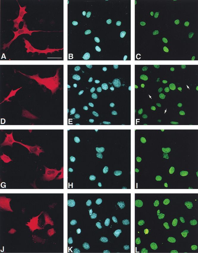
0.53]% increase of cells in G0/G1 phase and [⫺16.8 ⫾ 2.97]% proteins indicated.1804 GUARDAVACCARO ET AL. MOL. CELL. BIOL. FIG. 5. Inhibition of cyclin D1 nuclear immunofluorescence staining by ectopic PC3 in NIH 3T3 and Rb⫺/⫺ cells. (A to L) Representative immunofluorescence photomicrographs of cyclin D1 expression in NIH 3T3 (A to F) and Rb⫺/⫺ 3T3 (G to L) cells transfected with PC3 (or control -Gal). NIH 3T3 or Rb⫺/⫺ 3T3 cells (0.8 ⫻ 105) were seeded onto coverslips in 35-mm-diameter dishes and transfected with the expression vector pSCT--Gal or pSCT-PC3 (1.5 g each). After 60 h, cells were fixed, permeabilized, and stained. -Gal and PC3 proteins were detected with anti--Gal (A and G) or anti-PC3 (D and J) polyclonal antibodies, followed by goat anti-rabbit TRITC-conjugated antibody. Nuclei were stained by Hoechst 33258 dye (corresponding photomicrographs B, H, E, and K). Cyclin D1 was visualized by anti-cyclin D1 mouse monoclonal antibody, followed by goat anti-mouse FITC-conjugated antibody (corresponding photomicrographs C, I, F, and L). Arrows indicate
VOL. 20, 2000 CYCLIN D1 DOWN-REGULATION BY p53-INDUCIBLE GENE PC3 1805 independent stimuli. We also checked whether in Rb⫺/⫺ 3T3 rescence staining the expression of cyclin D1 and cyclin A in cells the inhibitory effect of PC3 on G1-S progression could be cells transfected with pSCT-PC3 or with the control vector reinstated after reintroduction of pRb by transfection (Fig. pSCT--Gal (Fig. 5A to L). Cells expressing ectopic PC3 were 2O). In fact, the percent changes in the cell populations in G1 detected by using the anti-PC3 antibody A3H (72). Again, it or S phases induced by PC3 cotransfected with exogenous pRb was observed that in both NIH 3T3 and Rb⫺/⫺ 3T3 cells (filled bars, Fig. 2O) attained about the same level seen in NIH expressing ectopic PC3 the cyclin D1 nuclear immunostaining 3T3 cells transfected with PC3 alone, if the basal effect of was detectable at a frequency significantly lower (fivefold) than exogenous pRb is subtracted (open bars, Fig. 2O). that in cells expressing ectopic -Gal (Fig. 5A to L and M, Cyclin D1 expression reverses the PC3-induced cell cycle showing the frequency values for cyclin D1 nuclear staining). block. As a whole, the above results indicate that PC3 impairs On the other hand, cyclin A expression in both NIH 3T3 and G1-to-S-phase progression by means of an active pRb, also Rb⫺/⫺ 3T3 cells transfected with PC3 remained the same as in given the ability of cyclin-CDKs to reverse the PC3-induced control cultures transfected with -Gal (Fig. 5N). dephosphorylation of pRb (Fig. 1B). PC3 down-regulates the transcription of the cyclin D1 gene. To further elucidate this aspect of the mechanism by which The above findings raised the question whether the reduction PC3 blocks cell cycle progression, we examined whether over- of cyclin D1 protein levels elicited by PC3 was a consequence expression of cyclins could overcome the inhibitory effect of of down-regulation of cyclin D1 transcription. To this aim, we PC3 on G1/S progression. To this aim, NIH 3T3 cells were analyzed the cyclin D1, A, and E mRNA levels in PC3-express- transiently transfected with PC3 in either the presence or the ing cells by semiquantitative RT-PCR. Cells expressing exog- absence of cyclins and CDKs, and the entry of cells into S enous PC3 were enriched as previously indicated by selecting phase was monitored by means of BrdU incorporation (Fig. 3). the cell population expressing the CD20 marker cotransfected While cyclins A, D3, E, B1, and B2 only partially counteracted with PC3. We observed that cyclin D1 mRNA levels were the impairment of DNA synthesis elicited by PC3 (with an significantly reduced by PC3, about 2.5- and 3-fold, compared increase of BrdU incorporation ranging from 16 up to 25 to to the levels of NIH 3T3 and Rb⫺/⫺ 3T3 control cells, respec- 40% of the basal level), cyclin D1 led to a significant (80%) tively (Fig. 6A and B). The mRNA levels of cyclin E appeared recovery of the basal level (86% when cyclin D1 was coex- to be not significantly decreased in NIH 3T3 cells and to be pressed with CDK4). These data point to cyclin D1 as an slightly increased by PC3 in Rb⫺/⫺ 3T3 cells (1.4-fold), essential component in the pathway(s) responsible for the PC3 whereas those of cyclin A were not significantly increased in inhibitory activity on the cell cycle. Generally, coexpression of both cell types (1.2-fold [Fig. 6A and B]). Thus, to assess the CDKs with cyclins and PC3 led to a recovery of BrdU incor- existence of transcriptional regulation by PC3, we analyzed the poration that was very similar to that brought about by cyclins effect of PC3 on the activity of the cloned cyclin D1 promoter, alone. This is consistent with the previous observation that transiently transfected into NIH 3T3 cells. We used the con- CDK2 and CDK4 alone did not modify the PC3-dependent struct prCD1-1810, which contains 1,810 nucleotides 5⬘ to the hypophosphorylation of pRb (Fig. 1B). transcription start in front of the luciferase reporter gene in the The ectopic expression of PC3 down-regulates cyclin D1 vector pGL2 (see reference 117). The activity of the cyclin D1 levels. The observation that cyclin D1 was able to rescue the promoter in cells cotransfected with PC3 was compared to that cell-growth-inhibitory effect of PC3, taken together with the of cells cotransfected with the empty vector. As a control of the well-established requirement for cyclin D1 in G1 progression efficiency of transfection, we measured the amount of plasmid (7), suggested that the cell cycle block imposed by PC3 could DNA present in each cell extract by dot blot hybridization, be consequent to a reduction of cyclin D1 levels. according to a previously described procedure (1). We ob- Thus, we wished to evaluate the effects of ectopic PC3 on the served that PC3 reduced the activity of cyclin D1 promoter up endogenous levels of cyclins and CDKs, by immunoblotting. to threefold, with a concentration-dependent effect (Fig. 6C). Given that the cells which took up and expressed the tran- A similar effect on the cyclin D1 promoter was also observed siently transfected PC3 were not more than 10 to 20% in our for E2F-1, known to inhibit the cyclin D1 promoter (113), used experimental conditions, to improve the detection of protein as an internal experimental control. levels the population of cells successfully transfected was en- A further analysis was performed to verify whether PC3, in riched up to 90% by flow cytometry, using the cotransfected addition to its effects on cyclin D1 transcription, could also CD20 antigen as a marker protein. It turned out that cyclin D1 affect the stability of cyclin D1 protein. To this aim, we mea- indeed was reduced by PC3 expression, in both NIH 3T3 and sured the half-life of Flag-tagged cyclin D1, cotransfected in Rb⫺/⫺ 3T3 cells (Fig. 4), about threefold, as judged by densi- NIH 3T3 cells with either pSCT-PC3 or pSCT control vector. tometry scanning. The other cyclins and CDKs analyzed did Cells were metabolically labeled with [35S]methionine, and the not show significant changes in their levels, except for a slight lysates were immunoprecipitated with an antibody to Flag (Fig. reduction of cyclin E in Rb⫺/⫺ 3T3 cells. As expected, the 7A). We observed that PC3 did not produce significant differ- levels of the different cyclins in control transfections were ences in the turnover kinetics of Flag-tagged cyclin D1 protein, similar for both NIH 3T3 and Rb⫺/⫺ 3T3 cells (43, 61). given its half-life of 23.3 ⫾ 5 and 21.5 ⫾ 4 min in the absence As an independent assessment, we verified by immunofluo- and the presence of PC3, respectively (average values ⫾ SEM the positions of nuclei negative for cyclin D1 staining. Bar, 40 m. (M) Percentage of NIH 3T3 and Rb⫺/⫺ 3T3 cells positive for cyclin D1 immunofluorescence staining after transfection with pSCT--Gal (open bars) or pSCT-PC3 (filled bars). Values are calculated as the percentages of cells positive for cyclin D1 nuclear staining, detected between cells positive for -Gal and those positive for PC3, whose total number within each experiment was assumed to be 100%. Means ⫾ SEM of three independent experiments, performed as described above for panels A to L, which are a representative field, are shown. ⴱ, P ⫽ 0.0000 versus the corresponding control (Student’s t test). The number of cells counted for each group is indicated at the top of each bar. (N) Percentage of cyclin A-positive cells by immunofluorescence staining after transfection with pSCT--Gal (open bars) or pSCT-PC3 (filled bars). Transfection and detection of PC3 and -Gal were performed as described for panels A to L. However, in order to distinguish the reactivity to the rabbit polyclonal anti-cyclin A antibody from that to either anti-PC3 (A3H) or anti--Gal (all rabbit polyclonal antibodies), cells were incubated first with anti-cyclin A and then with a mouse anti-rabbit antibody, washed, and fixed. Incubation with A3H (or anti--Gal) followed. Anti-cyclin A and anti-PC3 (or anti--Gal) antibodies were detected by goat anti-mouse FITC-conjugated and goat anti-rabbit TRITC-conjugated antibodies, respectively. Values are the means ⫾ SEM of three independent experiments. The number of cells counted for each group is indicated at the top of each bar.
You can also read