The Role of Transient Receptor Potential (TRP) Channels in the Transduction of Dental Pain - MDPI
←
→
Page content transcription
If your browser does not render page correctly, please read the page content below
International Journal of
           Molecular Sciences

Review
The Role of Transient Receptor Potential (TRP)
Channels in the Transduction of Dental Pain
Mohammad Zakir Hossain 1, * , Marina Mohd Bakri 2 , Farhana Yahya 2 , Hiroshi Ando 3 ,
Shumpei Unno 1 and Junichi Kitagawa 1
 1    Department of Oral Physiology, School of Dentistry, Matsumoto Dental University, 1780 Gobara Hirooka,
      Shiojiri, Nagano 399-0781, Japan; shumpei.unno@mdu.ac.jp (S.U.); junichi.kitagawa@mdu.ac.jp (J.K.)
 2    Department of Oral and Craniofacial Sciences, Faculty of Dentistry, University of Malaya,
      Kuala Lumpur 50603, Malaysia; marinab@um.edu.my (M.M.B.); farhanayahya@um.edu.my (F.Y.)
 3    Department of Biology, School of Dentistry, Matsumoto Dental University, 1780 Gobara, Hirooka, Shiojiri,
      Nagano 399-0781, Japan; hiroshi.ando@mdu.ac.jp
 *    Correspondence: mohammad.zakir.hossain@mdu.ac.jp; Tel./Fax: +81-263-51-2053

 Received: 26 December 2018; Accepted: 24 January 2019; Published: 27 January 2019

 Abstract: Dental pain is a common health problem that negatively impacts the activities of daily
 living. Dentine hypersensitivity and pulpitis-associated pain are among the most common types
 of dental pain. Patients with these conditions feel pain upon exposure of the affected tooth to
 various external stimuli. However, the molecular mechanisms underlying dental pain, especially
 the transduction of external stimuli to electrical signals in the nerve, remain unclear. Numerous ion
 channels and receptors localized in the dental primary afferent neurons (DPAs) and odontoblasts
 have been implicated in the transduction of dental pain, and functional expression of various
 polymodal transient receptor potential (TRP) channels has been detected in DPAs and odontoblasts.
 External stimuli-induced dentinal tubular fluid movement can activate TRP channels on DPAs
 and odontoblasts. The odontoblasts can in turn activate the DPAs by paracrine signaling through
 ATP and glutamate release. In pulpitis, inflammatory mediators may sensitize the DPAs. They
 could also induce post-translational modifications of TRP channels, increase trafficking of these
 channels to nerve terminals, and increase the sensitivity of these channels to stimuli. Additionally,
 in caries-induced pulpitis, bacterial products can directly activate TRP channels on DPAs. In this
 review, we provide an overview of the TRP channels expressed in the various tooth structures, and
 we discuss their involvement in the development of dental pain.

 Keywords: dental pain; dentine hypersensitivity; pulpitis; TRP channels; dental primary afferent
 neurons; odontoblasts; transduction mechanism

1. Introduction
     Dental pain or odontogenic pain is the pain that initiates from the teeth or their supporting
structures. The most common cause of dental pain is dental caries or tooth decay, the worldwide
prevalence of which is very high. It was reported that in 2010, dental caries in permanent teeth
remained the most prevalent global health problem, affecting 2.4 billion people, and dental caries in
deciduous teeth constituted the tenth most prevalent health condition, affecting 621 million children
worldwide [1]. Untreated dental caries lead to pulpitis (inflammation of the dental pulp) [2–6].
Typically, pulpitis is caused by invasion of the commensal oral microorganisms into the pulp due to
caries [2,3]. Irritation of the dental pulp by mechanical, chemical, thermal or electrical stimuli may
also cause pulpal inflammation [2–6]. Other causes of pulpitis include trauma, cracks on the tooth and
periodontal infections [4,6]. Symptomatic pulpitis can be an extremely painful condition and is one of

Int. J. Mol. Sci. 2019, 20, 526; doi:10.3390/ijms20030526                           www.mdpi.com/journal/ijms
Int. J. Mol. Sci. 2019, 20, 526                                                                      2 of 31

the main reasons that patients seek dental treatment [4,6,7]. It is often associated with intense lingering
pain to thermal stimuli. The pain can be spontaneous, diffuse or referred [4,6,7].
      Dentine hypersensitivity is another common odontogenic pain condition, the prevalence of which
varies widely, ranging from 3–98% [8–14]. It is characterized by short, sharp pain arising from exposed
dentine in response to stimuli—typically, thermal, evaporative, tactile, osmotic or chemical—and which
cannot be ascribed to any other form of dental defect or disease [13–15]. The dentine can be exposed by
chemical erosion, mechanical abrasion/attrition of enamel, and by loss of cementum following gingival
recession [6,13–15]. The modern lifestyle increases the consumption of acidic foods and drinks that
can result in significant tooth wear and exposure of dentine on any aspect of the tooth surface [13–17].
Dentine hypersensitivity is a special condition where dental pain arises in response to non-noxious
stimuli on the exposed dentine that normally does not elicit pain in healthy teeth [6,13–15]. Even light
tactile stimuli (weak air puff or water spray directed to the exposed dentine), which may only produce
light touch sensation on the oral mucosa or skin, provokes abrupt intense pain [6,13–15]. There are
three widely-held theories on the pathogenesis of this type of pain: (1) Dentinal fluid hydrodynamic
theory, in which it has been proposed that external stimuli cause movement of the dentinal fluid that
ultimately excites the nerve fibers in the pulp and initiates pain; (2) Neural theory, in which it has
been suggested that the nerve fibers that project into the dentinal tubules directly respond to external
stimuli; (3) Odontoblast transducer theory, in which odontoblasts themselves have been suggested
as pain transducers [13–15,18–20]. Among these, the dentinal fluid hydrodynamic theory is the most
widely accepted, although not without controversy [16,18–27]. In one study, water application onto
human dentine did not evoke pain; however, it caused dentinal tubular fluid movement in vitro [24].
Another study demonstrated a lack of correlation between dentinal fluid flow and pain in patients
after cold stimulation of the exposed dentine, suggesting that cold-sensitive receptors might also be
involved in pain transduction [26]. Recently, based on their findings, Shibukawa et al. proposed the
“odontoblast receptor hydrodynamic theory”, in which they posit that the movement of the dentinal
fluid mechanically stimulates mechanoreceptors in both odontoblasts and the nerve fibers in the
pulp [27]. Odontoblasts, movement of dentinal tubular fluid and nerves in the dental pulp may
all be involved in dentine hypersensitivity; however, the underlying mechanisms are not yet fully
understood [15].
      In addition to painful pulpitis and dentine hypersensitivity, pain may also occur when intense
thermal stimuli are applied on the surface of a normal intact tooth [28–31]. In the clinic, thermal pulp
testing (applying heat or cold onto the tooth surface) is routinely used to test the vitality of the dental
pulp of a tooth [28,29]. Thermal pulp testing induces a localized sharp pain in the tooth being tested
if the tooth is vital [28–31]. Drinking/eating of very cold or hot drink/food may also induce dental
pain [28,30,31].
      Although dental pain is a common health problem, its molecular and cellular pathophysiology,
particularly, how the external stimuli (e.g., physical, chemical or thermal) are transduced into electrical
signals in the nerve that are ultimately perceived as pain, remain unclear. Various ion channels
(e.g., voltage-sensitive Na+ , mechanosensitive K+ , L-type voltage-dependent Ca2+ channels) have
been reported to be expressed in the dental pulpal afferents and in the odontoblasts, and may play an
important role in the transduction process [32–35]. In recent decades, TRP channels have also been
detected in odontoblasts and dental primary afferent neurons (DPAs), and have been implicated in the
transduction of external stimuli into pain signals in the tooth [15,27,35,36]. This review focuses on the
involvement of TRP channels in the transduction of dental pain.

2. Dental Innervation
      Teeth are highly vascularized and richly innervated structures [6,37,38]. The nerves of a tooth
are mainly confined to the dental pulp [37,38]. The sensory stimuli-detecting nerve networks in the
dental pulp differ in many ways from those in the skin or oral mucosa [34,39]. The various types
of stimuli (e.g., mechanical, chemical or thermal) to the dental pulp or exposed dentine generally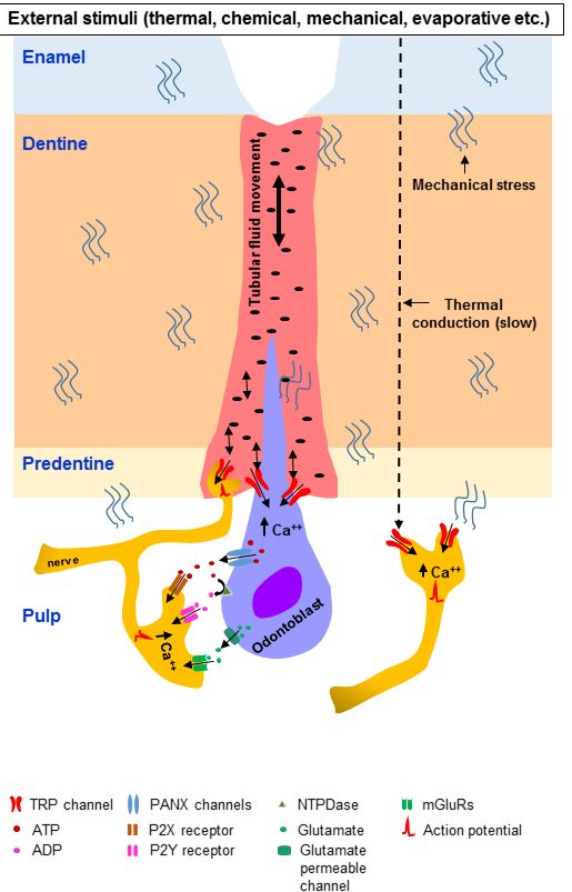
Int. J. Mol. Sci. 2019, 20, 526 3 of 31
elicit only pain sensation, while these stimuli applied to the skin or oral mucosa produce distinct
Int. J. Mol. Sci. 2018, 19, x FOR PEER REVIEW 3 of 31
types of sensation [20,34,35,39,40]. The majority of the axons of the nerves that innervate a tooth
enter the dental
dental pulp through
pulp through the apexthe apex [37,38,41,42].
[37,38,41,42]. ElectronElectron microscopic
microscopic analysis
analysis of of the
the nerve nerve
fibers fibers
within
within the dental pulp show that around 70–90% of the axons are unmyelinated
the dental pulp show that around 70–90% of the axons are unmyelinated (C-fibers) [40,41]. The (C-fibers) [40,41].
Theremaining
remaining axons
axons arearemostly
mostly thinly
thinly myelinated
myelinated (Aδ-fibers),
(Aδ-fibers), andand a very
a very small
small amount
amount areare thickly
thickly
myelinated
myelinated (Aβ-fibers)
(Aβ-fibers) [37,38,40,41]. However,
[37,38,40,41]. animal
However, studiesstudies
animal in whichin retrograde labeling techniques
which retrograde labeling
techniques
were used to were used the
evaluate to evaluate
size and thehistochemical
size and histochemical composition
composition of pulpalof pulpal sensory
sensory neurons neurons
within
within the trigeminal ganglion (TG) suggest that the parent axons of the dental
the trigeminal ganglion (TG) suggest that the parent axons of the dental pulpal nerves are largely pulpal nerves are
largely myelinated
myelinated (Figure 1)(Figure
[42–44].1) [42–44].
Figure 1. Innervation of a tooth. The cell bodies of the dental primary afferent neurons are located
Figure 1. Innervation of a tooth. The cell bodies of the dental primary afferent neurons are located in
in the trigeminal ganglion (TG). The axons of the afferent neurons project to the dental pulp through
the trigeminal ganglion (TG). The axons of the afferent neurons project to the dental pulp through the
thetwo
twomajor
majorbranches
branchesofofthe
thetrigeminal
trigeminalnerve,
nerve,namely,
namely,thethemandibular
mandibular (shown
(shown in in
thethe figure)
figure) and
and
maxillary
maxillary nerves. A large number of the parent axons of the afferent neurons before entering into thethe
nerves. A large number of the parent axons of the afferent neurons before entering into
dental pulp
dental areare
pulp myelinated.
myelinated.After
Afterentering
enteringthe
the dental
dental pulp, they extend
pulp, they extendbranches
branchesandandgradually
gradually lose
lose
their myelin sheath. In the crown area, the axons branch extensively to form the plexus
their myelin sheath. In the crown area, the axons branch extensively to form the plexus of Raschkow. of Raschkow.
Many
Manyaxons terminate
axons terminate very
veryclose
closetotothe
theodontoblasts
odontoblasts and sub-odontoblasticcells,
and sub-odontoblastic cells,and
andsomesome enter
enter thethe
dentinal tubules for a short distance into the inner part of the dentine.
dentinal tubules for a short distance into the inner part of the dentine.
It has been
It has observed
been observedthat
thatthe
theratio
ratioof
ofmyelinated
myelinated axons to unmyelinated
axons to unmyelinatedaxons
axonsisisreduced
reducedin in
nerves closer to teeth, compared with more distant
nerves closer to teeth, compared with more distant sites, indicating progressive loss of the myelin
indicating progressive loss of the myelin
sheath as as
sheath axons course
axons coursetoward
towardthe
thetooth
tooth[44,45].
[44,45]. Using electron
electronmicroscopic
microscopicanalysis,
analysis,a study
a study reported
reported
that the percentage of unmyelinated axons was higher in the crown area compared with the root areaInt. J. Mol. Sci. 2019, 20, 526 4 of 31
that the percentage of unmyelinated axons was higher in the crown area compared with the root area
in rat molars [46]. In that study, the unmyelinated axons of the dental pulp showed immunoreactivity
to a marker of myelinated nerves, neurofilament (NF)-200, and the percentage of NF-200-positive
unmyelinated axons was greater in the crown area than in the root area [46]. The conduction velocity
in the nerve fibers outside the dental pulp was higher than in the nerve fibers located inside the
pulp [47,48]. A study on human teeth also found that unmyelinated nerve fibers within the dental pulp
show immunoreactivity to markers for myelinated nerves (neurofilament-200, neurofilament-52) [49].
Furthermore, studies have reported that the majority of parent axons before entering the dental pulp
are thinly myelinated (Aδ), with a higher conduction velocity compared with nerve fibers inside the
dental pulp [44,47–49]. These findings suggest that many parent axons in the dental pulp afferents
are myelinated. Consistently, studies have reported that the cell bodies of the majority of the afferent
fibers from the dental pulp that are located in the TG are medium-sized (Aδ-neurons), while a minority
are large (Aβ-neurons) or small (C-neurons) [42,43]. After entering the dental pulp through the apex
of the tooth, the parent axons of the afferent nerves extend branches and gradually lose their myelin
sheath [43,46,49,50] (Figure 1). In the radicular pulp, the axons give off only a few branches, but in
the coronal pulp, the axons branch extensively to form the plexus of Raschkow (Figure 1) [6,37,38,41].
The axons lose their myelin sheath mostly in the coronal pulp and emerge as free nerve endings (brush
or fan-shaped) [49–51]. Many axons terminate very close to the odontoblasts, as well as sub- and
peri-odontoblastic cells, and some enter the dentinal tubules and continue along odontoblast processes
for a short distance, no further than 0.2 mm into the inner part of the dentine [51]. Some axons make
endings in both the pulp and the dentinal tubule [51]. It has been reported that around 30–70% of
odontoblast processes of a tooth are in close association with nerve endings in the inner part of the
dentine [50]. The dental pulp also contains sparsely distributed sympathetic postganglionic efferent
nerve fibers (unmyelinated), which mostly innervate the blood vessels, while parasympathetic nerve
fibers have not been observed [40,41,52,53].
Cytochemically, the DPAs are distinct from the afferent neurons of the skin [34,39,43]. Generally,
in the skin, the afferent neurons can be clearly divided into two major groups—peptidergic
and non-peptidergic [54,55]. The peptidergic neurons express a variety of neuropeptides and
signaling proteins, including calcitonin gene-related peptide (CGRP), substance P, nerve growth
factor (NGF) receptor and tyrosine kinase A (TrkA) receptor, and these neurons are responsive to
NGF. In comparison, the non-peptidergic neurons express isolectin B4 (IB4), glial cell line-derived
neurotrophic factor (GDNF) family receptor alpha-1 (GFRα1) and receptors for other GDNF family
members, and these neurons are responsive to GDNF [32,55–57]. The non-peptidergic neurons also
express purinergic receptors (P2X3, a receptor for ATP) [58–60]. The pulpal afferents cannot be
categorized into these two broad groups. They display the cytochemical features of both peptidergic
and non-peptidergic neurons, which is rare for skin somatosensory afferents [61,62]. Although
most pulpal afferents do not express IB4, they express other markers of non-peptidergic neurons
such as GFRα1 and P2X3 [61,62], and a large group of pulpal afferents also express markers for
peptidergic neurons such as CGRP, NGF receptor, substance P and TrkA receptors [63–65]. Another
unique feature of dental pulp afferents is that they express markers for both mechanoreceptors and
nociceptors [39,43,49,61,62]. It has been observed that pulpal nerve fibers express various cytochemical
markers of mechanosensitive nerve fibers, such as neurofilament (NF) markers (e.g., NF-200,
NF-52), parvalbumin, calbindin, epithelial sodium channels (ENaCs), acid-sensing ion channel 3
(ASIC3) and mechano-gated potassium channels [32,34,39,43,61,62,66,67]. They also express various
cytochemical markers of nociceptive nerve fibers in the skin, such as CGRP, GFRα1, TrkA and substance
P [39,43,61,68,69].
3. TRP Channels and Their Presence in Dental Tissues
TRPs are integral pore-forming membrane proteins that function primarily as non-selective ion
channels [70–73]. They have a putative six-transmembrane-spanning protein domain with a poreInt. J. Mol. Sci. 2019, 20, 526 5 of 31
region localized between transmembrane segments 5 and 6 [70–73]. They were discovered in the fruit
fly (Drosophila) in studies of their phototransduction (light detection) mechanism [74–76]. Later, they
were found in vertebrates, and to date, seven TRP subfamilies have been identified: TRPA (ankyrin),
TRPC (canonical), TRPM (melastatin), TRPML (mucolipin), TRPP (polycystin), TRPN (Drosophila
no mechanoreceptor potential C or NOMPC) and TRPV (vanilloid) [70–73,77]. Recently, the TRPY
subfamily was reported in yeast [78]. TRPN and TRPY are absent in mammals [70–73,77]. The TRPV
subfamily has six members (TRPV1–6), TRPA has one member (TRPA1), TRPC has seven members
(TRPC1–7), TRPM has eight members (TRPM1–8), TRPML has three members (TRPML1–3), TRPP has
three members (TRPP1–3), and TRPN has one member (TRPN1, found in fish) [70–73,77]. Among the
28 TRP channel genes that have been identified in mammals, 17 have been detected in the mouse TG
at the mRNA level [79]. Many TRP channel genes have also been identified in the human TG [80].
To date, to our knowledge four members of the TRPV subfamily (TRPV1, TRPV2, TRPV3, TRPV4),
four members of the TRPM subfamily (TRPM2, TRPM3, TRPM7, TRPM8), two members of the TRPC
subfamily (TRPC1, TRPC6), and one member of the TRPA subfamily (TRPA1) have been detected in
dental tissues (e.g., DPAs, odontoblasts).
3.1. TRPV
Among the six members of the TRPV subfamily, TRPV1–4 are weakly Ca2+ -selective cation
channels [70–73]. They can be activated by thermal stimulation, and are therefore referred to as
thermo–TRPs [70–73,81]. In vitro studies show that TRPV1–4 can be activated by temperatures ranging
from ~34 (TRPV4) to ~52 ◦ C (TRPV2) [81]. The temperature range for channels activation is not strict,
and a thermal threshold is not the optimal parameter to describe thermo-TRPs, because, the sensitivity
for thermal activation of these channels is substantially modified by cellular and environmental
factors [82–84]. For example, TRPV1 (normal activation temperature ~43 ◦ C) can be activated at
much lower temperatures when the membrane depolarized than when it hyperpolarized [82,83].
TRPV5–6 are highly Ca2+ -selective and are not activated by heat. They play an important role in Ca2+
homeostasis [70–73].
TRPV1, which is also known as the capsaicin receptor, was the first member of the TRPV
subfamily to be isolated [78]. It is activated by various exogenous and endogenous stimuli, such as
capsaicin (found in hot chili peppers), acids (pH < 5.9), heat, inflammatory mediators (e.g., bradykinin,
prostaglandins), NGF, anandamide (arachidonoyl ethanol amide), arachidonic acid metabolites
(e.g., N-arachidonoyl-dopamine), lipoxygenase products (e.g., 12-hydroperoxyeicosatetraenoic acid),
adenosine and ATP [85–91]. The threshold for activation of TRPV1 is dynamic [87,90,91]. For example,
the threshold is decreased by inflammatory mediators, but after activation by capsaicin, there is a
sustained refractory state (desensitization) [70–73,87,90,91]. Activation of TRPV1 has been shown to
promote the release of neuropeptides such as substance P and CGRP [70–73,87,91]. TRPV1 is expressed
predominantly in C-fibers and, to a lesser extent, in Aδ fibers [70–73,87,91]. In addition to sensory
neurons, TRPV1 expression has been detected in various other tissues, including keratinocytes of the
skin and oral mucosa, epithelium of the respiratory system, digestive tract, urinary bladder, cardiac
muscle, vascular smooth muscle, and the endothelium of blood vessels [87,92]. TRPV1 has been
reported to play an important role in thermal nociception [70–73,87,91]. The importance of TRPV1
in pain sensation has been demonstrated by studies in TRPV1 knockout mice [93]. It plays a role
in many physiological functions, including satiety, olfaction, gastrointestinal motility, and energy
homeostasis [72,87,92–95].
TRPV2 channels share 50% sequence identity with TRPV1 [70,73,81,96]. This channel has been
reported to function as a thermoreceptor, mechanoreceptor and osmoreceptor [70,73,81,96,97]. TRPV2
is activated by noxious temperature (~52 ◦ C) in vitro [97–99]. However, in vivo, TRPV2 knockout
mice exhibit a thermal response similar to wild-type mice [100]. TRPV2 is expressed in the peripheral
and central nervous systems [70,73,81,97,99,101–104]. In the spinal dorsal root ganglia (DRG) and
in the trigeminal ganglia (TG), expression of TRPV2 was observed in the medium to large-diameterInt. J. Mol. Sci. 2019, 20, 526 6 of 31
primary afferent neurons [97,99,102–104]. In culture, one-third of TRPV2-expressing DRG neurons
are CGRP-positive, and activation of these neurons by a TRPV2 agonist results in CGRP release [102].
In the central nervous system, TRPV2 expression has been observed in a number of regions that are
involved in osmoregulation and other autonomic functions [97,99,101].
TRPV3 can be activated by innocuous temperature (31–39 ◦ C), various natural compounds (such as
camphor and carvacrol), synthetic agents (such as 2-aminoethoxydiphenyl borate), and the endogenous
ligand farnesyl pyrophosphate (FPP) [105–108]. TRPV3 expression in sensory neurons of the peripheral
nervous system is not consistent across species, and it is minimally expressed in sensory neurons in
the rodent; however, the channel is relatively abundantly expressed in skin keratinocytes [109–112].
TRPV3 is also expressed in the epithelial cells of the oral and nasal cavities, as well as the cornea, where
it is reported to be involved in wound healing and thermo-sensation [111,112]. TRPV3 in keratinocytes
is reported to be involved in nociception mediated by ATP [110]. Indeed, increased pain sensitivity is
observed in transgenic mice overexpressing TRPV3 in skin keratinocytes [113].
TRPV4 was identified as an osmoreceptor [97,114–116]. Later, it was shown to be a polymodal
receptor that can be activated by various stimuli, including innocuous warm temperature (27–35 ◦ C)
and mechanical stimuli (membrane stretch and shear stress) [97,117–121]. This channel can
be activated by arachidonic acid metabolites, synthetic chemical agents such as 4α-phorbol
12,13-didecanoate, GSK1016790A, and the plant extract bisandrographolide A [70,72,73,122]. TRPV4
has also been implicated in nociception, itch, inflammatory pain, neuropathic pain and visceral
pain [70,72,73,123–127]. Expression of TRPV4 is observed in DRG neurons [123,126,127] and
TG [121,128–130] neurons. It is also expressed in satellite glial cells (cells surrounding the neurons) in
the DRG that regulate neuronal excitability in pain and inflammatory conditions [131]. Inflammatory
mediators (e.g., prostaglandins and proteases) may sensitize TRPV4, resulting in hyperalgesia [123,132].
Furthermore, escape latency in the hot plate test is increased following tissue injury and inflammation
in TRPV4 knockout mice, suggesting involvement of TRPV4 in thermal hyperalgesia [124].
3.1.1. TRPV in DPAs
TRPV channels have been detected in the cell bodies of DPAs located in the TG [133–138]. Among
the six members of the TRPV subfamily, TRPV1 has been the most extensively studied [133–141].
In animals, the percentage of TRPV1-expressing dental pulp TG afferent neurons varies among
the published papers from 8% to 85%, and TRPV1 expression is observed in small, medium and
large neurons [133–138]. In 2001, Ichikawa & Sugimoto reported that approximately 8% of rat
DPAs express TRPV1, and that 20% of these co-express the neuropeptide CGRP [133]. They also
compared TRPV1-immunoreactive afferent neurons in the dental pulp and the facial skin [133].
The percentage of TRPV1-immunoreactive neurons was lower in the dental pulp (17%) than in the facial
skin (26%) [133]. The same year, Chaudhary et al. reported TRPV1 mRNA expression in the cell bodies
of DPAs in the TG, and 65% of these cells were excited by capsaicin. The capsaicin-evoked excitation
was attenuated by a TRPV1 antagonist (capsazepine) [134]. Similar to Ichikawa & Sugimoto [133],
Chung et al. reported that approximately 10% of the dental pulpal afferent neurons expressed TRPV1,
and that the majority of these were small to medium neurons [135]. They also observed that TRPV1
expression was upregulated by application of lipopolysaccharides (a product of Gram-negative
bacteria) into the dentine [135]. Stenholm et al. reported that 21–34% of dental pulpal afferent
neurons were TRPV1-immunoreactive, slightly higher than among gingival primary afferent neurons
(21–26%) [136]. Gibbs et al. found that approximately 17% of the afferent neurons in the TG from
the dental pulp expressed TRPV1, lower than the percentage of TRPV1-immunoreactive afferent
neurons from the periodontal ligament (26%) in the rat [104]. They also found that 70% of the
TRPV1-immunoreactive neurons were myelinated, and that a majority (82%) of these were medium to
large-diameter neurons [104]. Furthermore, 60% of the TRPV1-immunoreactive neurons co-expressed
CGRP [104]. A high percentage of TRPV1-immunoreactive DPAs was observed by Kim et al. [137] and
Park et al. [138]. Kim et al. showed that 85% of the retrograde-labeled rat DPAs were immunoreactiveInt. J. Mol. Sci. 2019, 20, 526 7 of 31
for TRPV1, and that 71% of them produced inward cationic currents in response to application of a
TRPV1 agonist (capsaicin) [137]. Park et al. observed that 45% of the DPAs expressed TRPV1 [138].
They also showed that application of a TRPV1 agonist increased the intracellular Ca2+ concentrations
and produced inward currents in these neurons. Temperature changes (>42 ◦ C) also increased the
intracellular Ca2+ concentrations and produced inward currents [138].
Expression of TRPV1 has been reported in the nerve fibers within the dental pulp in animal and
human studies [137,139–141]. Kim et al. [137] found TRPV1-immunoreactive nerve fibers within the
rat dental pulp; however, Gibbs et al. did not observe TRPV1-immunoreactive nerve fibers within the
dental pulp of rat molars [104]. TRPV1 immunoreactivity has also been detected in the dental pulp
nerve fibers of the human permanent teeth [139–141], and immunoreactivity is significantly increased
in carious teeth compared with non-carious teeth [141]. Interestingly, TRPV1 expression tended to be
increased in painful carious teeth compared with non-painful carious teeth, although the difference
was not significant [141].
TRPV2 is expressed in the nerves within the dental pulp and in the cell bodies of the DPAs
located in the TG [103,104,136]. In 2000, Ichikawa & Sugimoto reported TRPV2-immunoreactive nerve
fibers within the rat dental pulp [103]. In the root area, TRPV2 immunoreactivity was observed in
the nerve bundles, and in the crown area, the TRPV2-immunoreactive nerve fibers were ramified
and extended to the base of the odontoblastic cell layer [103]. These investigators also observed that
approximately 37% of the dental primary afferent neurons retrogradely traced in the TG expressed
TRPV2, and that most of these were medium to large neurons [103]. In addition, 45% of these
neurons co-expressed CGRP, and 41% co-expressed parvalbumin (a marker of proprioceptors) [103].
Furthermore, the percentage of TRPV2-immunoreactive afferent neurons (37%) was greater than the
percentage of TRPV2-immunoreactive facial skin afferent neurons (9%) [103]. Stenholm et al. reported
that 32–51% of DPAs expressed TRPV2, somewhat higher than the percentage of TRPV2-expressing
gingival afferent neurons (41%) [136]. Gibbs et al. reported that a higher percentage of rat DPAs
expressed TRPV2 compared with TRPV1; 50% were immunoreactive for TRPV2, while 17% were
immunoreactive for TRPV1 [104]. They also observed that TRPV2 expression was higher among
DPAs (50%) compared with primary afferent neurons from the periodontal ligament (41%) [104].
Approximately 83% of the TRPV2-immunoreactive neurons were myelinated, and the majority of these
were medium to large neurons [104]. Around 33% of the TRPV2-immunoreactive neurons co-expressed
CGRP [104].
TRPV3 and TRPV4 are expressed in the TG on afferent neurons from the facial skin and
temporomandibular joint [105,129,130]. TRPV4 expression is also observed on the nerves of human
dental pulp, and expression is upregulated by chronic inflammation of the pulp [142].
3.1.2. TRPV in Odontoblasts
In 2005, Okumura et al. reported expression of TRPV1 on odontoblasts in neonatal rat
teeth [143]. They also showed a capsaicin-induced inward current in the odontoblasts using the
patch clamp technique that was inhibited by capsaizepine [143]. However, in acutely isolated
adult rat odontoblasts, Yeon et al. did not observe expression of TRPV1 by immunohistochemistry,
and intracellular Ca2+ concentration in the odontoblasts was not increased by application of heat
(42 ◦ C) or a TRPV1 agonist (capsaicin) [144]. They also did not detect TRPV1 or TRPV2 mRNA
in the odontoblasts [144]. In contrast, Tsumura et al. reported expression of TRPV1 in adult
rat odontoblasts, on the cell membrane and on their processes [145]. Application of an agonist
(capsaicin/resiniferatoxin/low pH solution) increased the intracellular Ca2+ level, which was inhibited
by application of antagonists [145]. They also reported that the TRPV1 channels in odontoblasts are
functionally coupled with cannabinoid receptor 1 and Na+ –Ca2+ exchangers, and that this coupling
is mediated by cyclic adenosine monophosphate (cAMP) [145]. TRPV1 has also been detected on
the odontoblasts of extracted healthy caries-free human premolar teeth [139]. mRNAs for TRPV1,
TRPV2, TRPV3 and TRPV4 were detected in acutely isolated cultured odontoblasts by Son et al. [146].Int. J. Mol. Sci. 2019, 20, 526 8 of 31
They also showed that when odontoblasts were stimulated by heat above 32 ◦ C, the intracellular
Ca2+ concentration increased, suggesting functional involvement of TRPV1, TRPV2 and TRPV3 in
the transduction of heat stimuli [146]. In addition, the intracellular Ca2+ concentration was increased
when hypotonic solution was applied, suggesting involvement of TRPV4 channels [146]. Sato et al.
reported expression of TRPV2 and TRPV4 proteins in rat odontoblasts [147]. They also showed
that extracellular hypotonic solution-induced membrane stretching in cultured mouse odontoblast
lineage cells produces inward currents and increases intracellular Ca2+ concentration, which can be
inhibited by application of TRPV1, TRPV2 and TRPV4 channel antagonists, suggesting that these
channels function as mechanoreceptors in odontoblasts [147]. TRPV1 and TRPV4 expression has
been reported by immunohistochemistry, PCR and western blot analysis in human odontoblast-like
cells derived from the dental pulp of a permanent tooth [148,149]. Application of agonists to these
channels increases the intra-odontoblast Ca2+ concentration. Hypotonic solution-induced membrane
stretch also increases the intra-odontoblastic Ca2+ concentration, which can be reduced by channel
antagonists [148,149]. Brief (10 min) application of the pro-inflammatory cytokine tumor necrosis factor
(TNF)-α enhances the response to chemical agonists and hypotonic solution-induced membrane stretch,
suggesting that inflammation increases TRPV4 channel activation [148,149]. TRPV4 immunoreactivity
has been detected in odontoblasts and their process in extracted healthy caries-free immature human
third molar teeth [149]. Wen et al. reported expression of TRPV1, TRPV2 and TRPV3 in native
human odontoblasts as well as cultured odontoblast-like cells from healthy human third molars
by immunohistochemistry, quantitative real-time polymerase chain reaction (qRT-PCR), western
blotting and immunoelectron microscopy [150]. Immunoelectron microscopy revealed expression of
TRPV1, TRPV2 and TRPV3 in odontoblastic processes, mitochondria and endoplasmic reticulum [150].
Egbuniwe et al. reported expression of TRPV1 and TRPV4 mRNAs in odontoblast-like cells derived
from human immortalized dental pulp cells. They observed that the application of a TRPV4 agonist
increased the intra-odontoblastic Ca2+ concentration. They also observed that the agonist caused
release of ATP from the odontoblasts, which was blocked by pretreatment with the antagonist [151].
In odontoblast-like cells obtained from cultured dental pulp cells from newborn rats, mechanical
stimulation increases the intracellular Ca2+ concentration, and this effect can be attenuated by
application of TRPV1, TRPV2 and TRPV4 antagonists, suggesting that these channels function as
mechanosensors in these cells [27].
3.2. TRPM
TRPM8 has been detected in DPAs and pulpal fibroblasts. TRPM3 and TRPM7 are expressed in
odontoblasts, while TRPM2 is expressed in pulpal fibroblasts. TRPM2 is activated by cellular stress
and participates in various cellular functions, including cytokine production, cell motility and cell
death [152,153]. It can be activated by cytosolic adenosine diphosphate ribose (ADPR), oxidative stress
and moderate heat in various cell types [152–155]. TRPM2 expression has also been detected in afferent
sensory neurons in the DRG and TG [79]. This channel is implicated in pathogenic pain [156].
TRPM3 is expressed in non-neural (e.g., epithelium of the kidney, pancreatic β cells) and
neural tissues, including sensory neurons in the DRG and TG [157–161]. TRPM3 can be
activated by extracellular hypo-osmolarity, noxious heat (>30 ◦ C) and chemical compounds, such
as pregnenolone sulfate (endogenous excitatory neurosteroid) and nifedipine (a drug used for
hypertension) [157,160,161]. TRPM3 knockout mice exhibit significant attenuation of thermal
hyperalgesia under inflammatory conditions [160].
Similar to other TRP channels, TRPM7 is expressed in a wide variety of tissues, including the
brain, heart and hematopoietic tissues [162–166]. TRPM7 has been implicated in multiple cellular
and physiological functions, including embryonic development, Mg2+ homeostasis, cell growth and
viability, synaptic transmission, and neuronal degeneration [163–166].
TRPM8 is activated by innocuous cooling (~26−15 ◦ C) as well as by noxious cooling (Int. J. Mol. Sci. 2019, 20, 526 9 of 31
small-diameter sensory neurons in the TG and DRG [169–172]. TRPM8 mRNA is more abundantly
expressed in the sensory afferents of the TG (especially those that innervate the tongue) compared with
the DRG [169–172]. While TRPM8 knockout mice do not lack sensation to noxious cold, they exhibit an
attenuation of avoidance behavior to moderately cold temperatures [173,174]. These mice also display
reduced cold hypersensitivity following nerve injury or complete Freund’s adjuvant (CFA)-induced
inflammation [175].
3.2.1. TRPM in DPAs
TRPM8 is expressed in rat DPAs in the TG [137,138,176]. By immunohistochemistry, Park et al.
observed that 13% of rat DPAs expressed TRPM8 [138]. Application of menthol (a TRPM8 agonist) or
exposure to temperatures less than 25 ◦ C increase intracellular Ca2+ concentrations and evoke inward
currents in the TRPM8-expressing neurons [138]. In one study, TRPM8 mRNA expression was detected
in 58% of rat DPAs [137]. However, Michot et al. detected TRPM8 expression in only 5.7% of mouse
DPAs, similar to that in afferent neurons innervating the facial skin and the buccal mucosa [176].
3.2.2. TRPM in Odontoblasts
Expression of TRPM3, TRPM8 and TRPM7 in odontoblasts has been reported in numerous
studies [146,148,177–182]. Son et al. detected mRNA expression of osmo-sensitive TRPM3 channels
in acutely isolated cultured odontoblasts from neonatal mice [146]. They also suggested that TRPM3
in these cells are involved in the transduction of osmotic stimuli, based on their finding of increased
intra-odontoblastic Ca2+ concentration following exposure to hypotonic solution [146]. However,
these investigators did not observe functional expression of TRPM8 [146]. TRPM8 expression was
also not observed by Yeon et al. in acutely isolated adult rat odontoblasts at the mRNA level [144].
They showed that application of cold stimuli (12 ◦ C) or menthol (TRPM8 agonist) did not increase the
intra-odontoblastic Ca2+ concentration [144]. In contrast, Tsumura et al. reported functional expression
of TRPM8 in acutely isolated adult rat odontoblasts [177]. TRPM8 immunoreactivity was observed
in the odontoblasts and their processes [177]. Chemical agonists (e.g., menthol, WS3, WS12) and
temperature changes (22 ± 1 ◦ C) also increased the intra-odontoblastic Ca2+ levels, and these changes
could be reduced by an antagonist [177]. TRPM8 expression was also reported in odontoblast-like
cells from the dental pulp of a human permanent tooth at both the mRNA (using PCR) and protein
levels (using western blotting and immunohistochemistry) [148]. Chemical agonism also increased
the intra-odontoblastic Ca2+ concentration in an antagonist-sensitive manner [148]. TRPM8 has also
been detected in freshly isolated human odontoblasts at the mRNA and protein levels as well as in
odontoblasts and their processes in extracted healthy caries-free human permanent teeth [178].
By single-cell RT-PCR and immunocytochemical analysis, TRPM7 was detected in acutely isolated
rat odontoblasts [179,180]. Functionality of TRPM7 as a mechanoreceptor in acutely isolated rat
odontoblasts was reported by Won et al. [180]. In their study, hypotonic solution-induced membrane
stretch or chemical agonism for TRPM7 caused a transient increase in intra-odontoblastic Ca2+
concentration, which was blocked by a non-selective mechanosensitive channel blocker or a TRPM7
blocker [180]. TRPM7 in odontoblasts has also been reported to play an important role in dentine
mineralization by regulating intracellular Mg2+ and alkaline phosphatase activity [181,182].
3.3. TRPA
The only member of the TRPA subfamily, TRPA1 is generally involved in pain, thermal and
chemical sensation [70,73,91,97]. It is expressed in many tissues, including the brain, heart, small
intestine, lung, bladder and joints [183–185]. It is highly expressed in small and medium neurons in
the DRG and TG [91,183,186–188]. TRPA1 channels are co-localized with TRPV1, CGRP, substance
P and bradykinin receptors [186,189–191]. TRPA1 is activated by noxious cold (Int. J. Mol. Sci. 2019, 20, 526 10 of 31
cinnamaldehyde (found in cinnamon), allicin (found in garlic) and acrolein (present in diesel
exhaust) [191–193].
3.3.1. TRPA in DPAs
In rat DPAs (labeled using retrograde dye applied to the dental pulp), TRPA1 was observed
in 11% of neurons [138]. Chemical stimulation by icilin (a TRPA1 and TRPM8 agonist) and cold
stimulation (Int. J. Mol. Sci. 2019, 20, 526 11 of 31
3.4. TRPC
TRPC1 and TRPC6 have been reported to be expressed in odontoblasts [179,197,198]. TRPC1 plays
an important role in store-operated Ca2+ entry (SOCE) in a variety of tissues and cell types [199,200].
TRPC1 is highly expressed in the hippocampus, amygdala, cerebellum, substantia nigra and inferior
colliculus [201–203], and participates in important neuronal processes related to synaptic transmission
and plasticity [203]. TRPC1 expression is observed on large myelinated sensory neurons in the
DRG [204], and it and TRPC6 play critical roles in mechanosensation, hearing [205–208] and cell
membrane stretch [206,207]. TRPC1 and TRPC6 are co-expressed with TRPV4 in the DRG, where they
are implicated in mechanical hypersensitivity in inflammatory and neuropathic pain conditions [208].
TRPC in Odontoblasts
TRPC1 and TRPC6 mRNA expression has been reported in acutely isolated rat odontoblasts,
where they have been suggested to play a role as mechanoreceptors [179]. TRPC1 and TRPC6
immunoreactivity is also detected on odontoblasts in human permanent teeth. TRPC6 protein
expression increases with time during odontoblast differentiation from pulp tissue. Furthermore,
differentiation is inhibited by downregulation of TRPC1 and TRPC6 expression, indicating involvement
of these channels in the odontogenic differentiation of human dental pulp cells [197,198].
3.5. TRP Channels in Pulpal Fibroblasts and Blood Vessels
Expression of TRPV1 mRNA and protein has been reported in human dental pulp fibroblasts
cultured from the pulp of third molar teeth [209], and application of the agonist capsaicin induces
production of IL-6 (an inflammatory cytokines) in these cells, which is dose-dependently inhibited
by the TRPV1 antagonist capsazepine, indicating involvement of this channel in fibroblast-mediated
pulpal inflammation [209]. TRPV1 expression was also observed in pulpal fibroblasts of healthy
caries-free human permanent teeth [139]. Karim et al. reported mRNA and protein expression of
TRPA1 and TRPM8 in human dental pulp fibroblasts [210]. Application of the TRPM8 agonist menthol
or cool temperature (22 ± 3 ◦ C) in combination with the TRPA1 agonist cinnamaldehyde or noxious
cold temperature (12 ± 2 ◦ C) increased the intra-fibroblastic Ca2+ concentration, suggesting that TRPA1
and TRPM8 in pulp fibroblasts are involved in sensing environmental stimuli [210]. TRPM2 has also
been detected in the fibroblasts of human dental pulp, and its expression is upregulated in teeth with
signs of irreversible pulpitis [211].
4. Involvement of TRP Channels in the Transduction of Dental Pain
TRP channels in odontoblasts and DPAs are suggested to function as polymodal receptors
involved in the transduction of various external stimuli. TRPV1, TRPV2, TRPV4, TRPM7 and TRPA1 in
odontoblasts have been implicated as mechanoreceptors [27,146–149,151,180]. In an odontoblast/TG
co-culture preparation, mechanical stimulation of the odontoblast-like cells increased intracellular Ca2+
concentration in the mechanically-stimulated odontoblasts as well as in neighboring neuron-like
cells [27]. The increase in intracellular Ca2+ in these cells was almost completely abolished by
application of a cocktail of TRPV1, TRPV2, TRPV4 and TRPA1 channel antagonists [27]. In addition,
hypotonic solution-induced mechanical stretching of the cell membrane increases intracellular Ca2+ in
cultured odontoblast-like cells [146–149,177,180], which is inhibited by antagonists of TRPV1, TRPV2,
TRPV4 [146,148], TRPA1 [177] and TRPM7 [180], further suggesting that these channels function as
mechanoreceptors and/or osmoreceptors.
Mechanosensitive TRP channels along with other mechanoreceptors expressed in odontoblasts
and DPAs can be activated by application of thermal stimuli to an intact tooth. A study on human
subjects showed that when a thermal stimulus was applied to a tooth surface, the subjects rapidly
sensed pain (1.28 s for hot stimuli and 1.49 s for cold stimuli), even before a noticeable change in
temperature at the wall of the dental pulp (3.68 s) [28,30]. Thermal stimulation on the tooth surfaceInt. J. Mol. Sci. 2019, 20, 526 12 of 31
can induce fluid movement in the dentinal tubules because of thermal expansion or contraction of
the fluid [18,212]. Interestingly, fluid movement was observed before a change in temperature in the
dentine, where the dentinal tubules are located [21,22]. A temperature gradient between the enamel
and dentine is observed when a thermal stimulus is applied on the surface of a tooth [21–23,25].
Tooth structures expand or contract because of this thermal gradient, which produces mechanical
stresses in these structures [21–23,25,28]. Mechanical deformation of the dentine (including pulpal
wall dentine) precedes the temperature changes in the dentine/dentine–enamel junction following
thermal stimulation of the surface a tooth [22,25]. These observations suggest that intense thermal
stimulation-induced mechanical deformation of the dentine may exert mechanical stresses on the
odontoblasts as well as on the pulpal tissues. In turn, the mechanical stresses may directly activate the
mechanosensitive TRP channels and other mechanoreceptors present in the odontoblasts and pulpal
nerve fibers (Figure 2).
The sharp pain perceived soon after thermal stimulation of the tooth surface may be at least partly
attributed to the mechanical stress-induced activation of mechanoreceptors on odontoblasts and DPAs.
Temperature changes in the dentine caused by thermal stimulation of the surface of an intact tooth
or a tooth with exposed dentine may also cause expansion/contraction and movement of dentinal
tubular fluid, which can activate mechanosensitive TRP channels (along with other mechanoreceptors)
on odontoblasts and nerve fibers within/near the dentinal tubules (Figure 2).
Intense thermal stimulation of the tooth surface may cause vasodilation and changes in pulpal
blood flow [213–215]. A study using dogs found that pulpal blood flow increased slightly and
gradually when the tooth surface temperature was raised from 35 to 40 ◦ C, and that it increased
sharply when the temperature was raised from 35 to 55 ◦ C [213,214]. The increase of pulpal blood flow
by thermal stimulation can increase the pressure within the pulpal tissue [213,214], which can excite
mechanoreceptors, including TRP channels, in the pulpal nerves.
Patients with dentine hypersensitivity may feel pain caused by osmotic changes. Osmotic
stimulation of the exposed dentine may induce movement of dentinal tubular fluid [18,19], which can
excite mechanosensitive TRP channels on nearby odontoblasts and nerve fibers (Figure 2). It has been
observed that dentine covered with a smear layer is much less responsive to hypertonic solutions than
dentine devoid of a smear layer, suggesting that fluid movement is greater when the smear layer is
absent [216].
Patients with dentine hypersensitivity may also feel pain after very light mechanical stimulation,
such as air puffs or water spray, on the exposed dentine, which may induce very little movement
of the dentinal tubular fluid. This suggests the presence of low-threshold mechanoreceptors on
odontoblasts and DPAs [39]. Fried et al. proposed that many DPAs may contain low-threshold
mechanoreceptors [39]. They termed these “algoneurons” [39]. The researchers suggested that these
algoneurons may transduce the nociceptive signal in teeth [39]. Indeed, activation of these neurons
causes activation of trigeminal brainstem neurons that deliver a pain message to higher brain centers,
resulting in the sensation of pain [39]. Similar low-threshold mechanoreceptors in the skin or oral
mucosa generally signal tactile sensation (e.g., light touch) [217]. Air puff on the exposed dentine
may also dehydrate the dentine surface and cause outward flow of the dentinal tubular fluid, which
can activate mechanosensitive receptors on odontoblasts and nerve fibers within/near the dentinal
tubules [18,19]. Mechanosensitive TRP channels may function as mechanoreceptors [218] to transduce
these types of stimuli.
When intense thermal stimuli are applied on the tooth surface, they can increase the temperature
at the dentine–pulp border (albeit slowly), which may excite the thermosensitive TRP channels on
odontoblasts and DPAs (Figure 2). Indeed, a slow increase in temperature to >43 ◦ C on the tooth
surface activates intra-pulpal C-fibers in the cat [219,220]. Another study showed that intra-dental
A-δ and C-fibers respond to intense cooling of the tooth surface [31]. Thermosensitive TRP channels
expressed on the A-δ and C-fibers in the dental pulp can be activated by high-temperature stimulation
of the tooth surface. Heat and cool/cold stimuli increase intracellular Ca2+ concentrations and elicitInt. J. Mol. Sci. 2018, 19, x FOR PEER REVIEW 12 of 31
Int. J. Mol. Sci. 2019, 20, 526 13 of 31
stimulation-induced mechanical deformation of the dentine may exert mechanical stresses on the
odontoblasts as well as on the pulpal tissues. In turn, the mechanical stresses may directly activate
the mechanosensitive
inward TRP DPAs
currents in cultured channels and
[138], other mechanoreceptors
indicating present
that these channels in as
function thethermoreceptors
odontoblasts and
in
pulpal
these nerve fibers (Figure 2).
cells.
Figure2.2. Mechanisms
Figure Mechanisms by by which
whichTRPTRPchannels
channelsmay maytransduce
transduce dental
dental pain when
pain whenexternal stimuli
external are
stimuli
are applied
applied on the
on the exposed
exposed dentine
dentine or onortheonsurface
the surface
of an of an intact
intact tooth. tooth.
ExternalExternal
stimuli stimuli on the
on the exposed
exposed
dentine dentine maymovement
may create create movement (indicated
(indicated by doubleby double way arrows)
way arrows) in the dentinal
in the dentinal tubulartubular fluid
fluid which
which can activate
can activate the mechanosensitive
the mechanosensitive TRP channels
TRP channels on odontoblasts
on odontoblasts and pulpaland pulpal
nerves.nerves.
Intense Intense
thermal
thermal stimulation
stimulation on the on the surface
surface of anofintact
an intact
toothtooth
may may induce
induce mechanicalstresses
mechanical stresseswithin
within the tooth
tooth
structures
structuresthat
thatultimately
ultimatelyexcite
excitethe
themechanosensitive
mechanosensitiveTRP TRPchannels.
channels. In Inaddition,
addition,temperature
temperaturemay may
conduct through the dental structures (relatively slow) to activate the thermosensitive
conduct through the dental structures (relatively slow) to activate the thermosensitive TRP channels. TRP channels.
Odontoblasts
Odontoblasts maymay communicate
communicate with with the
the pulpal
pulpalnerves
nervesthrough
throughparacrine
paracrinesignaling
signalingmechanisms
mechanisms
using 2+
using ATP
ATPandandglutamate.
glutamate. Ca Ca2+ enters
enters (indicated
(indicated by by single
single way
way arrows)
arrows) odontoblasts
odontoblasts through
through the the
activated
activatedTRP
TRPchannels.
channels.ATPATPmaymaybe bereleased
released(indicated
(indicatedbybyaasingle
singleway
wayarrow)
arrow)from
fromthetheodontoblasts
odontoblasts
through
throughpannexin
pannexin(PANX)
(PANX)channels
channelsand
andcancanactivate
activate P2X
P2X receptors
receptors expressed
expressed on on the
the pulpal
pulpal nerves.
nerves.
ATP
ATP can be converted (indicated by a curve arrow) by NTPDases to ADP, which canactivate
can be converted (indicated by a curve arrow) by NTPDases to ADP, which can activateP2YP2Y
receptors
receptorsexpressed
expressedon onthe
thepulpal
pulpalnerves.
nerves.Furthermore,
Furthermore,glutamate
glutamatereleased
released(indicated
(indicatedby byaasingle
singlewayway
arrow)
arrow) from
fromodontoblasts
odontoblasts through
throughglutamate-permeable
glutamate-permeable channels
channels cancan excite
excite the
the pulpal
pulpalnerves
nervesviavia
metabotropic glutamate receptors (mGluRs).
metabotropic glutamate receptors (mGluRs).Int. J. Mol. Sci. 2019, 20, 526 14 of 31
The findings described above provide ample evidence that external stimuli can activate the TRP
channels expressed in odontoblasts. The odontoblasts may in turn activate the sensory nerves of the
dental pulp. Recent studies have begun to provide insight into the mechanisms by which pulpal
nerves are activated by stimulated odontoblasts. Activation of TRP channels and other receptors on
the odontoblasts by external stimuli increases the intra-odontoblastic Ca2+ concentration [27,145–148].
These odontoblasts may then release ATP [27,140,151] and glutamate, which act on their receptors
on adjacent nerve fibers of DPAs (Figure 2). ATP plays an important role in pain signaling through
activation of purinergic receptors expressed on peripheral sensory nerve fibers [221,222]. Indeed,
in an odontoblast/TG neuron co-culture preparation, mechanical stimulation of odontoblast-like
cells increases the intracellular Ca2+ concentration in these cells as well as in the neighboring
neuron-like cells [27]. Application of an inhibitor of ATP-permeable pannexin-1 (PANX-1) channels
and ATP-degrading enzyme abolished the increases in intracellular Ca2+ in the neuron-like cells,
but not in the mechanically stimulated odontoblast-like cells [27]. This suggests that ATP released
through PANX-1 in stimulated odontoblasts excites neurons [27]. PANX-1 immunoreactivity in
the cell bodies and the processes of odontoblasts has been observed [27]. In addition, purinergic
receptor (P2X3/P2Y1/P2Y12) antagonists attenuate the increase in intracellular Ca2+ in neurons, but
not in the mechanically-stimulated odontoblasts, suggesting that neuronal purinergic receptors are
activated by the ATP released by the odontoblasts and its metabolite ADP [27]. Using the same
odontoblast/TG neuron co-culture preparation, Sato et al. showed that mechanical stimulation of
odontoblast-like cells evokes inward currents in medium-sized neuron-like cells that express NF-200
immunoreactivity (a marker of myelinated neurons), but not IB4 [223]. Medium-sized TG neurons
(A-δ) have been implicated in sharp pain associated with dentine hypersensitivity [20,34,38]. Sato et
al. also observed that the mechanical stimulation-induced currents were attenuated by application
of a cocktail of TRPV1, TRPV2, TRPV4 and TRPA1 channel antagonists, suggesting involvement
of these channels in transduction of mechanical stimuli applied to the odontoblast-like cells [223].
Furthermore, application of a P2X3 antagonist attenuated the induced currents in the neuron-like
cells, suggesting activation of P2X3 on the neurons by ATP released from mechanically-stimulated
odontoblasts [223]. PANX channels have also been detected in the dental pulp [224]. Expression of
PANX-1 [27,224] and 2 [224] was observed in odontoblasts and their processes. ATP is hydrolyzed to
ADP and other metabolites by ectonucleoside triphosphate diphosphohydrolases (NTPDases) [225].
NTPDase expression has been detected in the odontoblasts and Schwann cells that surround the
myelinated pulpal nerves [226]. Functional NTPDase enzymatic activity is observed in odontoblasts
and their processes, the sub-odontoblast layer, blood vessels and Schwann cells that surround the
myelinated pulpal nerves, suggesting that ATP and its metabolites are present in the dental pulp [224].
Using an in vitro human tooth perfusion model, it has been demonstrated that mechanical or cold
stimulation of the exposed dentine releases ATP from the dentine pulp complex, and that application
of a PANX channel blocker reduces ATP release [224]. ATP can activate the ionotropic purinergic
receptor P2X [227]. A variety of P2X receptor subtypes have been detected in the neurons of the
TG [228,229]. Among these, P2X3 expression is comparatively high [229], and it has been detected
in the nerves of the dental pulp [229–232]. ADP can activate the metabotropic purinergic receptor
P2Y [227]. Expression of P2Y12 receptors is detected in the neurons of the TG [233].
Glutamate may also function as a signaling molecule in odontoblast–TG neuron
communication [234]. In odontoblast/TG neuron co-culture preparations, the odontoblast mechanical
stimulation-induced increase in Ca2+ in the neighboring neuron-like cells is attenuated by the
application of a cocktail of antagonists of metabotropic glutamate receptors (mGluRs) [234]. When an
ATP-degrading enzyme was incorporated into this cocktail, the neuronal Ca2+ increases were further
suppressed, suggesting that both ATP and glutamate released by the odontoblasts act in a paracrine
manner to signal neighboring neurons [234]. The Ca2+ increase in the neighboring neurons, but not in
the stimulated odontoblasts, was also reduced by application of antagonists of glutamate-permeable
anion channels, suggesting that glutamate may be released from the stimulated odontoblasts throughInt. J. Mol. Sci. 2019, 20, 526 15 of 31
these channels [234]. Furthermore, in the same study, odontoblast-like cells were observed to express
group I, II and III mGluRs [234]. Glutamate is detected in the odontoblasts and nerve fibers of the
rat dental pulp [235]. The nerve fibers also express mGluR5, which is upregulated following dentine
injury [235]. The expression of mGluR5 has also been reported on TRPV1-immunoreactive nerve fibers
in the human dental pulp [139]. These observations suggest that glutamate released from stimulated
odontoblasts signal through mGluRs expressed on nearby nerve fibers (Figure 2).
TRP channels in the DPAs may play an important role in pain transduction under inflammatory
conditions of the dental pulp (pulpitis) (Figure 3).
In symptomatic pulpitis, the tooth is hypersensitive to external stimuli and pain persists after
removal of the stimuli (lingering pain). The tooth can be spontaneously painful [6,7]. The pain
may be caused by the sensitization of DPAs. Upregulation of various channels, including TRPs,
in the odontoblasts and DPAs may lead to hyperexcitability of the nerves. Indeed, TRPV1 in
the DPAs is up-regulated by LPS, a product of Gram-negative bacteria [135]. Upregulation of
TRPV1 [141] and TRPA1 [149,196] in the nerve fibers of the dental pulp is observed in carious
human teeth. In addition, upregulation of TRPA1 [195] and TRPV4 [142] in the pulpal nerve fibers
is observed in teeth with signs of pulpitis. TRPA1 expression is also increased in the TG following
experimental exposure of the dental pulp [194]. In caries-induced pulpitis, the various structures
of the dentine–pulp complex (e.g., odontoblasts, fibroblasts, dendritic cells, resident mast cells,
endothelial cells in blood vessels, nerve fibers) sense the invading pathogen-associated molecular
patterns shared by microorganisms through specialized pattern recognition receptors, such as toll-like
receptors (TLRs) and nucleotide-oligomerization binding domain (NOD)-like receptors [236–239],
leading to the initiation of an immune response. Vasodilation and extravasation ensue, leading to the
infiltration of blood-borne immune cells, such as neutrophils, monocytes and T-lymphocytes, into
the pulp [240,241]. Various inflammatory and immune mediators (e.g., prostaglandins, bradykinin,
histamine, cytokines, chemokines) are released from these cells (Figure 3). Activated odontoblasts,
fibroblasts and mast cells also release inflammatory mediators [240,241]. These inflammatory
mediators act on their receptors expressed on the nerve fibers, leading to sensitization of peripheral
nerves [32,33,240–243]. The inflammatory mediators can also modulate the sensitivity of TRP channels
to external stimuli [88,244–246]. They signal through their receptors on the sensory nerves, leading
to the activation of intra-neuronal signaling pathways (e.g., protein kinases A and C, Src kinase,
phospholipase C (PLC), extracellular signal-regulated kinase (ERK)) that induce post-translational
modifications on multiple TRP channel proteins, thereby affecting their trafficking to the membrane,
channel gating and sensitivity to various stimuli [88,244,246]. Additionally, several growth factors
produced during inflammation (e.g., NGF) increase the production and transport of TRP channels
to peripheral nerve terminals [88,244,246]. Growth factors may also directly increase the sensitivity
of the nerves to stimuli [88,244,246]. The sensitized nerves in turn release various neuropeptides,
such as substance P, CGRP and vasoactive intestinal peptide [240–243,246]. Neuropeptides, such as
substance P, are also released from fibroblasts [247–250]. Expression of the mRNAs for substance P and
its receptor neurokinin-1 has been reported in pulpal fibroblasts, suggesting that substance P may be
released from and signal in an autocrine manner in these cells [247]. Substance P can also be released
from various immune cells [251]. Local elevation of CGRP and substance P enhances vasodilation and
immune cell invasion, further increasing the release of inflammatory mediators, thereby perpetuating
and exacerbating the neurogenic inflammation (Figure 3) [32,242,247–250]. Under this inflammatory
state, the threshold for activation of TRP channels may decrease, causing hypersensitivity of the tooth
to external stimuli.You can also read