A novel and compact review on the role of oxidative stress in female reproduction
←
→
Page content transcription
If your browser does not render page correctly, please read the page content below
Lu et al. Reproductive Biology and Endocrinology (2018) 16:80
https://doi.org/10.1186/s12958-018-0391-5

 REVIEW                                                                                                                                          Open Access

A novel and compact review on the role of
oxidative stress in female reproduction
Jiayin Lu, Zixu Wang, Jing Cao, Yaoxing Chen* and Yulan Dong*

  Abstract
  In recent years, the study of oxidative stress (OS) has become increasingly popular. In particular, the role of OS on
  female fertility is very important and has been focused on closely. The occurrence of OS is due to the excessive
  production of reactive oxygen species (ROS). ROS are a double-edged sword; they not only play an important role
  as secondary messengers in many intracellular signaling cascades, but they also exert indispensable effects on
  pathological processes involving the female genital tract. ROS and antioxidants join in the regulation of
  reproductive processes in both animals and humans. Imbalances between pro-oxidants and antioxidants could
  lead to a number of female reproductive diseases. This review focuses on the mechanism of OS and a series
  of female reproductive processes, explaining the role of OS in female reproduction and female reproductive
  diseases caused by OS, including polycystic ovary syndrome (PCOS), endometriosis, preeclampsia and so on.
  Many signaling pathways involved in female reproduction, including the Keap1-Nrf2, NF-κB, FOXO and MAPK
  pathways, which are affected by OS, are described, providing new ideas for the mechanism of reproductive diseases.
  Keywords: ROS, Oxidative stress, Reproductive diseases, Antioxidants, Imbalance, Female fertility, Signaling pathways

Background                                                                            peroxidase (GPx), catalase (CAT) and glutathione reduc-
Oxygen is a necessary element of aerobic life, and oxida-                             tase (GSR), which can cause reduction of H2O2 to water
tive metabolism represents a principal source of energy.                              and alcohol. Non-enzymatic antioxidants are known as
Cells have a defense system against ROS under aerobic                                 synthetic antioxidants or dietary supplements, including
conditions, and in healthy biology, there is an appropri-                             vitamin C, vitamin E, β-carotene, selenium, zinc, taurine,
ate balance between pro-oxidants and antioxidants. OS                                 glutathione and so on [7].
occurs with the generation of excessive ROS or when                                     OS is considered to be responsible for the initiation or
the antioxidants’ defense mechanisms are weakened                                     development of pathological processes affecting female
[1–3]. The most important biologically ROS are                                        reproductive processes [8, 9], such as embryonic resorp-
superoxide anion (O2−•), hydroxyl radical (•OH), per-                                 tion, recurrent pregnancy loss, preeclampsia, intrauter-
oxyl (ROO•), alkoxyl (RO•) and hydroperoxyl (HO2•).                                   ine growth restriction (IUGR) and fetal death [10].
Free radical species are unstable and highly reactive,                                However, the relationship between ROS-induced OS and
but they can become stable by acquiring electrons                                     diseases is unclear and cannot be adequately investigated
from lipids, nucleic acids, proteins, carbohydrates or                                in human pregnancies because of self-evident ethical
nearby molecules, causing a cascade of chain reactions                                reasons. Therefore, animal models of both normal and
and resulting in cellular damage and disease [4–6]. There-                            disturbed pregnancies are essential for filling these im-
fore, OS can cause DNA damage, lipids peroxidation and                                portant gaps in our knowledge. The normal level of ROS
protein damage. Under normal circumstances, there are                                 plays an important regulatory role through various sig-
two types of antioxidants in the body: non-enzymatic anti-                            naling transduction pathways in folliculogenesis, corpus
oxidants and enzymatic antioxidants. Enzymatic antioxi-                               luteum oocyte maturation and feto-placental develop-
dants include superoxide dismutase (SOD), glutathione                                 ment [11]. However, ROS can sometimes exert dam-
                                                                                      aging effects when overabundant. They have a close
* Correspondence: yxchen@cau.edu.cn; ylbcdong@cau.edu.cn                              relationship with reproductive events, so tightly con-
Laboratory of Neurobiology, College of Animal Medicine, China Agricultural            trolled ROS generation is an important process. It is one
University, Haidian, Beijing 100193, People’s Republic of China

                                        © The Author(s). 2018 Open Access This article is distributed under the terms of the Creative Commons Attribution 4.0
                                        International License (http://creativecommons.org/licenses/by/4.0/), which permits unrestricted use, distribution, and
                                        reproduction in any medium, provided you give appropriate credit to the original author(s) and the source, provide a link to
                                        the Creative Commons license, and indicate if changes were made. The Creative Commons Public Domain Dedication waiver
                                        (http://creativecommons.org/publicdomain/zero/1.0/) applies to the data made available in this article, unless otherwise stated.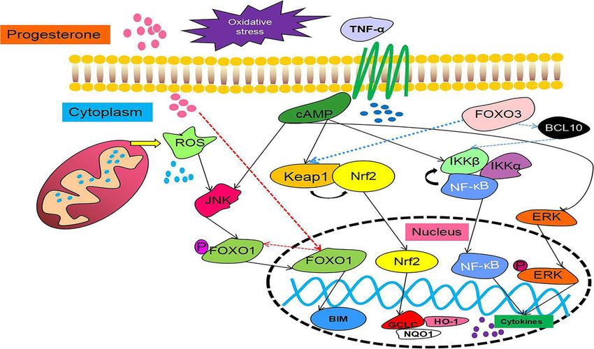
Lu et al. Reproductive Biology and Endocrinology (2018) 16:80 Page 2 of 18
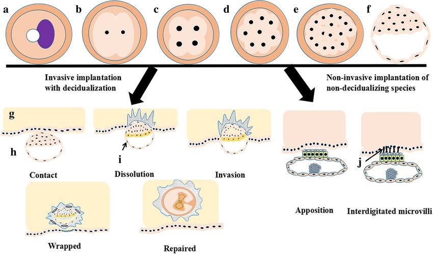
of the central elements of cell signaling, gene expression, contact, dissolution, invasion, wrapping and repair (Fig. 5).
maintenance of redox homeostasis and signal transduc- Ovarian function and blastocyst development from ovula-
tion pathways involved in cell function, growth, differen- tion to implantation are common in many mammalian
tiation and death [12]. When keywords were searched in species after ovulation (Fig. 5) [14].
the NCBI and Web of Science databases, there were
more than 100,000 articles on reproduction and oxida-
tive stress, but there were only approximately 20,000 ar- Oxidative stress
ticles on the relationship between female reproduction Reactive oxygen species (ROS)
and oxidative stress. There were more than 3000 arti- ROS are a double-edged sword: they not only play im-
cles about the mechanism, but there were only ap- portant roles as secondary messengers in many intra-
proximately 800 articles on uterine and ovarian cellular signaling cascades, but they also exert
diseases and oxidative stress. There is very little re- indispensable effects on pathological processes involving
search on the mechanism of uterine and ovarian dis- the generation of excessive ROS. The three major types
eases and oxidative stress, only 30 articles, and review of ROS are superoxide anion (O2−•), hydrogen peroxide
articles are rare. This review not only sheds light on (H2O2), and hydroxyl (•OH).
the mechanism of action of oxidative stress under Most ROS are produced when electrons leak from
normal physiological conditions, but it also explores the mitochondrial respiratory chain, also referred to
and speculates on the mechanisms of joint reproduct- as the electron transport chain (ETC) [15]. According
ive diseases, providing readers with more comprehen- to an estimate, up to 2% oxygen consumed can be
sive content. The 133 articles selected in this article diverted to the production of ROS formation by mito-
have a greater impact on the fields of reproduction chondria, especially at complexes I and III [16]. The
and stress. By summarizing previous studies, a free radical superoxide anion (O2−•) is formed by the
convincing review is offered. addition of one electron to ground state dioxygen,
The previous discussion of reproduction and oxidative but it is unstable in aqueous solutions due to its be-
stress was limited to individual diseases. This review ing able to react spontaneously with itself, producing
aims to provide a comprehensive discussion of the role hydrogen peroxide (H2O2) and molecular oxygen (O2)
of oxidative stress in female reproduction, and it specu- (reaction 1). It can reduce Fe3+ to Fe2+and transform
lates on new mechanisms of action. This review mainly into O2 (reaction 2). H2O2 is not a free radical, but it
examines the available evidence for the involvement of is very harmful to cells because it is able to cross bio-
cellular ROS-induced OS in pregnancy-related diseases, logical membranes and break down into the highly
and it explores the new signaling pathways between OS reactive hydroxyl radical (•OH).
and female reproduction. The main source of hydroxyl radical is the metal-cata-
lyzed Haver-Weiss reaction (reaction 3), the second of
which is the Fenton-type reaction (reaction 4).
Reproductive processes
It is well known that the development of ovarian follicles O2 − • þ O2 − • þ 2Hþ →H2 O2 þ O2 ð1Þ
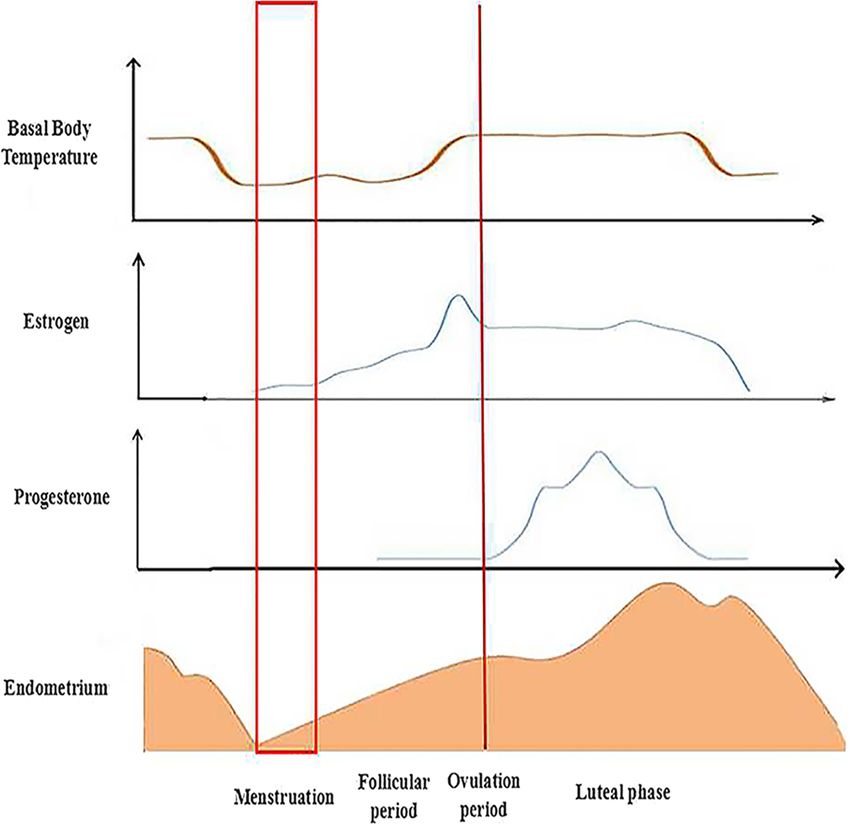
is a continuous process (Fig. 1). There are five stages in
female mammals. During these stages, the structure of O2 − • þ Fe3þ →O2 þ Fe2þ ð2Þ
the endometrium undergoes some changes. Estrogen
O2 − • þ H2 O2 →O2 þ OH− þ •OH ðHaver−Weiss reactionÞ ð3Þ
and progesterone are secreted via the ovaries and uterus
and undergo changes during the estrus cycle. In Fe2þ þ H2 O2 → Fe3þ þ OH− þ •OH ðFenton−type reactionÞ ð4Þ
addition, the basal body temperature also changes, while
the thickness of the endometrium undergoes the corre-
sponding transformation [13] (Fig. 2). The defense mechanism against oxygen free radicals
The combination of sperm and egg consists of three Primary defenses
steps: corona radiata dissolution; zona pellucida dis- As we all know, SOD, CAT, GPx and GSR belong to the
solution; and egg fertilization and cortical reaction primary defense mechanism (Fig. 6). SOD catalyzes O2−•
(Fig. 3). The process occurs in the ampulla portion of dismutation to produce H2O2 and O2 at a rate 104 times
the fallopian tube. Pregnancy starts when the fertil- higher than spontaneous dismutation at the physio-
ized egg is formed. Human chorionic gonadotropin logical pH [17, 18]. CAT is the enzyme that removes
(HCG) increases first and then decreases. Both estro- H2O2 from the cell when the latter is at high concentra-
gen and progesterone are increased during pregnancy tions (reaction 5) [19]. GPx is an enzyme that catalyzes
(Fig. 4). It is noted that the process of implantation is the reduction of H2O2 and organic free hydroperoxides
significant in reproductive events. The process includes requiring glutathione as a co-substrate (reaction 6 and 7)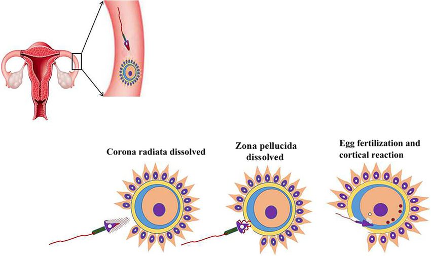
Lu et al. Reproductive Biology and Endocrinology (2018) 16:80 Page 3 of 18
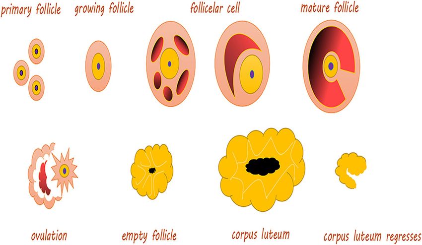
Fig. 1 The development of ovarian follicles. Primary follicle: The center has an oocyte, and there is a flat layer of follicular cells on its periphery.
Growing follicle: Including the primary growth follicle and secondary growth follicles. Primary growth follicle: One or more layers of cuboidal follicular
cells between the egg cells, and follicular cells demonstrate red-stained zona pellucida, while the follicular periphery appears like connective tissue
follicular membrane. Secondary growth follicle: Follicular cells appear in the follicular cavity, and some follicular cavities are large, forming a cumulus
of oophores. Follicular cells are located on the inner wall of follicles and are arranged in layers, called granular layers. The follicular membrane includes
the inner and outer membrane layers. Mature follicle: The follicle cavity is very large, and cumulus oophores are obvious. Follicular endometrial cells
appear close to the follicular granule layer. There is a layer of basement membrane between the granule layer cells and follicular endometrial cells;
endometrial cells are polygonal, with clear cytoplasm and round nuclei; cells can be seen between many capillaries, and outer membrane cells are
located in the outermost layer, mostly spindle shaped with the surrounding connective tissue boundaries not obvious. Ovulation: Mature follicles
develop to a certain stage, obviously protruding from the ovarian surface; with the follicular fluid increasing sharply, the pressure increases so that the
prominent part of the ovarian tissue becomes thinner and finally ruptures; secondary oocytes and their peripheral zona pellucida and radiation crowns
are discharged together with the follicular fluid. Empty follicle: At this time, the follicle is empty, indicating that the corpus luteum starts
to form. Corpus luteum: The residual follicle wall collapses after ovulation; the connective tissue of the follicular membrane and capillaries
stretches into the particle layer, and as the role of LH evolves, it evolves into a larger volume cell cluster, rich in capillaries and endocrine function and
fresh yellow in color
[20]. GSR is a cytosolic protein with a tissue distribution to be defense systems against oxidative injury by oxygen
similar to that of GPx. The enzyme reduces oxidized free radicals [23]. Vitamin E, the major lipid-soluble anti-
glutathione, utilizing NADPH generated by various sys- oxidant present in all cellular membranes, protects against
tems (reaction 8) [21]. lipid peroxidation. The tocopheryl radical might also be
directly reduced by the ascorbic acid-GSH redox couple.
2H2 O2 →2H2 O þ O2 ð5Þ
β-carotene exerts the most efficient scavenger action with
H2 O2 þ 2GSH→GSSG þ 2H2 O ð6Þ vitamin E, while β-carotene acts at low oxygen pressures.
However, vitamin E protects conjugated double bonds of
ROOH þ 2GSH→GSSG þ ROH þ H2 O ð7Þ β-carotene from oxidation.
GSSG þ NADPH þ Hþ →2GSH þ NADPþ ð8Þ In all, OS plays an important role in the pathophysi-
ology of complicated pregnancies. OS was described as
an imbalance in the generation of ROS [1]. These ROS
are oxygen free radicals produced by the reduction in
Secondary defense molecular oxygen and generated as byproducts of aer-
The existence has been reported of an enzyme with perox- obic respiration and metabolism. These molecules are
idase activity called phospholipid hydroperoxidase GPx, capable of activating and modulating various signaling
which is capable of reducing lipid hydroperoxides without pathways, including those involved in cell growth, differ-
the action of phospholipase A2 [22]. In addition, different entiation and metabolism [24]. They can also induce cel-
oxidoreductases that catalyze reduction reactions of thiol lular oxidative damage by interacting with DNA and
and other protein groups when these molecules are oxida- intracellular macromolecules, such as protein and mem-
tively damaged are protective enzymes against oxygen free brane lipids, so they can lead to cellular malfunction that
radicals. Nuclear enzymes for DNA repair are considered can initiate pathological processes.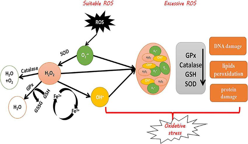
Lu et al. Reproductive Biology and Endocrinology (2018) 16:80                                                                                      Page 4 of 18

 Fig. 2 The changes of biology during different estrus cycle. Estrogen and progesterone are secreted via the ovary or uterus and undergo changes
 during the estrus cycle. In addition, the basal body temperature also changes, while the thickness of the endometrium has corresponding transformations.
 After menstruation, the new estrus cycle starts to develop. During the follicular period, the level of the basal body temperature and estrogen gradually rise.
 The thickness of the endometrium also increases. The levels of basal body temperature and estrogen maintain certain concentrations until the ovulation
 period. Thus, progesterone starts to increase. With the appearance of the luteal phase, all changes are restored until the end of menstruation

 Fig. 3 Fertilization processes of most viviparous and ovoviviparous animals. In most viviparous and ovoviviparous animals, the sperm and oocyte
 combine at the fallopian tube ampulla. In the picture, the first zygote shows a radiation crown dissolving; the second zygote shows the zona
 pellucida dissolving; the last zygote shows fertilized eggs and cortical response
Lu et al. Reproductive Biology and Endocrinology (2018) 16:80                                                                 Page 5 of 18

                                                                              follicular fluid microenvironment are major sources of
                                                                              ROS. ROS in the follicular fluid join in follicular growth,
                                                                              oocyte maturation, and ovarian steroid biosynthesis [15].
                                                                              At the same time, a critical process for ovarian folliculo-
                                                                              genesis, dominant follicle selection, CL formation and
                                                                              embryo formation is angiogenesis [30, 31], which is a
                                                                              complex process. It is promoted by estrogens that regu-
                                                                              lates some cellular factors, such as VEGF [32]. ROS pro-
                                                                              duced from NADP(H) oxidase were shown to be
                                                                              significant for angiogenesis in vivo and VEGF signaling
                                                                              in vitro [33]. Accordingly, ROS are involved in follicular
                                                                              growth in part by regulating angiogenesis.
                                                                                The appropriate amount of ROS is required for ovula-
                                                                              tion. ROS produced by the preovulatory follicle are con-
                                                                              sidered critical inducers of ovulation, and inhibition of
                                                                              ROS has been confirmed to disturb ovulation [27, 34].
                                                                              Oxygen deprivation stimulates follicular angiogenesis,
                                                                              which is important for abundant growth and develop-
                                                                              ment of ovarian follicles [35]. The development of folli-
                                                                              cles from the primordial stage to antral follicles is
                                                                              accompanied by a marked increase in the metabolic
                                                                              function of granulosa cells, especially a large increase in
                                                                              cytochrome P450 activity with steroid biosynthesis [36].
                                                                              Large amounts of ROS are produced during electron
 Fig. 4 The trends of HCG, estrogen and progesterone during pregnancy.        transport, indicating that functional granulosa cells are
 The yellow line represents the change in HCG. The green line represents      related to the pro-oxidant state in the follicles. ROS are
 the change in progesterone. The red line represents the level of estrogen.
                                                                              induced in preovulatory follicles with oscillation of pros-
 The final results of ovulation include two impacts, one of which is output
 in the body, called menstruation, and the other of which is combines         taglandins, cytokines, proteolytic enzymes, and steroids,
 with sperm, called fertilization. The level of hormones changes after        resulting in blood flow alterations and eventual follicle
 fertilization; in particular, hCG immediately increases to the highest       rupture [37]. With the exception of dominant follicles,
 level. However, the levels of estrogen and progesterone slowly               which are released for fertilization, the other growing
 increase to stable concentrations, while hCG begins to drop to
                                                                              follicles all undergo apoptosis, and this process is pro-
 a certain extent
                                                                              moted by ROS. In parallel, follicle-stimulating hormone
                                                                              (FSH)-induced estrogen synthesis and upregulation of
Oxidative stress in ovary                                                     CAT and GSH in growing follicles resist the apoptotic
ROS affect a variety of physiologic functions of the ovary,                   process to maintain the balance during normal ovarian
including ovarian steroid genesis, oocyte maturation, ovu-                    function [38]. ROS are generated in the CL and are
lation, formation of blastocysts, implantation, luteolysis                    involved in functional luteolysis. ROS and antioxi-
and luteal maintenance in pregnancy. OS is an important                       dants are related to progesterone synthesis in the lu-
modulator of ovarian germ cell and stromal cell physi-                        teal phase [35] (Table 1).
ology [25]. Concentrations of ROS could also play a major                       However, excessive deprivation of oxygen will also
role in the implantation and fertilization of eggs, and a                     cause some damage to follicles, as we discuss in the fol-
relevant study showed localization of SOD in the ovary                        lowing. Cu/Zn-SOD is increased in the CL during the
and found that copper-zinc SOD (Cu-Zn SOD) was local-                         early to mid-luteal phase, but it is decreased during the
ized in the granulose cell of growing follicles and mature                    regression phase, which could explain the increase in
Graafian follicles, as well as manganese superoxide dis-                      ROS concentrations during regression, and this change
mutase (MnSOD) being localized in luteal cells of the cor-                    in activity is similar to that in progesterone concentra-
pus luteum in rats [26].                                                      tions. Other possible explanation for the decrease in
  ROS exert both negative and positive effects on mam-                        Cu-Zn SOD during the regression phase is an increase
malian ovaries [27]. ROS affect multiple physiological                        in prostaglandin PGF2α or macrophages; another reliable
and pathological activities in the ovaries, from oocyte                       explanation is a decrease in ovarian blood flow [35].
maturation to fertilization. In cycling ovaries, different                    Prostaglandin F2α stimulates production of the SO anion
markers of OS are negatively affected [28, 29]. Macro-                        by luteal cells and phagocytic leukocytes in the CL. The
phages, leukocytes, and cytokines present in the                              reduction of ovarian blood flow causes tissue damage by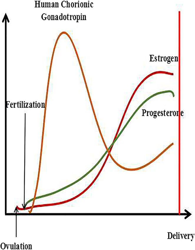
Lu et al. Reproductive Biology and Endocrinology (2018) 16:80                                                                                        Page 6 of 18

 Fig. 5 The process of implantation (including invasive implantation with decidualization and non-invasive implantation of non-decidualizing species)
 a: Zygote; b: 2 cells; c: 4 cells; d: 8 cells; e: Morula; f: Blastocysts; g: Endometrium; h: Uterine cavity; i: Trophoblast cells; j: Microvilli. Estradiol (E2)
 produced by the developing ovarian follicles interacts with progesterone produced by the CL to prepare the endometrium receptivity necessary for
 embryo implantation. The meeting of the oocyte and sperm and subsequent fertilization occur in the ampulla of the oviduct, followed by early
 embryo development within the oviduct, and the morula migrates to the uterus, where implantation occurs. The appearance of a fluid-filled inner
 cavity (blastocoel) is accompanied by cellular differentiation: the surface cells become the trophoblast and give rise to the extra-embryonic tissues,
 including the placenta, while the inner cell mass gives rise to the embryo and finally shedding of the zona pellucida, followed by orientation,
 apposition, attachment and adhesion of the blastocyst to the endometrium. If the blastocyst was not present, the CL would regress, and
 the uterus would start the cycle again. The time and chronological events of implantation differ among mammalian species irrespective
 of the length of gestation. In contrast to humans, horses, primates and rodents, in which implantation occurs shortly after the hatching
 of the blastocyst, the blastocyst in domestic ruminants and pigs elongates before implantation (the time to implantation: in pigs, the 14th day; in
 sheep, the 16th day; and in cattle, the 18th day), and this unique developmental event does not occur in the laboratory or in rodents or humans

ROS production. However, the concentration of                                       and luteal cells respond to hydrogen peroxide with
Mn-SOD in the CL during regression is increased, thus                               extirpation of gonadotropin action and inhibition of
scavenging the ROS produced in the mitochondria by                                  progesterone secretion. Hydrogen peroxide lowers both
inflammatory reactions and cytokines. Therefore,                                    cAMP-dependent and non-cAMP-dependent steroido-
complete disruption of the CL leads to a significant de-                            genesis [28, 43]. As we all know, OS influences the entire
crease in Mn-SOD in the regressed cells, and cell death                             reproductive process of women’s lives. ROS attacks the
is imminent [39]. In addition, ROS also participate in                              8th carbon atom of guanine in DNA to generate 8-hydro-
mammalian ovulation and follicular rupture. The gener-                              xy-deoxyguanosine (8-OHDG), which is an oxidized de-
ation of the two processes is the result of vascular                                rivative of deoxyguanosine, the levels of which are higher
changes or the proteolytic cascade. The crosstalk be-                               in aging oocytes [44]. 8-OHdG is the most familiar base
tween these two cascades is mediated by ROS, cytokines,                             modification in mutagenic damage. 8-OHdG causes base
and vascular endothelial growth factor (VEGF) [40, 41].                             mutation and mismatches in DNA replication, resulting in
Ad4BP, a zinc finger DNA-binding protein, was identi-                               G mutations to T and G:C to T:A transversion. Therefore,
fied as a transcription factor regulating steroidogenic                             8-OHdG has become a marker for OS.
P-450 genes in a cAMP-dependent manner [42], and a                                    OS has been implicated in different female diseases,
recent study showed that the correlation between Ad4BP                              including PCOS, which is the most common endocrine
and SOD expression suggested an association between                                 abnormality of reproductive-aged women and has a
OS and ovarian steroid genesis. Both human granulosa                                prevalence of approximately 18%. It is a disorder
Lu et al. Reproductive Biology and Endocrinology (2018) 16:80                                                                          Page 7 of 18

  Fig. 6 The defense mechanism against oxygen free radicals. SOD: Superoxide dismutase; GPx: Glutathione peroxidase; GSSG: Glutathione oxidase;
  GSH: Glutathione reductase; ROS: Reactive oxygen species; O2−•: Superoxide; H2O2: Hydrogen peroxide; •OH: Hydroxyl

characterized by hyperandrogenism, ovulatory dysfunc-                                antioxidant status. The decrease in mitochondrial O2
tion, and polycystic ovaries [45]. Various studies have                              consumption and GSH levels, along with increased ROS
reflected the presence of OS in PCOS patients. In a                                  production, explains the mitochondrial dysfunction in
study by Hilali et al., PCOS patients had increased serum                            PCOS patients. Physiological hyperglycemia generates
prolidase activity, as well as higher total oxidant status                           increased levels of ROS from mononuclear cells, which
and OS indices—the ratio of oxidants to total                                        activate the release of tumor necrosis factor-α (TNF-α)

Table 1 The role of oxidative stress in the female reproductive process
Function                    Reproductive process                                                                                Reference
Positive effect             Zn-Cu SOD↑ → Promotion of the development of follicles                                              [26]
                            Biosynthesis of ovarian steroids → P450↑ → ROS↑ → Blood flow↑ → Rupture of                          [36, 37, 40, 41]
                            follicles → Ovulation
                            ROS↑ → Promotion apoptosis of non-dominant follicles                                                [38]
                            FSH↑ → E2↑ → CAT and GSH↑ → Protection of cells from apoptosis
                            E2 and P ↓ → SOD↓ → OS↑ → Endometrial shedding and implant failure                                  [62]
                            ROS↑ → NF-κB↑ → PGF2α↑ → Luteum dissolution                                                         [35, 39, 61]
                            Sperm-ovum binding → ROS↑ → Corpus luteus functional↑                                               [39]
                            ROS↑ → Antioxidants↑ → Synthesis of progesterone
Negative effect             PCOS:                                                                                               [45, 46]
                            Serum proline activity↑, OS↑
                            Physiological hyperglycemia → ROS↑ (Monocytes) → TNF-α↑ → NF-κB↑ → Resistance of Insulin↑
                            Preeclampsia:                                                                                       [67, 76–81, 83–85]
                            Defective placenta → Hypoxia and reperfusion injury → OS↑ → Cytokines↑,
                            Prostaglandins↑ → TAS↓, GPx of placenta↓
                            VC↓ → Risk of preeclampsia↑
                            (MDA↑) ROS↑ → Vasoconstriction↑ → Coagulation activity↓
                            OS↑ → Vascular endothelial injury↑ → ROS↑ → TNF-α↑, ox-LDL↑ → Endothelial subtypes of
                            activated NAD(P)H oxidase → SO anion↑
                            Auto-antibodies of AT1-AA↑ → NAD(P)H oxidase↑ → ROS↑ → SO anion↑
                            Endometriosis:                                                                                      [71–73, 75]
                            In the peritoneal fluid, MDA↑, IL-6↑,TNF-α↑, IL-8↑, VEGF↑, MCP-1↑, ox-LDL↑.
                            In endometriotic lesions: OS↑ → NF-κB↑ → Inflammation↑
                            In endometriotic cells: MAPK↑
                            ROS↑ → IUGR, abortion, fetal malformation
The ‘→’ indicates that it has an effect on the next step. The ‘↑’ represents an increase and the ‘↓‘represents a decrease
Lu et al. Reproductive Biology and Endocrinology (2018) 16:80 Page 8 of 18
and increase the inflammatory transcription factor nu- [55–57]. During this process, placental tissue forms a
clear factor-kappa B (NF-κB). As a result, concentrations large amount of free radicals, and oxidation occurs.
of TNF-α, a known mediator of insulin resistance, are Intense stress results, but the placenta gradually adapts
further increased. The resultant OS creates an inflamma- to this environment and returns to normal under the ac-
tory environment that further increases insulin resist- tion of antioxidant activity [58, 59]. While physiological
ance [46], causing abnormal ovarian extracellular concentrations of endogenous glucocorticoids are sup-
remodeling, multiple cyst formation, and chronic anovu- portive of fetal development, excessive glucocorticoids in
lation, leading to infertility [47] (Table 1). utero (i.e., maternal stress) adversely affect mammalian
Recently, PCOS has been paid significant attention be- offspring by “programming” abnormalities that are pri-
cause it exerts severe effects on female reproduction. In marily manifested postpartum [60]. ROS are also be-
this review, we collect some interesting evidence that lieved to play a role in the different phases of the
implies a delicate relationship between stress and PCOS endometrial cycle. The late luteal phase is characterized
[48]. Its etio-pathogenesis and pathophysiology include by elevated levels of lipid peroxide and a decrease in the
roles for genetic, environmental and endocrine factors. antioxidant SOD. ROS stimulate the secretion of PGF2α
Franks et al. defined PCOS as a gene-dependent ovarian through activation of NF-κB [61]. Decreased levels of es-
pathology, characterized by the overproduction of an- trogen and progesterone lead to decreased SOD expres-
drogens and not uniformly represented by the inter- sion and hence generate OS in the uterus, resulting
action of genetic “propensities” with other genetic and ultimately in endometrial shedding and lack of implant-
environmental factors [49]. Thus, PCOS seems to be a ation. Controlled levels of ROS have, however, been as-
genetic disease, but after investigation by Escobar-Mor- sociated with angiogenic activity in the endometrium,
reale HF et al. found that it is caused by the interaction causing regeneration during every cycle. These studies
of susceptibility and protective genetic variants, and showed that limited levels of ROS are necessary to main-
these mutations could be chosen due to survival advan- tain physiological function, but when present in higher
tages in the evolution process, requiring an accurate concentrations, ROS can have deleterious effects [62]. In
study of environmental factors, including race, diet and other words, although a physiological balance between
lifestyle [50]. V. De Leo et al. reported that disruption of ROS and antioxidant activity is maintained in normal
the delicate balance between intra-ovarian and extra- pregnancies [63], an imbalance can increase OS. The
ovarian factors alters and impairs the formation of ma- placenta experiences a heightened level of OS in certain
ture oocytes, leading to infertility. In addition, insulin pathologic conditions of pregnancies, including gesta-
plays a particular role in PCOS, and in vitro studies have tional diabetes, fetal growth restriction, preeclampsia
demonstrated that insulin stimulates thecal cell prolifer- and miscarriage [64–66].
ation, increases secretion of androgens mediated by LH OS leads to endothelial cell dysfunction. In the
and increases cytochrome P450 expression of LH and uterus, endothelial cell dysfunction results in many
IGF-1 receptor [51]. As we all know, P450 can increase diseases, such as preeclampsia and endometriosis.
ROS. The above evidence indicates that ROS join in the There are many causes that induced endothelial cell
pathological process of PCOS. dysfunction. TNF-α, a plasma cytokine, has been
demonstrated to cause endothelial cell injury, but the
Oxidative stress in the uterus and placenta antioxidant Mn-SOD neutralizes SO anions generated
Pregnancy itself is a state of OS, arising from the in- by the cytokine TNF-α. This process is a
creased metabolic activity in the placental mitochondria self-protective mechanism against TNF-α-induced OS.
and increased ROS production due to the higher meta- In addition, defective placentation leads to placental
bolic demand of the growing fetus [52, 53]. Superoxide hypoxia and reperfusion injury due to ischemia, and
(SO) anions produced by the placental mitochondria the resultant OS triggers the release of cytokines and
appear to be a major source of ROS and lipid prostaglandins, resulting in endothelial cell dysfunc-
per-oxidation contributing to OS in the placenta [54], tion and playing an important role in the develop-
supported by mitochondrial production of lipid perox- ment of preeclampsia [67]. In addition, ROS
ides, free radicals, and vitamin E in the placenta, which generated from NADP (H) oxidase are critical for
increases as gestation progresses[55]. In the second tri- VEGF signaling in vitro and angiogenesis in vivo [33].
mester, the placenta gradually matures and increases in Small amounts of ROS are produced from endothelial
size, with less hairiness and wider blood vessels. The NADP (H) oxidase activated by growth factors and
cytotrophoblast becomes a single cell and gradually re- cytokines. ROS generated in and around the vascular
places the endothelial layer covering the smooth muscle endothelium could play a role in normal cellular sig-
of the spiral artery. Slowly, maternal blood penetrates naling mechanisms. They might also be important
from the mother’s spiral artery into the interstitial space causative factors in endothelial dysfunction.Lu et al. Reproductive Biology and Endocrinology (2018) 16:80 Page 9 of 18 Endometriosis is a benign, estrogen-dependent, Increased ROS concentrations in patients with pre- chronic gynecological disorder characterized by the pres- eclampsia have been proved by the increased levels of ence of endometrial tissue outside the uterus. There was MDA, an index of lipid peroxidation [76]. Under normal a report suggested that the elevated ROS causing OS are conditions, the impairment of circulatory homeostasis is produced by erythrocytes and apoptotic endometrioma caused chiefly by vascular endothelial dysfunction in cells, as well as the activated macrophages that are re- preeclampsia. It is characterized by the tendency to cruited to phagocytize apoptotic cells [10]. Additionally, cause vasoconstriction and low anticoagulant activity. the ROS producing enzyme xanthine oxidase, which is ROS seem to play a critical role in the endothelial dys- considered another contributor to excess ROS, are function associated with preeclampsia [77]. In other expressed in greater quantities in women with endomet- words, the pathologic event in preeclampsia is injury to riosis [68]. OS plays a large role in infertility. Another the vascular endothelium regulated by OS from in- way in which cells are damaged through OS is via creased placental ROS [78] or decreased antioxidant ac- lipid peroxidation, which is the oxidative destruction tivity [79]. of polyunsaturated fatty acids in the plasma mem- There are many reasons for the increase in ROS. For brane [69]. This leads to “increased membrane per- instance, neutrophil modulation occurring in preeclamp- meability, degraded membrane integrity, inactivated sia is an important source of ROS, resulting in increased enzymes and structural damage of the DNA; cell production of the SO anion and decreased levels of NO, death rapidly follows” [70]. In addition, OS induces ultimately causing endothelial cell damage in patients local inflammation, resulting in elevated levels of cy- with preeclampsia. Levels of TNF-α and oxLDL are in- tokines and other factors that promote endometriosis, creased in preeclampsia and have been shown to activate as discussed later [8]. the endothelial isoform of NAD(P)H oxidase, ultimately The peritoneal fluid of patients has been found to con- resulting in increased levels of the SO anion. These re- tain high concentrations of malondialdehyde (MDA), sults suggest that the consumption of antioxidants to pro-inflammatory cytokines (IL-6, TNF-α, and IL-1β), counteract heightened lipid per-oxidation might injure angiogenic factors (IL-8 and VEGF), monocyte chemo- the vascular endothelium and could be involved in the attractant protein-1 (MCP-1) [71], and oxidized LDL pathogenesis of preeclampsia [80]. (ox-LDL). Pro-inflammatory and chemotactic cytokines In addition, autoantibodies against the angiotensin re- play central roles in the recruitment and activation of ceptor AT1, particularly the second loop (AT1-AA), can phagocytic cells, which are the main producers of ROS stimulate NAD(P)H oxidase, leading to increased gener- and RNS. Activation of NF-κB by OS has been detected ation of ROS [81]. The AT1 receptor of preeclamptic in the endometriotic lesions and peritoneal macrophages women has been observed to promote both the gener- of patients with endometriosis [72]. Signaling mediated ation of the SO anion and over-expression of NAD(P)H by NF-ĸB stimulates inflammation, invasion, angiogenesis, oxidase in cultured trophoblasts and smooth muscle and cell proliferation, and it also promotes the apoptosis cells. Therefore, early placental development can be af- of endometriotic cells. Additionally, N-acetylcysteine fected by dysregulated vascular development and func- (NAC) and vitamin E are antioxidants that limit the prolif- tion secondary to NAD(P)H oxidase-mediated altered eration of endometriotic cells, likely by inhibiting activa- gene expression [82]. Preeclamptic women produce ROS tion of NF-κB [73]. A study indicated a therapeutic effect and exhibit higher NAD(P)H expression than those of NAC and vitamin E supplementation on endometriotic without the disease [35]. More specifically, it has been growth [74]. Similar to tumor cells, increased ROS and reported that women with early-onset preeclampsia pro- subsequent cellular proliferation in endometriotic cells duce larger amounts of the SO anion than women with activate of mitogen-activated protein kinase (MAPK) late-onset disease [83]. Affected women also have de- and extracellular regulated kinase (ERK1/2) [75]. creased total antioxidant status (TAS) and placental GPx More seriously, the increase in ROS in endometriosis [84] and low levels of vitamins C and E. Lack of vitamin patients can lead to adverse effects on embryos, such C intake seems to be associated with an increased risk of as IUGR, spontaneous abortion, or fetal dysmorpho- preeclampsia, and some studies have shown that peri- genesis [69] (Table 1). conceptional supplementation with multivitamins can Preeclampsia is a vascular pregnancy disorder that lower the risk of preeclampsia in normal or underweight often involves impaired placental development. It is a women [85]. complex multisystem disorder that can affect normoten- There have been studies focusing on the effects of re- sive women. It can cause the poor implantation and straint stress on uterine and embryo implantation in growth restriction observed in preeclampsia because OS pregnant mice. In these studies, uterine local micro-en- causes increased nitration of p38 MAPK, resulting in a vironment changes and uterine histomorphology re- reduction in its catalytic activity. search were emphasized. Liu Guanhui et al. reported
Lu et al. Reproductive Biology and Endocrinology (2018) 16:80                                                  Page 10 of 18

that the mice were subjected to restraint stress from em-       the Kelch-like ECH-associated protein 1 (Keap1)-Nuc-
bryonic day1 (E1). This study demonstrated that restraint       lear factor erythroid 2-related factor 2 (Nrf2) pathway,
stress increased the level of corticosterone (CORT) in          the Jun N-terminal kinase (JNK) pathway, the fork-
plasma, and uterine natural killer (uNK) cells in the endo-     head transcription factors of the O class (FOXO)
metrium were significantly increased, accompanied by the        family, and apoptosis.
decreased density of mast cells in the myometrium. In              Nrf2 is a key molecule activated in response to OS,
addition, restraint stress markedly decreased the CD3+CD4+      and it regulates antioxidant response to protect cell
T/CD3+CD8+ T cell ratio. Additionally, antioxidant ability      function [91]. Normally, Nrf2 binds to Keap1, is seques-
was compromised, and the concentration of MDA                   tered in the cytoplasm, and then is degraded by a prote-
was increased [86]. Moreover, restraint stress reduced          asome pathway [92]. After activation, it transfers to the
the weight of the uterus and ovary and the intake of            nucleus to activate a large number of antioxidant genes
food with reduction in weight, while the relative endomet-      [93]. In other words, transcriptional activation of anti-
rial area and uterine gland area were reduced after re-         oxidant defense genes and restoration of vascular redox
straint stress. In addition, restraint stress decreased         homeostasis are necessary when OS occurs. Importantly,
micro-vessel density and VEGF expression [87].                  the redox-sensitive Keap1-Nrf2 pathway plays a key role
                                                                in the process [94]. These studies also implied that Nrf2
The signaling molecules between oxidative stress                deficiency caused fetal DNA damage and neurological
and reproduction                                                deficits, and inactivation of Nrf2 has also been shown to
OS has led to a variety of signaling pathways, resulting        underlie inflammation-induced trophoblastic apoptosis.
in crosstalk among many protein factors in the body.            As studies have progressed, the literature has increas-
Especially in the female reproductive organs, OS leads to       ingly revealed that Nrf2 plays a significant role in preg-
a series of abnormal events in egg production and ovula-        nancy and has highlighted the important role of Nrf2 in
tion. During pregnancy, implantation will be impaired,          protecting the fetus in utero OS [95]. Nrf2 is sensitive to
leading to loss of embryos and changes in local immune          maternal immunological status. In normal pregnancy,
function in the uterus. Research on these signals is cur-       Nrf2 is only decreased after term vaginal delivery. How-
rently the most important concept in this field and is of       ever, notably, the expression of Nrf2 is significantly re-
great significance to the reproduction of female animals.       duced when the uterus is infected [96]. Furthermore, the
  Before this review, there were a number of reviews dis-       mechanism of Nrf2 antioxidant defense plays an import-
cussing the contact between reproduction and OS. For            ant role in adverse pregnancy priming. Nrf2 is a regulator
example, Perucci et al. proposed a hypothesis that the          of antioxidant defense in vascular dysfunction and oxida-
ADAMs pathway protects women from the inflamma-                 tive damage [95]. Many studies have shown that suitable
tory lesions of preeclampsia [88]. Wu et al. elaborated         OS increased Nrf2 and the expression of downstream tar-
on potential therapeutic approaches to placental stress         gets, such as heme oxygenase 1 (HO-1), NAD(P)H: quino-
by exploring the relationship between OS and apoptosis          neoxidoreductase (NQO1), and glutamate-cysteine ligase
and between OS and cellular autophagy, resulting in             subunit catalysis (GCLC), etc., to resist OS [97]. However,
speculation about a comprehensive therapeutic target            as described above, we speculate that the activity of Nrf2
[89]. Sultana et al. fully summarized the adverse preg-         significantly decreases, and Keap1 binds to Nrf2 more
nancy outcomes caused by aging placentas, explaining            strongly when excessive OS causes severe inflammation in
the mechanisms of telomerase and placental disorders            the uterus. Related studies have shown that FOXO3 par-
[59]. Wojsiat et al. explained the effects of OS on oocyte      ticipates in the interaction between Keap1 and Nrf2. Loss
and fertilization outcomes and the effect of overproduc-        of FOXO3 leads to severe inactivation of Keap1, which in
tion of active substances on in vitro fertilization [90].       turn cannot prevent the activation of Nrf2, which is a very
Nevertheless, our review not only summarizes the above          important finding in tumors. Research also revealed the
discussion but also makes reasonable assumptions about          important role of FOXO3 in the Keap1-Nrf2 axis. At the
the signaling pathways in reproductive diseases.                same time, it is not denied that, in the absence of FOXO3,
  Hypoxia and inflammation lead to the production of            Nrf2 is activated under the induction of AKT and protects
TNF-α, which induces the release of large amounts of            cells from damage due to OS by this form [98]. Therefore,
ROS from the mitochondria in cells. Excessive ROS               we hypothesize that, if OS induces inflammation in the re-
cause an imbalance between oxidation and antioxidation,         productive system, the changes in FOXO3 affect the inter-
leading to OS. The body’s signaling pathway will evince         action between Keap1 and Nrf2, which could be a marker
a series of changes following to exposure to the dual im-       of damaging OS in our study (Table 2).
pact of OS and inflammation. This article focuses on the           NF-κB is an active molecule in the immune system. In
collection of OS-induced reproductive disease-related           mammals, the NF-κB family is composed of five related
signaling pathways, including the p38 MAPK pathway,             transcription factors: c-Rel, p50, p52, RelB and RelA
Table 2 The important proteins in reproductive mechanisms
Protein Function and role in reproductive process Interaction between proteins Reference
Keap1-Nrf2 pathway Keap1: Keap1 binds to Nrf2 in cytoplasm. The right amount of OS → Nrf2↑ → HO-1↑,NQO1↑,GCLC↑ [91–93, 95–98]
Nrf2: Protects cells under moderate oxidative stress Excessive OS → FOXO3↑ → Ability of Keap1 to bind to Nrf2↑
A regulator of antioxidant defense in vascular dysfunction Deletion of FOXO3: OS↑ → AKT↑ → Nrf2↑
and oxidative damage.
Deletion of Nrf2 → Fetal DNA damage and nervous
system defects (the basis of trophoblast cell apoptosis).
Vaginal delivery and Uterine infection: Nrf2↓
NF-κB pathway An active molecule in the immune system; Inhibitory IκB protein family binds to NF-κB in cytoplasm [2, 99–104, 108, 109]
Redox-Sensitive transcription factors IKKB protein is degraded (IKKα, IKKβ,
Lu et al. Reproductive Biology and Endocrinology (2018) 16:80
Placental stress: OS → NF-κB↑ → Pro-inflammatory NEMO mediate) → NF-κB enters nuclear to regulate target gene.
Cytokines↑ → Placental apoptotic process is activated IKKβ↑ → p-FOXO3↑
Endometriosis: OS↑ → TNF-α↑ → NF-κB↑ Deletion of FOXO3 → NF-κB↓
In vitro: IL-1β → NF-κB↑ → MIF↑, TNF-α↑ FOXO3↑ → BCL10↑ → IKKB↓ → NF-κB↑ → Anti-apoptosis gene↑
FOXO family FOXO1(all tissues): Deletion of FOXO1, embryonic cell FOXO1↑ → WNT4↑,PRL↑,IGFBP1↑ [111, 112, 115–120]
death due to incomplete blood vessel development JNK↑ → FOXO1↑ → MnSOD↑, CAT↑ → Protecting cells
FOXO3(all tissues): Deletion of FOXO1, lymphocyte
proliferation, extensive organ inflammation
FOXO4 (muscle, kidney, colorectal): Deletion of FOXO4,
inflammation of the colon in response to inflammatory stimuli
FOXO6(brain, liver): Deletion of FOXO6, shows normal learning,
but memory consolidation is impaired
FOXO1↑ → Apoptotic pathway in decidual stromal cells,
and Inhibits endometrial epithelial cell growth
MAPK family JNK: Activated by stress and inflammation JNK↑ → FOXO1↑ → MnSOD↑, CAT↑ → Protecting cells [118, 121, 123–126, 129, 130]
P38 MAPK: Activated by stress and inflammation P450↑ → ROS↑ → ASK1-P-p38 MAPK
ERK: Activated by inflammation and growth factors
OS↑ → p38 MAPK↑ → Aging and premature aging of fetal tissue
Endometriosis: ERK↑
Endometrial stromal cells: time of p-ERK↑; OS↑ → ERK↑,H2O2↑ → p-ERK↑
The ‘→’ indicates that it has an effect on the next step. The ‘↑’ represents an increase and the ‘↓‘represents a decrease
Page 11 of 18Lu et al. Reproductive Biology and Endocrinology (2018) 16:80 Page 12 of 18 (a.k.a. p65) [99]. NF-κB is the nodal point of a primary (FOXOs) number four: FOXO1, FOXO3, FOXO4, and inflammation-stimulated signaling pathway that plays FOXO6. Further, FOXO1 and FOXO3 exist in nearly all a significant role in the immune response [100], while tissues. FOXO4 is highly expressed in the muscle and NF-κB is also a redox-sensitive transcription factor kidneys, and FOXO6 is primarily expressed in the [101]. Therefore, its effect is self-evident in OS, including brain and liver. They are involved in the processes of embryonic stresses. The NF-κB pathway is activated when proliferation, apoptosis, autophagy, metabolism, inflam- embryonic stresses occurs, and a variety of pro-inflamma- mation, differentiation and stress tolerance [111]. How- tory cytokines is increased. Then, the apoptotic process of ever, FOXO1 plays a significant role in reproduction. It the placenta is activated [2]. Therefore, this study indi- regulates cyclic differentiation and apoptosis in the normal cated that NF-κB controls cell survival through the en- endometrium [112]. Additionally, genome-wide expres- hancement of anti-apoptotic gene transcription. In most sion profiling demonstrated that FOXO1 knockdown per- cells, the NF-κB complex is inactive, and it is mainly turbs the expression of more than 500 types of genes in present in the cytoplasm by binding to the inhibitory IκB decidualizing human endometrial stromal cells [113]. In protein family. When the NF-κB pathway is activated, the the past, many studies of human endometrium provided IκB protein is degraded, NF-κB complex enters the nu- reliable evidence for this ability of FOXO transcription cleus to modulate the expression of target genes, and the factors to regulate diverse genes in response to change degradation of IκB protein is mediated through the IκB hormones [114]. However, the interaction between pro- kinase (IKK) complex, which consists of two catalytically gesterone and FOXO1 is even more striking. It is well active kinases (IKKα and IKKβ) and the regulatory scaffold known that progesterone exerts inhibitory effects on protein NEMO. In the activation pathway, IKKβ and endometrial epithelial growth, and a study revealed this NEMO are very necessary for activation of the complex mechanism and showed that siRNA inhibition of FOXO1 [102], while IKKβ also acts on other factors, such as Fork- significantly attenuated the effects of progestin in inhibit- head box O3, a transcription regulator. A study showed ing endometrial epithelial cell growth. Therefore, FOXO1 that FOXO3 is subject to IKKβ-mediated phosphorylation, is essential for the anti-proliferative effects of progesterone leading to the nuclear exclusion and degradation of on both endometrial stromal and epithelial cells [115]. FOXO3 [103]. In addition, a study reported that FOXO3 Further, FOXO1 is indispensable for the induction of was a positive regulator of NF-κB signaling and found that the most highly responsive decidual marker genes, in- over-expression of FOXO3 increased and knockdown of cluding WNT4, prolactin (PRL) and insulin-like growth FOXO3 repressed NF-κB activities. The study indicated factor-binding protein 1 (IGFBP1) [116]. It has been that FOXO3 activated NF-κB by inducing expression of found that FOXO1 activates apoptotic pathways in de- B-cell lymphoma/leukemia 10 (BCL10), an upstream cidual stromal cells. The pro-apoptotic Bcl-2 homology regulator of inhibitor of kappa B kinase (IKK)/NF-κB sig- 3 domain-only protein BIM is a major intermediate in naling [104]. In reproductive stress diseases, for example, this pathway [117]. It was validated that BIM is a endometriosis, increased expression of NF-κB has been FOXO1 target gene and is induced under the stimula- confirmed in cultured endometriotic stromal cells [105] tion of cAMP. Both cAMP and progestin promote in- and peritoneal macrophages isolated from women with creases in FOXO1, but BIM is only increased and cell endometriomas [106]. In any case, changes in NF-κB are death occurs when progestin disappears [120]. Addition- strongly associated with inflammation. Endometriosis is a ally, targeted phosphorylation of cytoplasmic FOXO fac- disease caused by OS during reproductive. OS leads to an tors by JNK promotes nuclear import and increases increase in TNF-α, which in turn causes inflammation, cellular protection against OS via the transcriptional ac- and the NF-κB pathway is activated. Additionally, in vitro tivation of MnSOD and CAT [118]. Thus, FOXO1 has evidence raised the possibility that the changes might be emerged as a major regulator of progesterone-dependent due to the endometriotic microenvironment. IL-1β stimu- differentiation of human endometrium and subsequent lates NF-κB with subsequent increased production of process (Fig. 7) [119]. Thoughtfully, FOXO1 is markedly inflammatory cytokines [107], including macrophage mi- induced upon decidualization both in vivo and in vitro, gration inhibitory factor (MIF) in endometrial stromal whereas FOXO3 expression is suppressed [120]. At any cells [108] and TNF-α in the immortalized epithelial (12Z) rate, FOXO1 plays a unique role either in reproduction cell line [109]. In conclusion, the NF-κB pathway is acti- or in OS (Table 2). vated when reproductive OS occurs (Table 2). The extracellular environment activates three path- FOXO1, the same family as FOXO3, is also involved ways, with ERK predominantly activated by inflamma- in the processes of OS and pregnancy. The FOXO sub- tion and growth factors, while JNK and p38 MAPK are family of Forkhead transcription factors is a direct predominantly activated by stress and inflammation downstream target of the PI3K/Akt pathway [110]. The [121]. Additionally, studies by Lee et al. showed that mammalian forkhead transcription factors of the O class ROS generated by dysfunctional electron transport in
Lu et al. Reproductive Biology and Endocrinology (2018) 16:80                                                                              Page 13 of 18

 Fig. 7 The signaling pathway of OS and pregnancy (a brief view). When the body, especially the maternal body, suffers from an imbalance between
 oxidation and antioxidant levels during pregnancy, in addition to changes in TNF-α, changes in progesterone cannot be ignored. First, TNF-α activates
 a series of signaling pathways in cells through cAMP, such as stimulation of the Keap1-Nrf2 signaling pathway, NF-κB signaling pathway, MAPK signaling
 pathway, etc., then promoting an increase in cytokines and changes in antioxidant-related genes. However, FOXO3 is involved in these signaling pathways.
 When FOXO3 is increased, it promotes the binding of Keap1-Nrf2, lowering the level of antioxidants and promoting the release of NF-κB by IKKβ
 by stimulating BCL10, thereby promoting the increase in cytokines and apoptosis. Finally, the mechanism underlying the changes in the
 FOXO family under the combined effects of both reproductive and oxidative stress remains unclear. It can only be demonstrated that JNK undergoes
 dephosphorylation of FOXO1 under the action of cAMP and ROS when oxidative stress occurs to induce it to enter the nucleus and promote apoptosis.
 When progesterone is reduced, nuclear translocation occurs in FOXO1, and it is phosphorylated

mitochondria activate the inflammatory ASK1-P-p38                              activation. H2O2 induces ERK phosphorylation in endo-
MAPK pathway [122]. The recent literature has identified                       metriotic stromal cells with a more serious induction
the physiologic aging of fetal tissues as a potential mech-                    compared with stromal cells from women who do not
anistic feature of normal parturition. This process is af-                     have endometriosis [130]. Today, although, no direct rela-
fected by telomere-dependent and p38 MAPK-induced                              tionship between phosphorylated ERK (p-ERK) activation
senescence activation (Fig. 7). Pregnancy-associated risk                      and OS is confirmed, an increase in OS markers is
factors can cause pathologic activation of this pathway,                       discovered in epithelial and stromal cells derived from
causing OS-induced p38 MAPK activation and leading to                          women with endometriosis in a similar pattern to
senescence and premature aging of fetal tissues [123, 124].                    p-ERK level (Table 2).
   It has been reported that the activation of ERK was in-
creased in endometriotic tissue, suggesting that ERK                           Conclusion
might play a role in endometriosis pathogenesis, and                           Based on the above, OS influences the entire reproduct-
phosphorylated ERK is increased in primary eutopic epi-                        ive process of woman. The production of excessive ROS
thelial cells [125, 126]. Prolonged phosphorylation of                         leads to OS events. ROS, including superoxide (O2−•),
ERK in endometrial stromal cells occurs in women with                          hydrogen peroxide (H2O2) and hydroxyl (•OH), cause
endometriosis, compared with women without endomet-                            DNA damage, lipid per-oxidation and protein damage.
riosis [127]. Therefore, the endometriotic microenviron-                       The antioxidative system is activated when slight OS oc-
ment could induce increased ERK activity in ectopic                            curred, such as SOD and GPx. In addition, when ROS
cells. Although only IL-1β-induced cyclo-oxygenase 2                           levels exceed the scavenging capacity of the system, the
(COX2) production and IL-8 secretion could be attenu-                          redox system can repair oxidized and damage molecules
ated by the ERK1/2-specific inhibitor PD98059, both                            using NADPH as an original electron source in such sit-
TNF-α and IL-1β activate ERK and induce the expres-                            uations. Thus, the maintenance of high redox potential
sion of IL-8 and IL-6 [128]. However, another study                            is a prerequisite for maintaining the reproductive sys-
found that the IL-1β-mediated COX2 expression was                              tems in a healthy state [15].
not affected when ERK inhibition occurs in endometrio-                           In this review, we mainly introduced the relative repro-
tic stromal cells, but it occurred rather through p38                          ductive diseases caused by OS and a series of signaling
MAPK activation [129]. OS may also contribute to ERK                           pathways,      including   in    PCOS,     endometriosis,
You can also read