The impact of PARPs and ADP-ribosylation on inflammation and host-pathogen interactions
←
→
Page content transcription
If your browser does not render page correctly, please read the page content below
Downloaded from genesdev.cshlp.org on May 18, 2020 - Published by Cold Spring Harbor Laboratory Press

SPECIAL SECTION: REVIEW

The impact of PARPs and
ADP-ribosylation on inflammation
and host–pathogen interactions
Anthony R. Fehr,1 Sasha A. Singh,2 Catherine M. Kerr,1 Shin Mukai,2 Hideyuki Higashi,2
and Masanori Aikawa2,3,4
1
 Department of Molecular Biosciences, University of Kansas, Lawrence, Kansas 66045, USA; 2Center for Interdisciplinary
Cardiovascular Sciences, Cardiovascular Division, Brigham and Women’s Hospital, Harvard Medical School, Boston,
Massachusetts 02115, USA; 3Center for Excellence in Vascular Biology, Brigham and Women’s Hospital, Harvard Medical School,
Boston, Massachusetts 02115, USA; 4Department of Human Pathology, I.M. Sechenov First Moscow State Medical University of
the Ministry of Health, Moscow 119146, Russian Federation

Poly-adenosine diphosphate-ribose polymerases (PARPs)                       and PARP5B promote PARylation, while most other mem-
promote ADP-ribosylation, a highly conserved, funda-                        bers (e.g., PARP3, PARP4, PARP6, PARP14, and PARP15)
mental posttranslational modification (PTM). PARP cata-                     catalyze MARylation (Hottiger 2015; Ryu et al. 2015;
lytic domains transfer the ADP-ribose moiety from NAD+                      Gupte et al. 2017). A new nomenclature has thus been pro-
to amino acid residues of target proteins, leading to mono-                 posed to call them the diphtheria toxin-like ADP-ribosyl-
or poly-ADP-ribosylation (MARylation or PARylation).                        transferases (ARTDs); e.g., ARTD1 for PARP1 (Hottiger
This PTM regulates various key biological and pathologi-                    et al. 2010). Differences between each PARP lead to diverse
cal processes. In this review, we focus on the roles of the                 functions for PARPs in biological processes such as the in-
PARP family members in inflammation and host–patho-                         nate immune response (Fig. 1).
gen interactions. Here we give an overview the current un-                     PARP family members contain a few structural do-
derstanding of the mechanisms by which PARPs promote                        mains, in addition to the catalytic domain. One of such
or suppress proinflammatory activation of macrophages,                      domains is the macrodomain that is contained in
and various roles PARPs play in virus infections. We                        PARP9, PARP14, and PARP15, for which they are called
also demonstrate how innovative technologies, such as                       “macro” PARPs. Macrodomains bind to, and in some cas-
proteomics and systems biology, help to advance this re-                    es hydrolyze, ADP-ribose in the free or protein-bound
search field and describe unanswered questions.                             form (“readers” of ADP-ribosylation) and influence
                                                                            many biological processes (Rack et al. 2016). Evidence
                                                                            has linked the MacroPARPs PARP9 and PARP14 in mul-
                                                                            tiple types of cancers, particularly lymphomas (Aguiar
                                                                            et al. 2000; Cho et al. 2009). PARP14 may also play an im-
                                                                            portant role in cell morphology (Vyas et al. 2013). We
Polyadenosine diphosphate-ribose polymerases (PARPs)
                                                                            found the interplay of PARP9 and PARP14 in the regula-
promote ADP-ribosylation, one of the fundamental post-
                                                                            tion of macrophage activation (Iwata et al. 2016), as de-
translational modifications (PTMs) (Gupte et al. 2017).
                                                                            scribed in this review.
This ubiquitous PTM regulates various key biological
                                                                               Different cellular distributions of PARPs may also
and pathological processes, including DNA repair, cell dif-
                                                                            indicate their distinctive targets and functions (Vyas et al.
ferentiation, gene transcription, signal transduction path-
                                                                            2013). While PARP1 is only found in the nucleus, PARP6,
ways, energy metabolism, and epigenetics. PARP catalytic
                                                                            PARP8, PARP12, PARP13, PARP15, and PARP16 are most-
domains transfer the ADP-ribose moiety from NAD+ to
                                                                            ly located in the cytoplasm. PARP2, PARP3, PARP4,
amino acid residues of target proteins, leading to mono-
                                                                            PARP5A, PARP5B, PARP7, PARP9, PARP10, PARP11,
or poly-ADP-ribosylation (MARylation or PARylation).
                                                                            and PARP14 are seen in both the nucleus and cytoplasm.
PARP members thus function as “writers” of ADP-ribose.
                                                                               ADP-ribolylation is reversed by “erasers” such as poly-
Among the 17 human PARPs, PARP1, PARP2, PARP5A,
                                                                            ADP-ribose glycohydrolase (PARG), ADP-ribosylhydro-
                                                                            lase 3 (ARH3), and macrodomains such as Mdo2 (Miwa
[Keywords: ADP-ribosylation; PARP; atherosclerosis; host–pathogen           and Sugimura 1971; Oka et al. 2006; Jankevicius et al.
interactions; immunity; inflammation; macrophage; vascular disease]
Corresponding authors: maikawa@bwh.harvard.edu, arfehr@ku.edu
Article published online ahead of print. Article and publication date are
online at http://www.genesdev.org/cgi/doi/10.1101/gad.334425.119. Free-     © 2020 Fehr et al. This article, published in Genes & Development, is
ly available online through the Genes & Development Open Access             available under a Creative Commons License (Attribution 4.0 Internation-
option.                                                                     al), as described at http://creativecommons.org/licenses/by/4.0/.

                  GENES & DEVELOPMENT 34:1–19 Published by Cold Spring Harbor Laboratory Press; ISSN 0890-9369/20; www.genesdev.org               1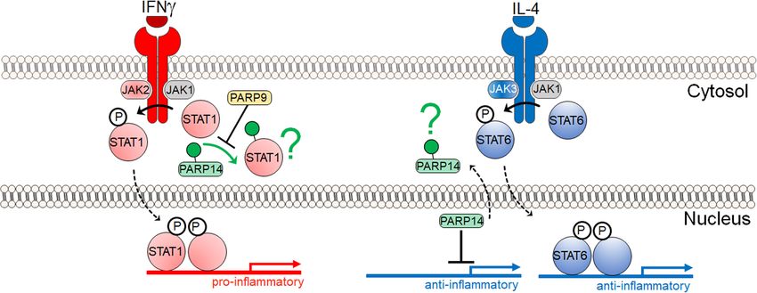
Downloaded from genesdev.cshlp.org on May 18, 2020 - Published by Cold Spring Harbor Laboratory Press
Fehr et al.
A B
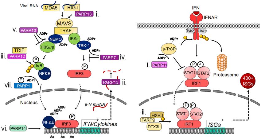
Figure 1. PARPs regulate the innate immune response at many different points. (A) Mechanisms used by MARylating and nonenzymatic
PARPs to modulate IFN and proinflammatory cytokine induction. (i) PARP13 can bind to RIG-I, which promotes its oligomerization and
the initiation of the cascade. (ii) PARP13 can also bind to IFN mRNA and target it for degradation. (iii) PARP12 was shown to bind TRIF
and enhance NFκB-dependent gene expression. (iv) PARP7 can ADP-ribosylate TBK-1, which inhibits it from phosphorylating IRF3.
(v) PARP10 can interact with and ADP-ribosylate NEMO, which prevents the activation of IKKs. (vi) PARP14 promotes H3K27 acetylation
and recruitment of Pol II to IFN promoters. (vii) Upon phosphorylation, PARP1 can poly-ADP-ribosylate NFκB and promote its activity.
(B) Mechanisms used by MARylating and nonenzymatic PARPs to modulate IFN-I signaling. (i) PARP11 binds to and ADP-ribosylates the
E3 ubiquitin ligase β-TrCP. This allows β-TrCP to interact with and ubiquitinate IFNAR, which targets it for proteasome-dependent deg-
radation. (ii) PARP9 and DTX3L interact with and ubiquitinate histone protein H2BJ, which leads to chromatin remodeling that enhances
expression of a subset of ISGs. (P) Phosphate group; (ADPr) ADP-ribose; (Ac) acetyl modification; (yellow ciricle) ubiquitin.
2013; Rosenthal et al. 2013). PARG is a potent enzyme 2019; Trott and Fadel 2019). Many investigations have fo-
that degrades poly-ADP-ribose, with several isoforms cused on the major role of macrophages in such contexts
that are derived from the single PARG gene through alter- and mechanisms for their proinflammatory activation
native splicing (i.e., 110-, 102-, 99-, and 60-kDa proteins). (Murray and Wynn 2011; Wynn and Vannella 2016; Gis-
The 110-kDa isoform, mostly seen in the nucleus, appears terå and Hansson 2017; Tabas and Lichtman 2017; Dec-
to play a dominant role in the PAR degradation. PARG ano and Aikawa 2018; Funes et al. 2018; Swirski and
cannot erase ADP-ribose when bound to proteins and Nahrendorf 2018; O’Rourke et al. 2019). Various sig-
leaves MARylated amino acid residues. PARG is a useful nal-transduction pathways participate in macrophage
tool that enables researchers the ability to enrich for activation, which are often regulated by PTMs such as
MARylated peptides for mass spectrometry analysis of phosphorylation and acetylation (Tietzel and Mosser
ADP-ribosylation (Higashi et al. 2019). 2002; Park et al. 2011; Zhou et al. 2014; Nakano et al.
2016; Vergadi et al. 2017; Dean et al. 2019). This section
focuses on the impact of PARPs and ADP-ribosylation in
macrophage activation and also summarizes their roles
PARPS in immune cells: a focus on inflammation
in the biology of other immune cells.
Immune cells serve an important role in the immune sys-
tem and differentiate into various subsets that perform a
PARP1 induces macrophage activation
spectrum of unique functions. The balance of the number
and inflammation
of different immune cell types and their activation levels
is crucial for health and disease. Overwhelming evidence Evidence suggests that ADP-ribosylation participates in
has associated chronic inflammation with various patho- inflammation (Bai and Virág 2012; Rosado et al. 2013;
logical conditions and their potential causes, including Kunze and Hottiger 2019). PARP1 has been implicated
atherosclerosis, cardiovascular events, cancer, autoim- in the mechanisms for responses (e.g., proinflammatory
mune diseases, metabolic disorders, neurological diseas- cytokine expression) of macrophages or macrophage-like
es, and aging (Johnston et al. 1987; Gisterå and Hansson cell lines to pathogen-associated molecular patterns
2017; Tabas and Lichtman 2017; de Vries and Quax (PAMPs), including lipopolysaccharide (LPS) (Hassa et al.
2018; Gomez et al. 2018; Aday and Ridker 2019; Bercovici 2005; Liu et al. 2012a; Yang et al. 2014; Minotti et al.
et al. 2019; Di Benedetto et al. 2019; Guner and Kim 2019; 2015; Bohio et al. 2019). Some responses involve the inter-
Horwitz et al. 2019; O’Rourke et al. 2019; Othman et al. play between PARP1 and nuclear factor κB (NF-κB), a key
2 GENES & DEVELOPMENT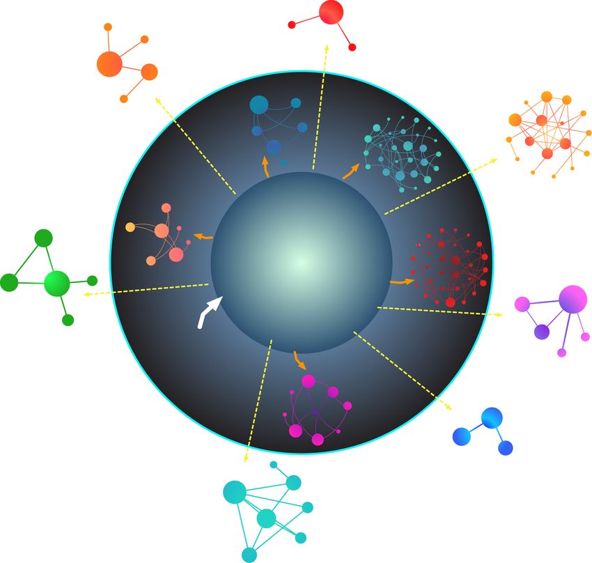
Downloaded from genesdev.cshlp.org on May 18, 2020 - Published by Cold Spring Harbor Laboratory Press

                                                                                           The role of PARPs on the immune response

transcription factor in immunity and various other biolog-         not involved in the differentiation of naïve T cells into T
ical processes (Hassa and Hottiger 1999; Hassa et al. 2005;        helper 17 (Th17) cells, it does impact Tregs, as these cells
Liu et al. 2012a; Minotti et al. 2015; Bohio et al. 2019;          are augmented in multiple organs in PARP1-deficient
Kunze and Hottiger 2019). Using PARP1-deficient mice               mice (Nasta et al. 2010). Using PARP1-deficient mice,
Oliver et al. (1999) provided evidence that PARP1 pro-             Nasta et al. (2010) demonstrated that PARP1 supresses
motes NF-κB activation in macrophages in vivo. This                the expression of Foxp3 and thus generation of Tregs via
study demonstrated that PARP1 deletion causes resis-               modulation of the chromatin structure and/or regulation
tance to LPS-induced endotoxic shock by NF-κB-depen-               of the transcription factors. Additional studies used
dent iNOS induction and NO production. Recent                      PARP1-deficiet mice further demonstrated mechanisms
evidence suggests that phosphorylation of PARP1 results            for PARP1-regulated suppression of Tregs via transform-
in PARylation of the NF-κB subunit p65/RelA, which in-             ing growth factor β (TGF β) receptors (Zhang et al. 2013).
duces the transcription of NF-κB-regulated genes (Fig.             Recent reports demonstrate the interactive role of
1A; Bohio et al. 2019). PARP1 also induces the release of          PARP1 and PARP2 in maintaining the number and func-
the high-mobility group box 1 (HMGB1), a proinflamma-              tion of T cells and promoting the development and func-
tory factor, from the nucleus to the cytoplasm in macro-           tion of B cells (Navarro et al. 2017; Galindo-Campos
phages, which requires its PARylation and subsequent               et al. 2019). Defective thymocyte maturation is observed
acetylation (Ditsworth et al. 2007; Yang et al. 2014).             in PARP1/PARP2-deficient mice, and accordingly T-cell
PARP1 also exerts proinflammatory effects on other mac-            numbers in peripheral blood are reduced (Galindo-Cam-
rophage-related cell types such as Kupffer cells in the fatty      pos et al. 2019). In PARP1/PARP2-deficient mice, the de-
liver and microglia in the injured brain (Ullrich et al. 2001;     velopment of bone marrow B cells is impaired, leading to
Mukhopadhyay et al. 2017). In some cases, PARP-1-in-               the reduction of transitional and follicular B cells (Nav-
duced activation of proinflammatory mediators, such as             arro et al. 2017). PARP1 also plays a role in the maturation
NF-κB, do not depend on its enzymatic activity, suggest-           and function of dendritic cells by regulating the produc-
ing mechanisms used by PARP1 to impact inflammation                tion of IL-10 and IL-12 (Aldinucci et al. 2007).
depend on the context or its targets (Hassa et al. 2005;
Minotti et al. 2015).
                                                                   PARP1 promotes experimental cardiovascular disorders
                                                                   A series of in vivo studies from the Boulares and Matter
The role of nicotinamide adenine dinucleotide (NAD±)
                                                                   groups (Oumouna-Benachour et al. 2007; von Lukowicz
in PARP1-mediated macrophage activation
                                                                   et al. 2008; Hans et al. 2009, 2011) used PARP1-deficient
PARPs catalyze the transfer of ADP-ribose from NAD± to             mice to demonstrate that PARP1 promotes the develop-
target proteins. Hence, NAD± is consumed by PARPs, and             ment of various cardiovascular disorders. Two studies
the activity of PARPs depends on the availability of NAD±          reported that PARP1 deficiency in apolipoprotein E-defi-
(Gupte et al. 2017). A recent report indicates that cell-          cient (ApoE −/−) mice reduces the size, macrophage and
autonomous production of NAD± via the kynurenine                   T-cell content, death of macrophage foam cells, necrotic
pathway (KP) is required to induce normal inflammatory             core, NF-kB activation, and adhesion molecule expression
macrophage activation and that the de novo NAD± syn-               in experimental atherosclerotic lesions, typical features of
thesis can be impaired in aged macrophages (Minhas                 human plaques prone to acute thrombotic events
et al. 2019). Another study proposed a mechanism linking           (Oumouna-Benachour et al. 2007; von Lukowicz et al.
the NAD± salvage pathway to LPS-induced PARP1 con-                 2008). Parp1 deletion attenuates dyslipidemia-induced
sumption of NAD± (Cameron et al. 2019). In LPS-stimu-              vascular dysfunction in ApoE −/− mice seemingly via
lated macrophages, an increase in reactive oxygen                  maintenance of eNOS activity (Hans et al. 2011). PARP1
species induces DNA damage, which in turn activates                deficiency furthermore improves delated cardiomyopha-
PARP1, leading to a reduction of available NAD±. Nico-             thy and concomitantly increases tissue inhibitor of metal-
tinamide phosphoribosyltransferase (NAMPT) is there-               loproteinase 2 (TIMP2) in ApoE −/− mice (Hans et al. 2009).
fore increased to maintain NAD± levels, which is crucial
for normal inflammatory macrophage activation.
                                                                   PARP9 and PARP14 regulate macrophage activation
                                                                   While many reports suggested multiple proinflammatory
PARP1 participates in the biology of other immune cells
                                                                   roles for PARP1, contributions of other PARP family
PARP1 modulates the differentiation of T cells into effec-         members in macrophage activation remain incompletely
tor T cells such as T helper 1 (Th1), T helper 2 (Th2), and        understood. We demonstrated that PARP9 and PARP14
regulatory T cells (Tregs) (Saenz et al. 2008; Nasta et al.        coregulate proinflammatory activation of human macro-
2010). PARP1 deficiency in murine T cells leads to the             phages (Iwata et al. 2016). In this study we took a sys-
increased expression of the Th1 cytokine interferon-γ              tems-biology approach involving unbiased proteomics,
(IFN-γ) and the decreased production of the Th2 cytokine           bioinformatics, and network analysis to identify potential
interleukin 4 (IL-4) (Saenz et al. 2008). IL-4 suppresses          molecular switches of the balance of proinflammatory
IFNγ secretion and Th1 differentiation, and PARP1 pro-             versus non/anti-inflammatory macrophage phenotypes
motes IL-4 expression via chromatin modifications at               as potential therapeutic targets. We performed proteo-
the IL-4 locus (Saenz et al. 2008). Although PARP1 is              mics of human and mouse macrophage-like cell lines

                                                                                                  GENES & DEVELOPMENT            3
Downloaded from genesdev.cshlp.org on May 18, 2020 - Published by Cold Spring Harbor Laboratory Press

Fehr et al.

treated with IFNγ or IL-4, which represent so-called proin-            stability and thus expression levels of tissue factor, a sur-
flammatory M1 versus non/anti-inflammatory M2 cells.                   face glycoprotein that plays a major thrombogenic role in
We processed our proteomics data of >5000 proteins                     macrophage-rich atherosclerotic lesions. PARP14’s asso-
with a conventional filtering method as well as our origi-             ciation with anti-flammatory STAT6 was first described
nal clustering method (Ricchiuto et al. 2015) to identify              using a yeast two-hybrid screen (Goenka and Boothby
molecules that increased with IFNγ and decreased with                  2006). Although subsequent studies were not done in
IL-4 (Iwata et al. 2016). Interestingly, the only protein              the context of macrophage activation, they indicated
that emerged from this stringent criteria was PARP14.                  that PARP14’s enhancement of IL-4 STAT6’s transcrip-
We also recognized that PARP9 showed similar responses.                tional activity may be more relevant to its promoter bind-
   The same study demonstrated in vitro that PARP14                    ing functions rather than its potential to ADP-ribosylate
suppresses proinflammatory IFNγ–STAT1 signaling and                    STAT6 itself (Fig. 2; Goenka et al. 2007; Mehrotra et al.
activates the anti-inflammatory IL-4-STAT6 pathway in                  2011). For instance, PARP14 can act as a transcriptional
primary human macrophages (Fig. 2; Iwata et al. 2016). Si-             repressor of STAT6 target genes, but activation by IL-4
lencing of PARP14 by siRNA accelerated the induction of                leads to its autoribosylation and dissociation of PARP14
proinflammatory cytokines and chemokines (e.g., TNFα,                  from a DNA-protein complex, thereby promoting STAT6
IL-1β, and CCL2/MCP-1) in IFNγ-treated macrophages                     binding instead (Fig. 2; Goenka et al. 2007; Mehrotra et al.
and suppressed anti-inflammatory molecules (e.g.,                      2011). Thus, despite the accumulating evidence that
MRC1and Arg1) in IL-4-treated cells. PARP9 silencing                   PARP14 promotes an anti-inflammatory state, there
generally exerted opposing effects. PARP9 also appeared                does not yet exist enough information to disentangle
to interfere with PARP14’s suppressive action on the                   PARP14’s enzymatic functions from that of its protein-in-
IFNγ–STAT1 axis, thus promoting proinflammatory mac-                   teraction functions, and how each of these functions may
rophage activation (Fig. 2). Cell-free enzyme reactions                maintain an anti-inflammatory response, irrespective of
with mass spectrometry as a read-out further indicated                 the cytokine or stimulus.
that ADP-ribosylation of STAT1 by PARP14 may reduce                       Evidence established that macrophages are a heteroge-
phosphorylation of this proinflammatory mediator. How-                 neous group of cells, as represented by the well-known
ever, the mechanisms used by PARP14 to interact with                   theory of M1 versus M2 polarization. Recent understand-
STAT1 and influences its ADP-ribosylation and phos-                    ing, however, suggests that macrophage heterogeneity is
phorylation requires further investigations.                           more complex and multidimensional than the M1/M2
   Due to the well-recognized knowledge that targets iden-             dichotomy (Murray et al. 2014; Nahrendorf and Swirski
tified in basic science often fail in the clinical stage, we           2016; Decano and Aikawa 2018). In our study, single-cell
performed network analysis that closely linked the net-                analysis demonstrated that IFNγ-elicited macrophages re-
work of PARP9, PARP14, and their first neighbor interac-               main largely heterogeneous, consisting of a few clusters
tors with the human coronary artery disease gene module                rather than uniformly “polarized” toward an activated
(Fig. 3). As predicted, in vivo studies in Parp14 −/− mice             phenotype (Iwata et al. 2016). Gene similarity maps dem-
demonstrated that PARP14 participates in the pathogene-                onstrated close interactions between PARP9, PARP14,
sis of arterial diseases. Consistent with in vitro studies,            STAT1, and STAT6, supporting our in vitro data described
PARP14 deficiency indeed mitigated lesion development                  above.
and inflammatory burden in models of coronary artery
disease in mice (Iwata et al. 2016).
                                                                       PARP14 regulates lymphocyte biology
   The anti-inflammatory role of PARP14 reported by us
are consistent with other reports. Iqbal et al. (2014) dem-            Several pieces of evidence demonstrate that PARP14 pro-
onstrated in macrophages that PARP14 reduces mRNA                      motes the differentiation of naïve T cells into Th2 cells by

                                                                                          Figure 2. A partial model of PARP14 and
                                                                                          PARP9 function in macrophage activation.
                                                                                          In vivo and in vitro studies pertaining to
                                                                                          IFNg signaling in primarily macrophages sug-
                                                                                          gest that PARP14 mitigates proinflammatory
                                                                                          phosphorylated STAT1 via ADP-ribosyla-
                                                                                          tion, and that PARP9 may act to inhibit
                                                                                          PARP14’s enzymatic activity (Iwata et al.
                                                                                          2016). In vitro studies pertaining to IL-4 sig-
                                                                                          naling in the context of B-cell biology suggest
                                                                                          that in nonstimulating conditions PARP14 is
                                                                                          a suppressor of STAT6 target genes. In re-
                                                                                          sponse to IL-4, PARP14 is thought to become
                                                                                          enzymatically active and dissociate from the
promoter(s), thereby allowing phosphorylated STAT6 to bind and activate target genes (Mehrotra et al. 2011). A green question mark in-
dicates that the fate of ADP-ribosylated substrates is not known. The IFNγ and IL-4 mechanisms appear distinct, but they may be partial
and complementary pictures of a complex biology.

4       GENES & DEVELOPMENT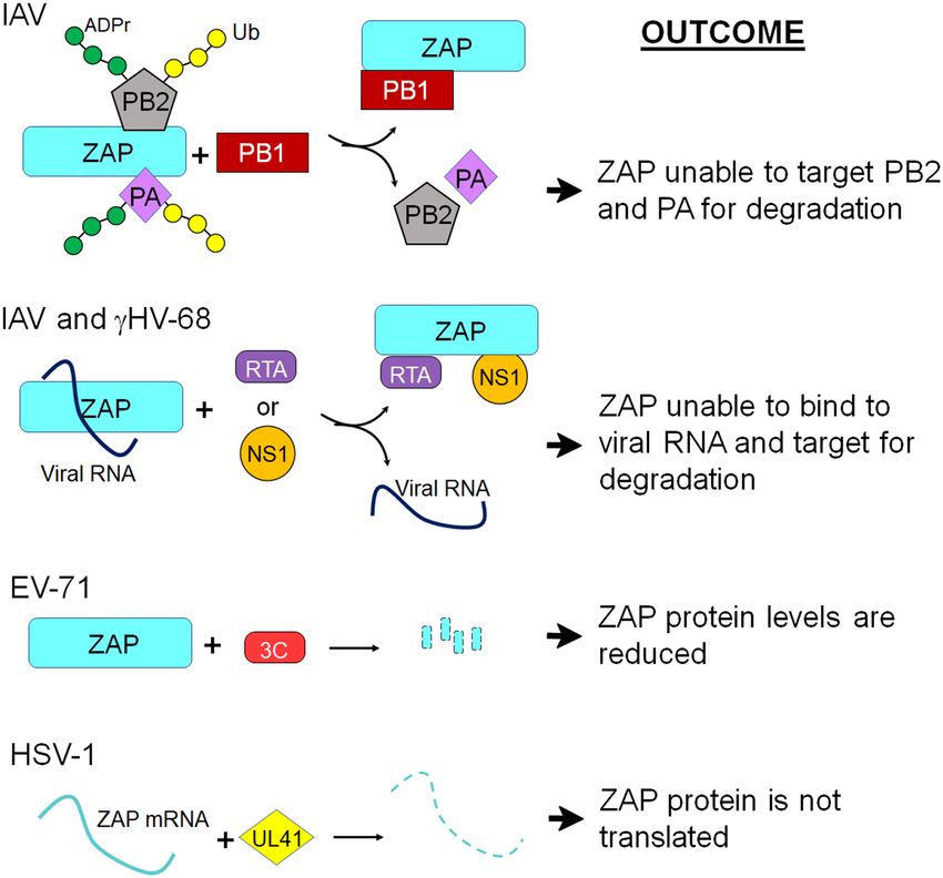
Downloaded from genesdev.cshlp.org on May 18, 2020 - Published by Cold Spring Harbor Laboratory Press

                                                                                                                                 The role of PARPs on the immune response

                                                     Hypertension                                                          Figure 3. Computational prediction of an
      Hypercholesterolemia                                                                                                 association between the PARP9–PARP14
                                                                                                                           network and human inflammatory diseases.
                                                     p=0.21
                                                                                                                           The network of PARP14 (blue)–PARP9 (pur-
                                                                                                                           ple) consists of proteins that directly interact
                                                                                                 Cardiomyopathy
                           p=0.21          Dermatomyositis                                                                 with these PARPs (blue and orange nodes, re-
                                                             Systemic lupus
                                                             erythematosus                                                 spectively). P-values indicate the significance
                                                                                                                           of closeness between the PARP14–PARP9
                                                                                                                           first neighbors in the interactome (the
                                          p=0.008            p
Downloaded from genesdev.cshlp.org on May 18, 2020 - Published by Cold Spring Harbor Laboratory Press

Fehr et al.

studies have begun to interrogate the complex biology of           sylated forms of PARP14 and PARP9, and interestingly
the PARP family members in the context of immunity.                SQSTM1, the ubiquitin pathway associated protein iden-
   Nearly 30 years ago, radioactive labeling strategies dem-       tified as a candidate PARP14 binding partner (Caprara
onstrated an increase in ADP-ribosylation signal in human          et al. 2018). These studies point toward ADP-ribosylation
monocyte-derived macrophages in response to IFNγ (Ber-             linking macrophage activation with protein homeostasis,
ton et al. 1991). In addition, there was no increase in            as indicated by the changes in ADP-ribosylation in nu-
PARP1 mRNA, implicating additional mechanisms rather               merous translational machinery; and by the emergence
than an increase in total PARP1 levels for the increase in         of proteins involved in ubiquitination (Higashi et al.
ADP-ribosylation. Using quantitative proteomics, we                2019).
made a similar observation in IFNγ-treated THP-1 and
RAW264.7 macrophage-like cell lines (Iwata et al. 2016).
Moreover, PARP14 and PARP9 exhibited the increased ex-             PARPS, ADP-ribosylation, and host–pathogen
pression following IFNγ exposure and had the opposite re-          interactions
sponse to IL-4, decreasing in abundance over the
stimulation period. These contrasting proinflammatory              ADP-ribosylation is well-known to play an important role
and anti-inflammatory responses by PARP9 and PARP14                in many host–pathogen interactions. For instance, many
were distinct from the other PARPs measured in the study.          important bacterial toxins are ADP-ribosyltransferases
More specifically, their responses to IFNγ were character-         (ARTs). Bacterial pathogens such as B. pertussis, V. chol-
istic of known cytokine inducible genes, STAT1, NMI (N-            era, P. aeruginosa, C. botulinum, S. aureus, and E. coli en-
myc interactor), OAS2, IFIH1, and IFIT3, among several             code for ARTs that target proteins such as elongation
others. This study thus provided a plausible scenario              factor 2, actin, and Rho GTPases that lead to cell death
where the increase in global ADP-ribosylation in macro-            (Holbourn et al. 2006). As discussed above, mammals
phages could be mediated in part by PARP14 (Iwata et al.           also encode for a diverse set of ARTs, most of which are
2016).                                                             termed PARPs, that impact infections. Here we discuss
   In a separate study, using quantitative proteomics in           mammalian encoded PARPs that are involved in host–
combination with shRNA-mediated knockdown experi-                  pathogen interactions, focusing on virus infections.
ments, PARP14 interactors were pursued in the context
of LPS signaling in RAW264.7 cells (Caprara et al. 2018).
                                                                   Mammalian PARPs display several properties indicative
A PARP14 coimmunoprecipitation of these RAW264.7
                                                                   of involvement in host–pathogen interactions
cells after LPS treatment found that SQSTM1 (Seques-
tome-1, a receptor linking autophagy and ubiquitylation),          PARPs interact with pathogens in many ways, and here
PARP9, DTX3L, and NMI were immunoprecipitated with                 we describe specific cases where they either promote or
a PARP14 antibody specifically in PARP14 wild-type but             restrict virus replication and the innate immune response.
not PARP14 knockdown cells, indicating that these pro-             This discussion is summarized in Table 1 and Figure 1.
teins form a PARP14–protein interaction network in re-             However, we start by discussing characteristics that indi-
sponse to LPS.                                                     cate an important role for PARPs in the host response to
   Protein–protein interactions alone do not clarify the           infection.
role of PARP enzyme activities in immunity. To date,                  First, many mammalian PARPs are stimulated by the
only one study has investigated ADP-ribosylated sub-               production of IFN (IFN-stimulated genes [ISGs]), and
strates in immune cells on a global level (Higashi et al.          thus are part of the mammalian antiviral defense system
2019). Since ADP-ribosylome studies are technically                (Atasheva et al. 2012; Zhang et al. 2015; Eckei et al.
challenging to perform; they rely on specialized proteo-           2017; Li et al. 2018; Grunewald et al. 2019a). Also, several
mic workflows to enrich and sequence ADP-riboyslated               mono-ADP-ribosylating PARPs are rapidly evolving, indi-
peptides (Martello et al. 2016; Larsen et al. 2017). We            cating ongoing conflict with pathogens. The PARP domain
used two independent approaches to enrich the ADP-                 of PARP13, a disordered region of PARP4, and the macro-
ribosylome of IFN-γ-treated THP-1 cells. One approach              domain(s) of the three macro-PARPs, PARP9, PARP14,
used an anti-PARylation antibody enrichment that pro-              and PARP15, are all under positive selection (Kerns et al.
vided candidate ADP-ribosylated proteins, and the other            2008; Daugherty et al. 2014). Furthermore, Parp14 and
enriched for MARylated peptides via their affinity for a           Parp15 have undergone multiple rounds of gene loss and
macrodomain to identify ADP-ribosylated proteins (Higa-            duplication, which creates novel gene products needed
shi et al. 2019). The majority of proteins identified from         for continual adaptation to new pathogens.
both approaches combined, comprised ribosome/RNA-                     Several PARP proteins are also present in stress gran-
binding proteins. These findings were not surprising               ules, which are important membrane-less organelles
since this protein class is highly abundant, and thereby           that function to restrict the translation of RNA when cells
more conducive to identifying ADP-ribosylated peptides.            are under stress, such as during a virus infection. They are
Other ADP-ribosylated proteins were associated with                often characterized by the presence of TIA1, TIAR, and
pathways involved in neutrophil degranulation and acti-            G3BP1, but are known to contain several hundred pro-
vation, IL-12 signaling, and glycolysis. Moreover, ADP-            teins (McCormick and Khaperskyy 2017). Interestingly,
ribosylation of a subset of the ribosome/RNA-binding               several proteins in stress granules are ADP-ribosylated,
proteins increased in response to IFNγ, as did ADP-ribo-           and PARPs, including 5a, 12, 13, 14, and 15, are known

6       GENES & DEVELOPMENT
Downloaded from genesdev.cshlp.org on May 18, 2020 - Published by Cold Spring Harbor Laboratory Press
The role of PARPs on the immune response
Table 1. PARPs with antiviral or proviral functions
Mono or Virus Proviral or
PARP/ARTH poly targeted antiviral Mechanisms References
PARP1/ARTD1 Poly HIV-1 Both Binds to TAR element to block HIV Gäken et al. 1996; Kameoka et al.
MLV Both transcription; binds to TR sequences 1999, 2004, 2005; Baekelandt
MPSV Pro in KSHV; ADP-ribosylates EBNA1 to et al. 2000; Ha et al. 2001;
EBV Anti prevent OriP from binding genome; Gwack et al. 2003; Ariumi et al.
KSHV Anti binds to and modified RTA protein of 2005; Parent et al. 2005;
γ-2 HV Anti γ-HVs to block transcription; binds to Tempera et al. 2010; Bueno
PRRSV Pro PRRSV nucleocapsid protein et al. 2013; Chung et al. 2015;
Grady et al. 2012; Liu et al.
2012a,b, 2015a,b
PARP5a/ARTD5 Poly HSV-1 Pro ADP-ribosylates EBNA1 to prevent OriP Deng et al. 2005; Grady et al.
EBV Anti from binding genome 2012
PARP7/ARTD14 Mono IAV Pro ADP-ribosylates TBK-1, which inhibits Atasheva et al. 2012, 2014;
SINV Anti IFN production; blocks cellular Kozaki et al. 2015, 2017;
VEEV Anti translation; binds to viral RNA and Yamada et al. 2016; Grunewald
RV Anti recruits exosomes to sites of et al. 2019b
MHV Pro replication
PARP12/ARTD12 Mono VEEV Anti Binds viral RNA and blocks translation; Atasheva et al. 2012, 2014; Liu
SINV Anti targets Zika virus proteins for ADP- et al. 2012b; Grunewald et al.
EMCV Anti ribosylation and degradation 2019a
VSV Anti
CHIKV Anti
ZIKA Anti
MHV Anti
PARP13/ARTD13 Inactive MLV Anti Binds RNA leading to RNA degradation Gao et al. 2002; Bick et al. 2003;
ALV-J Anti or translation inhibition; targets Guo et al. 2007, 2019; Muller
HIV-1 Anti influenza proteins PB2 and PA for et al. 2007; Hayakawa et al.
SINV Anti ADP-ribosylation and degradation; 2011; Zhu et al. 2011; Li et al.
SFV Anti binds to RIG-I and promotes its 2015, 2017; Liu et al. 2015a;
RRV Anti oligomerization; ZnF domains bind to Xuan et al. 2012, 2013; Mao
PRRSV Anti PRRSV nsp9 et al. 2013; Xie et al. 2018;
EBOV Anti Zhao et al. 2019; Takata et al.
MARV Anti 2017
EV-A71 Anti
γHV-68 Anti
HBV Anti
IAV Anti
PARP9/ARTD9 Monoa EMCV Anti With DTX3L, ubiquitinates histone Zhang et al. 2015
IAV Anti H2BJ and enhances ISG expression
SINV Anti
PARP14/ARTD8 Mono MHV Anti Enhances histone acetylation to Caprara et al. 2018; Grunewald
promote transcription of IFN-I genes et al. 2019a
PARP10/ARTD10 Mono VEEV Anti Blocks cellular translation Atasheva et al. 2012, 2014
PARP11/ARTD11 Mono VSV Pro Targets IFNAR for degradation, leading Guo et al. 2019
HSV-1 Pro to decreased IFN production
a
PARP9 is only known to be active when bound to DTX3L.
(Green) DNA-dependent PARPs; (blue) ZnF PARPs; (yellow) macro-PARPs; (orange) other-PARPs.
(HIV-1) Human immunodeficiency virus; (MLV) murine leukemia virus, (MPSV) myeloproliferative sarcoma virus; (EBV) Epstein-Barr
virus; (KSHV) Kaposi’s sarcoma herpesvirus; (PRRSV) porcine reproductive and respiratory syndrome virus; (HSV-1) herpes simplex
virus-1; (IAV) influenza A virus; (SINV) sindbis virus; (VEEV) Venezuelan equine encephalitis virus; (RV) rubella virus; (γ-2 HV) γ-2
herpesvirus; (γHV-68) murine γ-herpesvirus-68; (EMCV) encephalomyocarditis virus; (VSV) vesicular stomatitis virus; (CHIKV) chi-
kungunya virus; (ZIKV) Zika virus; (MHV) murine hepatitis virus; (ALV-J) avian leukosis virus subgroup J; (SFV) Semliki forest virus;
(RRV) Ross River virus; (EBOV) Ebola virus; (MARV) Marburg virus; (EV-A71) enterovirus strain A71; (HBV) hepatitis B virus.
constituents of stress granules (Leung et al. 2011). It has Some virus families encode for a macrodomain protein
been proposed that polyADP-ribose facilitates the concen- that reverses cellular ADP-ribosylation
tration of RNA-binding proteins in stress granules and
other nonmembranous structures and thereby promote Several decades ago, a conserved domain was identified in
their oligomerization (Leung 2014). all coronaviruses (CoVs), togaviruses, and hepatitis E virus
GENES & DEVELOPMENT 7Downloaded from genesdev.cshlp.org on May 18, 2020 - Published by Cold Spring Harbor Laboratory Press
Fehr et al.
that was termed the “X” domain (Gorbalenya et al. 1991; to the transactivation response element (TAR) and inhibit
Koonin et al. 1992). These domains are structurally ho- HIV-1 transcription by competing with TAR for binding
mologous to the nonhistone part of the macroH2A protein to p-TEF2b. The impact of PARP1 on HIV-1 infection re-
and are now known as macrodomains (Allen et al. 2003). mains controversial and is likely context-dependent.
Macrodomains from all three viral families bind mono- In addition to its role in regulating retrovirus replica-
and poly-ADP-ribose, and can efficiently remove tion, PARylation enhances and represses several other vi-
mono-ADP-ribose from proteins by hydrolysis, strongly ruses. This includes herpesviruses, where PARPs have a
indicating a role for ADP-ribosylation in either promoting wide range of effects. PARP1 and the tankyrase PARP5a
or inhibiting the replication of these viruses (Egloff et al. modify the EBV protein EBNA1. PARylation of EBNA1
2006; Li et al. 2016). Several studies on the CoV and alpha- causes it to dissociate from the dyad symmetry elements,
virus macrodomains have established that this protein which restricts OriP binding and impairs the maintenance
domain is critical for either replication or pathogenesis of the viral episome during latency (Deng et al. 2005; Tem-
(Eriksson et al. 2008; Park and Griffin 2009; Fehr et al. pera et al. 2010). PARP1 also binds to the TR sequences
2015, 2016; McPherson et al. 2017). Recent studies using in KSHV, which leads to reduced viral genome levels dur-
chikungunya virus (CHIKV) macrodomain mutants ing latency. PARP1 and the Ste-20-like kinase hKFC syn-
showed that macrodomain ADP-ribose binding facilitated ergistically bind to and ADP-ribosylate/phosphorylate the
initiation of virus replication, while hydrolase activity γ-2 herpesvirus replication and transcription activator
was essential for the amplification of replication complex- protein (RTA) (Fig. 4A). These interactions suppress
es (Abraham et al. 2018). Infection with ADP-ribosylhy- RTA-mediated transcriptional activation and KSHV lytic
drolase (ARH)-deficient CoVs, including severe acute reactivation (Gwack et al. 2003). Two mechanisms have
respiratory syndrome (SARS)-CoV and murine hepatitis been described by which γ-herpesviruses counter PARP1
virus (MHV), led to higher levels of IFN and other cyto- activity. First, it was found that ORF49 of γHV-68 binds
kines, indicating that it may block the innate immune re- to PARP1, preventing it from interacting with RTA (Fig.
sponse (Fehr et al. 2016; Grunewald et al. 2019a). The 4B). In addition, the processivity factor of KSHV and
ARH-deficient MHV replicates poorly in primary macro- γHV-68, PF-8, binds to and targets PARP1 for degradation,
phages, and importantly, this defect could be partially res- which reduces PARylated RTA and enhances virus repli-
cued by PARP inhibitors, directly indicating PARPs in the cation (Fig. 4C; Noh et al. 2012; Chung et al. 2015). In con-
antiviral response to CoVs (Grunewald et al. 2019a). How- trast to the antiviral effects of PARPs during γ-herpesvirus
ever, it remains unknown what proteins may be targeted infection, PARP activity seems to promote the replication
by the viral macrodomains (for reviews, see Fehr et al.
2018; Leung et al. 2018).
The roles of PARP1 and the Tankyrase PARPs in virus A
replication.
Some of the first reports of PARPs and ADP-ribosylation
impacting virus infection focused on the role of PARP1 B
on retrovirus and HIV-1 integration and replication.
Gäken et al. (1996) first demonstrated that PARP inhibi-
tors led to reduced retroviral integration into host chroma- C
tin. They further used antisense oligonucleotides and
overexpression of dominant-negative PARP1 to confirm
that PARP activity is required for integration of retroviral
vectors. Other groups further demonstrated the impor-
D
tance of PARP activity in retrovirus and HIV-1 integration
into host chromosomes using siRNA transfected and
PARP1-deficient cells (Ha et al. 2001; Kameoka et al.
2005). Mechanistically, it was suggested that PARP1
may help resolve a 4- to 6-bp gap in the genome produced Figure 4. Mechanisms used by herpesviruses to affect PARyla-
during integration (Ha et al. 2001). PARP1 activity may tion and their impact on replication. (A) PARP-1 can bind to
also impact HIV-1 transcription and replication (Kameoka and ADP-ribosylate the γHV RTA, which inhibits its ability to
et al. 1999, 2004). However, these results have been con- initiate lytic replication. (B) The γHV-68 protein ORF49 binds
founded by other reports that demonstrated either no evi- to PARP1 and prevents it from interacting with and ADP-ribosy-
lating RTA, which allows RTA to initiate viral gene transcrip-
dence that PARP1 was required for efficient HIV-1
tion. (C) The KSHV and γHV-68 PF-8 proteins bind to PARP1
integration or replication (Baekelandt et al. 2000; Ariumi
and target it for ubiquitination and degradation. This again pre-
et al. 2005), or evidence that PARP1 can repress HIV-1 vents ADP-ribosylation of RTA, which allows it to initiate lytic
or retrovirus infection (Parent et al. 2005; Bueno et al. replication. (D) The HSV-1 ICP0 protein targets PARG for ubiqui-
2013). Bueno et al. (2013) found that PARP1 inhibited ret- tination and degradation, resulting in enhanced PARylation dur-
roviral infection in a chicken B lymphoblastoid cell line, ing infection and increased replication. (ADPr) ADP-ribose; (Ub)
while Parent et al. (2005) showed that PARP1 could bind ubiquitin.
8 GENES & DEVELOPMENTDownloaded from genesdev.cshlp.org on May 18, 2020 - Published by Cold Spring Harbor Laboratory Press
The role of PARPs on the immune response
of HSV-1, the prototype α-herpesvirus. PARP5a (Tankyr- PARP13 (ZnF antiviral protein) PARP13, or ZnF antivi-
ase-1) expression was increased and it was translocated ral protein (ZAP), was one of the first PARPs identified to
to the nucleus during HSV-1 infection. Knockdown of have antiviral activities when, in a screen for antiviral
both PARP5a and PARP5b resulted in a threefold to four- ISGs, it was found to potently inhibit murine leukemia vi-
fold decrease in virus replication, and inhibition of their rus (MLV) replication (Gao et al. 2002). Somewhat surpris-
catalytic activity with XAV-939 resulted in a greater ingly, ZAP lacks the triad motif (H-Y-E) needed for
than 1-log reduction in virus replication (Li et al. 2012). catalytic activity and has no auto-ADP-ribosylating
To further indicate that PARP activity is important for vi- activity (Kleine et al. 2008). As such, most of its antiviral
rus replication, HSV-1 infection significantly increased activity is independent of ADP-ribosylation. Since its dis-
PARylation. The ICP0 protein targets nuclear forms of covery, ZAP has been shown to inhibit the replication of
PAR glycohydrolase (PARG), the enzyme that degrades several viral families, including retroviruses, alphaviruses,
PAR, for ubiquitination and degradation, providing a pos- filoviruses, picornaviruses, herpesviruses, arteriviruses,
sible mechanism for the dramatic increase in PARylation orthomyxoviruses, flaviviruses, and hepatitis B virus
during infection (Fig. 4D; Grady et al. 2012). (Bick et al. 2003; Muller et al. 2007; Zhu et al. 2011;
Finally, poly-ADP-ribosylation is implicated as having Wang et al. 2012; Xuan et al. 2012; Mao et al. 2013; Xuan
proviral activities in several viral systems. PARP inhibi- et al. 2013; Li et al. 2015, 2015a; Chiu et al. 2018; Xie
tors have led to greatly reduced infectivity of adenovirus- et al. 2018; Zhao et al. 2019). ZAP is transcribed into four
es, possibly through the ADP-ribosylation of their core different isoforms, with ZAPL and ZAPS being the most
proteins (Déry et al. 1986). PARP inhibitors also inhibit studied (Li et al. 2019). ZAPL contains the inactive PARP
JC virus replication (Nukuzuma et al. 2013). PARP1 binds or catalytic domain, while ZAPS does not. ZAPL tends
to the porcine reproductive and respiratory syndrome vi- to have greater antiviral activity, and this may be due, at
rus (PRRSV) nucleocapsid protein, and again, PARP inhib- least in part, to prenylation of the PARP domain (Charron
itors restricted the replication of PRRSV in cell culture et al. 2013; Schwerk et al. 2019). In addition to the PARP
(Liu et al. 2015b). The nucleocapsid protein of the related domain, ZAP contains four ZnF-binding domains and a
coronaviruses is also ADP-ribosylated, however the im- single WWE domain. ZAP uses its ZnF-binding domains
pact of this modification on virus replication or pathogen- to bind to viral RNA and recruits both the poly(A)-specific
esis remains unknown (Grunewald et al. 2018). Last, ribonuclease (PARN) and the RNA exosome to degrade the
PARP activity is required for efficient activity of the viral RNA (Guo et al. 2007). It also inhibits translation by
RNA polymerases derived from multiple strains of influ- blocking the interaction between eIF4G and eIF4A, and its
enza virus, indicating a potential proviral role for ADP- ability to block translation is required for it to degrade
ribosylation during influenza infection (Bortz et al. RNA. In addition, ZAP antiviral activity is enhanced by
2011). In summary, PARylation has a variety of different the ubiquitin ligase activity of TRIM25. TRIM25 binds
functions that can both repress and enhance virus to ZAP and ubiquitinates unknown proteins to enhance
replication. the antiviral activity of ZAP (Li et al. 2017; Zheng et al.
2017).
ZAP targets HIV RNAs for degradation, and prefers to
The roles of nonenzymatic and mono-ADP- target CG dinucleotides (Takata et al. 2017). Consistent
ribosylating PARPs in virus replication with this, an HIV-1 mutant with an increased CG content
and the antiviral response. replicated very poorly in MT4 cells, but that replication
was restored in ZAP-deficient cells. In addition, ZAP tar-
Nonenzymatic and mono-ADP-ribosylating PARPs have
gets the 3′ UTR of Japanese encephalitis virus (JEV), which
a variety of roles in promoting or inhibiting virus replica-
contains a high CG content (Chiu et al. 2018). Interesting-
tion. This class of PARPs include the zinc finger (ZnF)
ly, it appears many viruses maintain a low CG dinucleo-
PARPs (7, 12, and 13), the macrodomain-containing
tide level, and the level of ZAP sensitivity of several
PARPs (9, 14, and 15), and several PARPs that do not fit
viruses mildly correlates with their CG dinucleotide con-
into a specific category (4, 6, 8, 10, and 11). Here, we dis-
tent (Takata et al. 2017). However, ZAP sensitivity of a
cuss what is known about each of these PARPs in the in-
panel of alphaviruses does not correlate with the CG dinu-
nate immune response to viruses.
cleotide content found in their genome or individual viral
genes, suggesting that the CG dinucleotide motif is not
the only determinant for ZAP recognition (Li et al.
CCCH ZnF PARPs
2019). The localization of ZAP to stress granules also ap-
ZnF PARPs contain one or more ZnF domains. These do- pears to be functionally important for its antiviral activity
mains are small protein motifs that enable these PARPs to against alphaviruses, as ZAP mutants that do not localize
bind RNA. All three ZnF PARPs use this domain to inter- to SGs are unable to block SINV replication (Law et al.
act with viral RNA and inhibit either translation or 2019). In vivo, Zap knockout (Zc3hav1−/−) mice showed
degrade viral RNA, though the specific RNA sequence enhanced replication of SINV in 10-d-old mice as expected
that each PARP binds to is likely unique. In addition, all (Kozaki et al. 2015). Though surprisingly, in 23-d-old
three ZnF PARPs use either the enzymatic or nonenzy- weanling pups it was shown that a neurovirulent strain
matic functions in their PARP domain to impact the in- of SINV can use ZAP to decrease its replication in initially
nate immune response or virus replication. infected cells in vivo such that it prevents immune
GENES & DEVELOPMENT 9Downloaded from genesdev.cshlp.org on May 18, 2020 - Published by Cold Spring Harbor Laboratory Press
Fehr et al.
recognition, allowing the virus to spread to the central A
nervous system (CNS) and promote disease (Wang et al.
2016).
In addition, different studies have found conflicting re-
sults regarding the ability of ZAP to impact the innate im-
mune response. It was originally found that ZAPS, but not
ZAPL, potentiates RIG-I-dependent type I interferon (IFN-
B
I) production in human cells by binding to RIG-I via its
ZnF domains and promoting its oligomerization (Fig.
1A; Hayakawa et al. 2011; Chiu et al. 2018). However,
ZAP does not appear to enhance RIG-I-dependent IFN-I
production in mouse cells (Lee et al. 2013). More recent
data indicate that ZAPS may also reduce IFN mRNA by C
binding to the 3′ UTR of IFN mRNA and targeting it for
degradation (Fig. 1A; Schwerk et al. 2019). It is conceivable
that ZAP-S uses both functions but in a context-depen-
dent manner. D
While ZAPL does not contain an active PARP domain,
in some cases it is required for the ADP-ribosylation of
proteins. For instance, the C terminus of ZAPL binds to
the influenza virus polymerase proteins PB2 and PA, Figure 5. Viral mechanisms of ZAP antagonism. (A) IAV protein
which causes their subsequent poly-ADP-ribosylation, PB1 binds to ZAP, which prevents its interaction with the PA and
ubiquitination, and degradation (Liu et al. 2015a). It is un- PB2 proteins that otherwise would lead to PARylation, ubiquiti-
known which PARP and E3 ubiquitin ligase mediates nation, and degradation of these proteins. (B) IAV NS1 and γHV-
these effects. Knockdown of ZAPL modestly increased in- 68 RTA proteins interact with ZAP, preventing its association
with viral RNA. (C) The EV-71 3C protease cleaves ZAP to pre-
fluenza virus replication in cell culture, though it is not
vent it from accumulating. (D) HSV-1 UL41 protein cleaves
clear whether this is due to its ability to bind and target
ZAP mRNA to prevent its translation. (ADPr) ADP-ribose; (Ub)
PB2 and PA for degradation. Interestingly, this ZAPL ac- ubiquitin.
tivity was countered by the PB1 protein, which bound to
ZAPL preventing the ubiquitination of PB2 and PA, dem-
onstrating that the virus has evolved ways to neutralize
the antiviral activity of ZAP (Fig. 5A). In addition to types. This activity has been linked to the ZnF domains,
PB1, several other viral proteins have been found to coun- as mutations in the ZnF domains abrogated the ability
ter ZAP using multiple mechanisms (Fig. 5B–D). Influen- of PARP12 to move to stress granules (Welsby et al.
za A NS1 prevents ZAP-S from binding to its target RNA 2014). The translocation of PARP12 may also depend on
(Tang et al. 2017), γHV-68 RTA disrupts the intermolecu- PARP1 acting as a stress sensor in the nucleus, as an in-
lar interaction of ZAP (Xuan et al. 2013), HSV-1 UL41 de- crease in unconjugated PAR is a key factor that promotes
grades ZAP mRNA (Su et al. 2015), and, finally, the the recruitment of PARP12 to stress granules (Catara et al.
enterovirus (EV)-71 3C protease cleaves ZAP protein 2017). The translocation of PARP12 to stress granules is
(Xie et al. 2018). Due to its broad-spectrum antiviral activ- reversible, as it relocates back to the Golgi once the stress
ity, it is likely there are even more viral proteins that func- is relieved.
tion to counter ZAP. The antiviral role of PARP12 was first described in an
overexpression screen, where it mildly inhibited the repli-
PARP12 PARP12 is a mono-ADP-ribosyltransferase and cation of both VSV and MHV-68 (Liu et al. 2012b). Shortly
has four or five N-terminal CCCH-type zinc finger (ZnF) after this, another study found that PARP12 was differen-
domains that are important for RNA binding, one or two tially expressed in cells that cleared VEEV replication
WWE domains in the middle of the protein that are impor- compared with those that were persistently infected (Ata-
tant for ADP-ribose binding, and a PARP domain at the C sheva et al. 2012). Further analysis showed that PARP12L,
terminus, which provides the protein with mono-ADP- but not PARP12S, expression from a VEEV replicon or
ribosylating activity (Welsby et al. 2014). There are two virus restricts VEEV replication, as well as several other
splice forms of PARP12 mRNA, PARP12L, and PARP12S viruses including Sindbis virus (SINV), encephalomyocar-
(Atasheva et al. 2012). PARP12L contains both the ZnF ditis virus (EMCV), vesicular stomatitis virus (VSV), Rift
domains and the PARP catalytic domain, while PARP12S Valley fever virus (RVFV), and chikungunya virus
has the ZnF domains but lacks the PARP catalytic (CHIKV). Subsequently, this group showed that PARP12
domain. strongly inhibited both cellular and viral translation (Ata-
As described above, PARP12 translocates to cytoplas- sheva et al. 2014). Immunoprecipitation with PARP12
mic stress granules upon cell stress (Leung et al. 2011; identified several ribosomal proteins and translation and
Welsby et al. 2014). PARP12 initially localizes to the elongation factors, indicating that PARP12 interacts
trans-Golgi network (TGN) and translocates to stress with ribosomes. Interestingly, PARP12 required its enzy-
granules during stress stimuli in several different cell matic activity to block translation, but not to inhibit virus
10 GENES & DEVELOPMENTDownloaded from genesdev.cshlp.org on May 18, 2020 - Published by Cold Spring Harbor Laboratory Press
The role of PARPs on the immune response
replication, indicating that PARP12 may use distinct whether the ability of TiPARP to enhance MHV or IAV
mechanisms to block virus replication and cellular replication is tied to its ability to block the IFN-I response.
translation.
Additionally, PARP12 was identified in a screen for
Macrodomain-containing PARPs
ISGs that inhibit Zika virus (ZIKV) (Li et al. 2018). Using
both knockout cells and overexpression, the authors The macro PARPs contain two (PARP9/PARP15) or three
showed that PARP12 was both necessary and sufficient (PARP14) macrodomains that mediate binding to ADP-ri-
for the inhibition of ZIKV replication. PARP12 was re- bose, as described above. While some macrodomains can
quired for the ADP-ribosylation, ubiquitination, and cleave ADP-ribose from a substrate, it is likely that macro-
subsequent degradation of ZIKV NS1 and NS3 proteins. domains within these PARP proteins only bind ADP-ri-
This activity did not require either the ZnF or WWE do- bose. All three macro PARPs are rapidly evolving
mains of PARP, but did require its catalytic domain, as (Daugherty et al. 2014), and PARP15 has been identified
an inactive catalytic domain reversed the regulatory ef- in stress granules (Leung et al. 2011); however, direct evi-
fects of PARP12 on viral protein degradation. Interesting- dence of their involvement in virus infections is limited.
ly, NS1 and NS3 appeared to be poly-ADP-ribosylated, No study has identified a role for PARP15 in modulating
indicating that PARP12 may work with another PARP virus infection or the innate immune response, and thus
to mediate the poly-ADP-ribosylation of these proteins. it will not be discussed further.
PARP12 also has a role in the restriction of coronavirus
(CoV) replication, as siRNA knockdown of PARP12 PARP9 (BAL1) PARP9 was originally termed B-aggres-
partially restored replication of MHV lacking the ADP- sive lymphoma 1 gene (BAL1) as it was identified as a
ribosylhydrolase (ARH) activity of the CoV macrodomain risk factor for large diffuse B-cell lymphomas (Aguiar
(Grunewald et al. 2019a). The mechanism used by et al. 2005). It is catalytically inactive, at least when ex-
PARP12 to restrict MHV replication remains unknown. pressed by itself, but can ADP-ribosylate ubiquitin in
Finally, PARP12 was also shown to enhance NF-κB signal- the presence of an E3 ubiquitin ligase, DTX3L (Yang
ing, possibly by interacting with TRIF (Fig. 1A; Welsby et al. 2017). As described above, PARP9 promotes
et al. 2014). STAT1 phosphorylation, proinflammatory gene expres-
sion, and differentiation into M1-like macrophages (Iwata
PARP7 (TiPARP) PARP7, also known as tetrachlorodi- et al. 2016). In the antiviral response, PARP9 expression in
benzo-p-dioxin (TCDD)-inducible poly-ADP-ribose poly- malignant B-cell lymphoma lines can lead to widespread
merase (TiPARP), has a single ZnF domain that induction of ISG expression (Juszczynski et al. 2006).
mediates RNA binding. It also has a WWE domain and a Zhang et al. (2015) found that the complex of PARP9
PARP catalytic domain capable of mono-ADP-ribosyla- and DTX3L attached to STAT1 to mediate the hyper-re-
tion (Kozaki et al. 2017). sponsiveness of a mutant STAT1 protein (STAT1-CC).
Like PARP12, TiPARP was also shown to block VEEV The PARP9–DTX3L complex ubiquitinates histone pro-
replication and inhibit cellular translation when trans- teins, most notably H2BJ, which led to chromatin remod-
duced into cells by a VEEV replicon (Atasheva et al. eling and enhanced expression of at least a subset of ISGs
2014). In a separate study, siRNA knockdown of TiPARP (Fig. 1B). This interaction was necessary and sufficient to
in U373 human astrocyte cells led to increased replication inhibit the replication of multiple viruses, including
of SINV and rubella virus replication (Kozaki et al. 2017). EMCV, IAV, and SINV. The PARP9–DTX3L complex
The increase in SINV replication was also demonstrated also ubiquitinates the EMCV 3C protease, which led to
in TiParp −/− mice. TiPARP-mediated inhibition of SINV its degradation, but this effect was mostly, if not
was dependent on its ZnF domain, which binds to SINV completely, due to DTX3L activity.
RNA and recruits RNA degradation factors to sites of viral
replication. This suggests that TiPARP recognizes specific PARP14 (CoaST-6) PARP14 was originally identified as
virus RNAs for degradation, however the target specificity Collaborator of STAT6 (CoaST6) (Goenka and Boothby
of TiPARP is still unknown. 2006). It has a range of effects on cell physiology and im-
TiPARP also has proviral effects in addition to its anti- munity that were largely anti-inflammatory (Cho et al.
viral functions. TiPARP negatively regulates the type I 2011, 2013; Barbarulo et al. 2013; Vyas et al. 2013; Ian-
IFN response by ADP-ribosylating TBK1 (Fig. 1A; Yamada sante et al. 2015; Iwata et al. 2016; Krishnamurthy and
et al. 2016). The ADP-ribosylation of the kinase domain of Kaplan 2016). In the antiviral response, PARP14 instead
TBK1 suppresses IFN production. It was suggested that is required to enhance IFN-I production in RAW cells
negative regulation of IFN production by TiPARP may (transformed peritoneal macrophages) following LPS
help protect the cell from the harmful effects of type I treatment (Caprara et al. 2018), in primary macrophage
IFN. The same study found that the loss of TiPARP led cells during CoV infection, and following treatment of hu-
to decreased IAV replication, which strongly correlated man A549 cells with poly(I:C) (Grunewald et al. 2019a).
with increased IFN-I production. Our group has also found Following LPS treatment, PARP14-deficient cells showed
that siRNA knockdown of TiPARP led to decreased repli- similar levels of IRF-3 phosphorylation and nuclear trans-
cation of MHV and increased IFN-I production, further in- location, but had reduced levels of Pol II recruited to the
dicating TiPARP as a proviral factor for some viruses promoters of IRF-3-dependent genes (Caprara et al.
(Grunewald et al. 2019a,b). However, it remains unclear 2018). There was also a dramatic reduction in H3K27
GENES & DEVELOPMENT 11Downloaded from genesdev.cshlp.org on May 18, 2020 - Published by Cold Spring Harbor Laboratory Press
Fehr et al.
acetylation, a known marker of active promoters and en- protein (β-TrCP), which lead to the ubiquitination and
hancers (Fig. 1A). It is unclear whether this function of degradation of the interferon α/β receptor (IFNAR) (Fig.
PARP14 is dependent on its catalytic activity. It is also 1B; Guo et al. 2019). siRNA silencing of PARP11 or treat-
not yet known whether PARP14 has the same function ment with rucaparib, a pan-PARP inhibitor used in ad-
in the cellular response to virus infection or poly(I:C). vanced ovarian cancer, inhibited the replication of VSV
These two studies also found that PARP14 was required and HSV-1. While normally known to inhibit PARP1/2,
to restrict the replication of S. typhimurium and an at the concentrations of drug used in this study rucaparib
ARH-deficient MHV, though it is not known whether appeared to preferentially target PARP11. Interestingly,
the ability of PARP14 to inhibit these pathogens is tied following infection in vivo, rucaparib enhanced IFN-I sig-
to its role in up-regulating IFN production or whether naling, reduced VSV replication in multiple organs, and
these are distinct functions of PARP14. It is important led to better outcomes for the mice infected with either
to note that these experiments were done with undifferen- VSV or HSV-1. These data indicate that PARP11-specific
tiated or M0 macrophages, while other studies where inhibitors could be a useful means of treating specific viral
PARP-14 was found to have anti-inflammatory functions infections.
used M1 or M2 macrophages, differentiated by further In summary, several of the nonenzymatic or mono-
IFN-γ or IL-4 treatment. This suggests that PARP14 func- ADP-ribosylating PARPs are potent antiviral proteins
tion is likely context-dependent. that are able to inhibit viruses from several different viral
families. However, some do contain activities that pro-
mote virus replication. While some mechanisms are
Other PARPs
known, including blocking translation, degrading RNA,
The final class of PARPs do not fit into any of the other de- and targeting viral or host proteins for ubiquitination
fined classes of PARPs, have no similar domains other and degradation, many mechanisms are still unknown.
than the PARP domain, and are thus simply termed “oth- However, recent reports are making it clear that in
er PARPs.” These PARPs include PARPs 4, 6, 8, 10, 11, many, but not all cases, ADP-ribosylation is tied to pro-
and 16. PARP6 and PARP8 have no defined domains be- tein homeostasis, either through mediating translation
sides their PARP domain, and neither have a known role or ubiquitination-dependent protein degradation. In addi-
in the immune response. PARP4, while rapidly evolving tion, several studies have identified multiple points where
as described earlier, has not been reported to have any di- the innate immune response is modulated by PARPs and
rect antiviral or proviral activity. PARP16 promotes ER ADP-ribosylation (Fig. 1). The identification of similar and
stress responses by ADP-ribosylating IRE1α and PERK potentially novel processes mediated by PARPs in virus
(Jwa and Chang 2012), and also ADP-ribosylates Karyo- infections will likely be uncovered in the near future.
pherin β1, indicating a potential role in nuclear transport
(Di Paola et al. 2012). However, it also has no known anti-
viral activities or impact on the innate immune response. Final remarks
Here we focus on PARP10 and PARP11.
With the advent of new mass spectrometry techniques
PARP10 PARP10 contains both an RNA recognition and improved tools for detecting ADP-ribose, the last dec-
motif (RRM), nuclear import and export signals, and two ade has seen an explosion in our understanding of how
ubiquitin-interacting motifs (UIM) in addition to its cata- PARPs and ADP-ribose impact not just immunity, but
lytic domain (Verheugd et al. 2013) and is highly up-regu- biology in general. This technological development is ex-
lated by IFN (Eckei et al. 2017; Grunewald et al. 2019a). ponential, which will further facilitate PARP research in
Along with PARP12 and PARP7, it inhibits VEEV replica- the future. PARP inhibitors are being tested in the clinic
tion and blocks protein translation when expressed from a for chemotherapy, so it is likely that PARP inhibitors or
VEEV replicon (Atasheva et al. 2014). PARP10 also blocks agonists could be useful for treating immune disorders
NF-κB signaling and the production of proinflammatory as well. However, developing PARP-specific inhibitors
cytokines (Verheugd et al. 2013). Mechanistically, the or agonists will be challenging. There is still a long way
UIM of PARP10 interacts with K63-linked ubiquitin to go before we fully understand how PARPs function
chains and NEMO. PARP10 ADP-ribosylates NEMO both in cell culture and in vivo to target them for the treat-
and prevents its polyubiquitination, which ultimately ment of infections or immune diseases. Additional PARP-
blocks NF-κB from translocating to the nucleus to activate deficient animals and specific inhibitors are needed to
gene expression (Fig. 1A). It remains unknown whether gain a better knowledge of how PARPs impact pathogen-
this function of PARP10 impacts host–pathogen interac- esis from infection or immune-mediated diseases. While
tions or whether it functions in other contexts. PARPs are structurally and functionally distinct, specific-
ity and off-target effects of PARP inhibitors remain in-
PARP11 PARP11 is the second smallest PARP protein completely understood; thus, further characterization of
(PARP16 is the smallest), with only a single WWE domain each compound is necessary.
in addition to its ART domain, and is also highly up-regu- Accumulating clinical and scientific evidence supports
lated by IFN (Grunewald et al. 2019a). Recently, PARP11 a theory that inflammation promotes various global
was shown to block IFN signaling by ADP-ribosylating health threats such as myocardial infarction. However,
the E3 ubiquitin ligase β-transducin repeat-containing mechanisms of macrophage activation, for instance,
12 GENES & DEVELOPMENTYou can also read