Post-translational regulation of retinal IMPDH1 in vivo to adjust GTP synthesis to illumination conditions - eLife
←
→
Page content transcription
If your browser does not render page correctly, please read the page content below
RESEARCH ARTICLE

                                        Post-translational regulation of retinal
                                        IMPDH1 in vivo to adjust GTP synthesis
                                        to illumination conditions
                                        Anna Plana-Bonamaisó1,2, Santiago López-Begines1, David Fernández-Justel3,
                                        Alexandra Junza4,5, Ariadna Soler-Tapia1, Jordi Andilla6, Pablo Loza-Alvarez6,
                                        Jose Luis Rosa1,7, Esther Miralles8, Isidre Casals8, Oscar Yanes4,5,
                                        Pedro de la Villa9,10, Ruben M Buey3, Ana Méndez1,2,7*
                                        1
                                          Department of Physiological Sciences, School of Medicine, Campus Universitari de
                                        Bellvitge, University of Barcelona, Barcelona, Spain; 2Institut de Neurociències,
                                        Campus Universitari de Bellvitge, University of Barcelona, Barcelona, Spain;
                                        3
                                          Metabolic Engineering Group, Department of Microbiology and Genetics.
                                        University of Salamanca, Salamanca, Spain; 4CIBER of Diabetes and Associated
                                        Metabolic Diseases (CIBERDEM), Madrid, Spain; 5Metabolomics Platform, IISPV,
                                        Department of Electronic Engineering, Universitat Rovira i Virgili, Tarragona, Spain;
                                        6
                                          ICFO-Institut de Ciencies Fotoniques, The Barcelona Institute of Science and
                                        Technology, Castelldefels, Spain; 7Institut d’Investigació Biomèdica de Bellvitge
                                        (IDIBELL), Campus Universitari de Bellvitge, University of Barcelona, Barcelona,
                                        Spain; 8Centres Cientifics i Tecnològics (CCiTUB), University of Barcelona, Parc
                                        Cientı́fic de Barcelona, Barcelona, Spain; 9Physiology Unit, Dept of Systems Biology,
                                        School of Medicine, University of Alcalá, Madrid, Spain; 10Visual Neurophysiology
                                        Group-IRYCIS, Madrid, Spain

                                        Abstract We report the in vivo regulation of Inosine-5´-monophosphate dehydrogenase 1
*For correspondence:                    (IMPDH1) in the retina. IMPDH1 catalyzes the rate-limiting step in the de novo synthesis of guanine
mendezzu@idibell.cat                    nucleotides, impacting the cellular pools of GMP, GDP and GTP. Guanine nucleotide homeostasis is
                                        central to photoreceptor cells, where cGMP is the signal transducing molecule in the light
Competing interests: The
                                        response. Mutations in IMPDH1 lead to inherited blindness. We unveil a light-dependent
authors declare that no
                                        phosphorylation of retinal IMPDH1 at Thr159/Ser160 in the Bateman domain that desensitizes the
competing interests exist.
                                        enzyme to allosteric inhibition by GDP/GTP. When exposed to bright light, living mice increase the
Funding: See page 26                    rate of GTP and ATP synthesis in their retinas; concomitant with IMPDH1 aggregate formation at
Received: 26 February 2020              the outer segment layer. Inhibiting IMPDH activity in living mice delays rod mass recovery. We
Accepted: 30 March 2020                 unveil a novel mechanism of regulation of IMPDH1 in vivo, important for understanding GTP
Published: 07 April 2020                homeostasis in the retina and the pathogenesis of adRP10 IMPDH1 mutations.
Reviewing editor: Constance L
Cepko, Harvard Medical School,
United States
                                        Introduction
    Copyright Plana-Bonamaisó et
                                        Mutations in inosine monophosphate dehydrogenase 1 (IMPDH1), the enzyme responsible for the
al. This article is distributed under
                                        first and rate-limiting step in GTP synthesis, are associated to severe forms of inherited blindness. At
the terms of the Creative
Commons Attribution License,            least nine mutations have been associated to the RP10 form of autosomal dominant retinitis pigmen-
which permits unrestricted use          tosa, that primarily manifests as night blindness and gradually progresses to loss of central vision:
and redistribution provided that        R224P (Kennan et al., 2002); D226N (Bowne et al., 2002); R231P (Grover et al., 2004); T116M,
the original author and source are      V268I, G324D, H372P (Bowne et al., 2006a); K238E and K238R (Wada et al., 2005). Together they
credited.                               account for about 1% of adRP cases (Sullivan et al., 2013). IMPDH1 mutations have also been

Plana-Bonamaisó et al. eLife 2020;9:e56418. DOI: https://doi.org/10.7554/eLife.56418                                                    1 of 31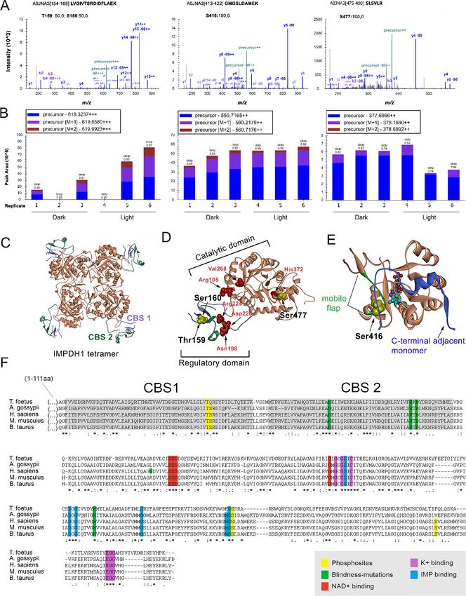
Research article                                                                    Biochemistry and Chemical Biology Neuroscience

                                    associated to rare autosomal dominant Lebers Congenital Amaurosis (adLCA), characterized by
                                    nearly complete blindness from an early age: R105W and N198K (Bowne et al., 2006a). Despite the
                                    ubiquitous nature of guanine nucleotide synthesis, clinical manifestations of IMPDH1 mutations are
                                    limited to the retina, for reasons that are not yet understood.
                                        RP10 mutations are allegedly ‘gain-of-function’ mutations, given that IMPDH1 knock-out mice
                                    present only a mild retinopathy (Aherne et al., 2004). Mutations do not directly affect IMPDH1 cata-
                                    lytic activity in vitro (Aherne et al., 2004; Mortimer and Hedstrom, 2005; Xu et al., 2008). IMPDH1
                                    capacity to bind single-stranded nucleic acids (Mortimer et al., 2008; Hedstrom, 2008) as well as
                                    IMPDH1 tendency to aggregate (Aherne et al., 2004; Tam et al., 2008) have been proposed to
                                    contribute to the pathophysiology. More recently, structural studies have provided a new framework
                                    to interpret the effect of mutations, by revealing the allosteric inhibition of eukaryotic IMPDHs by
                                    GDP/GTP binding to the Bateman domain (Buey et al., 2015). GDP/GTP binding sites at the Bate-
                                    man domain overlap with several of the residues mutated in adRP10 (Buey et al., 2015).
                                        There are two IMPDH isozymes, IMPDH1 and IMPDH2, that are 84% identical. While most human
                                    tissues show only basal IMPDH1 and high IMPDH2 expression, the retina is one of a few exceptions
                                    where IMPDH1 predominates (Hedstrom, 2009). In the retina, major unique IMPDH1 spliced forms
                                    outweigh the canonical 514aa protein (Bowne et al., 2006b).
                                        Purine nucleotide homeostasis is vital for many basic functions of the cell. Cells use different path-
                                    ways to synthesize purine nucleotides and balance the guanine and adenine pools (Figure 1). In de
                                    novo biosynthesis, a purine ring is assembled over ribose 5´-phosphate by sequential enzymatic
                                    steps that use precursors of the carbohydrate and amino acid metabolism (Zhao et al., 2013;
                                    Pedley and Benkovic, 2017). In this pathway, inosine monophosphate (IMP) is the first molecule
                                    with a complete purine ring and is the common precursor for GTP and ATP synthesis (Figure 1).
                                    IMPDH catalyzes the first committed step to GTP synthesis. In de salvage pathways, nucleotides are
                                    produced from recycled purine bases (Figure 1).
                                        IMPDH monomers consist of a catalytic and a regulatory domain. The catalytic domain is a (b/a)8
                                    TIM barrel that catalyzes two sequential reactions that convert IMP to xanthosine monophosphate
                                    (XMP) in a NAD dependent manner (Hedstrom, 2009). GMP acts as a competitive inhibitor of enzy-
                                    matic activity at this level, by binding to the IMP pocket (Hedstrom, 2009). The regulatory Bateman
                                    domain consists of two cystathionine b-synthase (CBS) repeats that act as ATP and GTP sensors to
                                    mediate allosteric modulation of catalytic activity (Buey et al., 2015; Labesse et al., 2013;
                                    Anthony et al., 2017; Buey et al., 2017; Fernández-Justel et al., 2019). In eukaryotes, binding of
                                    adenine nucleotides (ATP/ADP/AMP) induces the formation of extended octamers that remain fully

Figure 1. De novo and salvage pathways of purine synthesis in eukaryotic cells. Metabolites and enzymes of the de novo pathway in black, and the
salvage pathway in blue. R5P, ribose 5 -phosphate; PRPP, phosphoribosylpyrophosphate. The arrows from PRPP to IMP represent sequential enzymatic
activities of the purinosome complex. IMPDH1 is responsible for the rate-limiting and first committed step in de novo GTP biosynthesis.

Plana-Bonamaisó et al. eLife 2020;9:e56418. DOI: https://doi.org/10.7554/eLife.56418                                                     2 of 31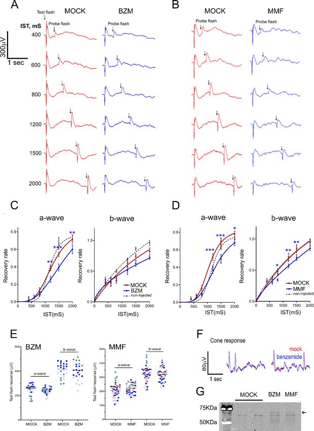
Research article                                                                  Biochemistry and Chemical Biology Neuroscience

                                    active, while GDP/GTP binding induces the formation of compacted octamers with significantly
                                    reduced catalytic activity (Buey et al., 2015; Labesse et al., 2013; Buey et al., 2017).
                                        The fact that most IMPDH1 mutations associated to RP10 overlap with GDP/GTP binding sites at
                                    the CBS domain and result in diminished GDP/GTP negative allosteric control of human IMPDH1 in
                                    vitro (Buey et al., 2015; Fernández-Justel et al., 2019) points to the physiological relevance of this
                                    allosteric mechanism in vivo.
                                        IMPDH can form mesoscale filamentous structures in mammalian cells (spicules or cytoophidia)
                                    under conditions that require higher IMPDH activity to keep with GTP demand (Liu, 2010;
                                    Chang et al., 2015; Aughey and Liu, 2016; Keppeke et al., 2018), like high rates of proliferation,
                                    or Gln/Ser/folate-metabolite deficiency (Chang et al., 2015; Keppeke et al., 2018; Calise et al.,
                                    2016). Formation of these reversible fiber-like subcellular structures is believed to transiently boost
                                    IMPDH activity (Chang et al., 2015; Keppeke et al., 2018). Reversible formation of IMPDH fibers
                                    could be recently induced in vitro from recombinant proteins, revealing that most adRP10 IMPDH1
                                    mutants form filaments but fail to disassemble them due to their inability to sense adenine and gua-
                                    nine nucleotides at the Bateman domain (Fernández-Justel et al., 2019).
                                        Little is known about the relevance of IMPDH1 catalytic activity in photoreceptor cells of the ret-
                                    ina or its physiological regulation in vivo. We here report the first posttranslational modifications of
                                    IMPDH1 in native retinal tissue, associated to dark/light states. We show that the predominant reti-
                                    nal splice form of IMPDH1 is phosphorylated at the Bateman domain (T159/S160) in response to
                                    light in vivo; and that this phosphorylation desensitizes the enzyme to GDP/GTP allosteric inhibition
                                    of catalytic activity in vitro. Together, these results reveal a novel light-dependent regulatory mecha-
                                    nism that impedes IMPDH allosteric inhibition, facilitating the elevated GTP levels required for pho-
                                    totransduction. Supporting the proposed mechanism, we show that exposure of living mice to
                                    bright light results in an increase in the global flux through de novo purine nucleotide synthesis in
                                    the retina, which correlates with a progressive accumulation of IMPDH1 aggregates at the outer seg-
                                    ment layer. Furthermore, inhibition of IMPDH activity in living mice by intravitreal injection of IMPDH
                                    inhibitors has a moderate but consistent effect on mass rod response recovery measured by electro-
                                    retinogram. Thereby, our results point to the de novo GTP synthesis being relevant to sustain the
                                    GTP pool during constant light exposure, likely to withstand the increased cGMP turn-over. This
                                    study gains insight into the complex in vivo regulation of IMPDH1 to maintain GTP homeostasis with
                                    changing illumination, central to rod dark/light physiology and relevant to understand IMPDH1-
                                    inherited retinal dystrophies.

                                    Results
                                    Dark/light-dependent phosphorylation of retinal IMPDH1
                                    Phosphoproteomic analysis of dark- and light-adapted bovine retinas (see Methods) revealed three
                                    phosphorylation sites in IMPDH1 (Figure 2A–B). One phosphorylation event was detected at the
                                    Bateman domain, assigned to Thr159 or Ser160 with a 50:50 probability (numbering corresponding to
                                    canonical bovine IMPDH1b of 514aa with Uniprot code A0JNA3). This numbering correlates with the
                                    canonical human IMPDH1b. The peak areas of the precursor ions for the monophosphorylated pep-
                                    tide 154–169 in 3 dark and 3 light biological replicates revealed a light preference of the observed
                                    phosphorylation event (light/dark log2 fold change = 1.67 with p=0.03). The light/dark log2 fold
                                    change of phosphopeptides representative of well characterized light- or dark-dependent phosphor-
                                    ylation events in rhodopsin (Wilden, 1995; Lee et al., 2002; Mendez et al., 2000), phosducin
                                    (Lee et al., 1990; Lee et al., 2004) and GRK1 (Lee et al., 1982; Palczewski et al., 1992) is shown in
                                    Figure 2—figure supplement 1 as a quality control of the phosphoproteomic analysis.
                                       Residues Thr159 and Ser160 map within the CBS motif 1 of the Bateman domain, in close proximity
                                    to Asn198, Arg224 and Asp226 mutated in adLCA or adRP10 (Figure 2C–D), and are directly involved
                                    in the binding of purine nucleotides at the allosteric nucleotide binding site 1 (Fernández-
                                    Justel et al., 2019).
                                       Phosphorylation at residue Ser416 was also identified, but observed to a similar extent in dark and
                                    light conditions (light/dark log2 fold change = 0.33 with p=0.08). This residue maps at the mobile
                                    flap in the catalytic domain (residues 412–432, in green in Figure 2E), required in the conformational
                                    transition step that activates the hydrolysis reaction during catalysis (Hedstrom, 2009).

Plana-Bonamaisó et al. eLife 2020;9:e56418. DOI: https://doi.org/10.7554/eLife.56418                                                3 of 31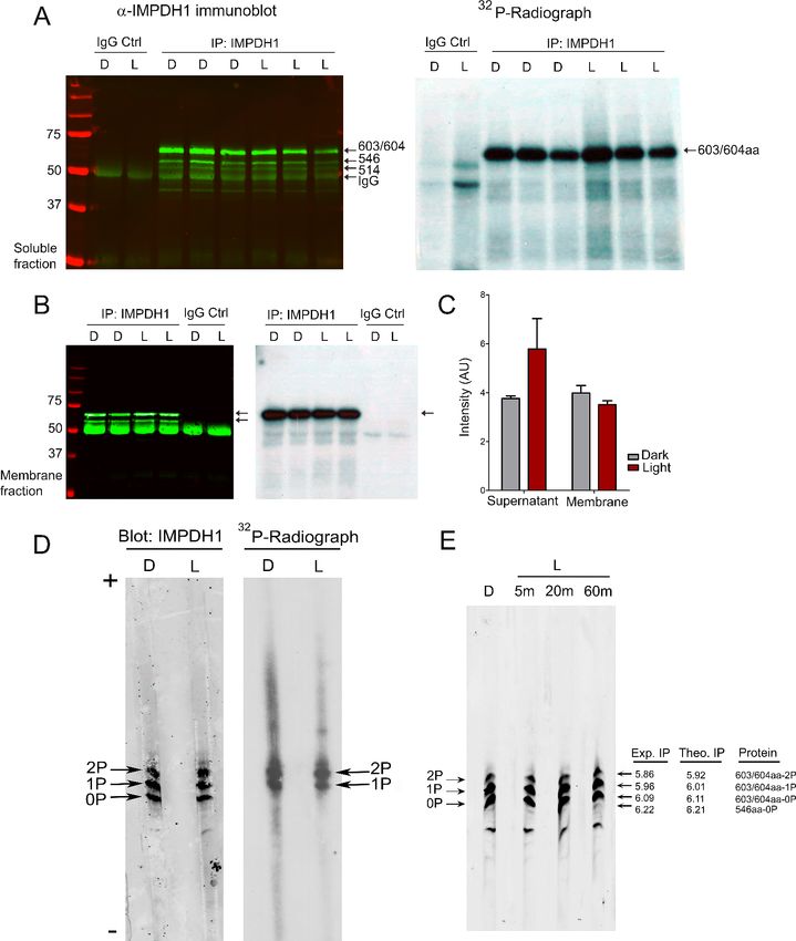
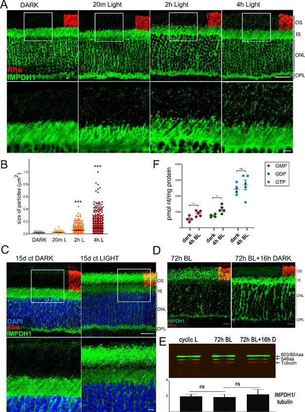
Research article                                                                        Biochemistry and Chemical Biology Neuroscience

Figure 2. IMPDH1 phosphorylation sites identified in a phosphoproteomic study of dark/light-adapted bovine retinas. (A) Mass spectra of identified
IMPDH1 phosphorylated peptides. Residue numbers refer to the canonical bovine isoform b (514aa) [Uniprot A0JNA3]. For peptide 154–169 only one
phosphorylation was detected, that could not be unequivocally assigned to T159 or S160. Phosphorylation at peptides 413–422 and 475–480 was at
S416 and S477, respectively. (B) Peak areas of the precursor ions corresponding to the identified phosphopeptides of IMPDH1. Samples 1–3 are dark-
adapted retinas; 4–6 light-exposed retinas (biological replicates). T159/S160 are preferentially phosphorylated in light, S477 in dark, and S416
indistinctly. (C) Ribbon diagram of IMPDH in its tetrameric conformation [pdb: 1jcn], showing the TIM barrel catalytic domain in coral, and the
regulatory Bateman domain with two copies of cystathione beta-synthase (CBS) sequence in slate blue and green. (D) Monomer conformation [pdb:
1jcn] showing T159 and S160 at CBS1 in the Bateman domain; and S477 at the COOH-terminus. Disease associated mutations, depicted in red, are
proximal to T159/S160 (Asn198; Arg224; Asp226) or to S477 (His372). (E) The catalytic domain of AgIMPDH, with the C319 loop in coral, the COOH-
terminus of an adjacent monomer in blue and the mobile flap in green. IMP depicted in space fill model [pdb:4xfi]. (F) Alignment of IMPDH from T.
foetus (prokaryotic), A. gossypii (filamentous fungus), H. sapiens, M. musculus and B. taurus IMPDH1 canonical isoforms. Phosphosites highlighted in
yellow.
Figure 2 continued on next page

Plana-Bonamaisó et al. eLife 2020;9:e56418. DOI: https://doi.org/10.7554/eLife.56418                                                          4 of 31
Research article Biochemistry and Chemical Biology Neuroscience
Figure 2 continued
The online version of this article includes the following figure supplement(s) for figure 2:
Figure supplement 1. Dark/light fold-change of peak areas from control phosphopeptides from phosducin, GRK1 and rhodopsin shown as a quality
control of the phosphoproteomic analysis.
Finally, phosphorylation of IMPDH1 at Ser477 occurred preferentially in the dark-adapted state
(light/dark log2 fold-change = 0.50 with p=0,19). Ser477 maps to a region that has not been
involved in the catalytic process. Remarkably, all the phosphorylated residues are conserved in mam-
malian IMPDHs (Figure 2F).
To further characterize IMPDH1 phosphorylation in vivo we performed an in situ metabolic label-
ing assay. Retinas from dark-adapted mice were dissected and incubated in Locke´s buffer with 32P-
inorganic phosphate for 90 m in the dark, to allow for 32P incorporation in the ATP pool. Retinas
were then kept in the dark or exposed to 2000 lux light for 5 m. IMPDH1 was immunoprecipitated
from supernatant and membrane fractions with an anti-IMPDH1 pAb generated against the canoni-
cal bovine IMPDH1b of 514aa (see Materials and methods). This antibody immunoprecipitated all
IMPDH1 isoforms in the murine retina (immunoblots in Figure 3A–B). 32P-incorporation was limited
to the largest and most abundant isoform in murine retinas, the 603/604aa retinal-specific spliced
form (Bowne et al., 2006b) (autoradiographs in Figure 3A–B). Phosphorylation of 603/604aa
IMPDH1 occurred to a high extent in dark- and light-retinas, both in the soluble and membrane frac-
tions, Figure 3C.
To obtain insight into the predominant phosphorylated species in dark and light, immunoprecipi-
tated IMPDH1 from 32P-labeled retinas was resolved by isoelectrofocusing (IEF), Figure 3D. Three
abundant species were detected in dark and light conditions: unphosphorylated IMPDH1-603/
604aa, IMPDH1-603/604aa-1P, and IMPDH1-603/604aa-2P.
The same species IMPDH1-0P, 1P and 2P were observed by IEF separation of retinal homoge-
nates from living mice that were either dark-adapted or exposed to 2000 lux light for 5, 20 and 60
m. Monophosphorylated forms are the predominant species in both dark and light conditions
(Figure 3E). Please note that the intensity of the di-phosphorylated band in the IEF radiograph
results from the incorporation of two 32P atoms. Therefore, a similar intensity of the mono- and di-
phosphorylated IMPDH1 bands actually reflects that the di-phosphorylated form is half as abundant
as the mono-phosphorylated form.
Measured isoelectric points (IP) of IMPDH1-603/604aa-0P, 1P and 2P correlated well with IP
predicted values, Figure 3E.
Taken together, our results show that the most abundant isoform of IMPDH1 in the retina, which
is a retina-specific spliced form, is phosphorylated to a high extent in vivo. Nearly two thirds of the
protein are phosphorylated in vivo, with mono- and di-phosphorylated forms being present but
monophosphorylated species predominating under dark or physiological light conditions.
In vitro effects of phosphorylation on IMPDH1 catalytic activity
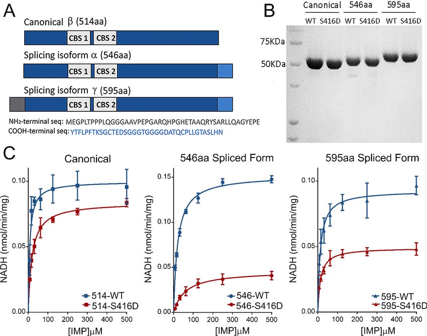
While IMPDH1-603/604aa is the prevalent isoform in murine retinas, two isoforms predominate in
human retinas: IMPDH1a (546aa) and IMPDH1g (595aa) (Bowne et al., 2006b). In order to study the
effect of phosphorylation on the catalytic activity of IMPDH1, the recombinant human IMPDH1a
(546aa) and its individual phosphomimetic mutants were expressed in bacteria and purified to homo-
geneity. The S160D-hIMPDH1a (546aa) and S477D-hIMPDH1a (546aa) mutants showed very similar
reaction kinetics and Michaelis-Menten parameters as the wildtype protein (Figure 4—figure sup-
plement 1), indicating that phosphorylation at these sites was unlikely to affect the Km or Vmax of
the enzyme. However, the S416D-hIMPDH1a (546aa) mutant showed a slower reaction (Figure 4—
figure supplement 1). This result was not surprising, given that S416 maps at the mobile flap that
determines the open or closed conformation of the enzyme during enzyme catalysis
(Hedstrom, 2009).
We confirmed the effect of Ser416 phosphorylation by measuring the effect on kinetics and IMP
dependence of the S416D substitution in the canonical and in both prevalent hIMPDH1 retinal iso-
forms: hIMPDH1a (546aa) and hIMPDH1g (595aa) (Figure 4A–B). Figure 4C shows that S416D sub-
stitution in the mobile flap had a significant effect at inhibiting the enzyme, that was higher for the
Plana-Bonamaisó et al. eLife 2020;9:e56418. DOI: https://doi.org/10.7554/eLife.56418 5 of 31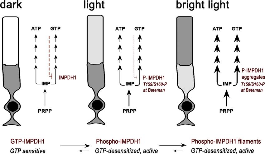
Research article                                                                         Biochemistry and Chemical Biology Neuroscience

Figure 3. The predominant retinal spliced variant of IMPDH1 is phosphorylated to a high extent in intact retinas. (A) Immunoblot of IMPDH1
immunoprecipitated from soluble fractions of 32P-labeled retinas, and corresponding autoradiograph. In situ metabolic labeling was performed in dim
red light, with retinas from dark-adapted mice incubated in Locke´s with 32Pi for 90 min at 37˚C, and then kept in the dark or exposed to light of 2000
lux for 5 min. IMPDH1 was immunoprecipitated in 3 biological replicates. 32Pi incorporation was observed at mIMPDH1-603/604aa isoform, in both dark
and light conditions (autoradiograph). (B) Immunoblot and autoradiograph of IMPDH1 immunoprecipitated from membrane fractions. 32Pi
incorporation was observed at IMPDH1-603/604aa, at dark and light conditions (2 biological replicates). (C) Average intensity of 32P-labeled 603/604aa
bands normalized by immunoblot signal, for dark and light samples. IMPDH1 is substantially phosphorylated under dark and light, both in soluble and
membrane fractions. A tendency to a higher phosphorylation in light in soluble fractions (p=0.180) and in dark in membrane fractions (p=0.308) was
apparent, with no statistical significance. (D) Isoelectrofocusing (IEF) of immunoprecipitated IMPDH1 samples from 32Pi-labeled retinas revealed the
presence of mono- and di-phosphorylated forms of IMPDH1-603/604aa in dark and light. Observed bands were: mIMPDH1-603/604-0P; mIMPDH1-603/
604-1P and mIMPDH1-603/604-2P. Please note that the di-phosphorylated form incorporates 2 radiolabeled 32P atoms per protein molecule, and
therefore the band intensity of the di-phosphorylated form in the autoradiograph reflects twice its abundance. (E) IEF of retinal homogenates (soluble
fractions) from 16 hr dark-adapted mice or mice that were exposed to 5, 20 or 60 min of 2000 lux light after pupil dilation. Measured isoelectric points
(IP) of bands correlated well with theoretical IP for the mIMPDH1 isoforms indicated.

Plana-Bonamaisó et al. eLife 2020;9:e56418. DOI: https://doi.org/10.7554/eLife.56418                                                            6 of 31
Research article                                                                        Biochemistry and Chemical Biology Neuroscience

Figure 4. Effect of S416D phosphomimetic substitution on enzymatic activity. (A) Main splice variants of hIMPDH1: hIMPDH1a (546aa) and hIMPDH1g
(595aa) contain extended sequences at the COOH- terminus (546aa) or both the NH2- and COOH-termini (595aa). (B) S416D mutant recombinant
proteins were rigorously normalized to their wildtype counterparts preceding enzymatic analysis. (C) Effect of S416D substitution on the Michaelis-
Menten kinetics of hIMPDH1b (514aa); hIMPDH1a (546aa); and hIMPDH1g (595aa) isoforms. Mutation S416D increased the Km for IMP and decreased
the Vmax significantly in hIMPDHa (546aa) and g (595aa) splice variants, with the effect being maximal in the hIMPDH1a (546aa) isoform. Kinetic
parameters were: hIMPDH1b (514aa) [Vmax = 0.099 ± 0.004, Km = 7.62 ± 1.69]; S416D/hIMPDH1b (514aa) [Vmax = 0.084 ± 0.002, Km = 19.13 ± 1.75];
hIMPDH1a (546aa) [Vmax = 0.152 ± 0.002, Km = 20.72 ± 1.32]; S416D/hIMPDH1a (546aa) [Vmax = 0.046 ± 0.002, Km = 59.59 ± 9.74]; hIMPDH1g (595aa)
[Vmax = 0.093 ± 0.003, Km = 12.86 ± 2.18]; S416D/hIMPDH1g (595aa) [Vmax = 0.049 ± 0.001, Km = 13.95 ± 1.71]. Results represent the media and S.E.M of
three independent experiments with three technical replicates each.
The online version of this article includes the following figure supplement(s) for figure 4:
Figure supplement 1. Effect of phosphomimetic substitutions on enzymatic activity.

                                    retinal spliced forms of 546 and 595aa than for the canonical form. However, because phosphoryla-
                                    tion at this residue was similar in dark and light conditions, we believe that the decrease in IMPDH1
                                    catalytic activity associated to Ser416 phosphorylation might occur in response to other signals such
                                    as nutritional stress but would have an effect unrelated to dark/light physiological conditions (see
                                    Discussion).
                                        It has recently been established that GDP and GTP allosterically inhibit the catalytic activity of
                                    eukaryotic IMPDH enzymes in vitro (Buey et al., 2015; Anthony et al., 2017; Buey et al., 2017; Fer-
                                    nández-Justel et al., 2019). Thereby, we then tested whether phosphorylation could affect allosteric
                                    inhibition in vitro.
                                        Figure 5A plots the normalized Vmax values (Vmaxapp in the presence of 2 mM GTP or GDP
                                    divided by Vmax in the absence of nucleotide) for all the hIMPDH1b (514aa) phosphomimetic mutants
                                    as well as for the wild-type IMPDH1 enzyme. As expected, phosphorylation at Thr159/Ser160 had an
                                    obvious effect on the allosteric inhibition of IMPDH1 in vitro (Figure 5A). We then assayed the cata-
                                    lytic activity of the T159E/D mutants at different concentrations of GDP (Figure 5B) and GTP

Plana-Bonamaisó et al. eLife 2020;9:e56418. DOI: https://doi.org/10.7554/eLife.56418                                                          7 of 31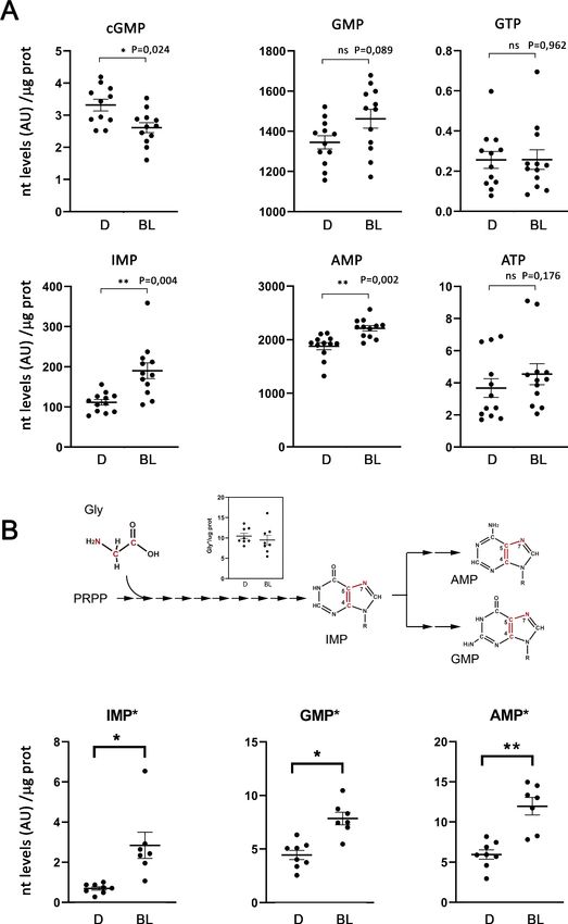
Research article                                                                       Biochemistry and Chemical Biology Neuroscience

                                    Figure 5. Effect of phosphomimetic mutations on GDP and GTP allosteric regulation of IMPDH1 activity.
                                    (A) Vmaxapp/Vmax for the indicated phosphomimetics mutants. (B) Vmaxapp/Vmax for the T159E and T159D mutants,
                                    as a function of GDP concentration. (C) Vmaxapp/Vmax for the T159E and T159D mutants, as a function of GTP
                                    concentration. Results shown are representative of two independent experiments.
                                    The online version of this article includes the following figure supplement(s) for figure 5:
                                    Figure supplement 1. HPLC determination of nucleotide levels in dark-adapted or light-exposed in situ retinas.

Plana-Bonamaisó et al. eLife 2020;9:e56418. DOI: https://doi.org/10.7554/eLife.56418                                                        8 of 31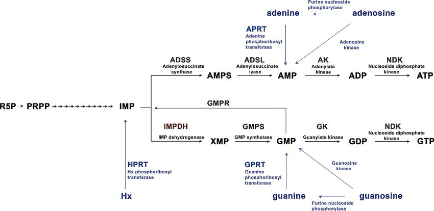
Research article                                                                       Biochemistry and Chemical Biology Neuroscience

                                    (Figure 5C), that yielded K1/2 values for enzyme inhibition around 5-fold higher than for the wild-
                                    type IMPDH1 (Table 1).
                                        Taken together our results show that phosphorylation at Thr159/Ser160 desensitizes the enzyme to
                                    the allosteric inhibition exerted by GTP and GDP. Given the light-dependence of this phosphoryla-
                                    tion event, these results indicate that IMPDH1 would be susceptible to GDP/GTP inhibition in dark-
                                    ness but desensitized to this allosteric regulation in light.
                                        In support of GDP/GTP allosteric regulation of IMPDH1 having physiological relevance, is the fact
                                    that photoreceptor cells present much higher GTP levels than most cells (Traut, 1994; Zhao et al.,
                                    2015; Sumita et al., 2016; Biernbaum and Bownds, 1979; Berger et al., 1980; Ostroy et al.,
                                    1990).
                                        We have assessed GTP levels (as a function of ATP levels) in dark/light retinas by high pressure
                                    liquid chromatography analysis using murine retinas obtained from dark-adapted mice, and kept in
                                    situ for 5 m in darkness or exposed to bright light (2000 lux light). It must be noted that although
                                    our nucleotide determinations were done in whole retinal extracts, guanine nucleotide levels largely
                                    reflect nucleotide content in photoreceptor cells, as cGMP and GTP levels are reduced to less than
                                    20% their values in retinas that lack the photoreceptor cell layer (rd1 mouse model Du et al., 2016).
                                    The levels of GMP, GTP, AMP and ATP in dark and light conditions are presented in Figure 5—fig-
                                    ure supplement 1, together with a representative HPLC chromatogram. Nucleotide peaks were
                                    assigned based on their retention time and absorbance spectrum, attained from nucleotide stand-
                                    ards. Our results showed that GTP levels are at least equimolar with ATP levels, both in darkness
                                    and under light conditions. Note similar GTP and ATP peaks in a representative chromatogram (Fig-
                                    ure 5—figure supplement 1). Another observation was that both AMP and GMP levels increased
                                    with light exposure.
                                        The fact that GTP is equimolar or higher than ATP in the retina further confirms that GTP levels
                                    are higher in photoreceptor cells than in most cell types, where the ratio of ATP:GTP is between 3:1
                                    to 5:1 (Traut, 1994; Zhao et al., 2015; Sumita et al., 2016).

                                    Constant bright light exposure results in IMPDH1 aggregation at the
                                    outer segment layer, and in increased flux towards de novo GTP and
                                    ATP synthesis
                                    It has been reported that mammalian IMPDHs can form mesoscale macromolecular assemblies in
                                    mammalian cells, denoted as cytoophidia, when an increase in IMPDH activity is required to keep
                                    with GTP demand (Aughey and Liu, 2016; Keppeke et al., 2018; Chang et al., 2015; Liu, 2016).
                                    Recently it has been proposed that IMPDH1 cytoophidia are more resistant to GDP/GTP-mediated
                                    allosteric inhibition, which indicates that they would allow a boost of GTP synthesis when required
                                    (Fernández-Justel et al., 2019; Keppeke et al., 2018; Duong-Ly et al., 2018a). IMPDH1 cytoophi-
                                    dia are not noticeable in the retina in mice reared in standard 12 hr dark/12 hr light cycles, although
                                    they can be induced and clearly detected if retinas are treated with mycophenolic acid (MPA), Fig-
                                    ure 6—figure supplement 1.
                                        We next intended to assess whether IMPDH1 cytoophidia assembly could be induced by physio-
                                    logical conditions that increased GTP demand, such as exposing living mice to bright light for
                                    increasing time periods (Figure 6). The premise was to activate cGMP phosphodiesterase maximally
                                    by bright light, so that cGMP synthesis increased accordingly during light adaptation, consuming
                                    GTP in the process. Figure 6A shows the characteristic immunolocalization of IMPDH1 in the retina.

                                    Table 1. K1/2 values (mM) for GDP and GTP.
                                    Enzyme kinetics data were fitted by non-linear regression to the Michaelis-Menten equation to derive
                                    Vmax and KM values. The Vmax values versus GTP/GDP concentration were then adjusted to a sigmoi-
                                    dal dose-response function using the GraphPad software package.
                                                                                                GDP                                         GTP
                                    HsIMPDH1b (514aa)-WT                                        0.45 ± 0.03                                 0.97 ± 0.1
                                    HsIMPDH1b (514aa)-T159D                                     2.19 ± 0.2                                  » 6

                                    K1/2 values are given in mM units (Mean ± SD), and are representative of two independent experiments.

Plana-Bonamaisó et al. eLife 2020;9:e56418. DOI: https://doi.org/10.7554/eLife.56418                                                         9 of 31
Research article Biochemistry and Chemical Biology Neuroscience Figure 6. Accumulation of IMPDH1 aggregates at the outer segment layer in living mice exposed to constant bright light. (A) Murine retinal sections from dark-adapted mice or mice exposed to 1600lux-light for 20 m, 2 hr or 4 hr were immunostained for IMPDH1 (green) and co-immunostained for rhodopsin (red). Confocal images are average z-projections of 5 optical slices with 0.13mm-step size. Magnified frames from the outer segment layer show gradual accumulation of IMPDH1 aggregates with time of bright light exposure. (B) Size and number of IMPDH1 aggregates with time (Dark vs 2 hr light, p
Research article                                                                        Biochemistry and Chemical Biology Neuroscience

Figure 6 continued
mice does not change, indicating that changes in IMPDH1 signal at the outer segment layer are due to protein translocation rather than to
transcriptional regulation. (F) Retinal GMP, GDP and GTP levels (pmol/mg prot) determined by HPLC from retinal extracts from mice that had been
dark-adapted or exposed to bright light (BL, 1600 lux) for 4 hr. GMP and GDP levels increased with light [p=0.021 for GMP and p=0.016 for GDP, with
n = 4] whereas the GTP levels were maintained [p=0.802 for GTP, with n = 4]. OS: outer segment, IS: inner segment, ONL: outer nuclear layer, OPL:
outer plexiform layer. Scale bar upper panels (20 mm) and bottom panels (5 mm).
The online version of this article includes the following figure supplement(s) for figure 6:
Figure supplement 1. Formation of IMPDH1 spicules in intact retinas in response to mycophenolic acid-treatment.

                                    IMPDH1 signal is much stronger in photoreceptor cell layers than at inner layers of the retina.
                                    IMPDH1 signal is particularly intense at the inner segment, outer nuclear and outer plexiform layers,
                                    and stronger in rods than cones. Strikingly, exposure of living mice to 1600 lux white light after pupil
                                    dilation led to the gradual accumulation of IMPDH1 aggregates at the rod outer segment layer, that
                                    increased in number and size with time (Figure 6A–B). In contrast to MPA treatment that induced
                                    cytoophidia formation at the outer nuclear and inner segment layers (Figure 6—figure supplement
                                    1), IMPDH1 aggregates induced by bright light accumulated at the rod outer segment layer.
                                    IMPDH1 accumulation was even more evident when mice were reared for 15d under constant light
                                    (1600 lux), Figure 6C. This light-dependent accumulation of IMPDH1 aggregates at the outer seg-
                                    ment layer did not involve a change in the protein levels of IMPDH1, and could be reverted by sub-
                                    sequent dark-adaptation (Figure 6D,E). This result indicated that IMPDH1 translocates from the cell
                                    soma to the outer segment layer under bright light exposure, in a reversible manner.
                                        To assess whether IMPDH1 catalytic activity increased in response to light in vivo, we determined
                                    the nucleotide levels in whole retinas from mice that had been dark-adapted or exposed to 4 hr of
                                    1600 lux light. First, nucleotide determinations were done by HPLC. Subsequently, nucleotide deter-
                                    minations were done by LC coupled to tandem mass spectrometry (LC-MS/MS), following the intra-
                                    vitreal injection of a stable isotope form of labeled glycine. Because the amino acid glycine
                                    contributes carbon and nitrogen atoms to the scaffold on which the purine ring assembles, labeled
                                    glycine allowed us to analyze whether the incorporation of labeled Gly atoms into IMP, AMP and
                                    GMP increased with light in vivo.
                                        The nucleotide determination by HPLC showed that GMP and GDP levels increased with 4 hr of
                                    light exposure, while GTP levels were maintained (Figure 6F, ,Table 2). No massive drop in the GTP
                                    levels was observed with bright light exposure, as reported in ex vivo retinas in other studies (see
                                    Discussion).
                                        For the metabolic flux analysis in vivo, 13C2;15N-Glycine was injected intravitreally in dark-adapted
                                    mice. Mice were then kept in the dark for 4 hr, or exposed to 1600 lux light for 4 hr. Whole retinal
                                    extracts were analyzed by mass spectrometry, to determine the levels of labeled and total cGMP,
                                    IMP, AMP, GMP, ATP and GTP, see Methods for experimental details.

                                    Table 2. Nucleotide levels in dark- or light-adapted murine retinas, determined by HPLC.
                                    Retinas were obtained from 16 dark-adapted mice that were either kept in the dark for 4 hr, or
                                    exposed to 4 hr bright light (1600 lux). HPLC determinations, with numbers indicating Mean ± S.E.M,
                                    with n = 4 biological replicates.
                                                                                 Pmol/mg protein
                                                                                 DARK                                                4 hr BL
                                    GMP                                          557.21 ± 88.5                                       916.31 ± 81.1
                                    GDP                                          762.57 ± 47.8                                       1174.11 ± 109
                                    GTP                                          2461.53 ± 178.1                                     2737.29 ± 310.6
                                    AMP                                          731.84 ± 125.4                                      1246.16 ± 69.8
                                    ATP                                          1579.69 ± 110.5                                     2759.18 ± 226
                                    GTP/ATP                                      1.56                                                0.99

Plana-Bonamaisó et al. eLife 2020;9:e56418. DOI: https://doi.org/10.7554/eLife.56418                                                       11 of 31
Research article Biochemistry and Chemical Biology Neuroscience Figure 7. Light exposure increases the flux through de novo purine nucleotide synthesis in the retina. Metabolic flux analysis through de novo purine nucleotide synthesis was performed by injecting 13C2;15N-Glycine intravitreally (to 4 mM final concentration in the vitreous) to dark-adapted mice, that were then kept in the dark or exposed to 1600 lux light for 4 hr. Total nucleotide levels and labeled nucleotide levels (that incorporated the labeled carbon and nitrogen atoms from labeled glycine) were determined by LC/MS-MS, and normalized per mg of protein in each sample. (A) Retinal levels of Figure 7 continued on next page Plana-Bonamaisó et al. eLife 2020;9:e56418. DOI: https://doi.org/10.7554/eLife.56418 12 of 31
Research article                                                                          Biochemistry and Chemical Biology Neuroscience

Figure 7 continued
total cGMP, IMP, AMP, GMP, ATP and GTP in dark-adapted mice (D); or bright-light-exposed mice (BL). Nucleotide levels are expressed in arbitrary
units (LC-MS/MS peak integration values) normalized per mg of protein in each sample. cGMP levels decreased with light exposure, as expected
[p=0,024, n = 12]. IMP levels showed a clear increase with light exposure [p=0004]; that was accompanied by an increase in AMP and GMP levels
[p=0002 for AMP; p=0089 for GMP]; whereas the ATP and GTP levels were maintained. (B) Retinal levels of labeled IMP, GMP and AMP, normalized per
mg prot. The labeled atoms from Gly* that are incorporated into the purine ring scaffold are shown in the diagram. The inset on the diagram shows that
the levels of Gly* measured at the retinas of injected eyes were similar in dark and bright light samples. Incorporation of Gly* into IMP, AMP and GMP
increased under bright-light exposure, indicative of an increase in the overall flux of de novo purine nucleotide synthesis [IMP*, p=0,020; AMP*,
p=0,003; GMP*, p=0,013, with n > 7]. In four eyes of the ‘dark’ group of mice, and five eyes of the ‘bright light’ group of mice the injection failed [the
injected Gly* did not reach the retina, and Gly*~0 in retinal extracts] and therefore could not be taken into account for determination of labeled
nucleotides.

                                         Figure 7A presents the levels of total cGMP, IMP, AMP, GMP, ATP and GTP in dark and bright
                                     light conditions (expressed in arbitrary units of LC-MS/MS peak integration, normalized per mg of
                                     protein) for 12 biological replicas. As expected, mass spectrometry analysis detected the decrease
                                     in cGMP that follows adaptation to a light steady-state (Barbehenn et al., 1986), as well as the
                                     increase in GMP and AMP levels that we had observed in HPLC determinations. GTP and ATP levels
                                     were maintained. Mass spectrometry allowed the determination of IMP levels in dark and light, strik-
                                     ingly revealing a substantial increase in IMP levels in the bright light condition. IMP levels could not
                                     be determined by HPLC, as IMP is masked by the GMP peak under the gradient required to
                                     unequivocally separate GTP and ATP (Sloan, 1984).
                                         Figure 7B shows the carbon and nitrogen atoms that glycine contributes to the purine ring, and
                                     presents the levels of labeled IMP, AMP and GMP in dark versus bright light conditions, normalized
                                     per mg of protein in each sample. Results are presented for 8 (dark) and 7 (light) individual retinas
                                     per group, because the intravitreal Gly* injection failed (Gly* did not reach the retina) in 4 (dark) or 5
                                     (light) of the 12 injected eyes in each condition. Interestingly, IMP*, GMP* and AMP* increased in
                                     the bright light condition to the same extent as the total nucleotides, indicating that the increase in
                                     the total amount of IMP, AMP and GMP was due to an increase in de novo nucleotide synthesis. Gly
                                     was uptaken to similar levels in dark and light conditions in the injected eyes, as shown in Figure 7B
                                     inset.
                                         Taken together, our results show that the global flux through the de novo synthesis of purine
                                     nucleotides increases with light. This increased flux could serve to maintain the ATP and GTP pools
                                     as their consumption increases in the phototransduction process.

                                     Inhibition of IMPDH catalytic activity delays mass rod recovery in
                                     electroretinogram responses
                                     Two processes that increase GTP consumption upon light exposure in photoreceptor cells would be
                                     GTP hydrolysis by the GTPase transducin at the activation step, and GTP conversion to cGMP syn-
                                     thesis during recovery of the light response (Mendez et al., 2001; Burns et al., 2002).
                                         To assess whether IMPDH1 catalytic activity was required to sustain the light response or rod
                                     mass response recovery, we recorded electroretinogram (ERG) responses simultaneously from both
                                     eyes of mice that were injected intravitreally with an IMPDH1 inhibitor (right eye) or control physio-
                                     logical saline buffer (left eye). The result of transiently inhibiting IMPDH activity on the rate of mass
                                     rod recovery after a saturating flash was tested using a paired-flash ERG paradigm (Lyubarsky and
                                     Pugh, 1996), and is shown in Figure 8A–F. Benzamide riboside (BZM) -converted to a dinucleotide
                                     in the cell after phosphorylation/adenylation- and mycophenolate mofetil (MMF) -an ester prodrug
                                     of mycophenolic acid (MPA)-, are both reversible selective noncompetitive IMPDH inhibitors. Inhibi-
                                     tors were injected intravitreously to an effective concentration of 80 mM (BZM); or 200 nM (MMF).
                                     Both drugs caused the characteristic SDS-PAGE mobility shift of the enzyme caused by drug binding
                                     (Ji et al., 2006), when retinas were obtained 20 m after intravitreal injection (Figure 7G), which dem-
                                     onstrated direct binding of the drugs to its target.
                                         IMPDH1 inhibitors did not significantly affect the amplitude of the a-wave or b-wave of the test
                                     flash [3 Cds/m2] in the high intensity range of mixed responses (Figure 8E), indicating no major
                                     effect on transducin activation. Following the paired flash paradigm, a test flash was triggered [3
                                     Cds/m2], and an identical probe flash was activated at 400, 600, 800, 1200, 1500 or 2000 ms

Plana-Bonamaisó et al. eLife 2020;9:e56418. DOI: https://doi.org/10.7554/eLife.56418                                                            13 of 31
Research article Biochemistry and Chemical Biology Neuroscience Figure 8. Effect of IMPDH inhibition on mass rod response recovery. (A) Simultaneous electroretinogram (ERG) recordings from both eyes of a mouse injected with benzamide (BZM, non-competitive IMPDH inhibitor) or drug dilution buffer (mock), in response to a probe flash triggered at increasing interstimulus times (IST) after a test flash. A slight but statistically significant delay in the recovery of the a-wave is observed at three interstimulus times. (B) A similar result is obtained when mycophenolate mofetil (MMF) -an ester prodrug of mycophenolic acid (MPA)- is injected instead of benzamide riboside. (C) Statistical analysis of mass rod response recovery (recovery rate plotted to interstimulus time, ERG paired flash paradigm) in benzamide- injected eyes versus corresponding control eyes. Overimposed black dashed lines are the curves obtained for control non-injected mice. Two-way Figure 8 continued on next page Plana-Bonamaisó et al. eLife 2020;9:e56418. DOI: https://doi.org/10.7554/eLife.56418 14 of 31
Research article                                                                        Biochemistry and Chemical Biology Neuroscience

Figure 8 continued
ANOVA (mock vs drug injections and IST as factors) with uncorrected Fisher´s test for multiple comparisons, a-wave benzamide vs mock [1200 ms
(p=0.0027); 1500 ms (p=0.0004); 2000 ms (p=0.0046), 4 mice analyzed]. (D) Statistical analysis for mycophenolate mofetyl (MMF)-injected mice: MMF vs
mock [1200 ms (p=0.0002); 1500 ms (p
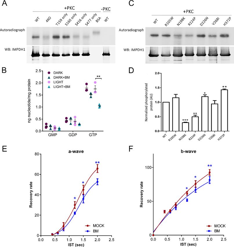
Research article                                                                         Biochemistry and Chemical Biology Neuroscience

Figure 9. PKC phosphorylates T159 and S160 of hIMPDH1 in in vitro phosphorylation assays. (A) In vitro phosphorylation assays with PKCa kinase of
hIMPDH1a (546aa); T159G/S160G/S416G/S477G-hIMPDH1a (546aa) (4KO); and forms of the protein with phosphorylation sites individually restored. 5
mg of purified protein were incubated with 4 mg/ml recombinant hPKCa in assay buffer with 0.05 mM PMA and 1 ml of 32P-g-ATP (3000 Ci/mmol), for 30
min at 30˚C. Samples were resolved by SDS-PAGE, transferred to a nitrocellulose membrane, exposed to an X-ray film (autoradiograph), and
immunoblotted for IMPDH1. PKC phosphorylated hIMPDH1a (546aa) selectively at T159 and S160. Results shown are representative of two
independent experiments. (B) GMP, GDP and GTP nucleotide levels (nmol nt/mg of retinal protein) determined by ion-pairing high performance liquid
chromatography (HPLC) in in situ dark-adapted or light-exposed retinas. Individual values and the Mean ± STDEV are indicated (3 biological replicates).
In light-exposed retinas, GTP levels decreased substantially in the presence of 50 mM bisindolylmaleimide (unpaired t-test p=0.007); while other
nucleotides did not show statistically significant changes between the dark and light conditions (n = 3). (C) In vitro PKC phosphorylation assay of
hIMPDH1a (546aa) disease mutants R105W; N198K; R224P; D226N; V268I; H372P. (D) Average intensity of autoradiograph signal normalized by the
immunoblot signal for blindness-associated mutations, expressed as a function of the wildtype levels. N198K and R224P mutations resulted in a
decrease of phosphorylation [p=0.0001 and p=0.004 versus the wildtype, n = 3 independent experiments]; while H372P led to an statistically significant
increase in phosphorylation [p=0.0013, n = 3 independent experiments]. (E–F) Mass rod response recovery (recovery rate plotted to interstimulus time,
ERG paired flash paradigm) in bisindolylmaleimide-injected eyes (to 1,25 mM final concentration in the vitreous) versus corresponding control eyes.
Two-way ANOVA (mock vs drug injections and IST as factors) with uncorrected Fisher´s test for multiple comparisons, a-wave bisindolylmaleimide vs
mock [1200 ms (p=0.0468); 1500 ms (p=0.0263); 2000 ms (p=0.0019), 4 mice analyzed]; and b-wave bisindolylmaleimide vs mock [1200 ms (p=0.0182);
1500 ms (p=0.0374); 2000 ms (p=0.0085), 4 mice analyzed].

Plana-Bonamaisó et al. eLife 2020;9:e56418. DOI: https://doi.org/10.7554/eLife.56418                                                          16 of 31
Research article                                                                 Biochemistry and Chemical Biology Neuroscience

                                    Discussion
                                    In this study we report that retinal IMPDH1 is phosphorylated at up to three residues in vivo: T159/
                                    S160 at the Bateman domain; S416 at the mobile flap in the catalytic domain; and S477 at the
                                    COOH-terminus. In the bovine system, in which calf eyes were dark-adapted in situ for 1 hr and reti-
                                    nas were processed in darkness or after 5 m of bright light exposure, T159/S160 was preferentially
                                    phosphorylated in response to light, S477 in the dark-adapted state, and S416 indistinctly at both
                                    conditions (Figure 2).
                                        The effect of these phosphorylation events on hIMPDH1 catalytic activity in vitro was determined
                                    in enzymatic assays with the corresponding phosphomimetic mutants. The S416D mutation signifi-
                                    cantly reduces retinal hIMPDH1 catalytic activity. However, because S416 phosphorylation does not
                                    appear to be regulated by light, we speculate that this phosphorylation event may respond to meta-
                                    bolic stress or nutrient deficiency (manifested in the phosphoproteomic analysis due to the 1 hr
                                    maintenance of the retinas in Locke´s -lacking Ser/Gly- during dark adaptation), to direct IMP
                                    towards ATP synthesis. In this respect, S416 maps at a region that resembles a consensus site for
                                    AMPK (Hardie, 2011), although the AMPK (A2/B2/G1) recombinant human isoform did not recog-
                                    nize hIMPDH1-546aa as a substrate in in vitro phosphorylation assays in this study. We believe that
                                    S416 phosphorylation would have a limited contribution to regulation of the enzyme by light in vivo
                                    under physiological conditions.
                                        Actually, isoelectrofocusing separation of retinal extracts from dark-adapted or light-adapted liv-
                                    ing mice revealed that >60% of the protein is phosphorylated under physiological conditions; with
                                    the predominant monophosphorylated form corresponding to phosphorylated T159/S160 in the
                                    light state and S477 in the dark state (based on the mass spectrometry data); and di-phosphorylated
                                    forms likely reflecting slow dephosphorylation kinetics of the reciprocal site.
                                        S477D substitution did not have a direct effect on enzymatic activity neither on GTP allosteric
                                    regulation of the enzyme. Thereby, we speculate that S477 phosphorylation might be involved in
                                    other aspects of enzyme regulation in vivo: for example inducing filament disassembly. To this
                                    respect, a recently reported structure of IMPDH2 polymers has revealed details of the interface that
                                    mediates cytoophidia assembly: the 12 amino-terminal residues of the canonical IMPDH2 isoform
                                    extend from the catalytic domain to bind into the adjacent molecule, in a shallow surface groove
                                    formed by a short helix (476-485), two beta strands (51-63), and two short loops (355-360, 379-380),
                                    (Johnson and Kollman, 2020). Strikingly, S477 maps into the short helix at the IMPDH2 cytoophidia
                                    longitudinal contact interface, allowing us to speculate that phosphorylation of S477 in IMPDH1
                                    might disrupt cytoophidia assembly. Additionally, S477 phosphorylation might also be implied in
                                    protein translocation from outer to proximal photoreceptor compartments during dark-adaptation
                                    after a period of bright light exposure, or regulating local IMPDH1 interactions with other proteins.
                                    Further experiments are needed to corroborate these hypotheses.
                                        We here demonstrate that the T159D and S160D (or T159E and S160E) substitutions increased
                                    about 5-fold the K1/2 for GDP/GTP inhibitory allosteric regulation of the enzyme in vitro, suggesting
                                    that light-dependent phosphorylation of these residues in vivo would effectively desensitize the
                                    enzyme to GDP/GTP allosteric control and promote enzyme activation.
                                        Light-dependent activation of IMPDH1 might seem at odds with the assumption in the field that
                                    GMP, generated from cGMP hydrolysis during light exposure, would act as a negative feedback reg-
                                    ulator of purine nucleotide synthesis by inhibiting IMPDH1, 5-phosphoribosyl 1-pyrophosphate syn-
                                    thetase (PRS), and the first enzyme of the purinosome Glutamine phosphoribosyl amidotransferase
                                    (GPA). However, by performing an in vivo metabolic flux analysis of purine nucleotide synthesis in
                                    dark/light conditions following an intravitreal injection of labeled glycine, we found that the purino-
                                    some activity was substantially increased with light, with IMP levels nearly doubling in light condi-
                                    tions (Figure 7). We found that IMP, GMP and AMP levels all increased with light; as did the
                                    incorporation of labeled atoms of glycine into these nucleotides. The overall de novo purine nucleo-
                                    tide synthesis increased in the retinas of living mice exposed to bright light.
                                        Considering these results, we believe that most of the GMP increase observed upon light expo-
                                    sure by HPLC actually reflects the light-driven overall increase in de novo purine nucleotide synthe-
                                    sis, rather than the GMP coming from cGMP hydrolysis alone. It is likely that the fraction of GMP
                                    coming from the hydrolysis of cGMP would be under detection by HPLC, given that cGMP itself is

Plana-Bonamaisó et al. eLife 2020;9:e56418. DOI: https://doi.org/10.7554/eLife.56418                                              17 of 31
Research article Biochemistry and Chemical Biology Neuroscience
under detection by HPLC in retinal extracts. cGMP levels in retinal extracts have been classically
determined by radioimmunoassay (Farber and Lolley, 1974).
By aligning our results with recent IMPDH structure/function studies (Buey et al., 2015;
Buey et al., 2017; Fernández-Justel et al., 2019; Johnson and Kollman, 2020), we propose the
model of IMPDH1 regulation outlined in Figure 10.
In the dark, IMPDH1 would be sensitive to GDP/GTP inhibitory allosteric control (negative feed-
back in red, Figure 10). We have observed that GTP levels in the retina are equimolar or higher than
ATP levels (Figure 5—figure supplement 1, Figure 6F, Table 2), as previously reported for frog rod
inner/outer segment suspensions (Biernbaum and Bownds, 1979) and bovine rod outer segments
(Salceda et al., 1982). This fact reflects that GTP levels are much higher in photoreceptors than in
most cell types, where ATP:GTP levels are between 3:1 to 5:1 (Traut, 1994; Zhao et al., 2015;
Sumita et al., 2016).
Upon light exposure, phosphorylation at T159/S160 would dramatically decrease the affinity for
GDP/GTP for the canonical site 1 in the Bateman domain, impeding IMPDH1 allosteric inhibition
(decreased negative feedback, Figure 10). This would be consistent with the increase in the overall
flux towards de novo purine nucleotide synthesis measured in light (thicker arrows, Figure 10). Fur-
thermore, structural models have shown that depletion of guanine nucleotides induces the assembly
of extended octamers into enzymatically active filaments in vitro, that are further stabilized at high
IMP levels (Johnson and Kollman, 2020). Accordingly, our results suggest that IMPDH1 filamenta-
tion is concurrent with the light-driven increase in IMP levels. We speculate that filament formation is
what leads to the gradual accumulation of IMPDH1 protein aggregates at the outer segment layer
of the retina upon constant light exposure (Figure 6). This aggregate formation in vivo is reversible
and does not correlate with changes in protein expression, which excludes that it results from tran-
scriptional regulation (Figure 6).
Figure 10. Proposed model of regulation of IMPDH1 activity in photoreceptor cells by light-dependent phosphorylation at T159/S160. In the dark,
IMPDH1 is sensitive to GTP. GTP can bind to the nucleotide binding sites at the Bateman domain and shift IMPDH1 conformation towards GTP-bound
IMPDH1 compacted octamers that are inactive (negative feedback regulation in red). Upon light exposure, phosphorylation at T159/S160 desensitizes
the enzyme to GTP, coinciding in time with the increase in IMP levels. We propose that these concurring conditions would cause filamentation of the
enzyme, leading to protein aggregation at the outer segment layer. In this way, light exposure would stabilize IMPDH1 in an active conformation
permissive to the increased flux towards guanine and adenine nucleotide synthesis (thicker arrows in light). This increased flux is likely required to
compensate for ATP and GTP consumption in the phototransduction process, to sustain the pools of ATP and GTP. IMPDH1 localization at the outer
segment layer in these macrostructures would be reverted by dephosphorylation and gradual decrease of IMP levels during dark-adaptation. The
model would predict a concerted modulation of ADSS/ADSL and the purinosome.
Plana-Bonamaisó et al. eLife 2020;9:e56418. DOI: https://doi.org/10.7554/eLife.56418 18 of 31Research article Biochemistry and Chemical Biology Neuroscience
IMPDH cellular aggregates are a landmark of cells or tissues with increased GTP demand and/or
circumstances of metabolic GTP deficiency that require stimulation of GTP biosynthesis
(Chang et al., 2015; Aughey and Liu, 2016; Keppeke et al., 2018; Calise et al., 2016; Liu, 2016).
A recent in vivo study has shown, for instance, that IMPDH1 formation of filaments is inherent to T
cell activation and proliferation, triggered at a post-translational level by NFAT or mTOR signaling,
and serves to increase guanine nucleotide levels (Duong-Ly et al., 2018b).
IMPDH1 aggregation upon light exposure points to a requirement to expand the GTP pool in the
light condition. Furthermore, the increased synthesis of IMP, AMP and GMP points to an expansion
of both ATP and GTP pools. In this context, the light exerted regulation on IMPDH1 could serve to
promote and stabilize an active conformation of the protein that is permissive to this increased flux
to purine nucleotides. Presumably IMPDH1 regulation would be part of a concerted global regula-
tion of the purine synthesis pathway in vivo, involving other metabolic enzymes.
The fact that light-dependent IMPDH1 aggregates accumulate at the outer segment layer sug-
gests that the light induced GTP demand is due to phototransduction. In light conditions, GTP at
the outer segment would be consumed in its conversion to GDP by transducin GTPase, and to
cGMP by retinal guanylate cyclase activity. By performing simultaneous electroretinogram record-
ings from both eyes of mice intravitreally injected with an IMPDH inhibitor or control saline buffer
we discarded that inhibiting IMPDH had a noticeable effect on activation of the light response (Fig-
ure 8). However, inhibiting IMPDH caused a delay in mass rod response recovery, that was modest
but statistically significant (Figure 8). Thereby, we conclude that de novo GTP biosynthesis contrib-
utes to cGMP replenishment during the recovery phase of the light response, even if cGMP can be
substantially sustained in dark-adapted responses by the salvage pathways.
In this respect, retinal GTP levels upon light exposure were reported to be maintained when it
was living mice that were exposed to light, or in situ retinas kept in supplemented tissue culture
medium (with glycine and serine), like in Du et al. study (Du et al., 2016) and this study. In contrast,
reports of light causing a clear decrease in GTP come from determinations performed in isolated
ROS/RIS preparations or retinas kept in Ringers, that cannot sustain one-carbon metabolism
(Biernbaum and Bownds, 1979; Salceda et al., 1982).
According to our model it might seem surprising that Impdh1-/- mice display only a slowly pro-
gressive retinal degeneration. However, despite maintenance of the retinal structure, these mice
present gradually diminished ERG responses from normal at 5 m to nearly extinguished at 13 m of
age (Aherne et al., 2004). Furthermore, compensatory changes like upregulation of IMPDH2 cannot
be excluded in this chronic scenario.
We here demonstrate that the light-dependent phosphorylated residues T159 and S160 in retinal
hIMPDH1 were selective in vitro substrates for PKCa kinase; but not for the other kinases tested
[PKA, PKC, PKG, CamKII, CKII, PKB-Akt and AMPK]. We also show that treatment of retinas with the
PKC inhibitor bisindolylmaleimide in situ led to a noticeable decrease of GTP retinal levels upon light
exposure (Figure 9); and its intravitreal injection caused a delay in mass rod recovery similar to that
caused by IMPDH inhibitors (Figure 9). We know from our phosphoproteomic analysis that the phos-
phorylation events that prime PKCa occur in the retina in response to light. Therefore, we propose a
model in which light triggers a signaling pathway that activates PKCa, that in turn phosphorylates
IMPDH1 at the Bateman domain, desensitizing the enzyme to GTP/GDP inhibition. None of the
tested kinases phosphorylated S416 or S477 residues of IMPDH1 selectively, so the signaling path-
ways involving phosphorylation at these residues remain unknown.
Our proposed model that GDP/GTP allosteric regulation of IMPDH1 is controlled by phosphoryla-
tion in vivo to adapt GTP synthesis to the illumination conditions supports our previous hypothesis
that IMPDH1 mutations would cause the pathology by resulting in higher than normal IMPDH1 activ-
ity; and/or by forming irreversible filaments. Abnormally high IMPDH1 activity could conceivably
result in abnormally high cGMP synthesis, which is one of the well-known causes of photoreceptor
cell damage (Wang et al., 2017); or could cause an ATP/GTP unbalance in darkness. On the other
hand, the formation of filaments –in moments of sporadic bright light exposure in the natural world-
would be reversible in healthy individuals, but irreversible in adRP10 patients by their impaired
capacity to sense GDP/GTP levels. This could result in the gradual accumulation of protein aggre-
gates that would eventually turn toxic.
Most IMPDH1 mutations in adRP10 and adLCA, including the mutations N198K, R224P and
D226N analyzed in this study, desensitize the enzyme to GDP/GTP allosteric inhibition (Fernández-
Plana-Bonamaisó et al. eLife 2020;9:e56418. DOI: https://doi.org/10.7554/eLife.56418 19 of 31You can also read