Mutant p53 Gain-of-Function: Role in Cancer Development, Progression, and Therapeutic Approaches - Frontiers
←
→
Page content transcription
If your browser does not render page correctly, please read the page content below
REVIEW
                                                                                                                                      published: 11 February 2021
                                                                                                                                    doi: 10.3389/fcell.2020.607670

                                            Mutant p53 Gain-of-Function: Role in
                                            Cancer Development, Progression,
                                            and Therapeutic Approaches
                                            Eduardo Alvarado-Ortiz 1,2† , Karen Griselda de la Cruz-López 2,3† , Jared Becerril-Rico 2 ,
                                            Miguel Angel Sarabia-Sánchez 4 , Elizabeth Ortiz-Sánchez 2 and
                                            Alejandro García-Carrancá 5*
                                            1
                                              Programa de Posgrado en Ciencias Biológicas, Universidad Nacional Autónoma de México, Mexico City, Mexico,
                                            2
                                              Subdirección de Investigación Básica, Instituto Nacional de Cancerología, Secretaría de Salud, Mexico City, Mexico,
                                            3
                                              Doctorado en Ciencias Biomédicas, Instituto de Investigaciones Biomédicas, Universidad Nacional Autónoma de México,
                                            Mexico City, Mexico, 4 Programa de Posgrado en Ciencias Bioquímicas, Departamento de Bioquímica, Facultad de
                                            Medicina, Universidad Nacional Autónoma de México, Mexico City, Mexico, 5 Laboratorio de Virus and Cáncer, Unidad de
                                            Investigación Biomédica en Cáncer, Instituto de Investigaciones Biomédicas, Universidad Nacional Autónoma de México and
                                            Instituto Nacional de Cancerología, Secretaría de Salud, Mexico City, Mexico

                         Edited by:
                     Marco Cordani,         Frequent p53 mutations (mutp53) not only abolish tumor suppressor capacities but
            IMDEA Nanociencia, Spain        confer various gain-of-function (GOF) activities that impacts molecules and pathways
                        Reviewed by:        now regarded as central for tumor development and progression. Although the complete
                        Patricia Muller,
         The University of Manchester,
                                            impact of GOF is still far from being fully understood, the effects on proliferation,
                      United Kingdom        migration, metabolic reprogramming, and immune evasion, among others, certainly
                  Simone Patergnani,        constitute major driving forces for human tumors harboring them. In this review
             University of Ferrara, Italy
                                            we discuss major molecular mechanisms driven by mutp53 GOF. We present novel
                 *Correspondence:
           Alejandro García-Carrancá        mechanistic insights on their effects over key functional molecules and processes
      carranca@biomedicas.unam.mx           involved in cancer. We analyze new mechanistic insights impacting processes such as
     † These   authors have contributed     immune system evasion, metabolic reprogramming, and stemness. In particular, the
                    equally to this work    increased lipogenic activity through the mevalonate pathway (MVA) and the alteration of
                                            metabolic homeostasis due to interactions between mutp53 and AMP-activated protein
                     Specialty section:
          This article was submitted to
                                            kinase (AMPK) and Sterol regulatory element-binding protein 1 (SREBP1) that impact
     Molecular and Cellular Oncology,       anabolic pathways and favor metabolic reprograming. We address, in detail, the impact
                 a section of the journal
                                            of mutp53 over metabolic reprogramming and the Warburg effect observed in cancer
   Frontiers in Cell and Developmental
                                 Biology    cells as a consequence, not only of loss-of-function of p53, but rather as an effect of
      Received: 17 September 2020           GOF that is crucial for the imbalance between glycolysis and oxidative phosphorylation.
      Accepted: 23 December 2020            Additionally, transcriptional activation of new targets, resulting from interaction of mutp53
       Published: 11 February 2021
                                            with NF-kB, HIF-1α, or SREBP1, are presented and discussed. Finally, we discuss
                               Citation:
                Alvarado-Ortiz E, de la
                                            perspectives for targeting molecules and pathways involved in chemo-resistance of
      Cruz-López KG, Becerril-Rico J,       tumor cells resulting from mutp53 GOF. We discuss and stress the fact that the status
Sarabia-Sánchez MA, Ortiz-Sánchez E
                                            of p53 currently constitutes one of the most relevant criteria to understand the role of
and García-Carrancá A (2021) Mutant
 p53 Gain-of-Function: Role in Cancer       autophagy as a survival mechanism in cancer, and propose new therapeutic approaches
      Development, Progression, and         that could promote the reduction of GOF effects exercised by mutp53 in cancer.
             Therapeutic Approaches.
       Front. Cell Dev. Biol. 8:607670.     Keywords: p53, gain of function, oncogenic pathways, metabolic reprogramming, stemness, chemo-resistance,
      doi: 10.3389/fcell.2020.607670        immune evasion

Frontiers in Cell and Developmental Biology | www.frontiersin.org                1                                        February 2021 | Volume 8 | Article 607670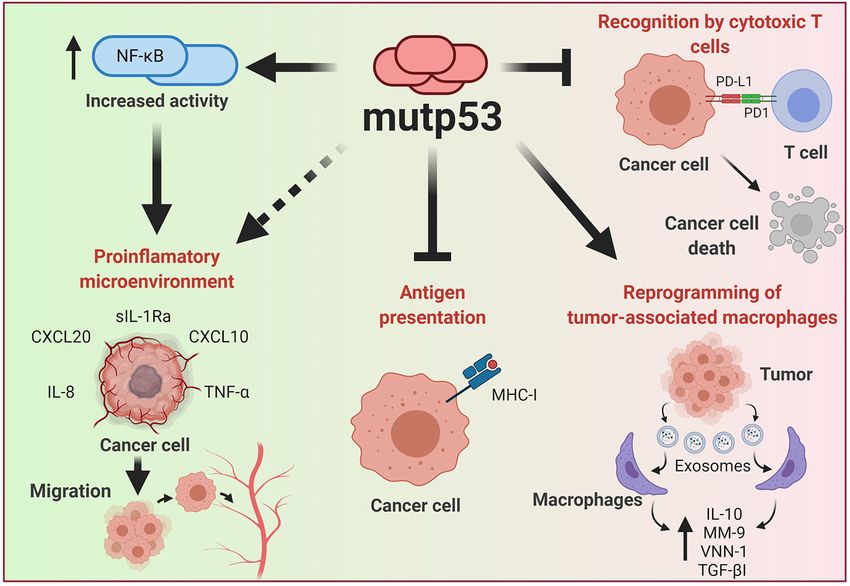
Alvarado-Ortiz et al.                                                                                      Gain-of-Function of Mutant p53 in Cancer

INTRODUCTION                                                               T antigen (TAg). Several groups used a monoclonal antibody
                                                                           to immunoprecipitate TAg from transformed cells. Although
Cancer is a complex set of diseases, all characterized by abnormal         they observed a 53–54 kDa protein in polyacrylamide gels,
cell growth, unresponsive to normal cellular and tissue controls.          the nature of this protein and its specific association with
It originates with wayward cells that once formed, grow, expand,           TAg was not evident (Chang et al., 1979). Simple experiments
and ultimately disseminate to other parts of the body, and in              revealed this as a cellular protein specifically associated with
many cases, when not detected early, will ultimately kill their            TAg and two seminal papers suggested that this protein, named
host. Cancer cells are characterized by dysregulated key elements          p53, represented a key element for viral transformation (Lane
and fundamental signaling pathways controlling proliferation,              and Crawford, 1979; Linzer and Levine, 1979). A few years
cell-death, interactions with the immune system, metabolic                 later, when a murine cDNA coding for TP53 was cloned and
changes, and response to drugs, among the most relevant                    shown to transform fibroblasts in culture, it was stated that
(Hanahan and Weinberg, 2011).                                              TP53 was “just another oncogene” and was recognized as
    The behavior and status of p53 is fundamental for cancer               such for a long time (Oren and Levine, 1983; Parada et al.,
development, progression, and for the fate of many cancer                  1984).
patients. p53 plays many important roles in cancer and is                      Rearrangements of the TP53 gene were found in several
considered a master regulator of intracellular functions, such that        human tumors and more importantly, loss-of-heterozygosity,
it has appeared on the covers of the most prominent science                a characteristic of tumor suppressor genes, was commonly
journals, like Science and, and has been awarded titles such as            observed (Masuda et al., 1987). Although these different lines
“the guardian of the genome” (Finlay et al., 1989; Soussi et al.,          of evidence strongly suggested that TP53 was not just another
1990; Yeargin and Haas, 1995).                                             oncogene, at the time, few envisioned that it would emerge as
    It is well established that altogether, around half of all human       prototype of all human tumor suppressor genes so far identified.
tumors exhibit alterations in TP53 alleles, either by inactivation,            Finally, when the effect of that same human gene on
loss or, importantly, mutations. Tumor cells containing mutant             transformed cells was studied, it clearly showed its nature as a
alleles of this gene generate mutant versions of the protein that,         tumor suppressor gene.
remarkably, mainly affect amino acids located within the DNA
binding domain (DBD) (Figure 1). These mutant versions of p53              CANONICAL FUNCTIONS
not only lead to loss of normal functions but surprisingly, confer
mutant proteins with new abilities that provide cancer cells with          A vast number of signals promote several p53-mediated
key gain-of-function activities (GOF’s).                                   functions, including cellular stress, DNA damage, hypoxia,
    Recently, the mechanisms and effects of these mutant alleles           nutritional stress, as well as differentiation signals. Activation
have been shown to affect key biological processes associated              of p53 (referred to as wtp53) drives a plethora of signals
with cancer progression, invasion, metabolic reprograming,                 that fire different fundamental responses such as cell-
and interactions with the immune system. The study of                      cycle arrest, apoptosis, senescence, regulation of cellular
such effects on central processes including proliferation,                 energy metabolism, antioxidant defense, and immune
migration, generation of an inflammatory microenvironment,                 system regulation (Figure 2). Relevant target genes of
metabolic reprogramming, stem-cell restricted characteristics,             the p53 transcription factor encode proteins, such as p21
and pharmacological resistance, has gained much attention.                 and p27 that induce cell-cycle arrest, PAI1 and CDKN1b
Although these processes are central for cancer, the molecular             involved in inducing senescence, PUMA, BAX, and
mechanisms involved and the precise targets acted upon by                  NOXA involved in apoptosis, or TIGAR and GLS2 for
mutp53 GOF’s, are only recently being elucidated.                          metabolic changes, among others (Kastenhuber and Lowe, 2017;
    Understanding the mechanisms involved and the effects of               Simabuco et al., 2018).
mutp53 GOF will be vital to better combat pharmacological                     One of best described roles for p53 is in the response
resistance of cancer cells that harbor mutp53, and to design               to DNA damage. Acting as a classic transcription factor,
effective therapies based on p53 status in different types                 p53 induces the expression of p21, which in turn inhibits
of cancer.                                                                 CDKs (cyclin-dependent kinases), resulting in cell-cycle arrest
    This review aims to integrate novel data on mechanisms and             (Deng et al., 1995).
targets involved in the effects of mutp53 GOF’s, stressing current            Another function involves regulation of oxidative
knowledge of the central pathways involved.                                phosphorylation and mitochondrial respiration through
                                                                           the induction of COX (cyclooxygenase) and GLS2 (glutaminase
DISCOVERY                                                                  2) expression. GLS2 is an enzyme involved in deamination
                                                                           of glutamine, allowing production of α-ketoglutarate, an
The product of the TP53 gene was first observed in the 1970’s by           intermediary metabolite of the TCA cycle (tricarboxylic acid
several groups when studying cellular transformation of rodent             cycle) (Hu et al., 2010). Antioxidant defense mechanisms
cells induced by a simian virus called SV40. Transformation was            modulated by p53 include increasing levels of GST (glutathione
observed when non-permissive cells were infected or rodents                S-transferase), an important enzyme implicated in avoiding
were injected with SV40, leading to tumor development and                  the deleterious effects of ROS (reactive oxygen species)
a strong host immune response against a viral protein called               (Puzio-Kuter, 2011).

Frontiers in Cell and Developmental Biology | www.frontiersin.org      2                                   February 2021 | Volume 8 | Article 607670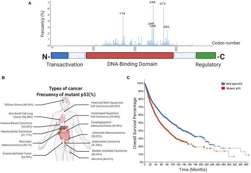
Alvarado-Ortiz et al.                                                                                   Gain-of-Function of Mutant p53 in Cancer

    Furthermore, key functions of p53 include regulation of             the DBD. Contact mutants generally produce structural changes
immune system interactions (Blagih et al., 2020). The main              in the p53 protein that directly affect DNA binding, while
effector that allows p53 to orchestrate these interactions is NF-       conformational mutants generate structural changes related with
kB (Komarova et al., 2005). NF-kB constitutes a key transcription       protein folding, both types of mutants have shown GOF activities
factor acting as one of the main regulators of pro-inflammatory         (Kim and Lozano, 2018).
activity in many immune responses. Its activation in leukocytes             The effect of mutp53 is reflected on tumorigenic ability.
promotes the expression of pro-inflammatory cytokines (IL-              Particularly, germline p53 mutants are mainly associated to Li-
1, IL-2, IL-6, TNF-α), chemokines (CXCL1, CXCL10, MCP-                  Fraumeni Syndrome, in which patients are more likely to develop
1), adhesion molecules (ICAM- 1, VCAM-1, ECAM-1), and                   tumors (Lang et al., 2004; Olive et al., 2004). Additionally, the
anti-apoptotic factors (BCL-2, c-Flip, survivin) (Liu T. et al.,        effect of mutp53 has been described in transgenic mice able to
2017). NF-kB activation is normally suppressed by binding to its        express mutp53 in a tissue-specific manner (Wijnhoven et al.,
inhibitors, a family of proteins known as IkB.                          2005). This versatility made it possible to determine that mutp53
    Following a pro-inflammatory stimulus, IKKs (IkB kinases)           can promote metastasis in a genetic context where mutant
are activated and phosphorylate IkB inhibitors, favoring their          versions of other proteins, such as oncogenic Ras, are present
degradation, thus enabling NF-kB transcriptional activity               (Morton et al., 2010). This evidence suggests that multiple
(Dresselhaus and Meffert, 2019). NF-kB regulates recruitment,           oncogenic effects drive tumorigenesis and metastasis. In the
survival, proliferation, activation, and differentiation of             case of colorectal cancer, even though loss of wtp53 improves
leukocytes, in response to antigen recognition or activating            the oncogenic capability of cancer cells through LOH, the
signals (Liu T. et al., 2017). Different reports have confirmed         presence of mutp53 in both alleles seems to be necessary to drive
the relationship between p53 and NF-kB, showing that wtp53              tumorigenesis (Nakayama et al., 2020). Moreover, intracellular
negatively controls the expression and activity of NF-kB (Gudkov        accumulation of mutant versions of p53 increases the effects of
et al., 2011; Blagih et al., 2020). This evidence indicates that        signaling pathways affected by new GOF activities (Pfister and
regulation of wtp53 over the immune system is mainly due to a           Prives, 2017).
mutual repression between wtp53 and NF-kB that promotes an
anti-inflammatory microenvironment.                                     Nuclear Effects of mutp53
    However, wtp53 immune regulatory functions are not limited          A consensus response element (RE) is required for wtp53 to
to repression of pro-inflammatory responses. For instance,              bind and regulate expression of target genes. However, the
MHC-I (Major Histocompatibility Complex Class I) is positively          mutations in the p53 DBD lead to disruption in the ability to bind
regulated by wtp53, promoting T cell recognition. Thus,                 this RE. Importantly, mutp53 can bind to novel non-canonical
alterations in TP53 also lead to deficient immune system                DNA binding sites of several genes and thus, can positively
responses (Wang B. et al., 2013; Blagih et al., 2020).                  or negatively regulate the expression of genes associated with
    Understanding wtp53 actions, it is possible to establish            malignancy (Kim and Deppert, 2007). This versatility of the
the effects derived from mutp53 GOF, being an important                 nuclear effects of mutp53 has just recently been described, and
criterion in biological processes ranging from proliferation,           it becomes relevant for the GOF associated with mutp53 (Göhler
migration, metabolism reprogramming, immune evasion, state              et al., 2005).
of differentiation, and chemoresistance, which are explained in             Even though DNA-binding of mutp53 does not depend
the following sections.                                                 on particular sequences, the mechanisms of mutp53-mediated
                                                                        transcription can be direct or indirect. Employing electromobility
                                                                        shift assays (EMSAS) and confocal fluorescence lifetime
IMPACT OF MUTp53 GAIN-OF-FUNCTION                                       microscopy it was shown that mutp53 (248P and 245S) can
                                                                        bind specifically and selectively to non-B DNA. Binding does
Although it is obvious that mutations in the TP53 gene should           not require the presence of specific sequence motifs but indeed
result in loss of canonical functions, in the last years it has         requires both the DBD and an intact p53 C-terminal regulatory
become evident that the most common mutant alleles acquire              domain (CRD). This mode of binding was termed as “DNA
new functions that fuel tumor progression.                              structure selective binding” (DSSB) (Göhler et al., 2005). In
   Most common GOF mutations in TP53 are located within the             support of this, several mutant versions of p53 have been
DBD, as have been observed in many different types of human             shown to bind preferentially to supercoiled DNA and by
solid tumors. Conversely, p53 mutations located in the regulatory       luciferase reporter assay demonstrated that the DNA topology
or tetramerization domains are less frequent. Common mutants            influences p53 regulation of BAX and MSP/MST1 promoters
have single missense mutations leading to single amino acid             (Brázdová et al., 2013). Moreover, it was shown that mutp53
substitutions at key “hotspots” that preponderantly include             binds efficiently to nonlinear DNA, and this can be increased
residues 175, 248, and 273, as shown in Figure 1A. These                by apurinic/apyrimidinic endonuclease 1/redox factor-1 (APE1)
high-frequency mutants represent non-random “hotspots” and              that stimulate DNA biding activity of numerous transcription
correlate with poor cancer-free survival (Figures 1B,C).                factors in a redox-dependent manner (Cun et al., 2014).
   There are two main types of mutant “hotspot” sites: contact              For instance, mutp53 can bind to DNA on non-canonical
mutants (R273H, R248Q, and R248W) and conformational                    sites from non-linear conformations (Göhler et al., 2005). It
mutants (R175H, G245S, R249S, and R282H), both affecting                is important to mention that there are physical interactions

Frontiers in Cell and Developmental Biology | www.frontiersin.org   3                                   February 2021 | Volume 8 | Article 607670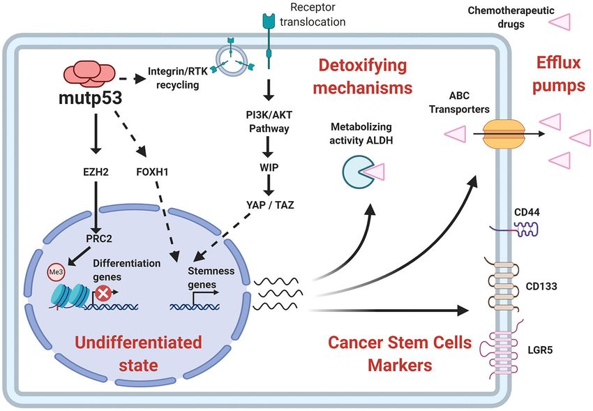
Alvarado-Ortiz et al.                                                                                                      Gain-of-Function of Mutant p53 in Cancer

 FIGURE 1 | Frequency of p53 mutations in human cancers. (A) Schematic picture showing the domain structure of the p53 protein, including the transactivation
 domain, DNA-binding domain and regulatory domain. The aligned graphs indicate the relative frequency of mutations across different domains of p53. p53 mutations
 are most frequently found in the DNA-binding domain, according to the IARC TP53 database. (B) Percentage frequency of TP53 gene alterations in different types of
 cancer. The data were obtained from TCGA PanCancer Atlas using a combined study (n = 10,967). (C) Overall survival for human cancer patients (N = 10,953
 patients from 32 studies) with mutp53 (red line) or wild type p53 (blue line). The graph was analyzed and obtained from cBioportal.

of mutp53 with remodeling complexes that cause changes                               tumor suppressor capacity such as p63 and p73, and modify
in the transcriptome, conferring plasticity in gene expression                       their transcriptional activity (Ferraiuolo et al., 2016). Recently, it
patterns. In this sense, it has been reported that binding of                        was shown that mutp53 interacts with the intracellular domain
mutp53 to particular motifs on non-B DNA conformations,                              of Notch1 to abrogate p63/p73 mediated repression of HES1
confers stability to mutp53, but also makes it more selective                        and ECM, promoting lymphomagenesis (Zhang et al., 2019). A
for modifying the activity of both transcription factors and                         more complete picture has been shown with newer evidence
chromatin remodelers (Göhler et al., 2005; Freed-Pastor and                          pointing out that mutp53 can act as a nuclear repressive factor
Prives, 2012). For instance, mutp53 has been related with the                        to downregulate pro-apoptotic responses, such as expression of
activity of the SWI/SNF chromatin complex, which increases                           the CD95 gene (Fas receptor), which is involved on apoptosis
histone modifications promoting an “open” state of chromatin,                        induction (Zalcenstein et al., 2003). Moreover, mutp53 is able to
influencing the global transcriptome and the expression of                           both positively and negatively regulate the activity of a variety
cancer-related genes. The fact is that >40% of gene expression                       of transcription factors, such as ETS2, NF-kB, HIF-1α, SMAD,
related to mutp53 can be explained by the effect on the SWI/SNF                      SREBP, or NF-Y (Kim and Lozano, 2018). For instance, mutp53
complex (Pfister et al., 2015).                                                      may directly cooperate with YAP1 (Yes-associated protein) and
   Moreover, overlap in the DNA binding sequence patterns was                        favor the transcriptional activity of NF-Y on proliferation-
observed through which mutp53 can act on response elements                           related genes, suggesting that mutp53 may act as a transcription
of other transcription factors, modulating gene expression                           cofactor to enhance GOF (Di Agostino et al., 2016). The nuclear
(Agostino et al., 2006). For example, mutp53 can regulate gene                       effect of mutp53 is an interesting field of study that is not
expression through physical binding to p53 family members with                       well clarified.

Frontiers in Cell and Developmental Biology | www.frontiersin.org                4                                         February 2021 | Volume 8 | Article 607670
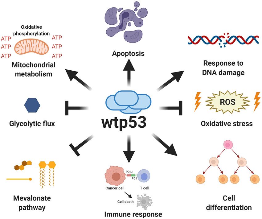
Alvarado-Ortiz et al.                                                                                                        Gain-of-Function of Mutant p53 in Cancer

 FIGURE 2 | Canonical functions of wild type p53. Wild type p53 is a major tumor suppressor whose functions are critical for protection against cancer. The canonical
 functions of wild type p53 include the induction of apoptosis, regulation of oxidative metabolism, and inhibition of glycolytic flux, as well as the response to DNA
 damage, increased antioxidant capabilities, regulation of immune response and differentiation processes.

Proliferation, Invasion and Metastasis                                                (DAB2-interacting protein). DAB2IP is a scaffold protein that
In the last decade, important contributions have allowed                              binds to and inactivates p85-PI3K, impairing its repressive
us to better understand the mechanisms involved and the                               functions over PI3K, promoting the intracellular effects of AKT1.
impact of mutp53 GOF on cell proliferation, invasion and                              Thus, growth factors, such as insulin, increase proliferation in
metastasis. The particular importance of mutp53 is in promoting                       prostate and breast cancer (Valentino et al., 2017).
proliferation, invasion and metastatic potential through                                  Recent findings reveal that mutp53 (R175H) exacerbate the
its effect on the endosomal pathway, leading to recycling                             oncogenic response of K-Ras. The active state of K-Ras (G12C)
of receptors and integrins. For instance, overexpression of                           is related to its GTP-binding form, while the GTPase-activating
mutp53 has been shown to increase translocation of EGFR                               proteins (GAPs) can favor the GDP inactive form. Importantly,
(Epidermal Growth Factor Receptor) and α5β1 integrin on the                           K-Ras activity is not enough to promote tumorigenic capacities
surface of cell membranes. This translocation is dependent on                         in models such as pancreatic ductal adenocarcinoma, since the
interaction with RCP (Rab-coupling protein) (Muller et al.,                           K-Ras mutant form cannot maintain the GTP-bound state.
2009). As a consequence, many of the intracellular pathways                           Nevertheless, mutp53 can regulate the splicing of GAPs through
associated with the regulation of endosomal pathways, including                       RNA-binding protein hnRNPK. The activity of mutp53 favors the
PI3K/AKT or MAPK cell signaling pathways, are activated by                            expression of GAP isoforms that cannot bind to Ras, abrogating
mutp53 (Figure 3).                                                                    the ability to decrease its activity and supporting the oncogenic
   Additionally, it has been reported that the R273H mutant                           effect of K-Ras. This mechanism reveals a synergism between
binds and represses the promoter region of miR-27a, a microRNA                        K-Ras and mutp53 through spliceosome effects, supporting
that negatively regulates the EGFR transcript. This reinforces the                    malignant progression through the effect of multiple oncogenes
evidence that the presence of mutp53 can favor the activity of                        like K-Ras (Escobar-Hoyos et al., 2020).
signaling pathways related to EGFR, as well as the downstream                             Additionally, it was found that mutp53 inhibits apoptosis
signaling mechanisms. Gastric cancer tumor samples corroborate                        associated with mitochondria and confers resistance to anoikis
this effect, showing reduced expression of miR-27a compared to                        (Tan et al., 2015), a type of cell death related with loss
normal tissue (Wang W. et al., 2013).                                                 of contact with the extracellular matrix or neighboring cells.
   Other effects attributed to mutp53 are mediated by the                             Apoptosis associated with mitochondria requires the dissociation
regulation of the PI3K/AKT pathway through binding DAB2IP                             of the proapoptotic protein BIM from the antiapoptotic

Frontiers in Cell and Developmental Biology | www.frontiersin.org                 5                                          February 2021 | Volume 8 | Article 607670
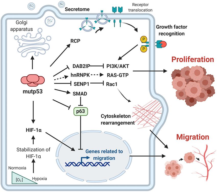
Alvarado-Ortiz et al.                                                                                                              Gain-of-Function of Mutant p53 in Cancer

 FIGURE 3 | Gain-of-function of mutant p53 over proliferation, invasion and metastasis. The principal GOF activities of mutant p53 have nuclear and non-nuclear
 effects. The nuclear effects involve binding to transcription factors such as HIF-1α or p63 and p73, while the non-nuclear effects are regulation of intracellular proteins,
 such as RCP, implicated in receptor translocation, DAB2IP scaffold protein implicated in the PI3K/AKT pathway, or SENP1, a protease related to Rac1 activity.

protein BCL-XL. However, the presence of the p53-R273H                                    transcription. MiR130b inhibits Zeb1 expression, a transcription
mutant suppresses BMF (BCL2-modifying factor) expression,                                 factor involved in regulating the expression of EMT markers.
which is a protein that induces cellular anoikis and apoptosis                            Thus, mutp53 represses transcription of miR130b and increases
by reducing the interaction between BIM and BCL-XL.                                       transcription of Zeb1, favoring invasion (Dong et al., 2013).
Knockdown of endogenous mutp53 restores sensitivity to                                        Studies employing immunoprecipitation assays have shown
apoptosis, highlighting the importance of mutp53 not only in                              that mutp53 interacts with the small GTPase Rac1 and inhibits
cellular proliferation, but in cell survival of lung, colon, and                          its interaction with SUMO-specific protease 1 (SENP1) favoring
breast cancer cell lines (Tan et al., 2015).                                              an active state of Rac1 (Yue et al., 2017). Thus, Rac1 activation
   Considering receptor recycling generated by RCP, it has                                is an important mechanism by which mutant p53 GOF promotes
been suggested that cellular scattering could be attributed to                            tumor metastasis.
HGF (Hepatocyte growth factor) as well as the presence of                                     Furthermore, accumulation of versions of mutant p53 seems
mutp53. Both elements potentiate MET (HGF receptor) activity,                             to favor GOF and the chaperone machinery mediated by
increasing its phosphorylation. The main biological responses                             Hsp90 partially explains mutp53 stabilization. Furthermore,
are cytoskeletal changes that allow cell motility (Jo et al., 2000).                      Hsp90 can be secreted by cancer cells, specifically those with
The presence of mutp53 not only exacerbates migratory abilities                           mutp53 (R175H), influencing ECM degradation as well as
through the MET receptor, but also promotes inhibition of p63, a                          migratory capacities. This effect is explained by the mutp53/RCP
key transcription factor regulating expression of anti-metastatic                         axis, favoring colonization to distant sites, such as the lung.
genes, evidencing that the effects of mutp53 are not limited to a                         Importantly, targeting the extracellular effect of Hsp90 decreases
particular mechanism (Muller et al., 2009, 2013).                                         the invasive capacities of mutp53 cancer cells (Zhang S. et al.,
   Moreover, in a model of endometrial cancer mutp53 can                                  2020). This evidence opens new avenues for the use of Hsp90
promote EMT (epithelial-mesenchymal transition). Studies on                               inhibitors in patients with mutp53.
miR130b, have determined that mutp53 is partially responsible                                 Part of the effects of mutp53 over migration rely on
for promoting an invasive phenotype through binding of mutp53                             other members of the p53 family, which include p63 and
to the promoter region of this microRNA, thereby repressing its                           p73 transcription factors. These transcription factors share a

Frontiers in Cell and Developmental Biology | www.frontiersin.org                     6                                            February 2021 | Volume 8 | Article 607670
Alvarado-Ortiz et al.                                                                                   Gain-of-Function of Mutant p53 in Cancer

conserved DBD that allows them to regulate the expression               isoforms, seem to depend on the interaction between mutp53
of a common pool of genes that are crucial for preventing               and the ribonucleoprotein complex composed by MALAT1
tumorigenesis. Although p63 and p73 form homo and hetero                lncRNA, SRSF1, and ID4, favoring splicing of VEGF pro-
tetramers, neither can bind to wtp53. Conversely, it has been           angiogenic isoforms. This being an important axis in breast
found that several mutp53 versions can interact with both p63           cancer cells (Pruszko et al., 2017). Recently, it has been
and p73, and inhibit their transcriptional activity. It was shown       reported that the effect of mutp53 reflects on morphological
that the recombinant core domain of some mutp53 proteins, but           alterations of the Golgi apparatus which lead to alteration of
not wtp53, binds and inhibits p63 by masking its DBD (Gaiddon           the secretome of cancer cells, promoting release of soluble
et al., 2001; Strano et al., 2001).                                     factors into tumoral microenvironment, including VEGF. From
    It is well accepted that GOF of mutp53 includes the ability         a mechanistic overview, this effect is explained by the dual action
to sequester the transactivation (TA) domain isoform of p63             of mutp53 and HIF-1α through miR-30d, under both hypoxia
and inhibit its interaction with its canonical DNA response             and normoxia conditions. The secretome alteration promoted by
element, thereby disrupting its downstream anti-metastatic              mutp53 exercises important effects on primary and distant sites
transcriptional networks (Strano et al., 2001). Additionally,           during carcinogenesis (Capaci et al., 2020).
Neilsen et al. (2011) demonstrated that mutp53 GOF activities
aberrantly alter the gene expression pattern of cancer cells to         Metabolic Reprogramming
promote oncogenesis, involving a collaborative approach with            Hyperactivation of oncogenic pathways directly regulates the
p63 transcription factor. They show that mutp53 uses p63 as a           metabolic pathways that support tumor growth. Interestingly,
molecular chaperone to bind to the promoter of target genes             mutp53 has been shown to enhance the Warburg effect, a
causing reprograming of the transcriptome. These genes are              process characterized by an increase in glucose uptake and
mainly associated with cellular invasion. These studies show that       lactate secretion even in the presence of oxygen (Levine
mutp53 can induce the secretion of pro-invasive factors to the          and Puzio-Kuter, 2010; Eriksson et al., 2017). It was shown
surrounding microenvironment (Neilsen et al., 2011).                    that mutp53 increases translocation of the glucose transporter
    Importantly, the effect of mutp53 over p63 is decisive for          GLUT1, without affecting total protein levels, favoring glucose
signaling pathways like TGF-β (Transforming Growth Factor               uptake. The mechanistic effect is explained by an upregulation
β), to determine whether they act as tumor suppressors or               of the RhoA pathway. RhoA is a protein involved in
promoters of cellular migration and metastasis. Extracellular           different intracellular pathways like the activation of the
TGF-β receptor ligands exert their actions through Smad 2/3             effectors, ROCK1/2, which has been demonstrated to improve
transcription factors. Under non-cancerous contexts, they act           the distribution of transporters to the cell membrane in
as cell growth suppressors, but in the presence of mutp53               different cell types. Impairment at different points of the
they improve migration ability, highlighting the pleiotropic            mutp53/RhoA/ROCK axis promotes an important decrease in
relevance of TGF-β in cancer. Interestingly, it was shown that          glycolytic flux in different types of cancer cell lines (Zhang
p63 is functionally inactivated when complexed with mutp53              et al., 2013). This constitutes one of the first reports that
and Smad in the presence of TGF-β ligands, this being critical          explains how the presence of mutp53 favors the Warburg effect.
for supporting metastasis. Moreover, this process is dependent          Moreover, other reports support this evidence, showing that
on mutp53 N-terminal phosphorylation by oncogenic Ras.                  wtp53 antagonizes the Warburg effect and favors oxidative
Mechanistically, mutp53 and Smad intercept p63 to form a                phosphorylation (Zhou et al., 2014; Hernández-Reséndiz et al.,
ternary complex in which the p63 transcriptional functions              2015).
are antagonized, offering an interesting explanation for the                Some authors have recently focused on the regulation of
migratory effects induced by TGF-β (Adorno et al., 2009).               the mevalonate (MVA) pathway implicated in lipid metabolism
Additionally, other reports revealed that mutp53 binds to the           and posttranslational modifications related to the malignant
MH2 domain of Smad3, promoting a decrease in canonical                  process. The biological effects of mutp53 on the regulation of
TGF-β pathway signaling (Ji et al., 2015).                              the MVA pathway explain various cellular processes ranging
    Moreover, studies have shown that part of the functions             from proliferation to fitness, or the regulation of the tumor
of mutp53 are involved with adapting to a hypoxic                       microenvironment, all of them with functional relevance for
microenvironment, which favors an invasive phenotype. In                tumorigenesis (Mullen et al., 2016; Ingallina et al., 2018).
this sense, there is dual participation between mutp53/HIF-1α,              Generation of MVA requires sequential action of
which allows for increased expression of extracellular matrix           enzymes, among which HMGCR constitutes a key element.
proteins, such as VIIa1 collagen and laminin-γ2, promoting              Transcriptional regulation of HMGCR is controlled by SREBP
an invasive phenotype in non-small cell lung cancer (Kamat              (Sterol Regulatory Element-Binding Protein), which recognizes
et al., 2007; Amelio et al., 2018). Other reports support this          sterol-response elements on its promoter region (Mullen
premise, since it has been shown that there is an increase in           et al., 2016). Importantly, simultaneous binding of mutp53
tumor vascularization, as reflected by VEGF expression, as well         and SREBP has been demonstrated on promoter regions
as an increase of ROS in cell lines with mutp53 status (Khromova        recognized by SREBP using ChIP assays of genes implicated
et al., 2009).                                                          in the MVA pathway, including HMGCR, in breast cancer
    This is reinforced by evidence suggesting that the expression       (Freed-Pastor et al., 2012). Thus, a great number of small
of pro-angiogenic isoforms of VEGF, but not anti-angiogenic             GTPases, such as Rho and Ras, whose post-translational

Frontiers in Cell and Developmental Biology | www.frontiersin.org   7                                   February 2021 | Volume 8 | Article 607670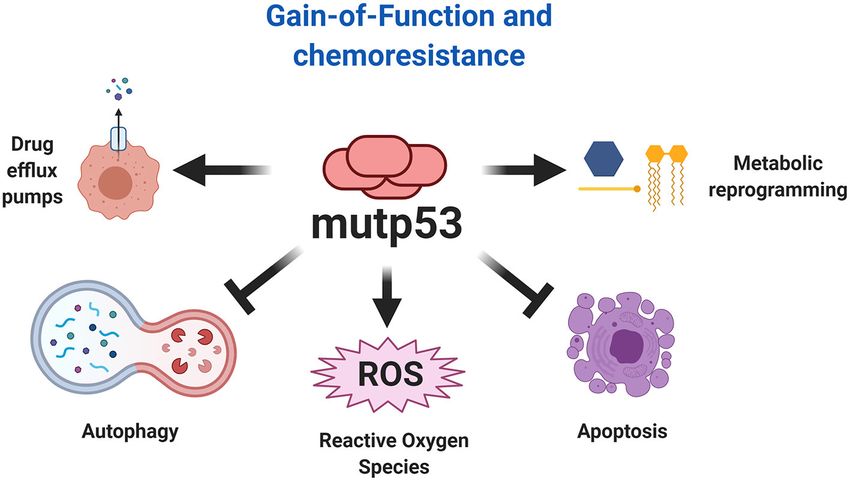
Alvarado-Ortiz et al. Gain-of-Function of Mutant p53 in Cancer
modifications are regulated downstream of MVA pathway can mechanisms for stress conditions (Tran et al., 2017; Ishak Gabra
be increased by mutp53 (Freed-Pastor et al., 2012; Parrales et al., et al., 2018).
2016).
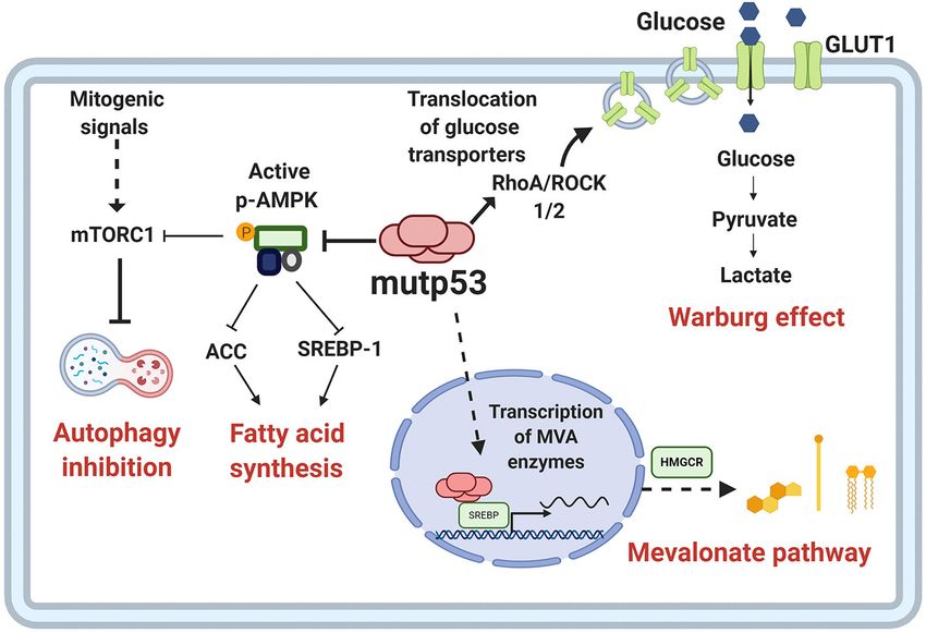
The increased activation of anabolic pathways is an essential Immune System Regulation
characteristic of cancer cells because they enable production of Genetic alterations of cancer cells induced by the malignant
the macromolecules required for replicative cell division and transformation process has an important effect in the ability to be
tumor growth. One of the proposed mechanisms through which recognized by the immune system. During recent years, p53 has
mutp53 favors the activation of anabolic pathways relies on emerged as one of the major regulators of cancer-immune system
AMPK inhibition, contrary to wtp53, which increases AMPK interactions (Blagih et al., 2020). The dynamic and bidirectional
activity (Feng et al., 2007). AMPK is a Ser/Thr kinase activated relationship between tumor cells and the microenvironment
by an increase in AMP levels, caused by energy stress (Zhou has been evidenced to be decisive for the establishment and
et al., 2014). AMPK decreases anabolic pathways such as fatty progression of tumors (Wang et al., 2017). One of the main
acid synthesis and protein synthesis, and promotes catabolic microenvironment components that allows the development
pathways including oxidation of fatty acids and autophagy. of tumor growth is the immune system; this relationship was
Mutp53 (R175H) can bind directly to the AMPKα subunit, already being contemplated in the nineteenth century as a
thereby inhibiting activation of AMPK by upstream kinases. The predisposing factor for cancer disease (Gonzalez et al., 2018). It is
consequences of this interaction, besides AMPK inhibition, is well accepted that under normal conditions, the immune system
that the downstream targets of this kinase are not being regulated, seeks to eliminate cells with aberrant characteristics, however,
and therefore, there is an increase in glycolytic flux, as shown in modifications in the functions of neoplastic cells not only prevent
Figure 4 (Zhou et al., 2014). elimination, but even benefit from the inflammatory functions of
Metabolic alteration and GOF related to mutp53 in cancer the immune system (Figure 5).
cells can increase the levels of reactive oxygen species (ROS). Tumor progression is generally associated with immune
Moreover, the presence of mutp53 decreases NRF2 (Nuclear system evasion, and loss-of-canonical function of wtp53 stands
factor erythroid 2–related factor 2) activity and glutathione as a crucial point for the generation of an inflammatory
synthesis, promoting ROS accumulation (Liu D. S. et al., microenvironment that not only limits the immune system
2017). Conversely, it has been widely demonstrated that response but indeed, benefits cancer cells (Blagih et al., 2020).
wtp53 has important role in regulating ROS levels and As previously discussed, wtp53 acts as a repressor of pro-
therefore, in determining the stress response. One mechanism inflammatory activity through the inhibition of NF-kB, which in
is through the regulation of TIGAR (TP53-induced glycolysis turn is also able to inhibit wtp53 activity, thus promoting cell
and apoptosis regulator), a transcriptional target of wtp53. survival and proliferation. This repressive function of wtp53 over
TIGAR shares sequence similarities with the bisphosphatase NF-kB is impaired by the presence of p53 mutants, acting in
domain (FBPase-2) of the bifunctional enzyme PFK-2/FBPase- an opposite manner, due to the stimulatory effect of mutp53 on
2 (6-phosphofructo-2kinase/fructose-2,6-bisphosphatase). These NF-kB activity after exposure to TNF-α (Webster and Perkins,
well-known functions lead to glycolysis blockage and favor 1999; Weisz et al., 2007). The pro-inflammatory and pro-
the production of NADPH through pentose phosphate. This tumorigenic effect of TNF-α, orchestrated by mutp53, results
mechanism represents an important mechanism for wtp53 from the interaction between mutp53 and NF-kB. Interestingly,
to favor antioxidant capacities, promoting ROS scavenging when both factors are bound on promoter regions of cancer-
(Bensaad et al., 2006) Thus, it is not surprising that presence of related genes, such as MMP9 and CCL2, they favor an active
mutp53 drives an imbalance between glycolysis and oxidative chromatin, improving transcriptional activity (Cooks et al., 2013;
phosphorylation, as well an increase in oxidative stress. Rahnamoun et al., 2017). The interaction between mut53 and
Importantly, TIGAR expression has been reported under NF-kB is persistent over time, generating inflammation-driven
conditions where mutant versions of p53 are present. Under colon cancer (Cooks et al., 2013; Uehara and Tanaka, 2018).
these conditions, TIGAR plays a key role in protecting cancer Thus, it is evident that mutp53 tumor cells have greater
cells from oxidative stress generated by sustained proliferation tumorigenic and migratory capabilities after a pro-inflammatory
(Cheung et al., 2013). This evidence supports a dynamism stimulus, as well as an increase in pro-inflammatory cytokine
between the functions of p53 to adapt to survival under expression. In addition to TNF-α, IL-8 is also increased in
stress conditions. mutp53 cancer cell lines. It is known that IL-8 is a pro-
Recently, it has been shown that cancer cells can adapt inflammatory cytokine that shows a high capacity for chemotaxis
to stress conditions. Availability of glutamine in the tumor toward neutrophils and whose expression is found to be
microenvironment allows cancer cells with mutp53 to generate dependent on NF-kB (Hidaka et al., 2005; David et al., 2016).
adaptive mechanisms to avoid apoptosis. However, although Consequences of IL-8 over-expression include an increase in
glutamine constitutes an important energy fuel for proliferation, tumorigenic properties, the EMT process, as well as improving
cancer cells with mutp53 (R288, R280) can adapt to stress stemness of tumor cells (Long et al., 2016). Additionally, it
conditions, even in the absence of glutamine. In accordance with has been observed that tumor cells carrying the endogenous
this, it was shown that mutp53 can reestablish canonical p53 R273H mutp53 suppress the activity of sIL-1Ra, an antagonist
transcriptional activity over a particular set of genes, such as of the IL-1R (interleukin 1 receptor), through an interaction
GLS1, CDKN1A, GAGG45A, and TIGAR, favoring new adaptive between mutp53 and the transcription factor MAFF (MAF bZIP
Frontiers in Cell and Developmental Biology | www.frontiersin.org 8 February 2021 | Volume 8 | Article 607670Alvarado-Ortiz et al. Gain-of-Function of Mutant p53 in Cancer FIGURE 4 | Metabolic reprogramming by mutp53. Mutp53 GOF activities are involved in different critical points of tumor metabolism. Mutp53 favors glucose uptake and hence the Warburg effect through membrane translocation of the glucose transporter, GLUT1, via the RhoA/ROCK1/2 axis. Moreover, mutp53 can induce the Warburg effect by directly inhibiting AMP-activated protein kinase (AMPK), a major cellular energy sensor and a master regulator of metabolic homeostasis. AMPK downregulates fatty acid synthesis by inhibiting transcription factor sterol regulatory element-binding protein 1 (SREBP1). Mutp53 increases the activity of SREBP1, a master regulator of fatty acids and cholesterol biosynthesis, and thus, the mevalonate (MVA) pathway. FIGURE 5 | Effect of mutp53 over the immune system. Mutp53 supports a pro-inflammatory microenvironment through the release of siL-1RA, CXCL20, CXXCL10, IL-8, or TNF-α, mainly by increasing the transcriptional activity of NF-kB. Additionally, the presence of mutp53 decreases MHC-I expression, avoiding recognition by T cells. Tumor cells can liberate exosomes that act over neighboring macrophages and improve IL-10, MM-9, VNN-1, and TGF-βI release, thus creating a microenvironment that improves cancer progression. Moreover, mutp53 can increase PD-L1, constituting an important mechanism for avoiding the oncolytic activity of T cells. Frontiers in Cell and Developmental Biology | www.frontiersin.org 9 February 2021 | Volume 8 | Article 607670
Alvarado-Ortiz et al.                                                                                        Gain-of-Function of Mutant p53 in Cancer

Transcription factor F), inhibiting its activity. In this manner, the        INF-γ receptors generates PD-L1 overexpression (Akinleye and
interaction of the R273H mutant with the MAFF transcription                  Rasool, 2019). However, in neoplastic cells, mutp53 has been
factor, prevents the suppressive action of sIL-Ra on IL-1R,                  shown to generate low levels of miR-34a, which enables PD-L1
amplifying the pro-inflammatory and tumorigenic activities of                overexpression (Cortez et al., 2016). Additionally, in melanoma,
IL-1 in colon and breast cancer cell lines (Kannan et al., 2012;             the presence of mutp53 also leads to PD-L1 overexpression and a
Ubertini et al., 2015).                                                      lower activity of cytotoxic T-cells over tumor cells (Thiem et al.,
   Among the pro-inflammatory cytokines expressed in tumor                   2019).
cells with mutp53 are those which recruit leukocytes, such                      Conversely, the positive regulation of wtp53 over MHC-I
as macrophages, neutrophils, dendritic cells, and lymphocytes.               establishes a relationship between p53 and oncolytic activity by
For example, overexpression of CXCL10, CX3CL1, and LTB                       T cells. Taking into account that mutp53 cells show low levels
chemokines in breast cancer, generates chemotaxis of T                       of MHC-I, this could represent an important barrier for T cell
lymphocytes, cytotoxic T lymphocytes, as well as NK (Natural                 recognition. Recently, it has been proposed that low doses of
Killer) cells, through a mechanism dependent on DAB2IP                       TNF (Tumor Necrosis Factor) can rescue the expression of MHC,
protein inhibition, thus promoting pro-inflammatory and cell                 making mutp53 cancer cells more sensitive to immunological
migration activities (Di Minin et al., 2014). This is derived from           therapy (Garancher et al., 2020).
the DAB2IP repressive activity on pro-inflammatory signaling                    This dual role of mutant p53 versions to induce both pro-
pathways such as NF-kB, and as previously mentioned, on                      inflammatory and anti-inflammatory environments becomes a
tumorigenic pathways such as the PI3K/AKT cell signaling                     challenge for the immunological eradication of cancer. It is
pathway. Therefore, the inhibition of DAB2IP through its                     possible that this process is related to the different tumorigenic
protein-protein interaction with mutp53 (R176H, R280K),                      stages of cancer, and therefore with different microenvironment
promotes the activation of these pathways (Bellazzo et al., 2017;            requirements for tumor progression or favoring certain cellular
Valentino et al., 2017). Additionally, in breast cancer cell lines,          subsets (Gonzalez et al., 2018).
overexpression of the CXCR4 receptor (whose ligand is the
CXCL12 chemokine) has been found in mutp53 cells, improving
their migratory capabilities (Mehta et al., 2007).                           Conferring Stemness
   It has recently been found that this GOF for cytokine release             It is now accepted that the majority of tumors exhibit a hierarchy
can be accomplished through exosome-mediated mechanisms.                     of cells within the tumor, where stem-like cells are positioned at
Under co-culture conditions of colon cancer cell lines (expressing           the top and are referred to as CSC (Cancer Stem Cells). Under
endogenous mutp53) with M0 and M2 macrophages, it has                        physiological conditions, tissues are subject to constant renewal,
been observed that the macrophages showed an increase in                     and decisions between self-renewal of tissue stem-cells or cell
the release of IL-10, MM-9 (metallopeptidase matrix 9), VNN-                 differentiation are associated with wtp53 activity (Solozobova,
1 (non-inflammatory vascular molecule 1), and TGF-βI, due                    2011).
to the action of miR-1246-containing exosomes secreted by                        The advancement of genetically modified models provides
tumor cells. This was corroborated in colon tissue samples                   information about the relevance of the relationship between
from patients with mutp53 (Cooks et al., 2018). This anti-                   p53 function and maintenance of the stem cell pool that
inflammatory microenvironment causes a failure in tumor cell                 might provide precursors for tumor initiation. For instance,
elimination by the immune system and, additionally generates                 transgenic mice harboring mutp53 developed malignant glioma,
activities that favor cellular metastasis due to the destruction of          mainly detecting cells in the corpus callosum and olfactory
the extracellular matrix and the formation of new blood vessels.             bulb, both migratory destinations for stem cells residing in
   This establishes that mutp53, not only generates a pro-                   the subventricular zone. This suggests that neural stem cells
inflammatory environment but also anti-inflammatory ones.                    or progenitors are mediating gliomagenesis caused by mutp53
Other studies show an over-expression of immunological                       (Wang et al., 2009).
checkpoints that facilitate immune system evasion by mutp53                      Another example was observed in hematopoietic stem cells,
tumor cells. In breast cancer patients, over-expression of                   where the R172H mutp53 promoted greater ability to self-renew
molecules associated with anti-inflammatory environments such                in vitro and in vivo compared to wtp53 loss, showing that
as CTL4, PD-L1, PD-L2, PD-1, LAG2, BTLA, and TIGIT was                       FOXH1, a regulator of stem cell factor receptor c-Kit and SCA-1
confirmed in tumors with mutp53, being associated with the                   (Stem Cell Antigen 1), was necessary for this phenotype in cells
prognosis of the disease (Liu et al., 2019).                                 expressing mutp53 (Loizou et al., 2019).
   Similarly, over-expression of the transmembrane protein PD-                   Following this notion, mice harboring the R248Q mutp53
L1 (Programmed Death-Ligand 1) has been found in mutp53                      favor tumor development, compared to mice with other
lung cancer and melanoma cells (Cortez et al., 2016; Thiem                   mutations, such as G245S, since R248Q alters the stem cell
et al., 2019). Its main function is the suppression of the pro-              compartments, by improving survival and self-renewal of
inflammatory activity of T cells after the recognition of their              hematopoietic and mesenchymal stem cells, putative primary
specific antigen by interaction of TCR (T cell receptor) with                malignant cells. This explains the similarity with Li-Fraumeni
MHC (major histocompatibility complex), being a regulatory                   patients, a familial cancer predisposition, in which the R248Q
mechanism of the inflammatory response (Akinleye and Rasool,                 mutp53 increases tumor initiation compared to other mutants,
2019). Additionally, the activation of the JAK-STAT pathway by               possibly because the R248Q mutp53 is able to co-aggregate

Frontiers in Cell and Developmental Biology | www.frontiersin.org       10                                   February 2021 | Volume 8 | Article 607670
Alvarado-Ortiz et al.                                                                                      Gain-of-Function of Mutant p53 in Cancer

into higher-order structures with other tumor-suppressor                   Furthermore, ALDH levels were upregulated in colorectal
transcription factors (Xu et al., 2011; Hanel et al., 2013).               tumor samples expressing p53 missense mutations and clinically
    Some types of cancer are originated through age-related                associated to higher aggressiveness. This poor prognosis seems to
mutations. This can be evidenced in C57BL/6, a type of old                 be linked to CSC-related capabilities, such as higher tumorigenic
mice vulnerable to developing fibrosarcoma, where it was                   potential and chemo-resistance, which agrees with the proposal
demonstrated that mesenchymal stem cells isolated and cultured             that mutp53 favors chemo-resistance and its absence leads to
in vitro were spontaneously transformed. The acquisition of                chemo-sensitivity (Figure 6).
tumorigenic potential was accompanied by the expression of                     Interestingly, mutp53 binds to promoter sequences of
stemness factors such as Klf4, Oct4, Sox2, c-Myc, as well as by            ALDH1A1, CD44, and LGR5. ALDH1A1 being implicated in
the expression of mutp53 (Li et al., 2007).                                mutp53-mediated chemo-resistance (Chen et al., 2016; Solomon
    The self-renewal capacity of undifferentiated populations              et al., 2018). ALDH1A1 belongs to the ALDH family of enzymes
requires a balance between “open” and “closed” state of                    and is capable of metabolizing, not only endogenous substrates
the chromatin. During the stemness of embryonic cells, a                   but also inactivates some drugs used in chemotherapy, especially
bivalent state has been identified that reflects posttranslational         aldophosphamides. Remarkably, Gui et al. (2020) found that
modifications of histones that can generate a transcriptionally            while the presence of wtp53 preferentially associates with a
inactive state. PRC1 and PRC2 (Polycomb Repressive Complex                 dominant ALDH isoform in tumors from HNSCC (Head
1 and 2) act in an orchestrated way to keep this repressive state,         and Neck Squamous Cell Carcinomas), mutp53 displayed a
defining specific lineages. However, in the case of cancer, these          different diversity of ALDH isoforms, thus severely influencing
mechanisms can regulate oncogenic functions through silencing              chemoresistance associated to ALDH.
of tumor suppressor genes (Laugesen et al., 2016). Interestingly,              Chemo-resistance involves capabilities such as efflux of
the presence of wtp53 seems to be determinant in controlling               exogenous agents. Therefore, artificial dyes are used to identify
these epigenetic modifications.                                            these cells in vitro and CSC are then referred to as SP (Side
    Recently, it has been found that mutp53 triggers self-renewal          Population). Analysis of SP in colorectal cancer-derived cell lines
of hematopoietic stem cells by increasing levels of H3K27me3               show that DLD-1 cells expressing mutp53 and Caco-2 (p53 null)
and therefore promoting a repressive chromatin state. In this              cells showed a SP, while in HCT116 cells harboring wtp53, it
study, three mutp53 versions (R248W, R273H and R175H)                      was hardly detected. This is in agreement with the evidence that
showed increased association with EZH2 (Enhancer of Zeste                  wtp53 inhibits MDR genes. Therefore, alterations in the p53 gene
Homolog 2), which is part of PCR2 (Polycomb Repressive                     would impact the drug efflux capacity of the cells (Allen et al.,
Complex-2), compared with wtp53, improving EZH2 binding                    2009).
to chromatin. Nonetheless, mutp53 promoted the presence of                     Functionally, CSC also are capable of serially forming spheres
H3K27me3 rather than altering genomic distribution (Chen                   in vitro and display higher tumorigenic potential under in vivo
et al., 2019). Moreover, mutp53 also indirectly upregulates EZH2           conditions. An example was confirmed by Zhao et al. (2019)
by attenuating miR-26a, a negative regulator of EZH2, supporting           where overexpression of R273H mutp53 in a p53-null cell line
another mechanism in the regulation of EZH2 activity (Jiang                showed elevated sphere formation and an increased expression
et al., 2015).                                                             of Sox2 and Nanog, favoring greater tumor initiating capacity.
    One of the main challenges in the study of CSC has been                Furthermore, normal astrocytes expressing mutp53 were able to
the development of appropriate tools that allow distinguishing             form spheres, emphasizing the potential of mutp53 in non-stem
them from the rest of the cancer cells. Surface protein markers            cells to trigger a CSC-like state (Escoll et al., 2017).
have allowed addressing this problem, predominantly employing                  Supporting evidence for molecular mechanisms that allow
CD44, LGR5, and CD133 (Barker et al., 2007; Keysar and Jimeno,             self-renewal in the presence of mutp53 are scarce. The canonical
2010; Alvarado-Ortiz et al., 2019). It was shown that wtp53                Wnt pathway represents the most likely candidate, since it is
inhibits CD44 expression in breast cancer cells, but R248H                 commonly associated with self-renewal of stem cells, and its
mutp53 increased CD44+ cells in colorectal cancer (Zeilstra et al.,        activity seems to be dependent on p53 status (Nusse and Clevers,
2013; Solomon et al., 2018).                                               2017). The β-catenin protein is the transcriptional cofactor
    Solomon and collaborators showed the relationship between              involved in Wnt signal transduction, and is modified post-
p53 functionality and CSC properties in colorectal cancer. They            translationally to regulate its functions.
showed that in the RKO cell line, which endogenously expresses                 It is well known that wtp53 acts negatively in regulating the
wtp53 but was transfected with R248H mutp53, there was an                  canonical Wnt pathway (Kim et al., 2011). It is noteworthy that
increase in the number of LGR5+ and CD44+ cells. Conversely,               mutp53 increases β-catenin levels (Cagatay and Ozturk, 2002),
knockdown of endogenous mutp53 in SW480 cells diminished                   possibly through Siah1 (Seven in absentia homolog 1) regulation,
CD44+ cells (Solomon et al., 2018).                                        a target of wtp53 that participates in β-catenin degradation
    In addition to surface proteins, CSC can also be identified            (Fiucci et al., 2004; Xie et al., 2009). Another mechanism related
through high enzymatic activity of proteins such as ALDH                   to β-catenin levels is mediated by mir-34a, a p53 transcriptional
(Aldehyde Dehydrogenase) (ALDHHIGH cells) (Toledo-Guzmán                   target, which downregulates β-catenin mRNA. In this regard,
et al., 2019). The ALDHHIGH population was augmented                       it is striking that mir-34a targets additional components of
in cells that overexpress mutp53, while it was reduced by                  the Wnt pathway, decreasing β-catenin-dependent transcription,
mutp53 knockdown (Solomon et al., 2018; Zhao et al., 2019).                including stemness-related genes (Kim et al., 2011).

Frontiers in Cell and Developmental Biology | www.frontiersin.org     11                                   February 2021 | Volume 8 | Article 607670
Alvarado-Ortiz et al. Gain-of-Function of Mutant p53 in Cancer
FIGURE 6 | Relationship of stemness properties with mutp53. Mechanisms related to self-renewal pathways favored by mutp53 include an increased EZH2 (subunit
of PCR2 complex) activity and improvement of epigenetic modifications associated with a repressive state of chromatin. Other pathways include YAP/TAZ activity, as
well as nuclear effects of FOXH1. Additionally, mutp53 increases CSC markers, such as CD44, CD133, LGR5, and the enzymatic activity of ALDH, contributing to
stemness and pharmacological resistance.
Therefore, the inhibitory role of wtp53 over the Wnt pathway gut. This became evident because mutp53 strengthened tumor
is consistent with an increase in transcription mediated by β- occurrence in distal sites, like colon, characterized by gallic acid
catenin in the presence of mutp53 (Cagatay and Ozturk, 2000). enrichment, while mutp53 diminished tumor progression into
Furthermore, c-Myc and Oct4 are upregulated in cells that proximal sites, where gallic acid is scarce. In the proximal site,
express mutp53, which could be explained by upregulation of β- the tumor-suppressive activity of mutp53 was independent of
catenin activity, but additional experimental data are required to canonical p53 transcription and more closely associated with
demonstrate this signaling axis (Hosain et al., 2016). suppression of the Wnt pathway. TCF4, the transcriptional factor
Some in vivo models have been employed that overactivate mediating β-catenin activity, was shown to be decoupled from
Wnt signaling to generate tumors in mice. Wnt-1 transgenic mice chromatin and the H3K4me3 active transcription epigenetic
develop mammary cancer, but when the mice are additionally marker was dropped from Wnt response targets. Noticeably, this
modified with a mutant version of p53 (R175H), a higher number mechanism of Wnt signaling inhibition was not observed in
of tumors in many mammary glands are observed. Additionally, crypts, in the presence of gallic acid, diminishing the protective
there is an increase in the pool of mammary epithelial stem cells, task of mutp53 against tumorigenesis and conversely, promoting
which is related to tumorigenic potential (Lu et al., 2013). tumor development, as reflected by canonical Wnt targets, such
Similarly, synergistic oncogenic properties of mutp53 have as CD44, c-myc, or Axin2. The overall data postulate a dual
been shown in two additional mouse models of Wnt- role for mutp53, in which the exogenous components and the
driven gastrointestinal cancer. In these studies, there are effects of the microbiome, though gallic acid, might decide the
exogenous conditions related to microenvironment, such as transition from tumor-suppressive to oncogenic function, or vice
gut microbiome, that determine the functions of mutp53 versa (Kadosh et al., 2020).
in two different anatomical sites, intestine and colon. The The YAP/TAZ complex (Yes-Associated
microorganisms of the intestinal tract and colon are interacting Protein/Transcriptional Co-Activator with PDZ-binding
with the host cells maintaining homeostasis. Recently, it motif), is a key component of mechanical stress. YAP/TAZ
was evidenced that a microbiome imbalance can promote complex is important for self-renewal and tumorigenic capacity
tumorigenesis, and its anatomical localization has proven to (Cordenonsi et al., 2011). Escoll et al. (2017) described that
be a key factor to promote the oncogenic effect of mutp53. mutp53 stimulates YAP/TAZ stability through phosphorylation
In this study, mutp53 GOF was dependent on gallic acid, a of WIP (WASP-Interacting Protein) by AKT2, in glial and
metabolite that simulates the effects of the microbiome in the breast cancer cells. In this context, mutp53 upregulated CSC
Frontiers in Cell and Developmental Biology | www.frontiersin.org 12 February 2021 | Volume 8 | Article 607670You can also read