HHS Public Access Author manuscript Compr Physiol. Author manuscript; available in PMC 2015 July 01 - HML Chiropractic & Functional Care
←
→
Page content transcription
If your browser does not render page correctly, please read the page content below
HHS Public Access
                                Author manuscript
                                Compr Physiol. Author manuscript; available in PMC 2015 July 01.
Author Manuscript

                    Published in final edited form as:
                     Compr Physiol. 2014 July ; 4(3): 1177–1200. doi:10.1002/cphy.c130051.

                    Autonomic Nervous System and Immune System Interactions
                    MJ Kenney and CK Ganta
                    Department of Anatomy and Physiology, Kansas State University, Manhattan, KS

                    Abstract
                         The present review assesses the current state of literature defining integrative autonomic-immune
Author Manuscript

                         physiological processing, focusing on studies that have employed electrophysiological,
                         pharmacological, molecular biological and central nervous system experimental approaches.
                         Central autonomic neural networks are informed of peripheral immune status via numerous
                         communicating pathways, including neural and non-neural. Cytokines and other immune factors
                         affect the level of activity and responsivity of discharges in sympathetic and parasympathetic
                         nerves innervating diverse targets. Multiple levels of the neuraxis contribute to cytokine-induced
                         changes in efferent parasympathetic and sympathetic nerve outflows, leading to modulation of
                         peripheral immune responses. The functionality of local sympathoimmune interactions depends on
                         the microenvironment created by diverse signaling mechanisms involving integration between
                         sympathetic nervous system neurotransmitters and neuromodulators; specific adrenergic receptors;
                         and the presence or absence of immune cells, cytokines and bacteria. Functional mechanisms
                         contributing to the cholinergic anti-inflammatory pathway likely involve novel cholinergic-
                         adrenergic interactions at peripheral sites, including autonomic ganglion and lymphoid targets.
Author Manuscript

                         Immune cells express adrenergic and nicotinic receptors. Neurotransmitters released by
                         sympathetic and parasympathetic nerve endings bind to their respective receptors located on the
                         surface of immune cells and initiate immune-modulatory responses. Both sympathetic and
                         parasympathetic arms of the autonomic nervous system are instrumental in orchestrating
                         neuroimmune processes, although additional studies are required to understand dynamic and
                         complex adrenergic-cholinergic interactions. Further understanding of regulatory mechanisms
                         linking the sympathetic nervous, parasympathetic nervous, and immune systems is critical for
                         understanding relationships between chronic disease development and immune-associated changes
                         in autonomic nervous system function.

                    INTRODUCTION
Author Manuscript

                    Autonomic Nervous System Regulation and Integrative Physiology: An Evolving State of
                    Cooperation
                                     The autonomic nervous system (ANS), composed of two primary branches, the sympathetic
                                     nervous system (SNS) and the parasympathetic nervous system (PNS), plays a critical role
                                     in regulating processes required for maintaining physiological homeostasis and responding
                                     to acute stressors, and has often been considered to function rather independently of other

                    Correspondence to: Michael J. Kenney, PhD, Department of Anatomy and Physiology, Kansas State University, Coles Hall 228,
                    Manhattan, KS 66506, Phone: 785-532-4513, Fax: 785-532-4557, Kenny@vet.k-state.edu.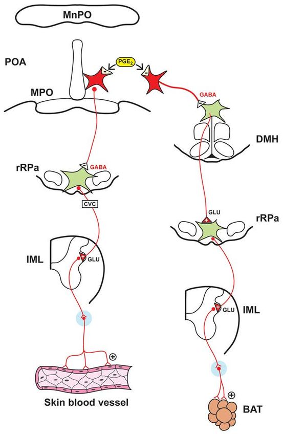
Kenney and Ganta                                                                                            Page 2

                                       adaptive systems. However, recent lines of inquiry have expanded the functional repertoire
Author Manuscript

                                       of the ANS by establishing an essential role for this system in regulating, integrating, and
                                       orchestrating processes between diverse physiological systems (49, 51, 71, 96, 112).
                                       Specifically, the results of many studies (71, 96, 97, 120, 121, 134, 135, 136, 150, 152, 181,
                                       200, 256, 281, 296, 297, 298) have established a critical role for the ANS in mediating
                                       interactions between the nervous and immune systems, two important adaptive systems that
                                       were originally considered to function independently of each other. The physiology of ANS
                                       function and regulation involves numerous complex, dynamic, and integrated steps (e.g.,
                                       neural outflow, transmitter synthesis, release and degradation, ganglionic regulation,
                                       receptor-mediated effects), many of which are likely involved in mediating neural-immune
                                       interactions.

                                       A key working principle for defining integrative autonomic-immune physiological
                                       processing is determining how signaling components of the immune system engage central
Author Manuscript

                                       autonomic neural circuits and regulate the level of activity in sympathetic and
                                       parasympathetic nerves, and how changes in autonomic regulation influence target immune
                                       organ and cell function. This review focuses on these physiological relationships with an
                                       emphasis on the results of studies focused on adult physiology that have used central
                                       microinjection and electrophysiological approaches, direct peripheral nerve recordings, and
                                       pharmacological and molecular biological techniques at both central and peripheral sites to
                                       investigate fundamental autonomic-immune interactions.

                       AUTONOMIC NERVOUS SYSTEM OVERVIEW
                       Sympathetic Nervous System and Parasympathetic Nervous System Regulatory
                       Components
Author Manuscript

                                       Sympathetic nerves innervating many target organs are tonically active. Direct recordings of
                                       the discharges of sympathetic nerves provide an output measure of central sympathetic
                                       neural circuits (148). The activity in sympathetic nerves contains multiple oscillations and,
                                       as reviewed by Barman and Kenney (12) and Gilbey (100), the sympathetic nerve discharge
                                       (SND) bursting pattern influences multiple physiological functions, including; regulating the
                                       level of efferent sympathetic nerve outflow, synchronizing or desynchonizing the activity in
                                       nerves innervating different targets, regulating target organ function, and generating
                                       differential patterns of sympathetic nerve outflow. SND pattern transformation is a
                                       consistent feature of SNS regulation. A fundamental regulatory strategy of the SNS involves
                                       selectively controlling the level and frequency components of SND bursts in nerves
                                       innervating different targets in response to a number of physiological conditions (148, 201).
                                       By altering important characteristics of efferent SND, including the level of activity, the
Author Manuscript

                                       bursting pattern, and the relationships between discharges in nerves innervating different
                                       targets, central sympathetic neural circuits regulate target organ responses.

                                       The PNS innervates multiple organ systems and plays an important role in regulating a
                                       diverse array of physiological functions, including; heart rate, hormone secretion,
                                       gastrointestinal peristalsis, digestion, inflammation, and immune function. The vagus nerve
                                       (10th cranial nerve) and its branches are extensively distributed through the body and are
                                       composed of approximately 80% afferent fibers (sensory) and 20% efferent fibers (motor)

                                            Compr Physiol. Author manuscript; available in PMC 2015 July 01.
Kenney and Ganta                                                                                             Page 3

                                       (69), thereby providing critical communicating pathways for relaying sensory information to
Author Manuscript

                                       central neural circuits and conveying efferent activity to peripheral targets. As reviewed by
                                       Tonhajzerova et al. (295), vagal nerve outflow can exhibit oscillatory fluctuations, indexed
                                       often by oscillations in cardiac vagal nerve activity and heart rate.

                                       Multiple sites in the central nervous system (CNS), including forebrain, brainstem and
                                       spinal neural circuits, regulate efferent sympathetic nerve outflow. The cell bodies of
                                       brainstem neurons whose axons project to sympathetic preganglionic neurons in the spinal
                                       cord are primarily confined to the rostral ventral lateral medulla (RVLM), midline medulla
                                       (rostral raphe pallidus, rRPa), and the A5 cell group in the pons (11, 58, 59, 179, 282, 283).
                                       The RVLM and rRPa play critical roles in SND regulation (11, 58, 59, 179, 282, 283).
                                       Forebrain nuclei (hypothalamic regions, anteroventral third ventricle region) also contribute
                                       to SND regulation (58, 59, 158, 179, 282). The paraventricular nucleus (PVN) of the
                                       hypothalamus is a major source of input to sympathetic preganglionic neurons and PVN
Author Manuscript

                                       neurons project to brainstem nuclei (e.g., RVLM) involved in SND regulation (58, 59, 158,
                                       179, 282). It is well-established that other hypothalamic nuclei, including the dorsomedial
                                       hypothalamus (DMH), ventromedial hypothalamus (VMH), and the preoptic area (POA) of
                                       the anterior hypothalamus, as well as the subfornical organ (SFO), are involved in SND
                                       regulation (58, 59, 158, 179, 282). The primary neurotransmitter released at SNS ganglionic
                                       sites is acetylcholine (ACh), whereas norepinephrine (NE) is the predominant
                                       neurotransmitter released from postganglionic SNS neurons at target organ sites.

                                       Peripheral vagal afferents terminate in the nucleus tractus solitarius (NTS), and projection
                                       neurons from the NTS make direct and indirect connections to a wide range of brain areas.
                                       Preganglionic parasympathetic neurons arise primarily from the caudal medulla, including
                                       the dorsal motor nucleus of the vagus (DMV) and the nucleus ambiguus (NA). The primary
Author Manuscript

                                       neurotransmitter released at both ganglionic and postganglionic sites in the parasympathetic
                                       system is ACh. It is generally considered that multiple CNS sites, including forebrain and
                                       brainstem nuclei, are involved in regulating the background level of activity and the
                                       responsivity of efferent parasympathetic nerve outflow (179), although central mechanisms
                                       remain to a large extent undefined.

                                       Multiple experimental methods have been employed for assessing SNS and PNS
                                       contributions to physiological states and pathophysiological conditions including:
                                       pharmacological blockade of nicotinic, adrenergic, and muscarinic receptors; molecular
                                       approaches involving genomic and proteomic analyses; direct recordings of afferent and
                                       efferent activity in sympathetic and parasympathetic nerves; and the use of time-domain,
                                       frequency-domain, and nonlinear methods to analyze specific components of directly-
Author Manuscript

                                       recorded nerve activity, as well as physiological indices of nerve activity, such as heart rate
                                       variability (46). Regarding the latter, analysis of heart rate variability provides an important
                                       noninvasive, clinically-relevant technique for studying ANS regulation (1, 46, 64, 111, 286).

                                            Compr Physiol. Author manuscript; available in PMC 2015 July 01.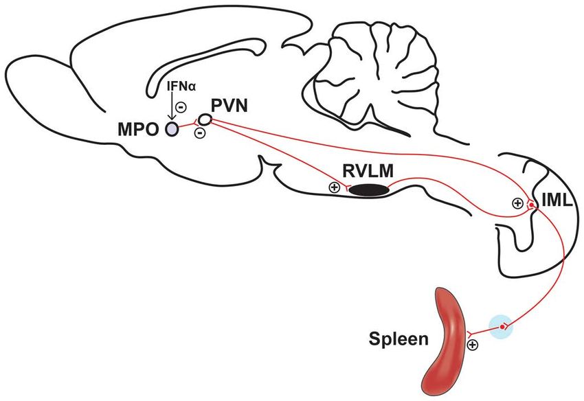
Kenney and Ganta                                                                                              Page 4

                       CYTOKINES, IMMUNE SYSTEM MEDIATORS AND AUTONOMIC NERVE
Author Manuscript

                       RESPONSES
                       Introduction
                                       Cytokines are signaling proteins that are synthesized and secreted by numerous types of
                                       cells, including; immune cells (e.g., macrophages, monocytes, lymphocytes), CNS neurons,
                                       microglia and astrocytes, and vascular endothelial cells (95). Cytokines and other immune
                                       cell products influence regulation of numerous physiological processes (e.g., sleep, fever
                                       production, neuroendocrine function, neuronal development, ANS responsiveness).
                                       Investigations studying interactions of immune mediators with the ANS have not been
                                       limited by the choice of cytokines or mediators to study. Indeed, interleukin-1β (IL-1β),
                                       interleukin-6 (IL-6), tumor necrosis factor-α (TNF-α), interferon-α (IFN-α), bacterial
                                       products such as lipopolysaccharide (LPS), and other inflammatory mediators, such as those
Author Manuscript

                                       from the cyclooxygenase pathway have been studied. This section reviews studies that have
                                       documented the effects of cytokines and other immune mediators on basal SNS and PNS
                                       activity, and autonomic neural responsiveness to acute experimental interventions.

                       Interleukin-1β
                                       The cytokine IL-1β mediates many of the diverse physiological responses of the acute-phase
                                       reaction. The intravenous IL-1β injection paradigm has been used to identify central
                                       pathways involved in cytokine-induced effects on stress-related neuroendocrine neurons (72,
                                       73). The results of previous studies demonstrate that IL-1β influences SND regulation in
                                       several ways. First, IL-1β administration (intravenous, intracerebroventricular,
                                       intracisternal) alters the level of activity in sympathetic nerves innervating visceral organs
                                       (kidney, spleen, adrenal gland) (129, 137, 210, 213, 246, 247). Central microinjections of
Author Manuscript

                                       IL-1β in the PVN increase renal SND (268). IL-1β receptors are located in the brain, cells in
                                       the CNS express mRNA for IL-1 receptors, and IL-1β mRNA is expressed in brainstem and
                                       hypothalamic areas under basal or nonactivated physiological conditions (56, 74, 77, 99,
                                       324). Renal and lumbar SND responses to intravenous IL-1β are dependent on the integrity
                                       of forebrain neural circuits (152). Second, IL-1β produces nonuniform changes in the level
                                       of sympathetic nerve outflow (210, 247). Specifically, intravenous IL-1β increases lumbar
                                       and splenic SND without significantly altering the level of renal and BAT SND in
                                       chloralose-anesthetized rats (247). Moreover, Niijima et al. (210) reported that intravenous
                                       IL-1β increased adrenal and splenic SND, but produced only transient increases followed by
                                       sustained reductions in renal SND in urethane-anesthetized rats. Third, the frequency-
                                       domain coupling of SND bursts can be influenced by IL-1β alone and in response to
                                       combined IL-1β and mild hypothermia (150), suggesting an additional mechanism via which
Author Manuscript

                                       IL-1β can influence sympathetic nerve outflow. Fourth, IL-1β can modulate responses to
                                       other physiological interventions. For example, IL-1β (intra-arterial administration)
                                       sensitizes the activity of visceral afferents recorded in the right thoracic sympathetic chain to
                                       ischemia and histamine (91), PVN IL-1β microinjection enhances the cardiac sympathetic
                                       afferent reflex as evaluated by renal SND responsiveness to epicardial application of
                                       bradykinin (268), and IL-1β markedly enhances BAT but not renal SND responses to
                                       hypothermia (151). These findings suggest that an important mechanism via which IL-1β

                                            Compr Physiol. Author manuscript; available in PMC 2015 July 01.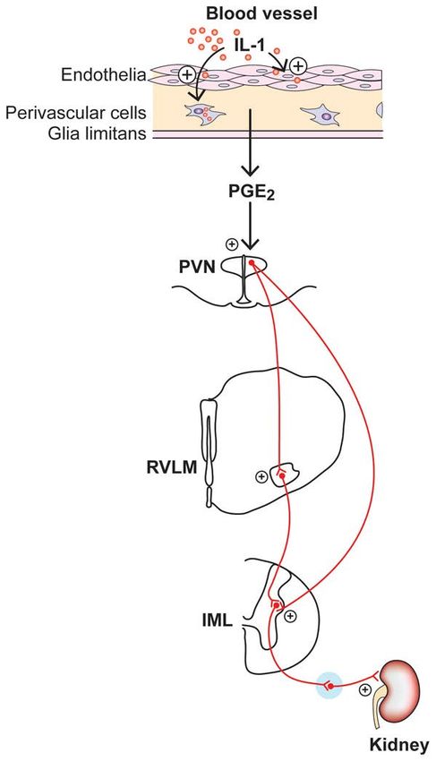
Kenney and Ganta                                                                                              Page 5

                                       influences SNS regulation is to modulate SND responses to acute physical stress. Fifth,
Author Manuscript

                                       Saindon et al. (247) reported renal, splenic and lumbar SND responses to IL-1β were
                                       augmented in vagotomized rats compared to rats with intact vagus nerves, suggesting the
                                       vagus nerve provides a tonic inhibition to visceral and peripheral SND in response to
                                       intravenous IL-1β, supporting the existence of complex interactions between IL-1β and SNS
                                       and PNS regulation.

                       Interleukin-6
                                       This cytokine belongs to a group of structurally related IL-6-type cytokines that includes
                                       IL-11, leukemia inhibitory factor, oncostatin M, ciliary neurotrophic factor, cardiotrophin-1,
                                       and cardiotrophin-like cytokine (119). Although IL-6 modulates numerous centrally-
                                       mediated physiological responses (e.g., fever, activation of the hypothalamic-pituitary-
                                       adrenal axis, immune regulation), the effect of either peripheral or central IL-6 on SND
Author Manuscript

                                       regulation remains poorly understood. Helwig et al. (120) reported central IL-6
                                       administration (intracerebroventricular; icv) activates splenic SND, and is associated with
                                       co-localization of IL-6 with the IL-6 receptor (IL-6R) in periventricular tissue at the level of
                                       the third ventricle, but is not widely distributed in brain parenchyma. IL-6 mRNA and IL-6
                                       receptor mRNA have been reported to be localized in several medial hypothalamic areas,
                                       including the dorsomedial, ventromedial, and medial preoptic nuclei (262). Vallieres and
                                       Rivest (301) reported low levels of IL-6 receptor mRNA under basal conditions in the
                                       ependymal cells of the ventricles, and several sites in the CNS, including the
                                       circumventricular organs (CVOs). In addition, central IL-6 administration induces cellular
                                       activation in numerous areas of the rat brain, including the ependymal cell layer, the
                                       ventricular walls, and CVOs (300).

                                       Haensel et al. (111) reviewed the results of studies examining relationships between heart
Author Manuscript

                                       rate variability and markers of inflammation in various cardiovascular diseases, whereas
                                       Huston and Tracey (128) reviewed the literature regarding interactions between the
                                       cholinergic anti-inflammatory pathway, heart rate variability, and potential therapeutic
                                       interventions. The existing literature provides support for the idea that R-R interval
                                       variability, interpreted in many studies as an index of cardiac vagal modulation, is inversely
                                       related to IL-6 and other inflammatory markers, including C-reactive protein, in both
                                       healthy subjects and patients with cardiovascular disease (10, 111, 128). These data suggest
                                       the existence of interactions between inflammation and ANS physiology and regulation in
                                       human subjects. As discussed by Haensel (111), measures of heart rate variability reflect
                                       PNS as well as SNS modulation of the heart, and the interpretation of correlations between
                                       inflammatory markers and autonomic function should consider both arms of the ANS.
Author Manuscript

                       Tumor necrosis factor-α and Interferon-α
                                       TNF-α has been shown to modulate SND regulation (213, 246, 331). Intravenous TNF-α
                                       reduces ear temperature (indicative of increased cutaneous SND) in anesthetized (246) and
                                       conscious rabbits (213), reduces renal SND in anesthetized rabbits (246), but increases renal
                                       SND in conscious rabbits (213). SND responses to TNF-α in conscious rabbits were blocked
                                       by indomethacin administration (213). Intravenous and intracarotid artery TNF-α
                                       administration in anesthetized rats increases renal SND, mean arterial blood pressure, and

                                            Compr Physiol. Author manuscript; available in PMC 2015 July 01.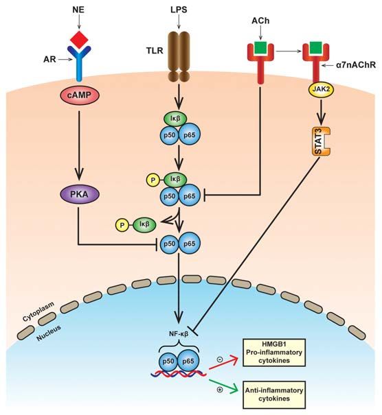
Kenney and Ganta                                                                                               Page 6

                                       neuronal firing of RVLM and PVN neurons (331). Responses to intravenous TNF-α were
Author Manuscript

                                       reduced following mid-collicular transection but not vagotomy (331). ICV administration of
                                       the cyclooxygenase inhibitor ketorolac reduced renal SND and PVN neuronal activation to
                                       intracarotid artery TNF-α administration, icv PGE2 increased the activity of PVN neurons
                                       and elicited renal sympathoexcitation, and PVN PGE2 microinjection increased RVLM
                                       neuronal activity and renal SND (331). These data suggest the excitatory influences of
                                       systemically administered TNF-α on renal SND are mediated by central actions of
                                       prostaglandins acting, at least in part, in the PVN (331). Additional central effects of TNF-α
                                       on SND regulation have been identified. For example, pentoxifylline (PTX, inhibitor of
                                       proinflammatory cytokine synthesis) administration attenuated the renal sympathoexcitation
                                       and decreased the PVN production of TNF-α in rats with congestive heart failure (109),
                                       whereas Shi et al. (268) reported that PVN TNF-α microinjection increased renal SND and
                                       enhanced the cardiac sympathetic afferent reflex.
Author Manuscript

                                       Several lines of evidence indicate that the cytokine IFN-α can affect the regulatory status of
                                       central sympathetic neural circuits. The icv administration of IFN-α in rodents increases
                                       splenic SND (141) and microinjection of IFN-α into the medial preoptic area (MPO) of the
                                       hypothalamus increases splenic sympathetic nerve outflow in anesthetized rats (140).

                       Granulocyte-Colony Stimulating Factor
                                       A novel regulatory axis connecting the endogenous growth factor and hematopoietic
                                       cytokine granulocyte-colony stimulating factor (G-CSF) and the SNS has recently been
                                       characterized (142, 168). G-CSF is a hematopoietic growth factor that was initially
                                       identified for its role in mediating the proliferation and differentiation of cells of the myeloid
                                       lineage (260, 261, 273, 274, 294). Functional effects are primarily dependent on the binding
                                       of G-CSF to its specific receptor (G-CSFR) (260, 261, 273, 274, 294), which is expressed in
Author Manuscript

                                       a variety of hematopoietic cells as well as on neurons, endothelial cells, glial cells, and
                                       numerous rat brain regions including forebrain, cerebellum, and the brainstem (260, 261).

                                       Several lines of evidence support the existence of regulatory interactions between G-CSF
                                       and the SNS. Yujiri et al. (330) reported that G-CSF administration significantly altered
                                       plasma levels of norepinephrine (NE) and epinephrine in human subjects. Hematopoietic
                                       stem and progenitor cell mobilization from bone marrow is enhanced following
                                       subcutaneous G-CSF administration in animals with an intact SNS (142). Importantly, G-
                                       CSF-induced mobilization of hematopoietic stem and progenitor cells from bone marrow
                                       was significantly reduced in animals that received 6-hydroxydopamine or were pretreated
                                       with the non-selective β-adrenergic receptor antagonist propranolol (142). Moreover,
                                       Katayama et al. (142) reported bone NE levels were markedly reduced whereas cardiac NE
Author Manuscript

                                       levels were only mildly reduced after the subcutaneous administration of G-CSF, suggesting
                                       that peripheral administration of G-CSF can produce rapid and possibly nonuniform effects
                                       on sympathetic nerve outflow.

                       Prostaglandin E2
                                       PGE2 is an important effector molecule for central neural inflammatory responses.
                                       Proinflammatory cytokines induce COX-2 expression in central neural sites, including

                                            Compr Physiol. Author manuscript; available in PMC 2015 July 01.
Kenney and Ganta Page 7
blood-brain barrier microvascular and endothelial cells, which in turn induces production of
Author Manuscript
PGE2 that influences centrally-mediated physiological responses, including activation of the
hypothalamic-pituitary-adrenal (HPA) axis, fever, and activation of the SNS (333). ICV
PGE2 administration increases PVN neuronal activity and renal SND (331, 333), and RVLM
neuronal activity is enhanced in response to microinjection of PGE2 in the PVN (331).
Renal sympathoexcitation to icv PGE2 is reduced by icv pretreatment with a prostaglandin E
receptor 3 (EP3) antagonist (333). PVN microinjection of PGE2 activates renal SND, an
effect that is reduced by the PVN microinjection of an EP3 receptor antagonist (333).
Central (both icv administration and microinjection into the medial preoptic area of the
hypothalamus) PGE2 increases brown adipose tissue (BAT) SND (186, 202). Consistent
with this response, Ootsuka et al. (216) reported that intravenous PGE2 increased BAT
SND, an effect that was prevented by inhibition of the medullary raphe region but not by
acute bilateral cervical vagotomy or truncal subdiaphragmatic vagotomy.
Author Manuscript
Lipopolysaccharide
The bacterial endotoxin LPS, a key ligand for toll-like receptors (182, 224, 239, 272, 276),
has been used widely in experimental studies to enhance the systemic and CNS synthesis of
various cytokines. Intravenous LPS or endotoxin administration increases renal and splenic
SND in conscious and anesthetized animals (184, 185, 213, 217, 307). LPS-induced splenic
sympathoexcitation is delayed by the icv administration of the nonsteroidal anti-
inflammatory indomethacin (185). Sayk et al. (257) reported LPS administration in human
subjects reduced muscle SND, blunted SND responsiveness to changes in arterial blood
pressure, and increased systemic levels of TNF-α and IL-6. LPS-induced elevations in
plasma levels of TNF-α were positively correlated to the relative reductions in muscle SND
whereas plasma IL-6 levels were negatively correlated to the relative reductions in muscle
Author Manuscript
SND (257). These data suggest that systemic inflammatory responses secondary to LPS
administration may influence central SND regulation.
Vayssettes-Courchay et al. (307) investigated the origin of SND responses to intravenous
LPS administration in a rodent model of sepsis. Intravenous LPS administration produced
renal sympathoexcitation in anesthetized rats, an effect that was not substantially altered
after lesion of the NTS. Intracisternal LPS directed to the surface of the ventral medulla
produced renal sympathoexcitation that was similar in magnitude to that observed after
intravenous LPS, suggesting a role for the ventral medulla in mediating SND responses to
LPS.
The expression of central cytokines and signaling pathways mediating renal
sympathoexcitation to central (icv) LPS administration has been studied by Zhang et al.
Author Manuscript
(332). LPS administration increased hypothalamic synthesis of TNF-α and
cyclooxygenase-2 (COX-2), levels of TNF-α and prostaglandin E2 (PGE2) in cerebrospinal
fluid, PVN production of superoxide, arterial blood pressure, and renal SND. LPS-induced
renal sympathoexcitation was reduced by blocking central NAD(P)H oxidase activity,
scavenging superoxide, or inhibiting p38 MAPK, and was eliminated by inhibiting COX-2
activity. In addition, LPS stimulated the hypothalamic expression of angiotensin II type 1
receptor mRNA. These results demonstrate that central LPS administration stimulates the
Compr Physiol. Author manuscript; available in PMC 2015 July 01.Kenney and Ganta Page 8
central production of pro-inflammatory cytokines, resulting in PGE2 synthesis, which in turn
Author Manuscript
activates sympathetic neural circuits and increases renal sympathetic nerve outflow.
PATHWAYS OF IMMUNE TO CENTRAL NERVOUS SYSTEM
COMMUNICATION
Multiple communication pathways and/or processes are known to transmit information
regarding peripheral immune status to the CNS. These physiological communication
pathways have been extensively researched, studied, and previously reviewed (10, 229,
230). There are generally two major pathways of communication relaying peripheral
immune status to the CNS, neural (sensory nerves) and non-neural.
The vagus nerve has been implicated as a component of the neural pathway transmitting
signals from the peripheral immune system to the brain (98, 103, 114, 229, 315). However,
Author Manuscript
as discussed by Saper et al. (256), the physiological relevance of the vagus nerve in immune
signaling to central neural circuits is not entirely clear and requires additional inquiry. Vagal
paraganglia demonstrate chemosensitive properties (212) and IL-1β receptors are present on
abdominal paraganglia (101). The intravenous administration of IL-1β increases the level of
afferent gastric vagal nerve activity in anesthetized rats (70), a response that is antagonized
by pretreatment with indomethacin (non-steroidal anti-inflammatory drug). Moreover,
cutaneous sensory afferents have also been implicated in providing a communication
pathway to central neural circuits regarding local immune cytokine formation (244).
MacGrory and colleagues (183) recently tested the hypothesis that vagal paraganglia play a
role in facilitating communication regarding peripheral levels of proinflammatory cytokines
and central nervous sites via changes in the discharge rate of vagal nerve ganglia. These
investigators reported that TNF-α and IL-1β administration did not acutely alter the
Author Manuscript
discharge rate of rat vagal paraganglia in vitro (183), results that question an immune role
for vagal paraganglia as an afferent signaling pathway to central neural circuits.
Mechanisms mediating immune to CNS communication via the non-neural/blood brain
barrier pathway have been extensively reviewed by Banks (10). Three mechanisms are
reviewed. First, the transport of cytokines can occur via saturable transport systems across
the blood brain barrier via complementary receptors, e.g., TNF/IL-1 transport has been
found to be mediated by TNF/IL-1 receptors (7, 48, 218). Second, the induction of blood
brain barrier secretions, such as prostaglandins, nitric oxide, and cytokines can occur
following systemic LPS or IL-1 challenge (76), and these molecules can subsequently gain
access to the brain parenchyma. For example, cyclooxygenase expression occurs in
perivascular macrophages (a subset of brain-resident macrophages) and endothelial cells of
Author Manuscript
the brain microvasculature in response to immune activation (36, 258, 265). Third, selected
brain structures lining the third and fourth ventricles are devoid of a blood brain barrier and
are collectively termed CVOs (132). These areas contain blood vessels with fenestrated
capillaries that lack tight junctions, allowing circulating molecules in the blood to enter into
CVOs (106, 166). Their anatomical location and unique structural morphology establish
CVOs as a point of communication between brain parenchyma, blood, and cerebrospinal
fluid (106). It is hypothesized that sensory CVOs, including the SFO, organum vasculosum
lamina terminalis, and area postrema (269), provide communication processes via the
Compr Physiol. Author manuscript; available in PMC 2015 July 01.Kenney and Ganta Page 9
presence of receptors and ligand binding sites for hormones, neurotransmitters, and immune
Author Manuscript
mediators (132). In support of the above studies, Maness et al. (193) and Peruzzo et al. (226)
demonstrated the tanycytic barrier in CVOs prevents cytokine leakage, and suggested that
immune to brain signaling at the CVOs may be linked to activation and release of local
cytokines from the adjacent immune cells (102). Consistent with this view, Komaki (163)
proposed that IL-1signaling involves specific receptors located in the CVOs.
In summary, both neural and non-neural pathways are likely involved in mediating
peripheral immune to brain communication, and each pathway may be activated individually
or together based on numerous factors including; the type of antigen or immune stimulant,
dose or concentration of the immune stimulant, and the route of antigen administration (104,
113, 115).
CENTRAL NERVOUS SYSTEM-IMMUNE SYSTEM-SYMPATHETIC NERVOUS
Author Manuscript
SYSTEM SIGNALING INTERACTIONS
Introduction
Much of the contemporary understanding of central regulation of SND has emanated from
studies using specific central neural microinjection and electrophysiological techniques
employed during a variety of physiological and pathophysiological conditions. Similar
experimental approaches have been used to investigate the role of the CNS in mediating
immune system-SNS interactions. The following sections highlight selected central SNS-
immune system and immune system-SNS lines of inquiry that provide a template for
establishing a conceptual framework to further understand fundamental strategies used by
these systems to regulate physiological function and sympathetic nerve outflow.
Author Manuscript
Activation of Central Sympathetic Nerve Outflow and Peripheral Immune Responses
Acute heat stress is an environmental stressor that markedly increases the level of activity in
numerous sympathetic nerves, including visceral SND (renal, splanchnic, splenic, adrenal)
(54, 125, 149, 153, 154, 155, 156, 157, 165, 211). Experimental interruption of RVLM
synaptic activation and axonal transmission during acute heating produce significant
reductions in hyperthermia-induced SND activation (125), suggesting the functional
integrity of RVLM neural circuits is required for sustaining a substantial degree of visceral
sympathoexcitation to increased internal body temperature (125).
The SNS innervation to the spleen provides a link between central sympathetic neural
circuits and splenic immunocompetent cells (81, 84), providing rationale for determining the
effect of heating-induced splenic SND activation on splenic cytokine gene expression. Ganta
Author Manuscript
et al. (96) reported the splenic sympathoexcitation to acute heating was associated with
increased splenic expression of IL-1β, IL-6, and growth-regulated oncogene 1 mRNA, levels
that were significantly higher than those observed in nonheated rats (Figure 1, adapted from
Ganta et al.; 96). Heating-induced increases in splenic IL-1β, IL-6, and GRO 1 mRNA
expression were significantly attenuated in splenic nerve-denervated compared with splenic
nerve-intact rats (Figure 1), an effect that was not secondary to differences in splenic blood
flow during heating (96). These data suggest that splenic sympathoexcitation, mediated via
Compr Physiol. Author manuscript; available in PMC 2015 July 01.Kenney and Ganta Page 10
activation of RVLM presympathetic neurons in response to increased internal body
Author Manuscript
temperature (125), modulates splenic cytokine gene expression, supporting the hypothesis
that physiological stimuli that increase splenic nerve outflow can affect peripheral immune
responses.
Angiotension II (Ang II) is known to exert prominent effects on central regulation of
cardiovascular function and sympathetic nerve outflow (9, 42, 191, 293, 334). Activation of
central sympathetic neural circuits, produced by icv administration of Ang II, and the
accompanying splenic sympathoexcitation, modulate splenic cytokine gene expression (97).
Splenic cytokine (IL-1β, IL-2, IL-5, IL-16, and transforming growth factor-β1) gene
expression was increased in response to central Ang II-induced increases in splenic SND in
rats with intact splenic nerves, responses that were significantly attenuated in splenic nerve-
denervated rats. Collectively, these findings provide experimental support for a functional
relationship between activation of central sympathetic neural circuits, increased levels of
Author Manuscript
splenic sympathetic nerve outflow, and modulation of splenic cytokine gene expression,
although the role of vagal contributions cannot be discounted.
A pathophysiological extension of this tenet, supported by the results of studies reviewed by
Catania et al. (44), is that acute brain injury exerts detrimental effects on peripheral immune
tissues and cells, responses that are, at least in part, secondary to activation of central
sympathetic nerve outflow. These authors highlighted a contributing role for brain injury
induced-central sympathoactivation in diverse immunological responses, including;
immunodepression, inflammatory responses in peripheral organs, alterations in intestinal
permeability, and acute-phase responses (44). Moreover, as reviewed by Dirnagl et al. (68),
there is emerging evidence that the SNS may contribute to the development of stroke-
induced immunodepression.
Author Manuscript
Central Inflammation, Sympathetic Neural Circuits, and Neurogenic Hypertension
Inflammation has been implicated as a contributing factor in numerous cardiovascular
diseases, including hypertension (89, 267). In a recent study, Kang et al. (135) established
that enhanced expression of nuclear factor kappaB (NFkB), activation of proinflammatory
cytokines, and production of reactive oxygen species contribute to hypertension produced by
chronic infusions of Ang II (135). Shi et al. (267) reported that chronic Ang II infusions
increased mean arterial pressure (MAP) and plasma NE levels, activated microglia in the
PVN, increased PVN mRNA expression of IL-1β, IL-6, and TNF-α, and reduced PVN
mRNA expression of IL-10. Moreover, central administration of an anti-inflammatory
antibiotic attenuated arterial blood pressure and NE responses, decreased activation of PVN
microglia, reduced PVN expression of mRNAs for proinflammatory cytokines, and
Author Manuscript
increased PVN expression of IL-10 mRNA in response to chronic Ang II. These data
support the hypothesis that activation of PVN microglial cells and the subsequent generation
of proinflammatory cytokines in the PVN play a critical role in mediating neurogenic
hypertension produced by chronic Ang II infusions.
The SFO, a forebrain circumventricular structure that lies outside the blood-brain barrier,
has been implicated in mediating Ang II-induced hypertension (86, 335, 336). The SFO is
considered a gateway for circulating Ang II to access AT1 receptors and activate neural
Compr Physiol. Author manuscript; available in PMC 2015 July 01.Kenney and Ganta Page 11
signaling pathways to areas of the brain involved in regulation of SND, including the PVN
Author Manuscript
(327). Young and colleagues (327) reported that in response to chronic Ang II, cells in the
subfornical region demonstrate enhanced biomarkers of endoplasmic reticulum stress,
alterations that can disrupt cellular function and contribute to chronic pathophysiological
states. Additional experiments demonstrated that genetic supplementation of the
endoplasmic reticulum chaperon protein 78-kDa glucose-regulated protein in the SFO
prevented the hypertension produced by peripheral administration of Ang II; supporting the
hypothesis that endoplasmic reticulum stress plays a role in the development of neurogenic
hypertension in this experimental model (327). Furthermore, arterial blood pressure and
renal SND were significantly increased following central administration of the endoplasmic
reticulum stress inducer, thapsigargin, indicating that central neural endoplasmic reticulum
stress can activate brain sympathetic neural circuits and increase arterial blood pressure
(327). Studies by Lob et al. (177, 178) have identified activation of NADPH oxidases, and
Author Manuscript
increased oxidant formation in the SFO as a contributing factor mediating the hypertension
induced by chronic Ang II infusion. Moreover, SND activation, mediated via the effects of
reactive oxygen species on central sympathetic neural signaling, may activate peripheral T-
cells thereby contributing to pathophysiological complications in vascular and renal tissues
(60, 177, 178), as well as providing a signal back to the SFO to further facilitate this cycle
(60).
Taken together the results of these studies suggest that circulating Ang II may activate AT1
receptors in the SFO, or other CVOs that lie outside the blood-brain barrier. Chronic
activation of cells in the subfornical organ can induce endoplasmic reticulum/oxidant stress,
thereby signaling neural projections to the PVN, which in turn may activate local PVN
microglia to enhance the expression and release of proinflammatory cytokines which
provides an excitatory signal to sympathetic premotor neurons to increase sympathetic nerve
Author Manuscript
outflow (Figure 2, adapted from Davisson and Zimmerman; 60).
Prostaglandin E2, Perivascular Macrophages, Paraventicular Nucleus Signaling, Heart
Failure and Sympathetic Nerve Discharge Regulation
PGE2 plays a key role mediating the effects of proinflammatory cytokines (e.g., IL-6, IL-1β,
TNF-α) on the CNS. Cyclooxygenase expression occurs in perivascular (a subset of brain-
resident macrophages) and endothelial cells of the brain microvasculature in response to
immune activation (36, 258, 265). Schiltz and Sawchenko (258) reported an enhanced
sensitivity for COX-2 in response to IL-1 or endotoxin treatment in perivascular
macrophages compared with endothelial cells. PGE2 can gain entrance to the brain and
activate neurohumoral systems (214, 237, 325, 333). PGE2 acts on at least four subtypes of
E-class prostanoid receptors to elicit physiological responses (8, 209, 214, 237, 325, 333).
Author Manuscript
Mechanisms involved in mediating PGE2 effects on central regulation of SND have been the
focus of several recent lines of inquiry (331, 333). Zhang et al. (333) reported icv PGE2
administration in anesthetized rats increased the discharge of PVN neurons and renal SND,
and PVN PGE2 microinjections increased renal sympathetic nerve activity. Renal
sympathoexcitatory responses to icv PGE2 administration and PVN PGE2 microinjections
were substantially attenuated by pretreatment with a selective EP3 receptor antagonist,
Compr Physiol. Author manuscript; available in PMC 2015 July 01.Kenney and Ganta Page 12
providing support for the hypothesis that EP3 receptors, including those within the PVN, are
Author Manuscript
involved in mediating central PGE2-induced sympathoexcitatory effects (333).
Recent lines of inquiry have established that myocardial infarction and heart failure induce
inflammation in central neural sites (90, 136, 328). Using a rat model of ligation-induced
myocardial infarction, Francis et al. (90) reported increased hypothalamic synthesis of TNF-
α within minutes following induction of myocardial infarction, a response that was sustained
for at least four weeks. Treatment with PTX, an inhibitor of proinflammatory cytokine
synthesis, prevented the increase in hypothalamic TNF-α. Consistent with these findings,
Kang et al. (136) reported that inhibition of central neural proinflammatory cytokine
synthesis in rats with heart failure reduced multiple indicators of hypothalamic activation,
including; proinflammatory cytokines, components of the renin-angiotensin system,
superoxide, and PGE2. The increased expression of COX-2 in the PVN of rats with heart
failure, as well as several proinflammatory cytokines and other indices of inflammatory
Author Manuscript
activation, are prevented by treatment with an NF-kappaB inhibitor (328), suggesting that
this transcription factor plays a key role in cytokine-mediated upregulation of PVN PGE2 in
rats with ischemia-induced heart failure (328).
Activation of centrally-mediated sympathetic nerve outflow is a hallmark of heart failure
(50, 66, 67, 85, 87, 88, 116, 126, 171, 288, 302, 314, 337). Rats with heart failure
demonstrate increased levels of plasma proinflammatory cytokines (110), NE, epinephrine,
and directly-recorded renal SND (134). Numerous indices of central neural excitation are
upregulated in heart failure rats, including; PVN levels of glutamate, NE, and tyrosine
hydroxylase (134). Moreover, levels of GABA, nNOS, GAD67 are reduced in the PVN of
heart failure rats (134). Importantly, icv treatment with PTX and etanercept (recombinant
TNF-alpha receptor fusion protein) prevented increases in PVN glutamate, NE, tyrosine
Author Manuscript
hydroxylase, and decreases in PVN GABA, nNOS, and GAD67 in heart failure rats (134).
In addition, PTX and etanercept prevented heart failure-induced increases in renal SND
(134). These data provide evidence that proinflammatory cytokines modulate PVN
neurotransmitters and contribute to the activation of renal SND in heart failure (134).
Perivascular macrophages have been implicated as providing an important link between
central cytokine-induced COX-2 activity, PGE2 production, and activation of SND. Yu et al.
(329) reported that elimination of perivascular macrophages in rats with increased
circulating levels of cytokines, secondary to an acute episode of myocardial infarction,
normalized expression of COX-2 in the PVN, reduced levels of PGE2 in cerebrospinal fluid,
and renal sympathetic nerve activity. Moreover, increases in arterial blood pressure and
renal SND in response to intracarotid TNF-α administration in normal rats were prevented
Author Manuscript
by the selective depletion of perivascular macrophages (329). These data suggest that
perivascular macrophages may provide a critical link between systemic inflammation,
circulating cytokines or immune mediators, and activation of central sympathetic neural
outflow (Figure 3, adapted in part from Serrats et al.; 265). In a similar manner, Helwig et
al. (120) reported icv IL-6 administration activates splenic SND, is associated with co-
localization of IL-6 with the IL-6 receptor (IL-6R) in periventricular tissue at the level of the
third ventricle, but is not widely distributed in brain parenchyma, suggesting that
Compr Physiol. Author manuscript; available in PMC 2015 July 01.Kenney and Ganta Page 13
periventricular tissue may be involved in transducing the IL-6 sympathoexcitatory signal to
Author Manuscript
sympathetic neural circuits, possibly at the level of the PVN.
Medial Preoptic Neurons Contribute to Cytokine- and Prostaglandin E2-induced Changes
in Sympathetic Nerve Discharge
The icv administration of IFN-α in rodents reduces splenic natural killer (NK) cell activity,
an effect that is not observed in splenic sympathetic nerve-denervated animals (139, 284),
supporting a critical role for the SNS in central IFN-α-induced modulation of peripheral NK
cell activity. ICV IFN-α administration increases splenic SND and electrical stimulation of
the peripheral end of the cut splenic nerve suppresses splenic natural killer cytotoxicity
(141). Microinjection of IFN-α into the hypothalamic MPO increases splenic SND whereas
activation of MPO neurons by glutamate microinjections or electrical stimulation of this
hypothalamic area reduces splenic SND (140). MPO microinjections of IFN-α suppress the
Author Manuscript
activity of MPO neurons (139), possibly via a pathway that involves PGE2, and the neural
projection pathway from MPO to PVN is considered to be primarily inhibitory to PVN cells
(139). Therefore, suppression of MPO neurons secondary to microinjection of IFN-α
mediates an increase in splenic SND resulting from disinhibition of PVN neurons from the
inhibitory MPO projection (139). Increased splenic SND reduces splenic NK cell activity
via activation of β-adrenergic receptors in the spleen (141). The physiological connectivity
linking central IFN-α, activation of sympathetic neural pathways (splenic SND in the
context of these studies) (Figure 4, adapted from Katafuchi et al.; 139), and reduced splenic
NK cell activity may be a functional component of the adaptive immune response that limits
overactivation of proinflammatory pathways and responses (139).
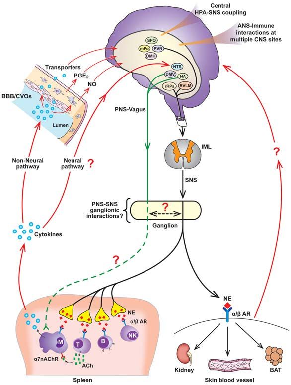
As has been extensively reviewed (94, 200, 205, 256), PGE2 in the POA of the
hypothalamus plays a critical role in mediating sympathoexcitatory responses that contribute
Author Manuscript
to fever development (Figure 5, adapted from Morrison; 200). PGE2 in the POA influences
the activation state of POA neurons that project to the DMH and the rRPa, thereby providing
a critical substrate for activation of BAT SND and cutaneous vasoconstriction sympathetic
outflow (94, 200, 205, 206, 256, 285, 326). It is likely that POA neurons that express EP3
receptors tonically inhibit DMH and rRPa neurons via GABAergic projections, and
inhibition of POA neurons secondary to binding of PGE2 to the EP3 receptor produces
disinhibition of DMH and rRPa neurons (200, 205, 206). The use of anatomical and
functional experimental approaches reveal that increased BAT SND is mediated by
activation of rRPa neurons secondary to disinhibition of DMH neurons, indicating that BAT
thermogenesis responses are dependent on activation of neurons in the dorsal hypothalamus
(94, 200, 202, 205, 206, 256, 285, 326). Cutaneous vasoconstriction produced by POA
PGE2 microinjection is mediated by inhibition of POA projection neurons that bypass the
Author Manuscript
dorsal hypothalamus and project directly to the rRPa (94, 200, 202, 205, 206, 256, 285,
326).
Summary
The results of studies using central microinjection and electrophysiological approaches,
peripheral nerve recordings, and molecular biological and pharmacological techniques have
advanced the understanding of mechanisms mediating sympathetic-immune interactions and
Compr Physiol. Author manuscript; available in PMC 2015 July 01.Kenney and Ganta Page 14
central regulation of sympathetic nerve outflow in several prominent ways. First, and
Author Manuscript
consistent with previous reviews regarding communication between the immune system and
the CNS (10, 230), sympathetic neural circuits are apprised of peripheral immune status, and
alterations in this status, via multiple pathways, including but not limited to; activation of
peripheral afferents with subsequent neural communication to central neural nuclei, access
of immune cell products and other immune mediators to central sympathetic sites via CVOs,
and induction of COX-2 activity and the synthesis of PGE2 in endothelial cells and
perivascular macrophages in the brain microvasculature. Second, the capability of cytokines
and other immune cell products to alter the level of activity in sympathetic nerves
innervating multiple targets (kidney, spleen, brown adipose tissue, tail) supports an
extensive integrative influence of immune system mediators on SND regulation and
physiological function. Third, cytokines and other inflammatory mediators produce changes
in sympathetic nerve activity, at least in part, via specific neural pathways. For example,
Author Manuscript
PGE2 in the POA contributes to fever generation by influencing the activation state of POA
neurons that project to the DMH, which in turn activate cell bodies in the rRPa, with
subsequent increases in the level of BAT SND (200). Fourth, SNS-immune system
interactions can influence peripheral immune responses to acute physiological stimuli. For
example, heating-induced activation of central sympathetic nerve outflow can modulate
splenic cytokine gene expression secondary to increased splenic SND. Fifth, immune system
mediators have been investigated as contributors to sympathetic neural alterations associated
with several pathophysiological states, including, but not limited to neurogenic
hypertension, heart failure, and fever (89, 90, 134, 135, 200, 267). Consistent with many
physiological processes and interactions, an appropriate level of cooperation and
physiological balance between immune cell mediators and sympathetic neural circuits is
important for maintaining homeostasis, and alterations in this balance can contribute to the
development and maintenance of pathophysiological conditions. Sixth, bacterial disease in
Author Manuscript
living organisms influences numerous sites and progresses in diverse ways, therefore, a
variety of experimental approaches and interventions have been used to characterize and
identify fundamental regulatory components relating the immune system and central
sympathetic neural circuits. For example, intravenous cytokine administration models
systemic bacterial infections, brain intraparenchymal cytokine microinjections model the
release of cytokines from CNS astrocytes, microglial cells, and neurons, and icv
administration of cytokines into the lateral ventricles has been used as an experimental
model for cytokines released from choroid plexus. Supporting the use of diverse
administrative approaches, Vallieres et al. (300) reported that systemic injection of IL-6
strongly activated cells in the CVOs, whereas a different pattern of cellular activation of
brain sites was observed after intracerebral injection of IL-6, suggesting that the response of
brain areas to IL-6 and other cytokines may be dependent on the origin of the cytokine
Author Manuscript
during an immune challenge.
Compr Physiol. Author manuscript; available in PMC 2015 July 01.Kenney and Ganta Page 15
PERIPHERAL SYMPATHETIC NERVOUS SYSTEM-IMMUNE SYSTEM
Author Manuscript
SIGNALING INTERACTIONS
Introduction
Complex interactions exist between the SNS and the immune system and it has been shown
that the SNS can both enhance and inhibit immune responses, depending on; experimental
conditions, the type of stress paradigm used, the specific pathophysiological state or disease
condition, activation state of the SNS, and types of immune cells activated. In addition, and
as reviewed previously, immune cell products can influence the functional state of the SNS.
This section reviews studies that have documented peripheral SNS-immune system
signaling interactions.
Sympathetic Innervation of Lymphoid Organs
Author Manuscript
Neuroanatomical and neurochemical studies using retrograde tracers and
immunocytochemical techniques have demonstrated that both primary and secondary
lymphoid organs, including; bone marrow, thymus, spleen, lymph nodes, and gut and
bronchiolar associated lymphoid tissue, are innervated by sympathetic nerve fibers (18, 19,
41, 79, 80, 82, 83, 208, 236, 299, 305, 320, 321). The sympathetic innervation of primary
and secondary lymphoid organs contributes to the maturation, development and regulation
of immune cells throughout the life of the animal (19). The sympathetic innervation to the
spleen is well-characterized (19). Post-ganglionic sympathetic nerves enter the spleen along
with the splenic artery after arising primarily from the superior mesenteric plexus and the
celiac ganglionic plexus (16, 47, 207). Sympathetic neural projections to the spleen include
a prominent innervation to the splenic white pulp concentrated around the central arteriole
and its branches (4, 15, 43, 78, 81, 84, 176). Studies using immunocytochemistry identified
Author Manuscript
that the sympathetic projections ultimately enter the periarterial lymphoid sheets ending near
T cells and surrounding supporting interstitial cells (4, 14, 15, 17, 19, 78, 81, 84, 320, 321).
The sympathetic innervation to the spleen also includes projections to the splenic red pulp
(4, 78, 81, 84, 176). High resolution studies of the splenic white pulp reveal the presence of
sympathetic nerve endings in close proximity (4–6 nm) to splenic T cells, B cells and
dendritic cells, forming synapses that have been termed neuroimmune junctions (78, 84,
278).
Adrenergic Receptor Expression on Immune Cells
Adrenergic receptors, as well as receptors for the binding of other ligands, including;
neuropeptide Y, β-endorphin, adenosine-3-phosphate and adenosine, are expressed on the
cell membranes of macrophages, T lymphocytes, B lymphocytes and NK cells (3, 28, 29,
Author Manuscript
33, 52, 55, 92, 122, 124, 131, 159, 164, 173, 180, 197, 198, 199, 227, 228, 231, 232, 243,
250, 275, 304, 306, 318, 322). Adrenergic receptors are seven-transmembrane, guanine
nucleotide-binding protein-coupled receptors, and ligand binding leads to activation of
adenylyl cyclase, intracellular accumulation of 3′-5′-cyclic adenosine monophosphate
(cAMP), and increased concentrations of protein kinase A (160, 196) or protein kinase C
(146). The protein kinases phosphorylate various transcription factors [e.g., nuclear factor
κB (NF-κB), mitogen activated protein kinase, Ras, Src] resulting in altered (increase or
Compr Physiol. Author manuscript; available in PMC 2015 July 01.Kenney and Ganta Page 16
decrease, based on the type of transcription factor activated) immune cell function, including
Author Manuscript
changes in cytokine secretion and adrenergic receptor expression (53, 57, 146, 167).
Functional and Regulatory Mechanisms Mediating Sympathoimmune Interactions
The presence of intimate anatomical connections between sympathetic nerve fibers and
immune cells, coupled with the expression of adrenergic receptors on immune cells, set the
stage for studying functional and regulatory mechanisms mediating sympathoimmune
interactions. A number of investigations have studied sympathoimmune interactions using
the spleen as the target organ (161, 190, 251, 280). Activation of post-ganglionic splenic
sympathetic nerve fibers leads to the release of NE at the neuroimmune junction (225). The
amount of NE released is dependent on several factors including: the level of sympathetic
nerve activity; expression of α/β adrenergic receptors on presynaptic nerve terminals and
immune cells (117, 310); and neuromodulators released by immune cells, such as cytokines
Author Manuscript
and neuropeptides. (6, 25, 30, 118, 161, 219, 245, 280).
Molecular mechanisms mediating sympathoimmune interactions have been extensively
studied (248, 249, 250, 251, 252, 253, 254). NE released from sympathetic nerve endings
binds to α and/or β adrenergic receptors (AR) expressed on immune cells (T cells, B cells,
NK cells and macrophages). Macrophages express α2ARs and β2ARs (3, 122, 124, 173,
243, 275, 304). Stimulation of α2ARs enhances macrophage activity via activation of
phospholipase C and G-protein dependent mechanisms, whereas stimulation of β2ARs
suppresses macrophage activity secondary to elevations in cAMP (3, 123, 130, 169, 235,
243, 304, 308, 317). T lymphocytes predominantly express β2ARs (28, 29, 33, 52, 92, 159,
164, 180, 197, 227, 228, 231, 232, 250, 306, 318, 322), and these receptors are expressed on
naïve T cells and T helper (Th)1 cells, but not on Th2 cells (232). Stimulation of β2ARs on
T cells increases cAMP and protein kinase A and inhibits T cell proliferation (13, 29, 33, 37,
Author Manuscript
52, 63, 65, 75, 159, 170, 192, 227, 250, 271, 309, 316, 322). Although not commonly
expressed under normal conditions, αARs can be expressed by T cells under certain
pathophysiological conditions states (203), and activation of T cell αARs often results in
negative regulatory function of T lymphocytes, including alterations in T cell maturation in
the thymus (105). β2ARs are predominantly expressed on B lymphocytes (28, 55, 92, 162,
164, 198, 199, 227, 306). Stimulation of these receptors increases cAMP and protein kinase
A (138) which in turn influences (increase or decrease, dependent on the type of mitogen
used for stimulation) the proliferation of B lymphocytes and subsequent antibody production
(19). NK cells express α1-, α2- and β2-ARs and activation of these receptors influences the
migration pattern of NK cells and their functional ability to cause cellular lysis. Activation
of NK cell β2ARs stimulates recruitment of these cells from the marginating pool to
circulating pools (21), and in vivo stimulation of the β2ARs receptors causes suppression of
Author Manuscript
NK cell activity (133, 266). Activation of α1- and α2-ARs has been shown to enhance the
cytotoxic potential of NK cells (323).
Previous studies have established a role for changes in the level of SND in regulating the
functional status of the immune system. Ganta et al. (96) provided support for the hypothesis
that central activation of splenic SND can modulate peripheral cytokine gene expression.
These investigators reported that whole body hyperthermia (96) and central angiotensin II
Compr Physiol. Author manuscript; available in PMC 2015 July 01.You can also read