IMMUNOLOGICAL EFFECTS OF ISOLATED REGIONAL PERFUSION IN MALIGNANT MELANOMA - Junko Johansson Department of Surgery Institute of Clinical Sciences ...
←
→
Page content transcription
If your browser does not render page correctly, please read the page content below
IMMUNOLOGICAL EFFECTS OF
    ISOLATED REGIONAL
  PERFUSION IN MALIGNANT
        MELANOMA

                  Junko Johansson

               Department of Surgery
            Institute of Clinical Sciences
   Sahlgrenska Academy, University of Gothenburg

                   Gothenburg 2019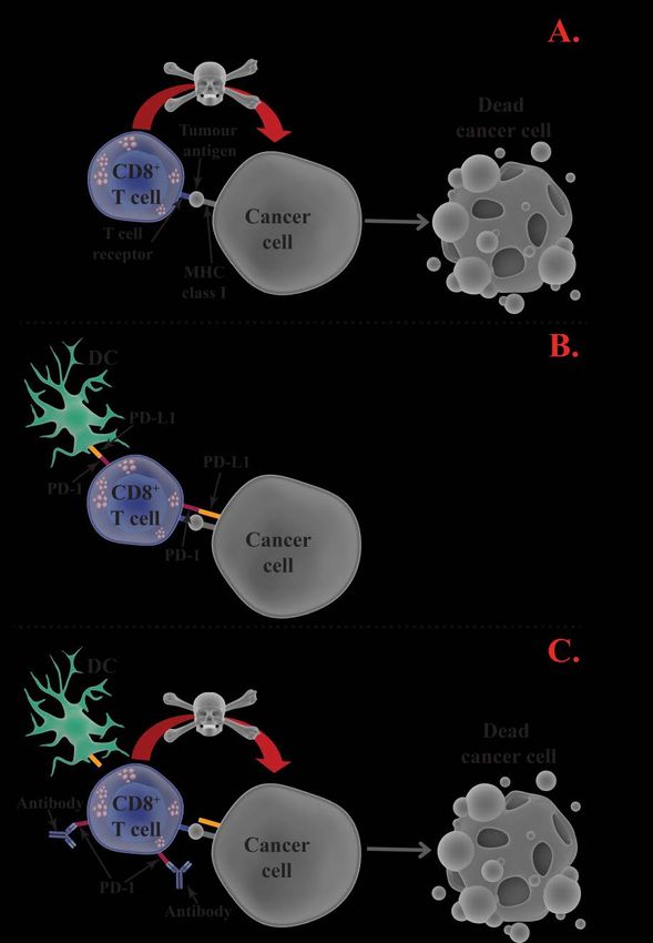
Cover illustration: Busted! by Junko Johansson

The cover shows s simplified illustration of an anti-tumoural immune
response wherein a dendritic cell directs cytotoxic T cells towards a
melphalan-exposed tumour.

Illustrations were made based on elements from Library of Science &
Medical Illustrations by somersault18:24 (somersault1824.com), licensed
under a Creative Commons license (CC BY-NC-SA 4.0), except otherwise
stated.

Immunological effects of isolated regional perfusion in malignant melanoma
© Junko Johansson 2019
junko.johansson@gu.se

ISBN 978-91-7833-422-3 (print)
ISBN 978-91-7833-423-0 (electronic)

Printed in Gothenburg, Sweden 2019
Printed by BrandFactory
Abstract
Malignant melanoma patients with metastatic disease confined to the limbs or
liver may be treated with hyperthermic isolated regional perfusion with a
chemotherapeutic agent, most commonly melphalan. This procedure enables
much higher tissue concentrations of the chemotherapeutic agent compared
with systemic administration. Isolated limb perfusion (ILP) is approved for
treatment of cutaneous metastatic melanoma, while the efficacy of isolated
hepatic perfusion (IHP) is under evaluation for the treatment of liver
metastases from uveal melanoma. Following ILP and IHP tumours often
gradually decrease in size during a period of several months, which might be
explained by a treatment-induced immunological anti-tumour response. This
thesis aimed at investigating the potential role of the immune system for
treatment response to ILP and IHP utilising in vivo analyses of patient
material and mice models and in vitro cell cultures. As reported in Paper I
and Paper II, patients who harboured a high fraction of activated and
antigen-specific T cells in blood prior to ILP were more likely to achieve a
complete disappearance of tumours following ILP. Furthermore, the in vitro
and in vivo assays showed that melphalan exposure enhanced the activation
of T cells and increased the numbers of intermediate and non-classical
monocytes. This may be due to the melphalan-induced upregulation of
immune-related stress markers on melanoma cells, which in turn stimulated
immune cells. In Paper III it was reported that high levels of interferon-
stimulated gene products in patient blood, including CXCL10, CCL2 and
PD-L2, were predictive of a favourable treatment response to ILP, and that
the receptors of these ligands increased on immune cells following treatment.
Paper IV describes different T cell immune profiles in blood between uveal
melanoma patients and healthy controls, and showed that melanoma patients
harboured a lower frequency of CD8+ T cells and more regulatory T cells.
Uveal melanoma patients achieved a longer progression-free survival
following IHP if they harboured a high fraction of activated T cells in blood.
In conclusion, the findings presented in this thesis point towards a role of the
immune system for treatment responses following both ILP and IHP,
suggesting that it may be beneficial to combine isolated regional perfusion
with immunotherapy.

Keywords: Melanoma, isolated regional perfusion, ILP, IHP, melphalan,
immunogenic cell death, T cells, monocytes, ISG
Sammanfattning på svenska
Malignt melanom är en av de vanligaste formerna av cancer i Sverige. Över
90% av alla fall av melanom i västvärlden är i form av hudcancer, men
melanom kan även uppstå på andra ställen i kroppen, exempelvis i ögonen.
De flesta som drabbas av hudmelanom botas lätt genom kirurgiskt
avlägsnande av tumören, men i de fall då metastaser, dvs. dottertumörer,
uppstår blir behandlingen mycket svårare och prognosen värre. För
ögonmelanom är metastasering ett stort problem då majoriteten av alla som
drabbas av ögonmelanom utvecklar dottertumörer, vilket inte är fallet för
hudmelanom. En behandlingsform för melanompatienter med metastaser
isolerade till ett område av kroppen, framförallt till extremiter vid
hudmelanom eller i lever för ögonmelanom, är hyperterm isolerad regional
perfusion med cellgiftet melfalan. Under en perfusionsbehandling kopplas
den drabbade kroppsdelens blodkärl till en hjärt-lungmaskin för att skapa ett
separat, isolerat cirkulationssystem som är skilt från kroppens systemiska
blodcirkulation. Till blodet i den isolerade kroppsdelen tillsätts höga doser av
cellgift för att cirkulera runt under en timme, innan cellgiftet sköljs bort och
kroppsdelen återigen kopplas till den systemiska cirkulationen. Detta upplägg
gör det möjligt att enbart behandla den del av kroppen som är drabbad av
tumörer, vilket minskar risken för generella biverkningar. Vissa av de
tumörer som försvinner efter behandlingen minskar gradvis i storlek under
flera månader innan de slutligen försvinner helt och hållet. En hypotes är att
det beror på att perfusionsbehandlingen med melfalan aktiverar kroppens
immunförsvar till att attackera de tumörer som överlever själva cellgiftet.
Syftet med denna doktorsavhandling var att undersöka om immunförsvaret
har en roll för behandlingsresponsen vid perfusion. Med hjälp av analyser av
blod från perfusionspatienter, cellkulturer och försöksdjur har vi kunnat visa
att melanompatienter med en stor andel aktiverade T-celler, immunceller
viktiga vid försvar mot cancer och virus, innan perfusion svarar bättre på
behandlingen. Perfusionsbehandlingen leder även till en högre aktivitetsgrad
av T-cellerna och en högre produktion av de signalsubstanser som används
för att locka T-celler och andra immunceller till tumörer. Detta kan bero på
att melfalan påverkar de överlevande melanomcellerna så att de börjar visa
upp och producera proteiner som aktiverar och lockar till sig immunceller.
Fynden som presenteras i denna avhandling tyder på att det kan vara
fördelaktigt      att   kombinera      isolerad   regional    perfusion     med
immunstimulerande behandling för att få en ökad behandlingseffekt.
List of papers
This thesis is based on the following studies, referred to in the text by their
Roman numerals.

  I.    Johansson, J., Kiffin, R., Andersson, A., Lindnér, P., Naredi, P.,
        Olofsson Bagge, R. and Martner, A. Isolated Limb Perfusion With
        Melphalan Triggers Immune Activation in Melanoma Patients.
        Frontiers in Oncology, 2018, 8(570)

 II.    Martner, A., Johansson, J., Ben-Shabat, I. and Olofsson Bagge, R.
        Melphalan, Antimelanoma Immunity, and Inflammation—Letter.
        Cancer Research, 2015, 75(24)

III.    Johansson, J., Kiffin, R., Aydin, E., Nilsson, M.S., Hellstrand, K.,
        Lindnér, P., Naredi, P., Olofsson Bagge, R. and Martner, A. Isolated
        limb perfusion with melphalan activates interferon-stimulated genes
        to induce tumor regression in patients with metastatic melanoma.
        Submitted

IV.     Johansson, J., Kiffin, R., Siarov, J., Mölne, J., Naredi, P., Olofsson
        Bagge, R., Martner, A. and Lindnér, P. Presence of activated T cells
        in peripheral blood correlates to longer progression-free survival in
        patients undergoing isolated hepatic perfusion for uveal melanoma
        liver metastasis.
        Manuscript

         Reprints were made with permissions from the publishers.

                                       i
Additional publication not part of this thesis:

i. Aydin, E., Johansson, J., Nazir, F. H., Hellstrand, K. and Martner, A. Role
of NOX2-Derived Reactive Oxygen Species in NK Cell–Mediated Control of
Murine Melanoma Metastasis.
Cancer Immunology Research, 2017, 5(9)

                                        ii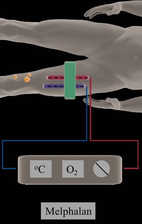
Content
ABBREVIATIONS ........................................................................................ V
1. BACKGROUND ......................................................................................... 1
   1.1. Cancer................................................................................................... 1
       1.1.1. Malignant melanoma ..................................................................... 2
           1.1.1.1. Cutaneous melanoma.............................................................. 2
           1.1.1.2. Uveal melanoma ..................................................................... 6
   1.2. Isolated regional perfusion ................................................................... 9
       1.2.1. The technique ................................................................................ 9
       1.2.2. Treatment response...................................................................... 10
       1.2.3. Melphalan .................................................................................... 11
       1.2.4. Hyperthermia ............................................................................... 11
       1.2.5. Tumour necrosis factor α ............................................................. 12
   1.3. The immune system............................................................................ 13
       1.3.1. Immune cells ............................................................................... 13
           1.3.1.1. Dendritic cells....................................................................... 14
           1.3.1.2. Monocytes and macrophages ............................................... 15
           1.3.1.3. T cells ................................................................................... 15
       1.3.2. Immune responses against pathogens .......................................... 17
           1.3.2.1. Extracellular pathogens ........................................................ 17
           1.3.2.2. Viruses and cancer ................................................................ 18
       1.3.3. Cancer immunology .................................................................... 20
           1.3.3.1. Immunoediting ..................................................................... 20
           1.3.3.2. Immunogenic cell death ....................................................... 21
           1.3.3.3. Immunotherapy..................................................................... 21
   1.4. Melanoma, isolated regional perfusion, and immunology ................. 26
2. AIMS ......................................................................................................... 27
3. METHODOLOGY .................................................................................... 29
   3.1. Isolated regional perfusion ................................................................. 29
   3.2. In vivo murine model .......................................................................... 29
   3.3. In vitro perfusion model ..................................................................... 30

                                                         iii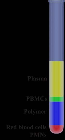
3.4. Methods .............................................................................................. 30
4. RESULTS AND DISCUSSION................................................................ 31
   4.1. Melphalan induces an immunogenic-type of cell death in melanoma
   cells............................................................................................................ 31
   4.2. Melphalan-exposed melanoma cells triggers induction of CD16+
   monocytes and interferon-stimulated gene products ................................. 34
   4.3. Favourable outcome after ILP is correlated with activated and antigen-
   specific T cells ........................................................................................... 37
   4.4. Favourable outcome after IHP is correlated with activated T cells .... 40
5. CONCLUDING REMARKS .................................................................... 43
6. ACKNOWLEDGEMENTS ...................................................................... 45
REFERENCES .............................................................................................. 49

                                                          iv
Abbreviations
ACT Adoptive cell transfer
APC Antigen-presenting cell
CR Complete response
CTLA-4 Cytotoxic T-lymphocyte-associated antigen 4
DAMP Damage-associated molecular pattern
DC Dendritic cell
HSP Heat-shock protein
ICD Immunogenic cell death
IFN Interferon
IHP Isolated hepatic perfusion
IL Interleukin
ILP Isolated limb perfusion
ISG Interferon-stimulated gene
MHC Major histocompatibility complexes
NK cell Natural killer cell
OS Overall survival
OVA Ovalbumin
PAMP Pathogen-associated molecular pattern
PBMC Peripheral blood mononuclear cell
PD Progressive disease
PD-1 Programmed cell death protein 1
PD-L1/2 Programmed death-ligand 1/2
PFS Progression-free survival
PMN Polymorphonuclear leukocyte
PR Partial response
vPRR Pattern recognition receptor
SD Stable disease
TCR T cell receptor
TNF Tumour necrosis factor
Treg Regulatory T cell
UV Ultraviolet
vi1. Background
To find a solution to a problem, you must first define the problem. To find a
treatment to cancer, you must first know what cancer is.
1.1. Cancer
Cancer is an umbrella term for a variety of diseases that all originate from
cells with uncontrolled growth and which spread and invade other parts of the
body. Often the cells will conglomerate in a mass known as a tumour. There
are both benign and malignant tumours, wherein benign tumours do not
invade nearby tissues or metastasise, e.g. spread, to other parts of the body, in
contrast to malignant tumours. Malignant tumours are thus cancers.
Depending on cell type and which tissue or organ the cells reside in, different
types of cancer arise.
Cell growth is heavily regulated. In order to maintain a functional
multicellular organism each cell needs to function in its proper way, for
example only dividing when needed and when ordered to. Cancer cells lack
or have circumvented these regulatory mechanisms, causing them to have
abnormal growth. This is due to an accumulation of mutations which causes
genetic instability of the cells [1]. It is often considered that a tumour
originates from a single abnormal cell, which produces daughter cells that in
each generation accumulate more and more mutations due to natural
selection.
Genetic instability is of paramount importance in the transformation of a
healthy cell to a cancer cell. It lays the foundation for eight underlying
principles, the “hallmarks of cancer”, in cancerogenesis: self-sufficiency in
growth signals (cancer cells develop their own signalling molecules that tell
them to grow), insensitivity to anti-growth signals (cancer cells ignore signals
that tell them to stop growing), evasion of apoptosis (evasion of self-
destruction), sustained angiogenesis (maintaining a constant blood supply),
limitless replicative potential (limitless amount of cell divisions), tissue
invasion and metastasis (spreading to other tissues and organs), deregulating
cellular energetics (altered energy metabolism), and avoiding immune
destruction (avoiding destruction by the immune system) [2, 3]. The latter is
of interest since another enabling characteristic for the hallmarks, in addition
to genetic instability, is inflammation induced and promoted by the tumour
itself [3]. Thus, cancer cells might promote one type of immunologic
response and at the same time manage to avoid destruction by immune cells.
11.1.1. Malignant melanoma
Melanoma is cancer of the melanocytes, which are cells producing the
pigment melanin. Melanocytes are originally derived from a structure called
the neural crest; a transient group of cells found during the early stages of the
development of an embryo [4]. The neural crest is a group of cells that are
part of the early structure of the pre-cursor to the central nervous system
(brain and spinal cord), the neural tube, but are pinched off during the
formation of the tube and differentiate into cells that do not remain part of the
central nervous system. Melanocytes are found throughout different parts of
the body, such as in the skin, the eye and in mucosal membranes in the nasal
cavity [5], the genital and urinary tract [6], and in the rectum [7].
Melanin protects the epidermis (the upper most layer of the skin) from
harmful ultraviolet (UV) light through absorbance and scattering of the
radiation [8]. Except for producing melanin, melanocytes have other
functions as well, such as by interacting with and regulating the immune
system [9, 10]. This may in part explain why melanocytes reside in parts of
the body that do not need protection against sun-derived UV damage.
Depending on where in the body the melanocytes reside, different forms of
melanoma arise. Despite originating from the same cell type, they differ in
their genetic and phenotypic composition and how the disease develops in the
patient. This thesis if focused on melanoma in the skin (cutaneous melanoma,
Papers I-III), and in the eye (uveal melanoma, Paper IV).
1.1.1.1. Cutaneous melanoma
When discussing skin cancer, care should be taken to define what type of
skin cancer that is the focus of discussion. Although all forms of skin cancer
have some things in common, such as arising in the skin and being induced
by UV radiation, there are big differences in incidence and mortality rates
between them. In general there are three different types of cancers that are
included in the term skin cancer; basal cell carcinoma, squamous cell
carcinoma and cutaneous melanoma [11]. The incidence rates of basal cell
carcinoma and squamous cell carcinoma are higher than for melanoma,
though cutaneous melanoma has a higher mortality rate [12].
1.1.1.1.1 Epidemiology and causes
Cutaneous melanoma is most common in countries with a predominantly
Caucasian population, such as in Europe, Northern America and in Oceania
[13]. In 2017 it was the fifth most common type of cancer among women and
the sixth most common among men in Sweden, and unfortunately the
incidence rate is steadily increasing [14]. Most melanoma patients in Sweden
are diagnosed when they are around 60 years old, and the most common site
2for the primary tumour is the extremities for women while it is the trunk for
men [15-18].
Exposure to UV radiation is a known risk factor for the probability to
develop cutaneous melanoma. In WHO’s list of different agents and their
carcinogenic hazards to humans, UV radiation is found in group 1, where
chemicals and other agents who have been shown to be carcinogenic belong
[19]. For a reference, tobacco smoke and plutonium are also in group 1. The
hazardous property of UV radiation is that it damages DNA.
Studies have shown that the risk of developing melanoma is highest during
intermittent sun exposure compared to chronic sun exposure [20]. Severe
sunburns and high sun exposure during childhood are also risk factors. The
UV radiation which is derived from the sun fall into three categories: UVA
(315-400 nm), UVB (280-315 nm) and UVC (100-280 nm). UVC is absorbed
by the ozone layer and does not affect humans, while UVB penetrates only
the uppermost layer of the skin (epidermis) and UVA reached down to the
middle layer (dermis). UVB causes direct DNA damage by absorption into
the DNA molecule where it induces molecular rearrangements while UVA
causes indirect DNA damage by the formation of reactive oxygen species
[21].
As for many other types of cancer, mutations in certain genes have been
correlated to a higher risk of developing melanoma. Examples of such genes
are the tumour suppressor genes CDKN2A and BAP1, and the oncogene
CDK4 [22, 23]. Another interesting gene where a mutated status has been
linked to melanoma is MC1R. MC1R encodes for the melanocortin-1-receptor
which regulates both hair and skin pigmentation in humans. People with red
hair and fair skin often have mutated variants of the MC1R gene [24], and
these features together with a high number of common and atypical naevi
(i.e. moles) have been linked to a higher risk of developing melanoma [25].
1.1.1.1.2 Subtypes and staging
Cutaneous melanoma is clinically classified into four different subtypes
depending on how and where the tumour grows [25]. The most common type
is superficial spreading melanoma, accounting for approximately 60% of all
cases in Sweden [15, 17, 18], which has a horizontal growth pattern in
contrast to nodular melanoma which almost exclusively has a vertical growth.
Nodular melanoma accounts for 20% of the Swedish cutaneous melanoma
cases [15, 17, 18]. Acral lentiginous melanoma can be found on e.g. the
fingers, the palm of the hand and the sole of the foot (acral meaning affecting
or belonging to peripheral body parts), and is uncommon among Caucasian
patients but is percental more common among patients of African, Asian and
Hispanic descent. Lentigo maligna melanoma is correlated with long-term
3UV exposure and increasing age and can grow down into the hair follicles of
the skin.
To classify the spread of the disease, melanoma is categorized into different
stages according to the TNM staging system [26]. T defines the depth of the
primary tumour and if the primary tumour has ulceration, N explains the
spread to nearby lymph nodes and M contains information about distant
metastasis and is defined according to anatomical site. Based on the TNM-
status the patient is then staged into a numerical staging system from stage 0
to stage IV, where the higher the number the more the metastatic spread.
Stage 0 is superficial and non-invasive, stage I and II is melanoma without
sign of metastatic spread, stage III indicates metastasis to lymph nodes and
stage IV is when the disease has distant metastasis and thus has spread to
other organs. Two other measurements that are sometimes utilised for
melanoma is the Breslow thickness (describing the vertical thickness of the
primary tumour) and the Clark level (how far down into the skin the primary
tumour has invaded) [26].
1.1.1.1.3 Prognosis and treatment
The prognosis for cutaneous melanoma
A short primer on survival:
depends a lot on the characteristics of the
tumour at the time of diagnosis and the state of • Overall survival (OS): Time
metastasis. The melanoma-specific and overall from diagnosis until death of
survival decreases rapidly with increasing any cause.
• Melanoma-specific survival:
depth of the primary tumour and a higher Time from diagnosis until
degree of metastatic spread [15-18]. Among death due to melanoma.
Swedish melanoma patients diagnosed with • Progression-free survival
stage I and stage II melanoma, 90% are alive (PFS): Time from start of
after five years without dying of their disease. treatment until progression
and/or recurrence of
For stage III patients, the corresponding melanoma.
percentage is 36% and for stage IV it is 25%
[17]. Worth noticing is that over 90% of all
patients are diagnosed with stage I or stage II [16, 17].
The standard treatment option for patients with stage I or II melanoma
without metastatic spread to lymph nodes is surgical excision of the tumour.
To prevent recurrence of the tumour a certain area around it is also often
removed; the margin of the excision is based on the thickness of the tumour.
In case of suspicion of metastatic spread to lymph nodes, which is usually the
first place of metastasis, a sentinel lymph node biopsy may be performed.
The sentinel lymph node is the first draining lymph node from the primary
tumour and is hypothetically the first lymph node metastases gather in. The
sentinel lymph node can be found by injecting a radioactive tracer or a dye
4near the primary tumour and then surgically remove the nodes that have
taken up the radioactive tracer or the dye. By removing and examining the
lymph node it is possible to evaluate the metastatic spread of the disease [27-
29].
For patients with metastatic disease, surgery is usually not curative but can be
utilised to prolong and improve the life of the patient. Depending on how
many metastases and where they are located, patients may or may not benefit
from surgical excision of their metastatic tumours [30]. Other treatment
strategies that may be employed are targeted therapies, locoregional therapies
and immunotherapies.
In addition to harbouring mutated versions of CDKN2A, BAP1 and CDK4,
40-60% of all melanoma patients do also have a changed version of BRAF
[31-33]. BRAF encodes for the protein B-raf which is a part of the
MAPK/ERK pathway; a signal-transduction pathway wherein signalling
molecules bind to a receptor on the cell surface and induce an intracellular
signalling cascade, ultimately regulating vital cell functions such as cell
growth and proliferation. The mutated BRAF common in melanoma encodes
for a version of the protein that is highly activated [31]. Another component
of the MAPK/ERK pathways is N-ras, encoded by the NRAS gene. It is also
frequently mutated in melanoma, with 15-20% of all melanomas harbouring a
mutated variant [32, 33]. Targeted therapies have been developed wherein
inhibitors target the most common mutated variant of the BRAF gene product
or other proteins in the same signalling pathway. Despite a relatively high
response rate, many patients will experience progression of their disease
since it is common to develop resistance to the inhibitors [34].
For in-transit melanomas, i.e. when the melanoma has started to metastasise
and spread from the primary tumour via the lymph vessels but have not yet
reached the nearest lymph nodes, locoregional therapies may be an option.
Locoregional therapies only treat the part of the body affected by metastatic
disease, compared to systemic therapies which affect the whole body. Two
locoregional treatment options are isolated regional infusion and isolated
regional perfusion with chemotherapeutic agents, the latter being the subject
of this thesis and will be described in detail in chapter 1.2.
Immunotherapy has during recent years been an important part in the
treatment of metastatic melanoma. Immunotherapy aims to boost the body’s
own immune system to attack the cancer cells, which might be achieved by
e.g. making cancer cells more attractive to immune cells or activating
immune cells. Immunotherapy in the treatment of cancer will be discussed
further in chapter 1.3.
51.1.1.2. Uveal melanoma
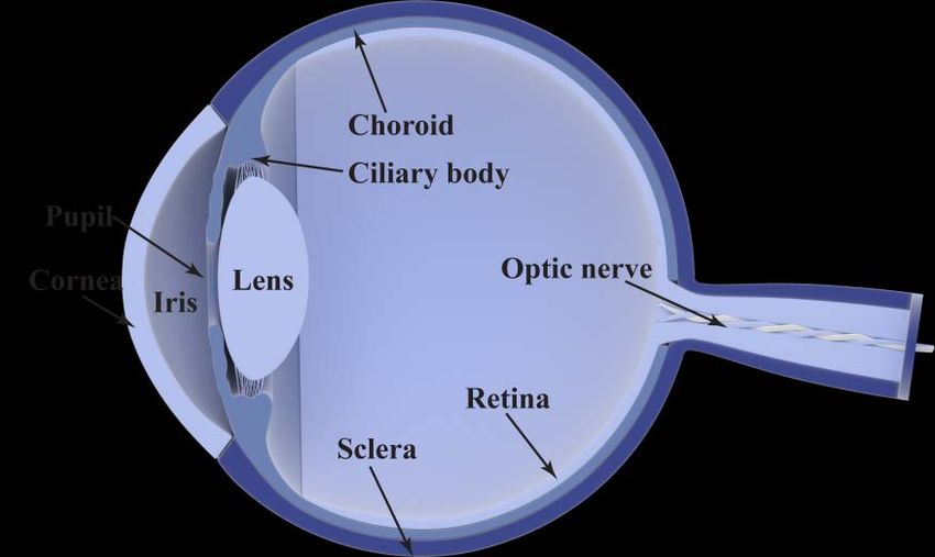
Uveal melanoma arises in the uvea of the eye. The uvea consists of three
parts; the iris which controls the size of the pupil and thus the amount of light
entering the eye, the ciliary body which holds the lens and produces the fluid
found between the cornea and the lens, and the choroid which is a vascular
layer of the eye containing connective tissues and blood vessels.
Figure 1. Schematic diagram of the human eye. The uvea consists of the iris, the ciliary body
and the choroid.
1.1.1.2.1 Epidemiology and causes
Despite being a rather rare form of cancer, uveal melanoma is the most
common intraocular tumour in adults and is the second most common form
of melanoma after cutaneous melanoma. It constitutes approximately 5% of
all melanoma cases [35, 36]. As with cutaneous melanoma, uveal melanoma
is most common in Caucasian populations [37, 38], and in Sweden there are
approximately 70 diagnosed uveal melanoma cases each year [12].
In contrast to cutaneous melanoma where exposure to UV radiation is a
known risk factor for the development of the disease, the relationship
between UV radiation and uveal melanoma is not as clear. Intermittent
exposure to sun-derived UV light has not been shown to increase the risk of
developing uveal melanoma [39], even though hypothetically melanocytes in
the eye should be affected as well. It is known that people with fair skin and
light eye colour, such as blue-eyed Caucasians, have a higher risk of
developing uveal melanoma [40].
Mutations in the BAP1 gene have shown to increase the risk of the
development of uveal melanoma [41-44]. Two other genes that have shown
6to be implicated in the development of cutaneous melanoma, CDKN2A and
CDK4, do not seem to increase the risk of uveal melanoma [45-48],
indicating differences in the genetic landscape of the two forms of melanoma.
1.1.1.2.2 Subtypes and staging
The TNM staging system is also applicable for uveal melanoma and can be
utilised to further categorise the spread of the disease in a numerical system
with stages from I to IV [49, 50]. If the affected eye is removed or a biopsy is
taken, the additional stage G might be added. G denotes the pathological
appearance of the tumour, i.e. what the tumour cells and the tissue look like
when observed with a microscope. Uveal melanoma tumours consist of cells
that can be classified as spindle cells or epithelioid cells [51]. An extremely
brief explanation of the difference between the two cell types is that spindle
cells look more slender and stream-lined, while epithelioid cells are bulkier.
1.1.1.2.3 Prognosis and treatment
The problem with uveal melanoma is that the risk of developing metastatic
disease is rather high. 25% of all uveal melanoma patients will have been
diagnoses with metastatic disease after five years, and among them the
majority will have developed metastases in the liver [52, 53]. Only 20% of all
uveal melanoma patients will be alive one year after being diagnosed with
metastatic disease, and after two years only 4-8% of all patients are alive [52,
53].
It is not fully known why uveal melanoma has such a high metastasis rate to
the liver. Uveal melanoma cells that form metastases travel through the
circulatory system of the blood, in contrast to cutaneous melanoma cells that
more often traffic the lymphatic system, which is due to the lack of
lymphatics in the eye. But this does not explain why uveal melanoma cells
end up in the liver, since there are other stops between the eyes and the liver
if you travel via the blood road, such as the lungs. Thus, it is thought that the
liver produces substances that attract and stimulate the growth of uveal
melanoma cells [54]. For example, the liver produces high amounts of C-X-C
motif chemokine 12 (CXCL12) which binds to the receptor C-X-C motif
chemokine receptor 4 (CXCR4) on e.g. melanoma cells and recruits them to
the liver.
Genetic studies of primary uveal melanomas have shown that the loss of one
copy of chromosome 3 is strongly correlated to a poor prognosis and higher
risk of developing metastatic disease [55-57]. Inactivating mutations in the
BAP1 gene usually occur together with loss of chromosome 3 [58]. It has
recently been revealed that primary uveal melanomas can be further divided
7into four genetically distinct subgroups with different prognoses; two groups
with loss of one copy of chromosome 3 and two groups without loss [59].
The major treatment options for primary uveal melanoma are either removal
of the eye (enucleation) or radiotherapy. Enucleation used to be the standard
treatment, but it has been shown that there is no difference in survival
between patients undergoing enucleation or more conservative treatments
such as radiotherapy [60, 61], resulting in enucleation now being utilised
only for big tumours where more conservative treatments will not be able to
save the patient’s vision [62]. One commonly used type of radiotherapy is
brachytherapy in which a sealed radiation source is placed next to the tumour.
Other types of conservative therapies where the affected eye is not removed
include photodynamic therapy, wherein light is used to induce the production
of radicals and reactive oxygen species, and thermotherapy, wherein heat is
used to treat the tumour [63].
Due to the high rate of liver metastasis formation, management of metastatic
disease is of outmost importance. Unfortunately, no specific treatment option
has of yet been proven successful in improving survival, despite many
different types of therapies being developed and tested [64]. As for cutaneous
melanoma, locoregional therapies, targeted therapies and immunotherapies
have been tested in various studies. Worth noting is that BRAF mutations are
not common in uveal melanoma, subsequently rendering B-raf inhibitors
useless. Instead, in uveal melanoma mutations in the genes GNAQ and
GNA11 are much more common, with over 90% of all primary tumours
harbouring mutations in GNAQ or GNA11 [65]. Both genes encodes for
proteins that are part of the intracellular IP3 pathway which regulates the
release of calcium inside the cell.
81.2. Isolated regional perfusion
Hyperthermic isolated regional perfusion with chemotherapeutic agents is a
locoregional chemotherapy wherein the treatment only affects the part of the
body where tumours are located. It is used for cancer patients with tumours
confined to a body part or an organ, such as a limb or the liver. The main idea
behind isolated regional perfusion is that it enables the administration of
much higher concentrations of a chemotherapeutic drug compared to regular
systemic chemotherapy; if the drug is only locally administered, systemic
side effects and toxicity can be avoided.
At the Sahlgrenska University Hospital in Gothenburg, the only centre in
Sweden where isolated regional perfusion is performed, isolated limb
perfusion (ILP) is utilised for cutaneous in-transit melanoma with metastatic
disease confined to the limbs, while the usage of isolated hepatic perfusion
(IHP) in the treatment of uveal melanoma liver metastases is currently being
investigated in a randomised clinical trial. Melphalan is the most commonly
used chemotherapeutic drug for ILP and IHP, and the procedure is conducted
under mild hyperthermia (40oC). Patients with bulky disease or who has
undergone repeated perfusions might also receive tumour necrosis factor α
(TNF-α).
1.2.1. The technique
Isolated regional perfusion is an old method, first described and used already
in the 1950’s [66]. Since then the method has been refined and adapted, and
today it is used in the treatment of malignant melanoma and other type of
solid cancers, such as soft-tissue sarcomas.
A detailed description of the technical aspects of ILP and IHP is beyond the
scope of this thesis but can be found elsewhere [67]. The main idea is to
redirect the major blood flow to the limb or the liver by clamping the artery
and veins, insert cannulas and connect the cannulas to a heart-lung machine,
thus creating an isolated circulatory system. The blood that is perfused
through the limb or the liver is oxygenated and supplied with high doses of
the chemotherapeutic agent melphalan, and the treated tissue is held at a
temperature of 40oC. Radioactive isotopes are added to the perfusate to
enable monitoring of potential leakage to the systemic circulation. The total
perfusion time is 60 minutes, followed by rinsing of the limb or the liver with
a salt-based solution to get rid of the melphalan residues.
9Figure 2. Schematic representation of the set-up for ILP.
1.2.2. Treatment response
Clinical response to the treatment regarding tumour burden is evaluated after
three months according to WHO’s criteria as complete response (CR), partial
response (PR), stable disease (SD) and progressive disease (PD) [68]. CR is
defined as disappearance of all tumours, PR is a decrease of more than 50%
of total tumour burden, PD is an increase of more than 25% in existing
tumours or the appearance of new tumours, and SD is where none of the
criteria for CR, PR or PD are met. ILP with melphalan has a CR rate of 50-
70%, with over 85% of all patients achieving an overall response (CR or PR)
[69-71]. IHP with melphalan has a CR rate of 20%, with an overall response
rate of 70% [72]. Five years after treatment around 30-40% of ILP patients
will still be alive [69, 70], while the effectiveness of IHP in improving
overall survival is currently being investigated in a clinical study. The
SCANDIUM trial (ClinicalTrials.gov identifier number: NCT01785316) is a
10clinical study evaluating if IHP improves overall survival compared to best
alternative care for uveal melanoma patients with liver metastases [73].
1.2.3. Melphalan
Chemotherapy is the usage of chemical compounds which inhibit cell growth
and induce cell death in the treatment of cancer. Chemotherapeutic agents
were first used in cancer treatment around the time of World War II. During
World War I mustard gas was effectively used as a chemical warfare agent,
and during World War II there was continued interest in researching mustard
gas. There was an academic interest in the cytotoxic properties of the gas, and
the question arose whether or not it could also be used to kill cancer cells.
The first time a derivative of mustard gas was intravenously used to treat
cancer was in 1942, and the rest, as they say, is history [74-76].
Melphalan, also known as phenylalanine mustard, is a
nitrogen mustard derived from mustard gas. It is an
alkylating agent and adds an alkyl group to the guanine
base of DNA, causing linkage between DNA strands and
subsequently inhibition of DNA synthesis and cell
death. Is it used for systemic treatment in multiple
myeloma and ovarian cancer [77], but systemic
administration of melphalan is not feasible for
melanoma since the effective dose is higher than what
the body tolerates [78]. Thus, melphalan is only
administered with ILP or IHP in the treatment of
malignant melanoma. Figure 3. Structural
formula for melphalan.
1.2.4. Hyperthermia
ILP and IHP with melphalan is often performed under mild hyperthermia
(40oC). There are synergistic effects in play between hyperthermia and
melphalan, resulting in enhanced death of the cancer cells [79-81]. There are
many underlying factors behind the synergy, such as a higher uptake of
melphalan in tumour tissue during hyperthermia [82] and an increase in the
amount of DNA crosslinks produced by melphalan [79].
Furthermore, hyperthermia has in itself anti-tumoural properties. In addition
to the direct killing of cancer cells by heat, hyperthermia inhibits
angiogenesis, thus preventing tumours from forming new blood vessels
which are critical for their survival [83, 84]. Hyperthermia has also been
indicated to induce anti-tumoural immune responses, as reviewed in [85, 86].
This is due to many different factors, for example the increased expression of
heat-induced stress ligands, such as heat-shock proteins (HSPs), which bind
11to and activate immune cells such as dendritic cells (DCs) and natural killer
(NK) cells.
1.2.5. Tumour necrosis factor α
Tumour necrosis factor α (TNF-α) is a cytokine, a cell signalling protein, and
a major driver of the inflammatory response. It has a dual role in the context
of cancer; it is anti-tumoural since it directly can kill cancer cells by
triggering apoptosis (programmed cell death) in cancer cells, and it is pro-
tumoural due to activation of the transcription factor NF-κB and by
sustaining an inflammatory tumour environment (remember, inflammation is
one of the enabling characteristics for the hallmarks of cancer) [87-91].
TNF-α is sometimes utilised in ILP and IHP due to its ability to enhance the
uptake of melphalan into tumour cells by affecting the tumour vasculature
and increasing the blood vessel permeability [92, 93]. It is most often utilised
for patients requiring multiple perfusions or for patients with big and bulky
tumours where uptake of melphalan might be limited due to the tumour size.
121.3. The immune system
The immune system is the body’s own defence against disease. It protects us
from foreign pathogens, such as bacteria and viruses, but does also provide
protection against diseases arising from within the body itself, most notably
against cancer. The immune system is very complex and comprises several
levels of protection, from the small proteins of the complement system via
immune cells to the skin and mucosa, which are our first and biggest barriers
against invading pathogens.
There is both innate and adaptive immunity. Briefly described, innate
immunity is the part of the immune system which first reacts to an invading
pathogen. It provides a swift response with broad specificity. In contrast,
adaptive immunity is highly specific and contains immune cells which upon
the first encounter with a pathogen develop a “memory” of the pathogen,
prompting the cells to react much faster and stronger subsequent times the
pathogen is encountered [94].
1.3.1. Immune cells
The immune cells (white blood cells or leukocytes) are cells dedicated to
protect the body and are found circulating in blood or stationed in tissues. All
blood cells (immune cells, red blood cells and platelets) are produced in the
bone marrow and are derived from the same hematopoietic stem cell. The
stem cell forms immune cells of two different lineages; lymphoid cells and
myeloid cells [94]. The cells have different functions and
mature cells can be found in different parts of the body.
Another common classification of immune cells purified
from peripheral blood is into peripheral blood
mononuclear cells (PBMCs) and polymorphonuclear
leukocytes (PMNs). A very common way to separate
immune cells from whole blood is by density gradient
separation in which blood is placed on top of a
hydrophilic polymer solution in a tube and then
centrifuged. Due to differences in density between
different cells, some cells will sediment through the
polymer while others will not, creating a layered solution
in the tube after centrifugation. The bottom layer
contains red blood cells and PMNs, the layer above is
the polymer, on top of the polymer is a thin layer with
PBMCs and the uppermost layer contains plasma [95]. Figure 4. Diagram of
density gradient separation
of whole blood.
13Table 1. Cheat sheet for different classifications of the most common immune cells found in
blood.
Lymphoid Myeloid Adaptive Innate PBMC PMN
B cell X X X
Basophil X X X
Dendritic
X X X
cell (DC)
Eosinophil X X X
Monocyte X X X
Neutrophil X X X
Natural killer
X X X
(NK) cell
T cell X X X
There are many different types of immune cells and many of them cooperate
during an immunological response. Below follows descriptions of the three
major immune cell populations of relevance for this thesis; dendritic cells,
monocytes and T cell.
1.3.1.1. Dendritic cells
Dendritic cells (DCs) are of myeloid lineage and are found in blood in an
immature form and in tissues and lymph nodes as more mature cells. They
are relatively rare in blood and constitute1.3.1.2. Monocytes and macrophages
Monocytes do also belong to myeloid-lineage cells, and they share some
similarities with DCs. They constitute approximately 10% of all immune
cells in blood [101]. Monocytes are APCs and can during certain conditions
be converted into DCs. They are also potent cytokine producers.
Furthermore, when migrating into tissues they are transformed into
macrophages; large immune cells which are stationed in tissues, patrolling
the area looking for invading pathogens. Macrophages and monocytes are
excellent phagocytes, meaning that they are cells capable of engulfing and
subsequently destroying cells and structures detrimental to a healthy body,
such as pathogens, cell debris and dead cells (macrophage actually means
“big eater” in Greek).
Monocytes are found in blood while macrophages are present in tissues.
Monocytes are usually divided into three different subtypes based on the
expression of the cell surface receptors CD14 and CD16 [101-103]. Classical
monocytes (CD14++CD16-) are the most common type of monocyte,
constituting around 80-90% of all monocytic cells, and often migrate into
tissues to become macrophages. They can also convert into the intermediate
(CD14++CD16+) and nonclassical (CD14+CD16++) monocytes which
accumulate during infections and inflammatory conditions and produce high
levels of the pro-inflammatory cytokines TNF-α and interleukin 1 β (IL-1β).
The different monocyte populations are discussed in Paper I.
The majority of tissue-resident macrophages are not descendants of
monocytes, but are instead developed before birth from their own pre-cursor
cells and are present throughout the whole adult life. During inflammatory
conditions the pool of tissue-resident macrophages are maintained through
the addition of monocyte-derived macrophages. Macrophages have different
functions and different gene signatures depending on which tissue or organ
they reside in, suggesting that they are highly adapted to their surrounding
environment [104]. They are also given different names depending on tissue
type, such as Kupffer cells in the liver, microglia in the brain and osteoclasts
in bone.
1.3.1.3. T cells
T cells are lymphocytes essential for adaptive immunity. They activate and
regulate various type of lymphocytes (including themselves), and are
important in the eradication of cancer cells and virus-infected cells. The “T”
in T cell stands for thymus. Like all other immune cells T cells are first
developed in the bone marrow, but they mature and are transformed into
functional T cells in the thymus. The fraction of T cells in peripheral blood
during healthy conditions varies a lot between different individuals, but is
15typically around 60-80% of all PBMCs. T cells are of importance for Papers
I-IV in this thesis.
There are two major T cell subpopulations; helper T cells and cytotoxic T
cell. They are distinguished and denoted by the receptors CD4 for helper T
cells and CD8 for cytotoxic T cells.
What’s up with all the CDs?
In immunology CD is an abbreviation for “cluster of differentiation” and not for a disc-
shaped storage device. It is basically a naming system for cell surface molecules used for
identifying different types of cells. A CD structure is usually a receptor or a ligand found
on the cell surface, and often their function and relevance to the cell is known. For
example, all T cells express CD3, which is a co-receptor to the main T cell receptor which
T cells need to recognise antigens. By combining different CD markers it is possible to
identify different immune cells, e.g. a cell expressing both CD3 and CD4 but not CD8
(written as CD3+CD4+CD8-) is a helper T cell.
1.3.1.3.1 CD4+ helper T cells
CD4+ T cells are regulators of immunity; activating or inhibiting responses
from e.g. CD8+ T cells and the antibody-producing B cells by receptor
interactions and secretion of cytokines. The CD4+ T cells are further
classified into different groups based on which cytokines they produce and
which transcription factors that are responsible for the development of that
particular CD4+ T cell subtype. Th1 cells produce interferon γ (IFN-γ) and
interleukin 2 (IL-2) which activate CD8+ T cells and macrophages. They are
important for the development of immune responses against intracellular
pathogens. In contrast, Th2 cells mediate responses against extracellular
pathogens by the production of e.g. interleukins 4 and 5 (IL-4, IL-5) which,
among many things, activate B cells, basophils and eosinophils. Th17 cells
primarily produce interleukin 17 (IL-17) which recruits and activates
neutrophils to help in the eradication of extracellular bacteria and fungi. T
regulatory cells (Tregs) are another member of the CD4+ T cell family.
Compared to the other CD4+ T cells, Tregs have a different function; instead of
activating an immune response, Tregs are responsible for suppressing it. Tregs
are responsible for curbing the activity of other immune cells, particular other
T cells, that otherwise would lead to damage; such as overreactive T cells or
T cells that mount a response against healthy tissue, causing autoimmune
disorders. This is done through many mechanisms, one is the secretion of
immunosuppressive cytokines such as transforming growth factor β (TGF-β)
and interleukin 10 (IL-10) [105, 106].
CD8+ cytotoxic T cells
While CD4+ T cells regulate and facilitate the induction of an immune
response, CD8+ T cells directly kills abnormal cells. After being activated
16and told which cell to target (via antigen-presentation by APCs), the CD8+ T
cell kill the target cell through induction of apoptosis via the release of
cytotoxic molecules or direct cell-to-cell contact. They contain and release
perforin, which creates pores in the cell membrane of the target cell, and
granzyme B, which enters the target cell through the pores and induces
apoptosis. Alternatively, the structure Fas ligand (FasL) on the surface of
activated CD8+ T cells can bind to the receptor Fas on the target cell. Upon
binding the FasL/Fas complex triggers the activation of an intracellular
signalling cascade, ultimately leading to apoptosis of the target cell [107].
1.3.2. Immune responses against pathogens
What really happens during an infection or when the immune system mounts
a response against cancer cells? Do the immune cells just circulate
throughout the body and attack everything that seems suspicious? The truth is
that the immune system is a highly complex and dynamic system, where
different cells have different roles. The threats the immune cells need to
recognise and eliminate are of widely different natures; from foreign bacteria
and parasites, to virus that infiltrate the body’s own cells and to cancer cells
which are altered host cells, meaning that different kinds of immune
responses need to be initiated. One thing that is common for all immune
responses is that they always include collaborations between different kinds
of immune cells.
1.3.2.1. Extracellular pathogens
During an infection by foreign pathogens, e.g. bacteria, the bacteria are first
recognised by innate immune cells patrolling the area which happened to be
infected. Among the first responders are the tissue-resident macrophages that
try to eliminate the pathogens by e.g. phagocytosis. Macrophages, and other
myeloid cells, express pattern recognition receptors (PRR) on the cell surface
which bind to certain patterns, or structures, found only on microbes, so
called pathogen-associated molecular patterns (PAMPs). The stimulation of
PRRs triggers the activation of transcription factors in the myeloid cells,
which among other things leads to production and secretion of cytokines that
regulate other parts of the immune system. One important group of cytokines
are chemokines, which are proteins stimulating other cells to migrate to the
area of infection. By following increasingly higher concentrations towards
the source of the chemokine, immune cells stationed far from the site of
infection can find their way to the pathogen. Innate myeloid cells, such as
neutrophils, are abundant in blood but not in tissues. However, during an
infection neutrophils rapidly respond and migrate to the area of infection and
can rapidly start eliminating the pathogen [94]. In contrast to the myeloid
cells, the activation and recruitment of T cells is much more complex.
17Naïve T cells are mainly present in secondary lymphoid organs such as
lymph nodes or the spleen (the primary lymphoid organs are the bone
marrow and the thymus where immune cells are created and mature), waiting
to be activated. An APC, for example a DC, needs to take up a part of the
pathogen (the antigen), travel through the lymph vessels to the lymph node
and present the antigen for the T cell. By presenting the antigen on cell
surface structures called major histocompatibility complexes (MHC), the
antigen becomes recognisable by the T cells. T cells have different T cell
receptors (TCRs) that recognise different antigen-MHC complexes, and if a T
cell with the correct TCR recognises the antigen in the context of MHC, the T
cell becomes activated and starts to proliferate. This is a process called
antigen-presentation. There are two kinds of MHC; class I and class II. All
APCs express MHC class II and use it to present antigens to CD4+ T cells,
while CD8+ T cells can only recognise antigens presented on MHC class I.
After being activated towards its particular antigen for the first time, T cells
differentiate into effector and memory T cells. The memory cells are long-
lived and will react rapidly the second time they encounter the same antigen,
thus enabling them to mount a faster response [94].
1.3.2.2. Viruses and cancer
In contrast to the restricted expression of MHC class II, almost all cells in the
body have MHC class I. This is because all cells need to be able to send a
message to the immune system in case they become intracellularly infected,
for example by viruses. By constantly sampling their own insides and
presenting peptides on the MHC class I molecules, infected cells show
circulating memory CD8+ T cells what is inside them. If the presented
peptide is a “normal” peptide, the T cells will not interfere. But if the
presented peptide is of foreign origin, e.g. virus-derived, and the CD8+ T cell
has been pre-activated against this peptide by an APC, the CD8+ T cell will
start to eradicate the infected target cell [94].
While normally only intracellular peptides are presented on MHC class I,
APCs can present both extracellular and intracellular antigens on MHC class
I. In order for the naïve T cells, which have no memory functions, to
recognise an intracellular antigen from a target cell for the first time, an APC
must present the peptide (which for the APC is extracellular) on MHC class I
to a naïve CD8+ T cell. DCs are the APCs that most effectively can do this
kind of antigen-presentation, which are known as cross-presentation [94].
Cross-presentation is of importance for the eradication of tumours since
cancer cells do not have any “foreign” antigens that may trigger an immune
response.
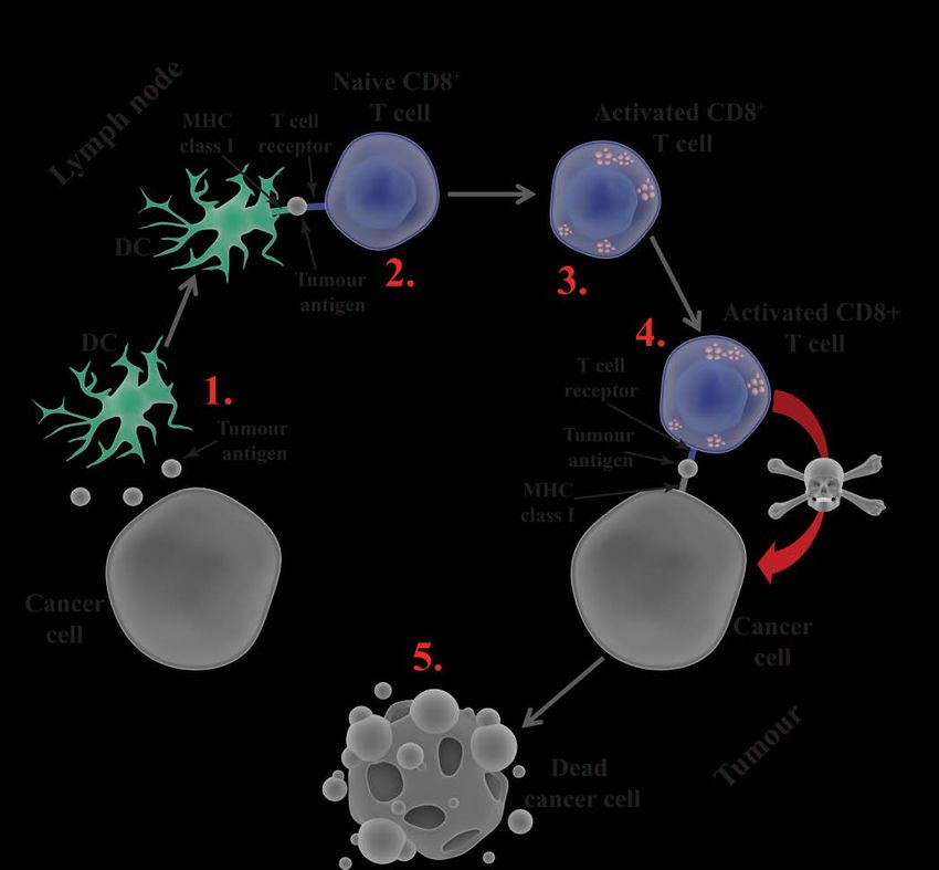
18Figure 5. Very simplified depiction of initiation and implementation of an anti-tumour
response. 1) An APC, here a DC, takes up a tumour antigen (e.g. a DAMP) from a cancer cell
and travels to the lymph node where 2) it cross-presents the antigen to a naïve tumour-specific
CD8+ T cell. 3) The T cell becomes activated and travels to the tumour where it searches for
the same antigen presented on MHC I by the tumour. 4) When the T cell finds the same
antigen it will 5) kill the cancer cell.
One form of antigen that DCs can take up from the tumour microenvironment
and cross-present to naïve CD8+ T cells to start an immune response is a
damage-associated molecular pattern (DAMP). In contrast to the PAMPs
which are extracellular structures found on microbes, DAMPs are
intracellular molecules that are released in the extracellular environment or
expressed on the cell surface when the cell is stressed or injured. DAMPs are
generally structures which during normal conditions only can be found inside
the cell, such as RNA/DNA, the energy-carrying molecule adenosine
triphosphate (ATP) and the calcium-binding protein calreticulin [108-110]. If
a cell dies from non-immune related causes, it may release, for example, its
DNA into the environment.
19During a particular type of cell death called immunogenic cell death (ICD),
dying cells expose and release high amounts of DAMPs, which stimulate the
recruitment and activation of DCs and facilitate the cross-presentation to
naïve CD8+ T cells, thus initiating an anti-tumour response [108, 109].
Certain chemotherapeutic agents are known to induce ICD, and this will be
further discussed in the next section.
1.3.3. Cancer immunology
During recent years it has been increasingly more known that the immune
system is important in the fight against cancer. Different immunotherapies
boosting and regulating the immune response have successfully been applied
to the treatment of different cancers. The academic journal Science awarded
cancer immunotherapy the prestigious “Breakthrough of the Year” award for
2013 [111], and the Nobel Prize in Physiology or Medicine 2018 was
awarded jointly to James P. Allison and Tasuku Honjo “for their discovery of
cancer therapy by inhibition of negative immune regulation” [112].
1.3.3.1. Immunoediting
Immunoediting is a balance between tumour progression and immune-
derived tumour suppression. It is a dynamic process showing how immune
cells can both suppress and promote tumour growth, and it also shows how
cancer cells are both negatively and positively affected by the inflammatory
environment they reside in. Immunoediting consists of three phases, called
the three E’s of cancer immunoediting: elimination, equilibrium and escape.
Elimination, also known as cancer immunosurveillance, is when the immune
system actively fights cancer cells, as previously described. During the
elimination phase the immune system might succeed in eradicating all cancer
cells, in which the immunoediting process finishes and does not progress to
equilibrium. If some malignant cells survive, the process enters the
equilibrium phase. During equilibrium the immune cells are trying to
eradicate the remaining cancer cells, succeeding in containing them but not
fully destroying them. This phase might persist for many years; illustrating
that the time lag from the appearance of malignant cells to the formation of
tumours may be very long. Due to the high selection pressure exerted by the
immune system on the cancer cells, some cancer clones arise which are
immunologically resistant. These clones escape the immunological attack,
proliferate and form tumours which cannot be contained by the immune
system. One commonly employed immune escape mechanism is the
downregulation of MHC class I on cancer cells, thus hindering CD8+ T cells
from recognising the cancer cells. Moreover, tumours may actively suppress
the immune system by secreting immunosuppressive cytokines, express
ligands to inhibitory receptors found on immune cells, and recruit
20You can also read