The cancer metabolic reprogramming and immune response
←
→
Page content transcription
If your browser does not render page correctly, please read the page content below
Xia et al. Molecular Cancer    (2021) 20:28
https://doi.org/10.1186/s12943-021-01316-8

 REVIEW                                                                                                                                            Open Access

The cancer metabolic reprogramming and
immune response
Longzheng Xia1†, Linda Oyang1†, Jinguan Lin1, Shiming Tan1, Yaqian Han1, Nayiyuan Wu1, Pin Yi1,2, Lu Tang1,2,
Qing Pan1,2, Shan Rao1, Jiaxin Liang1, Yanyan Tang1, Min Su1, Xia Luo1, Yiqing Yang1, Yingrui Shi1, Hui Wang1,
Yujuan Zhou1* and Qianjin Liao1*

  Abstract
  The overlapping metabolic reprogramming of cancer and immune cells is a putative determinant of the antitumor
  immune response in cancer. Increased evidence suggests that cancer metabolism not only plays a crucial role in
  cancer signaling for sustaining tumorigenesis and survival, but also has wider implications in the regulation of
  antitumor immune response through both the release of metabolites and affecting the expression of immune
  molecules, such as lactate, PGE2, arginine, etc. Actually, this energetic interplay between tumor and immune cells
  leads to metabolic competition in the tumor ecosystem, limiting nutrient availability and leading to
  microenvironmental acidosis, which hinders immune cell function. More interestingly, metabolic reprogramming is
  also indispensable in the process of maintaining self and body homeostasis by various types of immune cells. At
  present, more and more studies pointed out that immune cell would undergo metabolic reprogramming during
  the process of proliferation, differentiation, and execution of effector functions, which is essential to the immune
  response. Herein, we discuss how metabolic reprogramming of cancer cells and immune cells regulate antitumor
  immune response and the possible approaches to targeting metabolic pathways in the context of anticancer
  immunotherapy. We also describe hypothetical combination treatments between immunotherapy and metabolic
  intervening that could be used to better unleash the potential of anticancer therapies.
  Keywords: Metabolic reprogramming, Immunity, Oxysterols, TME, TIL, Immune checkpoint…

Introduction                                                                          oxidative phosphorylation (OXPHOS) under aerobic
Metabolism involves a network of biochemical reac-                                    conditions. When oxygen is scarce, the cells rely on
tions that convert nutrients into small molecules                                     glycolysis rather than oxygen-consuming mitochon-
called metabolites [1]. Through these conversions and                                 drial metabolism for energy supply. However, the
the resulting metabolites, cells generate the energy,                                 metabolic pattern of tumors is different from that of
redox equivalents and macromolecules (including pro-                                  normal cells [3]. As first observed by Otto Warburg,
teins, lipids, DNA and RNA) that they require to sur-                                 the phenomenon that cancer cells prefer to perform
vive and sustain cellular functions [2]. It is well                                   glycolysis in the cytosol even in the presence of oxy-
known that normal cells get energy via first glycolysis                               gen, was known as ‘‘Warburg effect’’ or “aerobic gly-
in the cytosol that is followed by mitochondrial                                      colysis” [4]. Because the infinite proliferation of
                                                                                      tumor cells requires faster energy supply, the ATP
                                                                                      production rate of glycolysis is much faster than that
* Correspondence: yujany_zhou@163.com; march-on@126.com
1
 Hunan Key Laboratory of Cancer Metabolism, The Affiliated Cancer Hospital            in the oxidative phosphorylation although efficiency
of Xiangya School of Medicine, Hunan Cancer Hospital, Central South                   in ATP production per molecule of glucose is much
University, 283 Tongzipo Road, 410013 Changsha, Hunan, China
                                                                                      lower via glycolysis [5, 6]. In fact, various central
Full list of author information is available at the end of the article

                                       © The Author(s). 2021 Open Access This article is licensed under a Creative Commons Attribution 4.0 International License,
                                       which permits use, sharing, adaptation, distribution and reproduction in any medium or format, as long as you give
                                       appropriate credit to the original author(s) and the source, provide a link to the Creative Commons licence, and indicate if
                                       changes were made. The images or other third party material in this article are included in the article's Creative Commons
                                       licence, unless indicated otherwise in a credit line to the material. If material is not included in the article's Creative Commons
                                       licence and your intended use is not permitted by statutory regulation or exceeds the permitted use, you will need to obtain
                                       permission directly from the copyright holder. To view a copy of this licence, visit http://creativecommons.org/licenses/by/4.0/.
                                       The Creative Commons Public Domain Dedication waiver (http://creativecommons.org/publicdomain/zero/1.0/) applies to the
                                       data made available in this article, unless otherwise stated in a credit line to the data.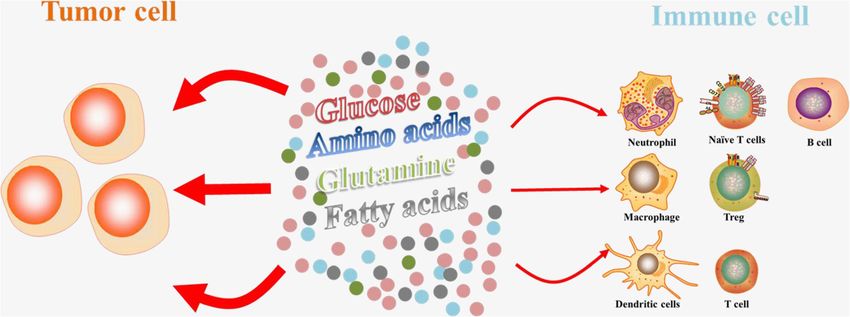
Xia et al. Molecular Cancer   (2021) 20:28                                                                     Page 2 of 21

metabolism pathways can be dysregulated in cancer              disease, and its heterogeneity in cells and structure gives
cells [2]. More importantly, emerging evidence indi-           it a complex metabolic pattern. In fact, tumor cells
cates that cancer cells are able to suppress anti-tumor        mainly use the glycolysis pathway to quickly provide
immune response by competing for and depleting                 ATP (adenosine triphosphate) for their own growth, and
essential nutrients or otherwise reducing the meta-            also provide biological macromolecules for cell replica-
bolic fitness of tumor-infiltrating immune cells [7, 8].       tion through the pentose phosphate pathway (PPP) and
  Both the innate and adaptive immune systems have now         serine metabolism pathway [2, 7]. For instance, under
established roles in the host defense against cancers          hypoxic conditions, tumor cells usually produce pyruvate
through various mechanisms, which are raising an unpre-        through the glycolysis pathway, which in turn produces
cedented development of modern cancer immunother-              lactic acid instead of entering mitochondria and convert-
apies. The innate immune system consists of different          ing to Acetyl-CoA to produce ATP [1]. However, even
populations of immune cells, containing macrophages,           under sufficient oxygen conditions, tumor cells still pref-
neutrophils, monocytes, eosinophils, basophils, and nat-       erentially use glycolysis to generate ATP, which is a
ural killer cells, which are responsible for innate immunity   well-known aerobic glycolysis (Warburg effect) (Fig. 1)
against pathogens to maintain homeostasis of the host [9].     [4]. At the same time, tumor cells will not only decom-
Indeed, immune cells are capable of sensing various sig-       pose glucose for providing ATP, but also use glutamine,
nals in the microenvironment and turning on specific           serine, arginine, fatty acids and lipid substances to pro-
immune functions in response. More and more evidences          mote their own proliferation [21]. Interestingly, tumor
have pointed out that the immune response is associated        cells will choose different metabolic methods to produce
with dramatic modifications in tissue metabolism, includ-      ATP and biological macromolecules for their own use
ing the depletion of nutrients, increased oxygen consump-      according to the concentration of external nutrients and
tion, and the generation of reactive nitrogen and oxygen       different stress conditions.
intermediates [10–12]. Similarly, many metabolites in the         For example, under the stress condition of nutrients
tumor microenvironment, in turn, also influence immune         deprivation such as glucose or glutamine, tumor cells ac-
cell differentiation and effector function [13, 14]. But       tivate oncogene c-Myc to support the survival of tumor
recent works reveal that immune cells compete with can-        cells by regulating the expression of metabolic enzymes
cer cells and other proliferating cells in the microenviron-   such as PHGDH, PSAT1, PSPH and other metabolic en-
ment for nutrients [15]. Therefore, it suggests that           zymes in the serine synthesis pathway and enhancing
metabolic interventions hold promise for improving the         the de novo synthesis of serine and maintaining redox
effectiveness of immunotherapies.                              homeostasis [22]. In addition, under hypoxic or
  The previous studies showed that the modifications of        nutrient-deficient stress conditions, tumor cells produce
cancer cell metabolism are, in part, due to the recruit-       acetyl-CoA by ingesting the smallest two-carbon fatty
ment of many inflammatory and immune cells [16]. Sub-          acid (e.g., acetoacetate), to provide energy for their own
sequently, more and more researchers have found that           survival and bio-macromolecules such as fatty acids to
the aberrant metabolites or intermediates of cancer            promote their own survival [23–25]. Similarly, the me-
metabolism could have an important role in regulating          tabolites produced by tumor cells through the decom-
the proliferation, differentiation, activation and function    position of ketone bodies could enter the TCA
of immune cells [17–19]. Recent studies found that our         (tricarboxylic acid) cycle to provide ATP for cell survival
immune system was closely related to other metabolic           [26]. Therefore, the metabolic mode of tumor cells is
functions (including cancer) in a way that has never           complex and changeable, and it will choose the optimal
been realized before. Moreover, therefore it is described      metabolic mode for its own survival according to its dif-
as a new field called immunometabolism [20]. However,          ferent environment.
the actual process of how metabolic reprogramming and
cancer immune response affect each other has not been          2. Metabolism of immune cells
understood. In this review, we will discuss this process       The immune system contains a variety of immune cells,
from the following aspects.                                    such as macrophages, neutrophils, monocytes, eosino-
                                                               phil, basophils, lymphocytes and natural killer cells.
Main text                                                      These cells are at rest when the body is in a steady state,
Overview of metabolism of tumor cells and immune cells         and will be quickly activated and respond when the body
1. Metabolism of tumor cells                                   is stimulated by infection, inflammation or other exter-
Energy metabolism reprogramming, which fuels fast cell         nal substances [27]. In the previous section, we briefly
growth and proliferation by adjustments of energy              introduced the complex metabolic modes of tumor cells.
metabolism, has been considered as an emerging hall-           Interestingly, these complex and varied metabolic
mark of cancer [4]. Because tumor is a heterogeneous           methods also exist in immune cells [28]. The recent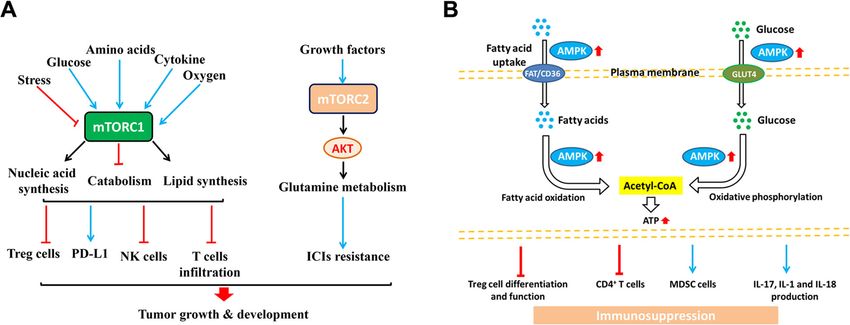
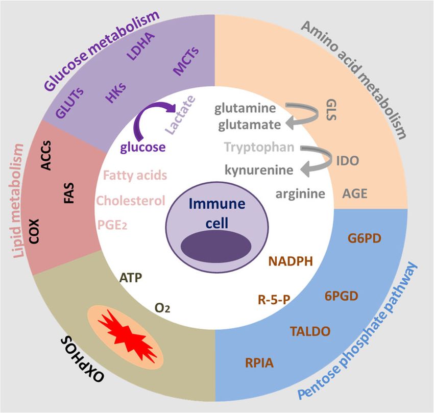
Xia et al. Molecular Cancer    (2021) 20:28                                                                                         Page 3 of 21

 Fig. 1 Regulation of glucose metabolism in cancer cells. Glucose metabolism mainly contains glycolysis and TCA cycle in the mitochondrion.
 These pathways are generally altered in tumor cells compared to normal cells

research shows that there is a significant difference in                  after the activation of B lymphocytes caused by LPS
energy use between immune cells in the resting state                      stimulation or antigen stimulation, glycolysis and mito-
and the activated state. Among many immune cells with                     chondrial metabolism are enhanced. But the glycolysis is
different functions, T cells play an important role in                    the major metabolism in activated B lymphocyte. Differ-
clearing pathogens and killing cancer. T cells will show                  ently, the regulatory T cells (Treg cells), M2 macrophage
completely different metabolic patterns according to dif-                 mainly rely on OXPHOS from fatty acid oxidation
ferent activation states [29]. For instance, the metabol-                 (FAO) to supply energy [32]. Different metabolic
ism of naïve T cell is basically static, showing zero                     patterns also affect the differentiation of different
proliferation, so only the most basic nutrient intake,                    immune cell subgroups (Table 1). Therefore, exploring
minimum glycolysis rate and minimum biosynthesis                          the metabolic reprogramming mechanism of immune
need to be maintained. ATP is mainly produced by                          cells and the effect of metabolism on immune cell func-
OXPHOS [30]. Once activated by an external stimulus                       tions will not only help to understand the essence of
into an effector T cell (Teff), it appears as a metabolic                 immune response and its regulatory mechanism.
activation state, increasing nutrient absorption, increas-
ing glycolysis rate, synthesis accumulation of protein,                   Competition for nutrients between tumor cells
lipid and nucleotide [30]. At the same time, the mito-                    and immune cells
chondrial oxygen consumption is reduced, and eventu-                      Metabolic transitions are not unique to cancer cells, but
ally T cells gain the ability to grow and proliferate and                 are also characteristic of other rapidly proliferating cells,
produce progeny cells to exert effect killing function.                   such as activated T cells, Treg cell, neutrophils and so on
   Interestingly, the metabolic pattern of memory T cells                 [28]. Glucose is the nutrient that tumor cells absorb and
is similar to that of the naïve T cells with maintaining                  consume the most, and is the most dependent, and it is
basic nutrient intake, lower glycolysis rate and relying on               also an important energy substance necessary for immune
OXPHOS to provide ATP [30, 31]. In addition, the acti-                    cell activation, differentiation, and function [51, 52]. The
vated neutrophils, M1 macrophages, iNOS-expressed                         tumor microenvironment (TME) is accompanied by dif-
dendritic cells (DCs) mainly rely on glycolysis for energy                ferent degrees and types of immune cell infiltration. Like
supply. Glycolysis plays an important role in the activa-                 cancer cells, tumor-infiltrating lymphocytes (TIL) require
tion of DCs; however, DC mainly uses oxidative phos-                      nutrients found within the TME to support proliferation
phorylation for energy metabolism in the resting state.                   and differentiation [53]. The researchers found that tu-
Meanwhile, the activation of DC has also changed the                      mors inhibited the function of tumor-infiltrating T cells
lipid metabolism and affected its function. Besides, aer-                 through competitive uptake of glucose, even when there
obic glycolysis and pentose phosphate pathways are the                    are enough tumor antigens for T cells to recognize [54].
main metabolic modes of neutrophils. Glycolysis regu-                     Indeed, several recent studies demonstrated that the
lated many important functions of neutrophils, such as                    glycolytic activities of cancer cells may restrict glucose
respiratory burst and chemo-taxis. More interestingly,                    consumption by TIL T cells, thereby inducing T-cell
Xia et al. Molecular Cancer     (2021) 20:28                                                                                          Page 4 of 21

Table 1 Different metabolic ways of immune cells
Type                                    Cell subtypes                             Metabolic patterns                                     Reference
T cell                                  Naïve T cell                              FAO                                                    [33–35]
                                                                                  OXPHOS
                                                                                  Glutamine metabolism
                                        Treg cell                                 OXPHOS                                                 [36]
                                                                                  FAO
                                        Effector T cell                           Glycolysis                                             [37, 38]
                                                                                  OXPHOS
                                        Memory T cell                             FAO                                                    [39, 40]
                                                                                  OXPHOS
B cell                                  B cell (resting)                          Glycolysis                                             [41, 42]
                                        Activated B cell                          Glycolysis
Macrophages                             M1                                        Glycolysis                                             [43, 44]
                                                                                  Pentose phosphate pathway
                                        M2                                        OXPHOS                                                 [45, 46]
                                                                                  FAO
Neutrophils                             -                                         Glycolysis                                             [47, 48]
NK cell                                 -                                         Glycolysis                                             [41]
DCs                                     DCs (resting)                             OXPHOS                                                 [49, 50]
                                        DCs (active)                              Glycolysis

exhaustion and immune escape [55]. For instance, an                         glucose under tumor microenvironment conditions is
accelerated glucose metabolism in renal cell carcinoma                      responsible for the damage to T cell function, the com-
(RCC) tissues is associated with a low infiltration of CD8+                 petitive uptake of amino acids, glutamine, fatty acids and
effector T cells, as demonstrated by an inverse correlation                 other metabolites or growth factors by tumor cells and
between GLUT1 expression and infiltrating T cell num-                       immune cells and the expression of corresponding trans-
bers in a tissue microarray analysis of RCC specimens                       porters on the cell surface are still important factors af-
[56]. In fact, a large amount of glucose uptake by the                      fecting the functioning of immune cells (Fig. 2) [15].
tumor in the microenvironment is bound to inhibit T cell                    Moreover, high levels of lactate and low pH, hypoxia
function by affecting T cell metabolic patterns.                            and high levels of ROS are likewise prevalent in the
  According to the literatures reported, glycolytic                         TME, eventually leading to cancer progression and im-
metabolites also have a deleterious effect on immune                        mune escape [57]. Thus, targeting these metabolic path-
function [57]. Although the competitive uptake of                           ways in tumors could be a promising strategy to

  Fig. 2 The nutritional competition between tumor cells and immune cells inside tumors. The competition-caused deficiency of glucose,
  glutamine, and fatty acids and a couple of amino acids are known to affect the function of immune cells, including Treg, macrophages, dendritic
  cell, NK cells and so on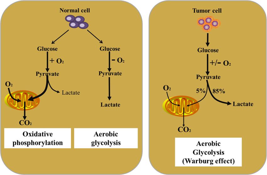
Xia et al. Molecular Cancer   (2021) 20:28                                                                     Page 5 of 21

overcome the deleterious effects of metabolic competi-        fibroblasts (CAFs) can be used by tumor cells as an al-
tion between the tumor and the immune system, and             ternative nutrient source [64].
enhance tumor immunogenicity [58]. However, further              In addition, lactic acid will also affect the function of
investigations are needed to test this hypothesis in the      NK cells, and then impair the secretion of IFN-γ [65, 66].
tumor setting both in preclinical models and in future        Mechanism research shows that excessive intake of patho-
human studies.                                                logical concentrations of lactic acid by NK cells can cause
                                                              intracellular acidification and inhibit the up-regulation of
                                                              the nuclear factor of activated T cells (NFAT) signal,
Effect of tumor metabolic reprogramming on
                                                              resulting in reduced production of NFAT-regulated IFN-γ
immune cells
                                                              and promoting apoptosis [67, 68]. More importantly, both
In addition to nutrient consumption, metabolites pro-
                                                              human and rat melanoma present the high levels of lac-
duced by cancer cells can have a profound effect on im-
                                                              tate. For instance, in immune-competent mice, reducing
mune cells in the microenvironment [57]. For example,
                                                              the production of lactic acid can slow the tumorigenic
lactate, a byproduct from elevated aerobic glycolysis in
                                                              ability and the infiltration of CD8+ T cells and NK cells se-
cancer cells, induces apoptosis via reduced expression of
                                                              creting IFN-γ in the tumor is significantly increased [67].
the autophagy factor FIP200 in naïve T cells in both pa-
                                                              However, in mice lacking lymphocytes and NK cells, there
tients with ovarian cancer and mouse models [59]. How-
                                                              was little difference in tumorigenic ability between the low
ever, the immune cells that function in the immune
                                                              lactate group and the control group. Interestingly, a recent
system are by no means only T cells and NK cells. And
                                                              research discovered that lactate produced by glycolysis of
metabolites produced by tumor metabolic reprogram-
                                                              tumor cells in the tumor microenvironment activates
ming are complex and changeable. Although tumor cells
                                                              mTOR pathway, thereby phosphorylating the transcrip-
are mainly powered by aerobic glycolysis of glucose,
                                                              tion factor TFEB and inhibiting its nuclear translocation,
there are other metabolic methods, such as amino acid
                                                              thereby inhibiting the expression of ATP6V0d2 (vacuolar
metabolism, glutamine metabolism, fatty acid metabol-
                                                              ATPase subunit) in macrophages. The inhibition of
ism, cholesterol metabolism, etc. We will discuss the ef-
                                                              ATP6V0d2 could mediate HIF-2α lysosomal degradation
fect of tumor metabolic reprogramming on immune cell
                                                              and program TAMs (tumor-associated macrophages) in
function from the following aspects.
                                                              the tumor microenvironment into immune cells that pro-
                                                              mote tumor growth [69]. Similarly, a negative correlation
1. The effect of tumor metabolites on immune cell             between intratumoral lactate concentration and overall
Lactate                                                       survival in patients with cervical cancer has been reported
 The aberrant glycolysis of tumor means that tumor cells      [68]. Together, these findings suggest that the higher lac-
consume a lot of glucose and produce large amount of          tate content in the tumor and the accompanying acidified
lactic acid even in the presence of sufficient oxygen, with   TME will suppress immune cell function and abrogate
a correspondingly low rate of OXPHOS. Lactic acid             immunosurveillance of cancer, ultimately leading to
accumulates in cells and then is exported into the extra-     immune escape.
cellular environment via activating monocarboxylate
transporters (MCTs) on cell membrane, in particular,          Glutamine
monocarboxylate transporter 4 (MCT4), which ultim-             Glutamine metabolism as a whole is a crucial element
ately results in establishing an acidic TME [60]. The low     of cancer cell metabolism. Glutamine is important for
pH of the TME has been shown to be beneficial for the         nucleotide synthesis, amino acid production, redox bal-
selection of more aggressive tumor cells and suppress         ance, glycosylation, extracellular matrix production,
tumor immunity to promote tumor progression [61]. In          autophagy, and epigenetics [70]. Subsequently, studies
fact, the previous and recent research both reported that     have shown that glutamine plays an important role in
both aerobic glycolysis and the resultant acidification of    the growth of normal cells and cancer cells [71]. Under
the TME have been shown too strongly influence T cell         conditions of nutritional deficiencies, cancer cells can
mediated antitumor immune responses and the activities        obtain glutamine by breaking down large molecules. For
of tumor-infiltrating myeloid cells [62]. In addition, the    example, excessive activation of the oncogene RAS can
recent clinical studies have found that the serum lactic      promote endocytosis, and cancer cells clear extracellular
acid level increases significantly with the increase of the   proteins and degrade into amino acids including glutam-
patient’s tumor burden [62]. Actually, lactate produced       ine, providing nutrients for cancer cells [72]. In addition
by tumor cells might contribute to tumorigenesis by           to tumor cells, glutamine has a high utilization rate in
promoting IL-23-mediated and IL-17-mediated inflam-           immune cells to support cell fate determination and im-
mation [63]. In addition to modulating immune                 mune responses, such as lymphocytes, macrophages, and
responses, lactic acid produced by cancer associated          neutrophils [73–75]. According to the study reported,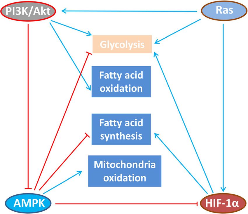
Xia et al. Molecular Cancer   (2021) 20:28                                                                   Page 6 of 21

glutamine deprivation can suppress T cell proliferation       response via inhibiting Th1 differentiation, B cell func-
and cytokine production. However, glutamine restriction       tion, T cell activation and allergic reactions, which
during T cell activation in vitro has been shown to pro-      would enhance the anti-tumor immunity [85]. In
mote memory CD8+ T cell differentiation [76].                 addition, PGE2 can also exert anti-inflammatory effects
  In fact, glutamine metabolism plays an important role       on natural immune cells, such as neutrophils, monocytes
in the activation of immune cells and regulating the          and natural killer cells [86, 87]. More importantly, the
transformation of CD4+ T cells to inflammatory sub-           accumulation of PGE2 secreted by tumor cells can trans-
types [74, 77]. It is now clear that the high utilization     form M1 macrophages with tumor suppressing effect to
rate of glutamine in many immune cells is related to the      M2 macrophages that promote cancer [88]. The PGE2
functional activity of immune cells, such as cell prolifer-   secreted by tumor cells also can stimulate bone marrow-
ation, antigen presentation, synthesis and secretion of       like cells to secrete cancer-promoting CXCL1, IL-6 and
cytokines, production of NO and peroxide, phagocytosis,       granulocyte-colony-stimulating factor (G-CSF), and in-
etc. These functions are all indirectly or directly           hibit the secretion of TNF-α and IL-12 in myeloid cells
dependent on NADPH reserves [78]. Moreover, glutam-           stimulated by lipopolysaccharide (LPS), and inhibit the
ine has a high utilization rate in macrophages, and neu-      activation of type I interferon-dependent innate immune
trophils. With the increase in utilization of glutamine,      cells, and inhibit T cells from targeting tumor antigens,
the apoptosis rate of immune cells was significantly re-      and then achieve the purpose of immune escaping and
duced. For instance, glutamine reduces neutrophil apop-       tumorigenesis [89].
tosis by reducing the expression of pro-apoptotic                According to literature reports, PGE2 plays an import-
proteins Bax and Bcl-xs [79]. The production and secre-       ant role in lymphocyte development and proliferation,
tion of pro-inflammatory cytokines (IL-6, IL-1, and           and participates in the body’s immune regulation, in-
TNF) through macrophages are also controlled by the           cluding inhibition of T lymphocyte proliferation, differ-
obtainability of glutamine [80]. Therefore, the metabol-      entiation and cytokine secretion in vitro. High levels of
ism of glutamine plays an essential role as a modulator       PGE2 can not only directly regulate the malignant bio-
and synergetic supporter for the activation of a macro-       logical behavior of tumor cells, but also prevent the pro-
phage. In addition, by blocking the glutamine pathway in      inflammatory response in various immune cells and me-
cancer cells to increase the content of amino acids in        diate the reprogramming of TME, making TME in an
the tumor microenvironment and enhance the killing ef-        immunosuppressive status [90]. Studies have shown that
fect of immune cells. Jonathan D. Powell and his team         PGE2 damages the malate-aspartate shuttle (MAS) sys-
found that blocking glutamine can induce different            tem via activating the cAMP-PKA pathway, causing the
metabolic processes, thereby overcoming the immune            decrease of aspartic acids and a variety of enzymes and
escape of tumors [81]. It suggests that targeting the glu-    intermediate metabolites and the growth arrest of CD8+
tamine metabolism will become a novel treatment of            T cells [91]. Subsequently, the researchers found that
cancer.                                                       PGE2 have shown a strong synergistic effect in inhibiting
                                                              the killing function of TIL cells or regulating the secre-
PGE2                                                          tion of TIL cell-related cytokines [92]. Some researchers
 Arachidonic acid is an important class of eicosapenta-       have reported that PGE2 could increase the expression
enoic acid related to essential fatty acids in tumor cells,   of the Treg-specific marker Foxp3 and stimulate the
and it is an important substrate for the synthesis of pros-   functions of Treg cells in vitro and in a mice lung cancer
taglandins. PGE2 is an important cell growth and regula-      model [93]. More importantly, the activated Treg cells
tion factor in the cell and it is also a kind of highly       also induced COX-2 expression and PGE2 production,
active inflammatory mediator in the inflammatory re-          which then support immune-suppressive functions by
sponse [82] and can also participate in the immune re-        themselves. Additionally, a previous study reported that
sponse as an immunosuppressive factor [83]. Cancer-           PGE2 could impair the function of tumor-associated
associated fibroblast (CAF)-derived PGE2 or other             DCs (dendritic cells) [94]. In this article, the authors
sources of PGE2 could induce cancer cell invasion and         pointed out PGE2 promoted tumor evasion through acti-
participate in tumor progression by stimulating angio-        vating β-catenin signaling to inhibit the function of
genesis, cell invasion and metastasis, and inhibits apop-     CD103+ DCs in the TME. Blocking the production of
tosis through a variety of signal pathways to promote         PGE2 by tumor cells may be a good anti-tumor im-
cell survival [84]. In addition, PGE2 can also affect other   munotherapy method.
cells in the microenvironment through autocrine and
paracrine methods, such as destroying the immune              Arginine
response [83]. For instance, PGE2 is also used as an anti-     Most tumor cells lack ASS1 (argininosuccinate syn-
inflammatory molecule to suppress the immune                  thetase 1), a key enzyme that produces arginine, and
Xia et al. Molecular Cancer (2021) 20:28 Page 7 of 21
therefore will cause the loss of intracellular arginine and utilized by tumor cells in a large amount, which will
synthesis capacity [95]. In this case, tumor cells will cause the lack of tryptophan and trigger the apoptosis of
use exogenous arginine to make up for the lack of T cells [17, 101]. Furthermore, the accumulation of tryp-
arginine caused by the lack of key metabolic enzymes tophan induced by systemic IDO inhibition promotes
in the cell. Interestingly, arginine metabolism also has cru- tumor regression by increasing the production of cyto-
cial roles in T cell activation and modulating immune re- kines, including IL-12 and IFN-γ, and tumor infiltration
sponses. For instance, supplementation of arginine of T cells and neutrophils in mouse metastatic liver
stimulates T cell and NK cell cytotoxicity and effector tumor and bladder tumor models. The IDO1 inhibitor
cytokine production in vitro and, in combination with has been shown to alleviate immunosuppression in the
anti-PD-L1 antibody treatment, significantly enhances an- TME and promote the activation of tumor-specific T
titumor immune responses and prolongs the survival of cells in preclinical models [101]. The outcomes of com-
osteosarcomabearing mice [96]. bined treatment with IDO1 inhibitor plus the immune
In the cell, arginine mainly produces urea, L-ornithine checkpoints inhibitors (ICIs) are being assessed in
and nitric oxide, L-melon through the reaction of argi- patients with melanoma. IDO inhibition might impair
nase (ARG) and nitric oxide synthase (NOS). Some stud- NAD+ generation not only in immunosuppressive and/
ies have reported that the accumulation of ARG1- or pro-tumor TAMs but also in CD8+ TILs. Importantly,
expressing immunomodulatory cells, including M2-like production of NAD+ is implicated as a crucial event that
tumor-associated macrophages (TAMs), tolerogenic DCs sustains T cell immune responses [102]. However, other
and Treg cells, in the TME might suppress antitumor IDO inhibitors are undergoing clinical testing, mostly in
immunity by degrading arginine and thus limiting the combination with ICIs.
availability of this amino acid to T cells [97]. More im-
portantly, studies have pointed out that the arginine Fatty acids
absorbed by tumor cells in the tumor microenvironment Tumor cells often have increased rates of de novo fatty
is mainly related to tumor provided by bone marrow acid synthesis to divert energy production into anabolic
cells (macrophages, monocytes, myeloid inhibitory cells, pathways for the generation of plasma membrane phos-
neutrophils, etc.). It means that tumor cells will consume pholipids and signaling molecules. Meanwhile, fatty acid
a large amount of arginine in the TME, which will cause synthesis provides cell membranes and other key lipid
the lack of arginine in the TME, so the activation of an- cell structures necessary for immune cell proliferation
titumor immune cells is bound to be inhibited [97]. [103]. The studies have reported that fatty acid synthesis
Therefore, replenishment of arginine and prevention of is necessary for inflammatory macrophages to differenti-
arginine degradation in the TME are attractive strategies ate and function. However, some studies pointed out
to re-invigorate T cell mediated and NK cell-mediated that effector immune cell grow rapidly and require lipids
immune responses [98]. Moreover, arginine supplemen- formed by fatty acid synthesis to build cell membranes
tation during in vitro expansion of T cells promotes during proliferation, while memory immune cells grow
their differentiation to central memory-like T cells with slowly and biosynthesis needs relatively little, so fatty
superior antitumor activity. acid oxidation (FAO) is the main [104]. Actually, the ab-
errant accumulation of lipid metabolites (e.g., short-
Tryptophan chain fatty acid, long chain fatty acid, cholesterol, etc.) in
Tryptophan is an essential amino acid necessary for tumor-infiltrating myeloid cells, including MDSCs, DCs
organisms to carry out protein synthesis and other life and TAMs, has been shown to skew these immune cells
metabolic activities. Actually, tryptophan degradation is towards immunosuppressive and anti-inflammatory phe-
mainly converted by two different dioxygenases IDO1 notypes via metabolic reprogramming [103]. Nowadays,
(indoleamine-2, 3-dioxygenase) and TDO2 (tryptophan- many scholars believe that the regulation mechanism of
2, 3-dioxygenase) to tryptophan to kynurenic acid [99]. fatty acids on immune cells is not that fatty acids change
Expression of high levels of these tryptophan-degrading the composition of fine membranes or become inflam-
enzymes in tumor cells promotes tumor progression and matory mediators, but that fatty acids directly participate
is correlated with a worse prognosis in patients with gas- in signal transmission in cells.
tric adenocarcinoma [100]. In fact, high levels of IDO
and TDO in tumors have been suggested to decrease Cholesterol
tryptophan availability in the TME, which in turn sup- Cholesterol is an important part of the surface of the
presses the tumoricidal functions of T cells. Because the cell membrane. Rapidly proliferating cells require more
activation of T cells is extremely sensitive to the concen- membrane structure and more cholesterol synthesis.
tration of tryptophan in the peripheral environment, the High expression of cholesterol in tumor cells can protect
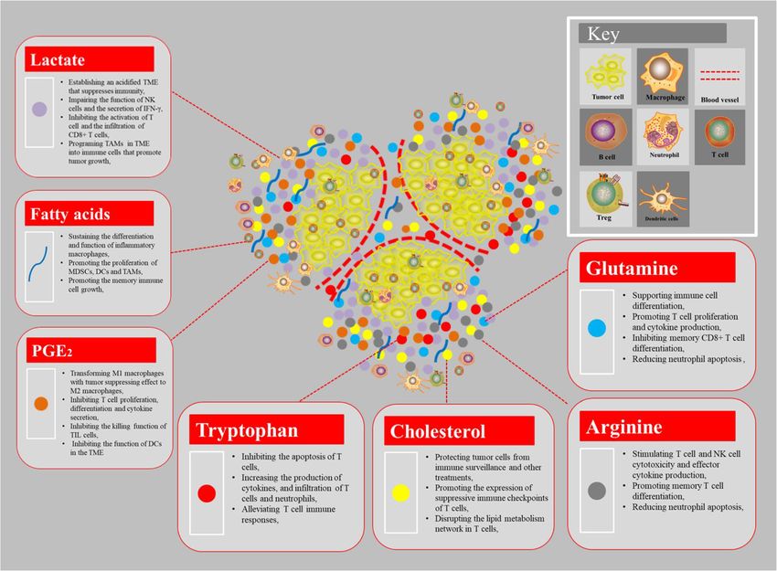
tryptophan in the microenvironment will be metabolized tumor cells from immune surveillance and otherXia et al. Molecular Cancer (2021) 20:28 Page 8 of 21 treatments [105]. Emerging evidence suggests the im- However, the products produced by tumor cell metabol- portance of cholesterol metabolism in innate immune ism are diverse, and their functions on immune cells are response. The high level of cholesterol caused by tumor also different. In the above, we focused on the impact of cell can promote the expression of suppressive immune lactate and other metabolites on immune cells (Fig. 3), checkpoints of T cells, thereby making it lose its anti- but the impact of other metabolites on immune cells still tumor effect [106]. More importantly, researchers found needs a lot of research. that the cholesterol concentration in tumor cells is much higher than immune cells, and the higher the cholesterol 2. The effect of metabolic enzymes on tumor immunity concentration in immune cells, the higher the expression Metabolites are the essential substrates for epigenetic of immune detection points such as PD-1, LAG-3, and modification enzymes to write or erase the epigenetic TIM-3, etc. Similarly, immune cells are also prone to blueprint in cells. Thus, the availability of nutrients and apoptosis, and the lower the cytotoxicity and prolifera- activity of metabolic pathways strongly influence the tion capacity of the cells. In fact, high cholesterol can function of metabolic enzymes. Consistent with findings disrupt the lipid metabolism network in T cells, thereby from tumor models, the results of several clinical studies exerting the function of suppressing immunity [107]. revealed that aerobic glycolytic activity in human tumors Oxysterols, the metabolites originating from autoxida- is negatively associated with host antitumor immune tion or enzymatic oxidation of cholesterols, are oxygen- responses and therapeutic outcomes of anticancer ated enzymatically (e.g., 25-hydroxycholesterol (25OHC), immunotherapy [114]. In fact, the elevated glycolysis is 27-hydroxycholesterol (27OHC) 22-hydroxycholesterol the metabolic basis for trained immunity to provide the (22HC) and 24S-hydroxychlesterol (24OHC)), while energy substrates for the enhanced activation of trained some are not produced enzymatically (e.g., 7alpha/beta- innate immune cells. For example, the three rate- hydroperoxycholesterol (7OOHC), and 7 ketocholesterol limiting enzymes in the glycolytic pathway, including (7KC)) [108]. They are present in very low concentra- hexokinase 2 (HK2), phosphofructokinase 1 (PFK1), and tions in mammalian systems, always accompanied by a pyruvate kinases type M2 (PKM2) acted as a hallmark of high excess of cholesterols [109]. The 27OHC is elevated liver cancer and is responsible for the regulation of im- in both breast tissues and tumors in patients with estro- mune evasion via regulating by many mechanisms, such gen receptor-positive breast cancer [110]. Elevating the as the AMPK, PI3K/Akt pathway, HIF-1α, c-Myc and level of 27OHC promotes cell proliferation and tumor noncoding RNAs in HCC (hepatocellular carcinoma) growth by modulating a series of gene expression includ- [115]. Besides the glycolytic enzymes, the enzymes about ing estrogen-receptor signaling genes, such as ARMT1 cholesterol metabolism and transport have important ef- and PARD6B, as well as genes involved in the GDFN– fect on immune cell function, production, and activity RET signaling pathway [11]. In addition, oxysterols also [116]. Researchers found that the cholesterol esterase have multiple functions in shaping the immunological ACAT1 in the T cell metabolism pathway is a good landscape. For instance, the accumulation of 22OHC regulatory target. Inhibiting the activity of ACAT1 can can recruit CD11bhighGr1high neutrophils in the condi- greatly improve the anti-tumor function of CD8+ T cells tioned medium of various cancer cells, which are emer- (also known as killer T cells) [117]. Because after ging as an important immunosuppressive population in ACAT1 is inhibited, the free cholesterol level on the the TME [111]. Besides, 24OHC and 27OHC also recruit killer T cell membrane increases which makes the T cell neutrophils in other cancer types. Especially, 27OHC has tumor antigen immune response more efficient. In been found to deplete cytotoxic CD8 T cells, thereby addition, the extracellular nucleotidase CD39 and CD73 promoting tumor metastasis [112]. Actually, cholesterols can significantly up-regulate the extracellular adenosine can be transformed into a variety of substances with im- content of tumors, and can maintain the expression of a portant physiological effects in the body, e.g., adrenal chronic innate immune response, leading to immune cortex hormones, androgens, estrogen, progesterone, 7- tolerance, and then leading to uncontrolled tumor dehydrocholesterol, vitamin D3, bile acids, etc. Among growth [118]. Although successive studies have pointed them, adrenal cortex hormones could suppress the func- out the important regulatory role of metabolic enzymes tion of all immune cells [113]. Other cholesterol-derived in tumor immunity, its specific regulatory functions and mediators have an important role in immune regulation, mechanisms still need a lot of research to support it. but different mediators have different regulating effects on immunity. However, it’s well known that decreasing 3. The effect of metabolic pathways on tumor immunity the high cholesterol concentration in TME helps to mTOR pathway relieve the immune suppression of T cells. Mammalian/mechanistic target of rapamycin (mTOR) Metabolites produced by cancer cells can have a pro- is a serine/threonine protein kinase that is highly con- found effect on immune cells in the microenvironment. served from yeast to mammals, and it plays an important
Xia et al. Molecular Cancer     (2021) 20:28                                                                                              Page 9 of 21

 Fig. 3 The regulation of metabolites produced by tumor cells in immune cellsThe metabolites are known regulators of immune cell function,
 such as lactate, fatty acid, PGE2 and arginine, etc. As a consequence, the accumulation of extracellular lactate and the amino acids arginine,
 tryptophan, and glutamine in the tumor environment could affect the proliferation, function and differentiation of immune cells, and the
 production of cytokines. The glucose-deprived, lactic acid-enriched TME impairs T cell and NK cell function and thus antitumor immune
 responses and polarizes tumor-associated macrophages (TAMs) towards a generally pro-tumor, M2-like phenotype. Competition for amino acids,
 including arginine and tryptophan, between immune cells and tumor cells can also suppress antitumor immunity. The availability and usage of
 fatty acids in immune cells within the TME are also influenced by competition with tumor cells. Notably, a high rate of cholesterol esterification in
 the tumor can impair immune responses and, therefore, disruption of cholesterol esterification in order to increase the concentration of
 cholesterol in the plasma membranes of immune cells might increase their proliferation and improve their effector function. The generation of
 prostaglandin E2 (PGE2), by tumor cells and other immunomodulatory cells is also implicated in the suppression of antitumor responses

role in regulating cell growth and metabolism [119].                         inhibitors to block the mTOR signaling pathway can
mTOR has two different structures and functions:                             promote the expression of Treg cells. The study of
mTOR complex 1 (mTORC1) and mTOR complex 2                                   mouse lung cancer model found that mTORC1 can pro-
(mTORC2). Actually, mTORC1 can promote anabolism                             mote the expression of PD-L1, so that tumor cells can
such as protein and nucleic acid synthesis, and inhibit                      avoid the killing of immune cells. Studies on melanoma
catabolism such as autophagy. In addition, it is notable                     have found that tumor cells secrete inhibitory cytokines,
that mTORC2 can be involved in glutamine metabolism                          which reduces the infiltration of T cells into the tumor
where it can increase the uptake of glutamine by regulat-                    microenvironment, making tumor cells resistant to im-
ing its cell surface transporters through activation of                      mune checkpoint inhibitors after activation of AKT in-
AGC kinases [119]. A recent study summarized that the                        duced by mTORC2 [119]. mTOR exerts an influence in
mTOR signaling pathway of tumor cells in the tumor                           the interaction of many kinds of cells and in many links,
microenvironment promoted the tumorigenesis and de-                          such as regulating the function of CD8+ T, thereby pro-
velopment by affecting tumor immunity (Fig. 4a) [120].                       moting the immune killing effect of T cells on tumor
For instance, gene knockdown or the use of rapamycin                         cells, and inhibiting the activation of NK cells, and
Xia et al. Molecular Cancer     (2021) 20:28                                                                                            Page 10 of 21

 Fig. 4 The regulation of metabolic pathway in immune cells. a The activation of mTORC1 induced by various factors (cytokines, glucose and
 oxygen, etc.) up-regulates PD-L1 expression, and inhibits the Treg cells, NK cells and T cell infiltration. In addition, the growth factors-induced
 mTORC2 activation could increase AKT expression and affect the glutamine metabolism, ultimately causing the ICIs (immune checkpoint
 inhibitors) resistance and promote the cancer growth and development. b AMPK activation enhances the uptake of fatty acids and glucose via
 FAT/CD36 and GLUT4 respectively. Meanwhile, the activation of AMPK increases the fatty acids oxidation and oxidative phosphorylation in
 mitochondria to elevate the intracellular ATP level. Ultimately, these alterations commonly affect the functions of immune cells, such as inhibiting
 Treg cells differentiation and function, decreasing CD4+ T cell activity, inducing MDSC cells (marrow-derived suppressor cells) and elevating the
 secretion of IL-17, IL-1, and IL-18, which ultimately lead to the immunosuppression

finally playing an immunosuppressive effect. The mTOR                        inflammatory macrophages towards the M2 anti-
pathway can directly act on the energy metabolism of                         inflammatory phenotype [124]. Moreover, AMPK activa-
tumor cells, or it can play a role by affecting immune                       tion regulates the signaling of the anti-inflammatory
cells [121]. In future studies, it is necessary to further                   cytokine in macrophages, e.g., enhancing the IL-10-
clarify the mechanism of action of the mTOR pathway                          induced suppression of LPS-stimulated cytokine produc-
in different tumor cells and corresponding immune cells,                     tion [125]. Several studies have also demonstrated that
and evaluate the overall therapeutic effect of mTOR                          the activation of AMPK has an important role in the dif-
inhibitors, so that it can be better used in tumor                           ferentiation and functions of T lymphocytes by regulat-
treatment.                                                                   ing their energy metabolism. These observations clearly
                                                                             indicate that AMPK signaling controls the balance
AMPK Signaling                                                               between energy metabolism and immune responses.
 AMPK (AMP-activated protein kinase) is a key mol-
ecule in the regulation of cell energy homeostasis. The                      Adenosine signaling pathway
activation of this kinase responds to the stress of cells                     The concentration of adenosine in tissues is markedly
depleting intracellular ATP, such as low glucose, hyp-                       increased within a few hours following tissue injury, as
oxia, and ischemia and heat shock. As a cell energy                          well as in hypoxic tissues and the TME. Accumulation
detector that responds to low ATP levels, AMPK actively                      of the nucleoside adenosine in the tumor microenviron-
regulates signaling pathways that replenish intracellular                    ment has been shown to inhibit the anti-tumor function
ATP supply, including fatty acid oxidation and autoph-                       of various immune cells, including cytotoxic T cells and
agy [122]. AMPK negatively regulates ATP-consuming                           natural killer cells, by binding to cell surface adenosine
biosynthetic processes including gluconeogenesis, lipid                      2A receptor (A2AR). The ectonucleotidases CD39 (also
and protein synthesis. Interestingly, AMPK activation                        known as NTPDase 1) and CD73 (5 -NT) are cell sur-
regulates cellular immunity in cooperation with immune                       face molecules with pivotal roles in controlling the pro-
signaling pathways and controlling energy metabolism                         duction of adenosine through the catabolism of ATP to
which consequently affects the activation of immune                          AMP and AMP to adenosine, respectively [126]. Treg
cells (Fig. 4b) [123]. There is convincing evidence that                     cells can express CD39 and contribute to immunosup-
AMPK activation prevents inflammatory responses                              pression in the TME via the adenosine-A2AR signaling
through the inhibition of pro-inflammatory signaling                         axis. Actually, adenosine signaling via A2AR negatively
pathways. For example, in macrophages, the activation                        regulated the production of type 1 cytokines and
of AMPK promotes the polarization of M1 pro-                                 enhanced the production of IL-10 by cAMP/protein
Xia et al. Molecular Cancer (2021) 20:28 Page 11 of 21
kinase A and caused signal transducer and activator of the needs of biosynthesis, so as to complete the prolifer-
transcription 5 (STAT5) dephosphorylation, which ation, differentiation and execution of effector functions.
resulted in reduced IL-2R signaling in T cells and inhib- Its metabolic pattern is completely different from that of
ition of the nuclear factor kappa B (NF-κB) pathway inactivated immune cells, which is very similar to the
[127]. More importantly, targeting CD39 and CD73 growth of tumor cells. The phenomenon of “metabolic
activity to inhibit adenosine production is an attractive reprogramming” has occurred [28]. At the same time,
strategy for enhancing antitumor immunity. E.g., Treg the phenotype and function of immune cells will be reg-
co-express CD39/CD73 on the surface and generate ulated by metabolism. Therefore, different metabolic
extracellular adenosine, contributing to immunosuppres- pathways regulate the growth, differentiation and func-
sive activities, because the activation of adenosine signal- tion of immune cells [2]. Among them, the research on
ing pathway mediated the suppression of Tregs by PGE2 glucose metabolism is the most in-depth. We will dis-
receptors expressed on T cells, leading to the up- cuss the effect of immune cell metabolic reprogramming
regulation of adenylate cyclase and cAMP activities on cancer immunity from the following aspects.
[125–127]. Therefore, tumors use the adenosinergic
pathway by increasing adenosine production to promote 1. Effect of immune cell metabolic pattern on immune
Treg activity, aiming at an immunosuppressive micro- cell functions.
environment to escape immune surveillance and pro- .
mote cancer growth. The known metabolic patterns of immune cells can be
There are several different signaling pathways involved divided into the following three categories: (1) the acti-
in cell metabolism that influence the cancer T cell vated immune cells are metabolized in a manner similar
immunity, such as PI3K/AKT, Ras, insulin receptor, to the Warburg effect without obvious oxidative phos-
MAPK, HIF-1α signaling pathway (Fig. 5) [128, 129], etc. phorylation, (2) glycolysis in effector T cells, (3) tricarb-
However, the concrete mechanism is not very clear and oxylic acid cycle and oxidative phosphorylation in
need further study. resting immune cell. Interestingly, some studies indi-
cated that fatty acid oxidation is also active in resting
Effect of immune cell metabolic reprogramming immune cell [32, 128, 129]. On one hand, immune cells
on tumor immunity in different activation states or differentiation stages
The activated process of immune cell requires a large exhibit different metabolic patterns. This active selection
amount of energy and metabolic intermediates to meet of metabolic pathways enables immune cells to adapt to
their functional requirements. On another hand, the
environment and metabolic state of organisms will affect
the phenotype and function of immune cells (Table 2).
Glycolysis
Glycolysis is the major metabolic pathway in neutro-
phils, M1 macrophage, dendritic cell, naïve T cell and
effector T cell [30], etc. For instance, CD8+ T cells
are a key component of adaptive immune response
and play an important role in anti-tumor immunity.
T cell antigen receptor (TCR) connection and subse-
quent co-stimulation trigger substantial changes in
cell metabolism and induce the activation and prolif-
eration of naïve CD8+ T cells. Although oxidative
phosphorylation (OXPHOS) is the main energy source
for naïve and memory CD8+ T cells, elevated glyco-
lytic metabolism is a prerequisite for cell growth and
expansion of the activated CD8+ T cell population
Fig. 5 Cross-regulation of immunometabolic signaling pathways in T
[130]. Besides the naïve T cell and effector T cell,
cell. Under hypoxia conditions, HIF-1α signaling activates anabolism-
associated programs, such as glycolysis and fatty acid synthesis; when the macrophages are polarized, the glucose me-
however, these mechanisms require additional investigation in tabolism pattern changes. For example, when M1-type
primary T cells. AMPK directly inhibits HIF-1α. During activation and/ stimulating factor IFN-γ and LPS co-stimulate macro-
or replete nutrient conditions, PI3K-AKT and Ras signaling promotes phages derived from mouse bone marrow, cellular
glycolysis, fatty acid oxidation. PI3K-Akt can reportedly inhibit the
glycolysis is enhanced. More importantly, glycolysis
AMPK, but this regulation is not yet reported to occur in T cells
can regulate the inflammatory response associatedXia et al. Molecular Cancer (2021) 20:28 Page 12 of 21
Table 2 Metabolic changing of immune cells
: Significantly up-regulated; : Up-regulated; : Down-regulated
with macrophages [131]. Under the stimulation of OXPHOS
LPS, the metabolic mode of mouse macrophages Regulatory T cells (Treg cells), M2 macrophages and
switches to glycolysis, resulting in the accumulation memory T cells mainly rely on FAO (fatty acid oxida-
of succinate, an intermediate product of the tricarb- tion)-sourced OXPHOS for energy supply. There is clear
oxylic acid cycle, which can be further activated data from several groups that perturbing the OXPHOS
AKT-mTOR-HIF1α signaling pathway induces the of immune cell can inhibit its functions [134]. For in-
expression of inflammatory factor IL-1β [132]. Gly- stance, resting NK cells depend mainly on OXPHOS for
colysis regulates many important functions of neutro- their survival. When it’s activated by some cytokines,
phils, activated NK cell and DC cells, therefore, such as IL-2, IL-12, and IL-15, both murine and human
people expect to control the tumor immunity by NK cells up-regulate OXPHOS to support IFN-γ pro-
regulating the glycolytic changes of immune cells, duction. Interestingly, some studies confirmed that
which is also an important direction for future OXPHOS inhibition showed little effect on NK cell cyto-
research [133]. However, this is far from actual clin- toxicity. Additionally, resting T cells also mainly depend
ical application and need further study. on OXPHOS, and antigen presentation and theXia et al. Molecular Cancer (2021) 20:28 Page 13 of 21
induction of the glycolytic pathway in a mitochondria- functions [141]. In resting neutrophils, the amount of
independent manner [135]. A recent study indicated that glucose metabolized via this route is only 2 to 3 percent
mitochondrial OXPHOS directs lineage commitment of of the total glucose consumed by the cells. The PPP is of
pathogenic Th17 cells and suppresses Treg cells by or- particular importance for neutrophils because it provides
chestrating distinct cellular and molecular events [136]. the NADPH needed for de novo fatty acid synthesis and
In this article, the authors pointed that it is possible that for NOX activity. NOX uses NADPH to reduce oxygen
Treg cells generated with OXPHOS inhibition under and generate superoxide (O2) [141, 142]. M1 type mac-
Th17 conditions may exhibit distinct functional and rophages transport a large amount of glucose from out-
transcriptional characteristics from classical Treg cells. side the cell and are enriched in the cell in the form of
That suggested that manipulating cellular metabolism, glycogen. When glycogen is decomposed, a large amount
and specifically mitochondrial OXPHOS, may provide a of glucose 6-phosphate flows to the pentose phosphate
new promising therapeutic intervention for modulating pathway, which increases the ratio of NADPH/NADP+
the balance between pathogenic Th17 and Treg cells in and promotes the production of GSH [143]. Blocking
autoimmune response. glycogen decomposition or inhibiting the oxidation stage
of the pentose phosphate pathway will cause M1 macro-
FAO phages to reduce the GSH/GSSG ratio, increase ROS,
It’s reported that the FAO is not the major metabolic and increase M1 macrophages apoptosis [144, 145].
pathway in many immune cells, such as memory T cell Therefore, interfering with the flow of glycogen to PPP
and activated DC cell [137]. However, the effect of FAO can promote the apoptosis of M1 macrophages mediated
in regulating the function of immune cell cannot be ig- by ROS.
nored. E.g., M2-like macrophages are dependent on FAO However, the roles of immune cells metabolic repro-
to fuel their bioenergetics demands to maintain the anti- gramming in modulating function of immune cells
tumor effect; however, whether inhibition of FAO im- remain to be explored. In the above, we only briefly
proves the antitumor activity of macrophages has not introduced the most important metabolic changes of
been established [138]. Moreover, the FAO plays an es- immune cells, but the role of other metabolic methods is
sential role in the generation and maintenance of Tm still unclear, such as amino acid metabolism, glutamine
(memory T cell) [139]. Researchers have confirmed that metabolism, cholesterol metabolism, etc. Therefore,
FAO is the metabolic energy basis for Tm to respond to research on the metabolic reprogramming of immune
antigen stimulation in a timely manner, and is conducive cells needs to be strengthened.
to Tm mitochondria to maintain normal functions and
long-term cell survival. Importantly, FAO is also very 2. Effect of immune cell metabolic reprogramming on
important for T cells, which can regulate the balance be- immune checkpoints
tween Teff (effector T cell) and regulatory T cells (Treg) Emerging evidence indicates that ICIs (immune check-
[140]. FAO inhibits Teff cell activation and up-regulates point inhibitors) also affect the metabolic fitness of T
the expression of the inhibitory programmed death 1 cells. A study indicates that PD-1 and CTLA-4 receptors
receptor and CPT1a, then weakening the secretion of decrease glucose uptake, inhibit glycolysis and impair T
IFN-γ. Conversely, the expression of genes involved in cell activation, whereas only PD-1 engagement promotes
FAO (including CPT1a) in Treg cells is up-regulated, FAO and enhances lipolysis [146]. Interestingly, PD-1
and the level of FAO increases, which promotes the gen- blockade reverses glucose restriction in TILs (tumor
eration of Treg cells [140]. Tolerant cells such as M2 killer cells), enhancing CD8+ T cells glucose influx and
macrophages or Treg cells mostly live in tissue microen- glycolysis via mTOR signaling, which allows IFN-γ pro-
vironments where nutrients are relatively lacking, so the duction, improving their effector anti-tumor function
efficiency of ATP production is crucial, so it is more [8]. It suggests that the immune checkpoints are closely
important to produce more ATP through FAO and associated with the function of immune cells. However,
maintain normal mitochondrial function. FAO plays a it is of great interest to define whether metabolic alter-
key role in regulating the innate and adaptive immune ations of immune cell could affect immune checkpoints.
response, which mainly depends on the different primary Since the metabolism of different types of immune cells
needs of different immune cells. Therefore, exploring is different, changing the metabolic mode of immune
the metabolism of different immune cells is essential for cells is bound to affect the expression of immune check-
a comprehensive analysis of FAO’s immune regulation. points. For example, naïve T cells are quiescent, and
generate ATP mainly through oxidation of pyruvate in
Pentose phosphate pathway the TCA cycle, OXPHOS and FAO [138]. After encoun-
Glucose utilization through the pentose phosphate tering antigens that are recognized by the T cell receptor
pathway (PPP) is required for several neutrophil (TCR) and simultaneously activate co-stimulatory signalsYou can also read