MODULATION OF CELLULAR SIGNALING PATHWAYS BY THE ONCOPROTEINS OF T-LYMPHOTROPIC HERPESVIRUSES REGULATION ZELLULÄRER SIGNALWEGE DURCH DIE ...
←
→
Page content transcription
If your browser does not render page correctly, please read the page content below
MODULATION OF CELLULAR SIGNALING PATHWAYS
  BY THE ONCOPROTEINS OF T-LYMPHOTROPIC
                     HERPESVIRUSES

REGULATION ZELLULÄRER SIGNALWEGE DURCH DIE
 ONKOPROTEINE T-LYMPHOTROPER HERPESVIREN

              Der Naturwissenschaftlichen Fakultät II
      der Friedrich-Alexander-Universität Erlangen-Nürnberg

                                 zur
                Erlangung des Doktorgrades Dr. rer. nat.

                             vorgelegt von

                          Kristin Katsch
                               aus Borna
Als Dissertation genehmigt
von der Naturwissenschaftlichen Fakultät II
der Friedrich-Alexander-Universität Erlangen-Nürnberg

Tag der mündlichen Prüfung:                      13. März 2012 ........................................................

Vorsitzende/r der Promotionskommission: Prof. Dr. Rainer Fink..............................................

Erstberichterstatter/in:                         Prof. Dr. J. Helmut Brandstätter.............................

Zweitberichterstatter/in:                        PD Dr. Brigitte Biesinger-Zwosta..........................
CONTENT
1     SUMMARY                                                                                     1

2     ZUSAMMENFASSUNG                                                                             2

3     INTRODUCTION                                                                                3
    3.1     T-LYMPHOTROPIC HERPESVIRUSES                                                          3
    3.2     THE ONCOPROTEINS OF HERPESVIRUS SAIMIRI (HVS) AND HERPESVIRUS ATELES (HVA)            5
    3.3     NUCLEAR FACTOR OF KAPPAB (NF-ΚB) SIGNALING                                            6
    3.4     SRC-FAMILY KINASES (SFK) AND RELATED SIGNALING                                        7
    3.5     THE MULTIFUNCTIONAL TRANSCRIPTION FACTOR SRF AND ITS COFACTORS                       11
4     RATIONALE                                                                                  15

5     RESULTS                                                                                    16
    5.1      TRANSFORMATION OF LYMPHOCYTES BY HVS AND HVA                                        16
       5.1.1   Transformation efficiency in marmoset and human lymphocytes                       17
       5.1.2   Relevance of NF-κB for the survival of transformed cells                          18
       5.1.3   Requirement of SFK for the survival of transformed cells                          19

    5.2      DIFFERENTIAL SIGNALING BY THE VIRAL ONCOPROTEINS IN HUMAN T CELLS                   21
       5.2.1   Surface expression of the T-cell activation marker CD69                           21
       5.2.2   IFN-γ secretion                                                                   22
       5.2.3   NF-κB activation                                                                  23
       5.2.4   Protein tyrosine phosphorylation                                                  25
       5.2.5   MAPK and related target gene activation                                           26

    5.3      TIP-MEDIATED SRF REPORTER ACTIVATION IN T CELLS                                     28
       5.3.1    Influence of MAPK inhibition and TCF binding site ablation                       28
       5.3.2    Requirement of actin-regulated MRTF and RhoGTPase activity                       30
       5.3.3    The role of SFK interaction and activity                                         34

    5.4     INFLUENCE OF T CELL RECEPTOR STIMULATION ON    SRF ACTIVITY                          35
6     DISCUSSION                                                                                 37
    6.1     THE VIRAL ONCOPROTEINS AS MEDIATORS OF T-CELL TRANSFORMATION                          37
    6.2     A DISTINCTIVE ROLE OF SFK-RELATED SIGNALING FOR VIRAL TRANSFORMATION OF HUMAN T CELLS 39
    6.3     A NOVEL PATHWAY OF SRF ACTIVATION IN T CELLS                                          43
7     MATERIAL AND METHODS                                                                       49
    7.1     BACTERIA CULTURE FOR PLASMID PREPARATION                                             49
    7.2     CELL CULTURE                                                                         49
    7.3     ELECTROPORATION OF EUKARYOTIC CELLS                                                  51
    7.4     CELL LYSIS                                                                           51
    7.5     PROTEIN METHODS                                                                      53
    7.6     CELL BASED METHODS                                                                   56
    7.7     NUCLEIC ACID METHODS                                                                 58
8     ABBREVIATIONS                                                                              63

9     REFERENCES                                                                                 65
1 SUMMARY
The T-lymphotropic tumorviruses, Herpesvirus saimiri (HVS) and the closely related
Herpesvirus ateles (HVA), persistently infect their natural hosts, the squirrel and the spider
monkeys, respectively, without causing any apparent disease. In other New World primate
species like marmosets they induce T-cell lymphomas. Induction of proliferation of marmoset
T cells in vitro is also known. Distinct pathogenic properties and a divergent genomic
sequence region coding for the viral oncoproteins classifies HVS isolates into three
subgroups: HVS-A, -B, -C. Only HVS-C is able to transform human T cells to permanent
proliferation in vitro. In general, growth transformation by HVS and HVA depends on the
expression of virus-specific oncoproteins StpA, -B, -C, Tip und Tio.
The first part of this thesis presents a comprehensive in vitro study revealing that all HVS
subgroups and HVA transform T cells of two different marmoset species, whereas human T-
cell transformation is restricted to HVS-C as well as HVA. In transiently transfected human
Jurkat T cells, comparative analyses demonstrate that NF-κB is activated by oncoproteins of
all virus strains. Furthermore, NF-κB activity is essential for survival of HVS and HVA
transformed cell lines. Likewise, Src-family kinase (SFK) activity is also required for
survival. However, SFK-related signaling in human Jurkat T cells is only induced by
oncoproteins of HVS-C and HVA, Tip and Tio. They are thereby distinguished from the other
viral oncoproteins. These results suggest that the unique potential of HVS-C and HVA to
transform human T cells correlates with the particular SFK targeting by their oncoproteins.
The second part of this thesis focusses on serum response factor (SRF) activation in Tip
expressing human T cells. Induction of an SRF luciferase reporter depends on the cofactor
MAL, which is known to be regulated by cytoskeletal reorganization. Accordingly, Tip-
mediated SRF induction requires RhoGTPase activity and actin polymerization. This pathway
is not only triggered by the viral oncoprotein Tip, but also by T-cell stimulation and
constitutively active RhoGTPases. Thus, Tip induces MAL:SRF activity and thereby may link
cytoskeletal modifications with target gene activation responsible for viral oncogenesis.

                                               1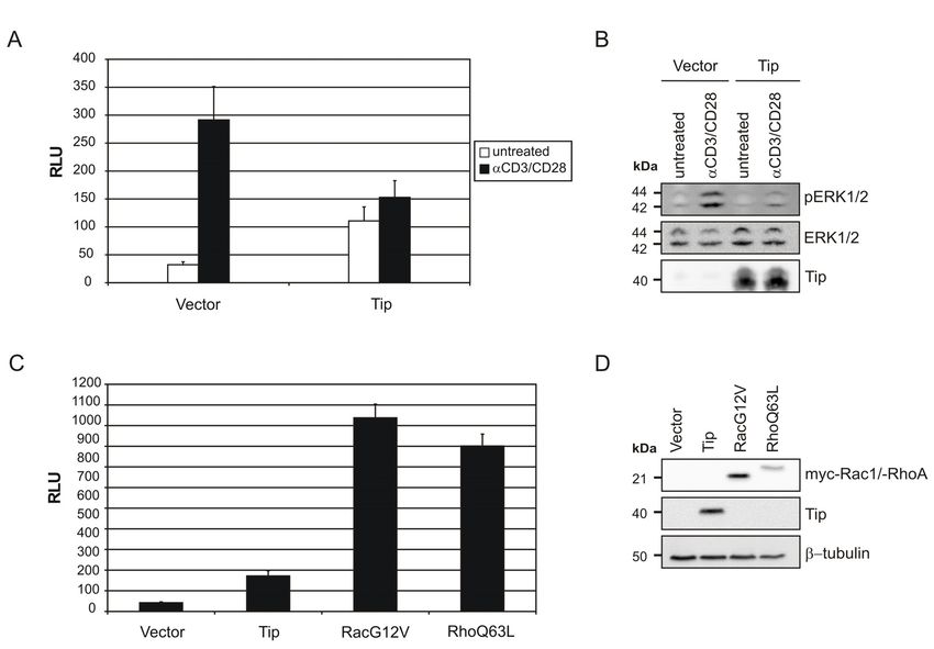
2 ZUSAMMENFASSUNG
Die T-lymphotropen Tumorviren Herpesvirus saimiri (HVS) und das nahe verwandte
Herpesvirus ateles (HVA) infizieren persistent ihre natürlichen Wirtspezies, die
Totenkopfäffchen bzw. Spinnenaffen, ohne ersichtliche Erkrankungen zu verursachen. In
anderen Neuweltprimaten wie Krallenaffen rufen sie T-Zell-Lymphome hervor. Die Induktion
anhaltender, Proliferation von T-Zellen aus Krallenaffen in vitro ist ebenfalls bekannt.
Aufgrund verschiedener pathogener Eigenschaften und divergierender genomischer
Sequenzen, welche die transformations-relevanten viralen Onkoproteine kodieren, werden
HVS-Isolate in drei Untergruppen eingeteilt: HVS-A, -B, -C. Nur HVS-C ist in der Lage
humane T-Zellen zu permanentem Wachstum in vitro zu transformieren. Generell ist die
Wachstumstransformation durch HVS und HVA abhängig von der Expression der Virus-
spezifischen Onkoproteine StpA, -B, -C, Tip und Tio.

Im ersten Teil dieser Arbeit zeigt eine umfassende in vitro Untersuchung, dass T-Zellen von
zwei verschieden Krallenaffen-Arten durch alle HVS-Untergruppen und HVA transformiert
werden, wohingegen humane T-Zell-Transformation auf HVS-C und HVA beschränkt ist. In
transient transfizierten humanen Jurkat T-Zellen zeigen vergleichende Analysen der
Onkoproteine, dass NF-κB von Onkoproteinen aller Virusstämme aktiviert wird. Weiterhin ist
NF-κB-Aktivität essentiell für das Überleben von HVS- und HVA-transformierten Zell-
Linien. Src-Familie-Kinase (SFK)-Aktivität wird gleichermaßen für deren Fortbestand
benötigt. SFK-assoziierte Signalweiterleitung wird jedoch nur beeinflußt durch die
Onkoproteine von HVS-C und HVA, Tip und Tio. Diese unterscheiden sich dadurch von den
anderen Onkoproteinen. Diese Daten sprechen für eine Korrelation zwischen der besonderen
Fähigkeit von HVS-C und HVA, humane T-Zellen zu transformieren, und der speziellen
Ansteuerung von SFK durch ihre Onkoproteine.
Der zweite Teil dieser Arbeit befaßt sich mit der Aktivierung des Serum Response Faktors
(SRF) in Tip-exprimierenden T-Zellen. Die Induktion eines SRF-Luziferase-Reportergens ist
abhängig von dem Kofaktor MAL, welcher durch Umstrukturierung des Zytoskeletts reguliert
wird. Entsprechend benötigt die Tip-vermittelte SRF-Induktion RhoGTPase-Aktivität und
Aktin-Polymerisierung. Dieser Weg wird nicht nur durch das virale Onkoprotein Tip
aktiviert, sondern auch durch T-Zell-Stimulation und konstitutiv aktive RhoGTPasen. Somit
induziert Tip die Aktivität von MAL:SRF und könnte dadurch Zytoskelett-Veränderungen mit
der Aktivierung von Ziel-Genen verbinden, welche verantwortlich sind für die virale
Onkogenese.

                                             2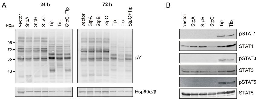
3 INTRODUCTION

3.1 T-LYMPHOTROPIC HERPESVIRUSES

Members of the family Herpesviridae are present in most animal species. They are assigned to
the subfamilies of α-, β- and γ-herpesviruses. Tumor induction in the hematopoietic system is
a common feature of γ-herpesviruses. Nevertheless, they are biologically and molecularly
heterogeneous and therefore classified into lymphocryptoviruses (γ1-herpesviruses) and
rhadinoviruses (γ2-herpesviruses). Epstein-Barr Virus (EBV) is the prototype of B-cell
lymphoma inducing γ1-herpesviruses in humans. The Kaposi sarcoma-associated Herpesvirus
8 (KSHV/HHV-8) is the first identified human pathogenic γ2-herpesvirus, causing several
neoplastic diseases of endothelial and lymphoid origin. For historical reasons, however, the
prototypic γ2-herpesvirus is the T-lymphotropic Herpesvirus saimiri (HVS), which has never
been reported to be pathogenic in humans.
HVS and the closely related Herpesvirus ateles (HVA) persist in T cells of their New World
primate host species, Saimiri sciureus and Ateles spp., without causing any apparent disease.
Both HVS and HVA are readily isolated from natural host T cells and amplified using owl
monkey kidney (OMK) cells. The cleared supernatants of these cultures contain high titers of
HVS, but only low titers of HVA, which remains mostly associated with cell debris (reviewed
in (Ensser & Fleckenstein, 2005)). Experimental inoculation of other New World primate
species, rapidly leads to fatal lymphoproliferative diseases classified as T-cell lymphoma and
leukemia. Experimental tumor induction in rabbits has also been described. Permanently
proliferating T-cell lines can be established ex vivo from blood and tissues of New World
primates and rabbits. Stable cell lines can also be generated by infection of isolated human
and non-human primate lymphocytes (reviewed in (Fleckenstein & Desrosiers, 1982; Ensser
& Fleckenstein, 2005).
The structurally identical double-stranded DNA genomes of HVS and HVA comprise a
coding AT-rich L-DNA (low density DNA) with 77 and 73 open reading frames (ORF),
respectively. This protein-coding DNA is stabilized by flanking GC-rich tandem repeats
termed H-DNA (high density DNA) (Bornkamm et al., 1976; Albrecht, 2000). Most of the
ORFs are conserved and code for proteins contributing to virus structure and replication or for
viral homologues of cellular genes, which manipulate cellular processes like cell cycle
(vCyclin), cytokine production (vIL-17) and apoptosis (vFLIP, vBcl-2).

                                              3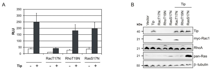
INTRODUCTION

A major sequence divergence at the left terminus of the L-DNA gave rise to the classification
of HVS isolates into the subgroups A, B, and C (Desrosiers & Falk, 1982; Medveczky et al.,
1984; Szomolanyi et al., 1987; Medveczky et al., 1989). This highly variable genomic region
determines viral oncogenesis and in vitro transformation, but is not required for viral
replication (Desrosiers et al., 1985; Desrosiers et al., 1986). It encodes distinct, subgroup-
specific proteins now recognized as HVS oncoproteins (Kamine et al., 1984; Murthy et al.,
1989; Biesinger et al., 1990; Geck et al., 1990; Duboise et al., 1998; Choi et al., 2000; Hör et
al., 2001). Sequence analysis of the HVA genome confirmed the close relationship to HVS
and identified a related oncoprotein (Albrecht et al., 1999; Albrecht, 2000).
The efficiency of tumor induction and in vitro T-cell transformation by HVS varies with the
HVS subgroup and with the infected host species. All HVS subgroups as well as HVA are
able to induce tumors and T-cell transformation in the New World primate species Saguinus
(S.) oedipus (cottontop marmoset); however, in vitro transformation by HVS-B is
controversial (Fleckenstein & Desrosiers, 1982; Reiss et al., 2002). Yet, only a few studies
directly compared different wild-type viruses in other species. In vitro infections of Callithrix
(C.) jacchus (common marmoset) lymphocytes with HVS strains of subgroups A, B, and C
reveals a restricted transforming potential of subgroup B (Desrosiers et al., 1986; Szomolanyi
et al., 1987). Inoculation of New Zealand White rabbits with high doses of HVS results in
lymphoproliferative lesions only in response to a subgroup C strain (Medveczky et al., 1989).
Transformation of human T lymphocytes to permanent, antigen-independent growth upon
infection with HVS is again restricted to subgroup C (Biesinger et al., 1992). Thus, HVS-C
possesses the highest oncogenic potential among all HVS subgroups. HVA was not included
in these previous studies and has not yet been successful in transformation of human T
lymphocytes. Nevertheless, an expression cassette for the HVA oncoprotein can substitute for
the cognate HVS-C oncoproteins in human T-cell transformation by recombinant HVS-C
(Albrecht et al., 2004).
Human peripheral blood mononuclear cells (PBMC), cord blood lymphocytes (CBL) and
thymocytes are susceptible to growth-transformation by HVS-C. The resulting human cell
lines consist of CD4+CD8- or CD4-CD8+ T cells. Their phenotype resembles activated mature
T cells expressing CD2, CD3, CD5 and CD7 at their surface. Transformed antigen-specific
T-cell clones retain many features of the primary parental cells like antigen-induced protein-
tyrosine phosphorylation and intracellular calcium mobilization (Mittrucker et al., 1993;
Weber et al., 1993; Bröker et al., 1993a). However, HVS-C transformed cells exhibit CD2
hyper-responsiveness and an altered cytokine profile with upregulated expression of

                                               4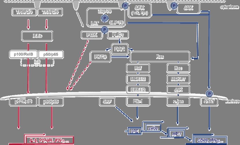
INTRODUCTION

interleukin 2 (IL-2) and interferon γ (IFN-γ) (Mittrücker et al., 1992; De Carli et al., 1993).
Transformed human T cells do not support lytic replication and release of infectious viral
particles. Viral persistence is maintained by high copy numbers of non-integrated viral
episomes, while the cellular karyotype is unaffected (Biesinger et al., 1992; Troidl et al.,
1994). In contrast, HVS- and HVA-transformed marmoset lymphocytes are semi-permissive
for virus production and therefore release low titers of viral particles (Falk et al., 1978;
Schirm et al., 1984; Desrosiers et al., 1986; Kiyotaki et al., 1986; Szomolanyi et al., 1987)

3.2 THE ONCOPROTEINS OF HERPESVIRUS SAIMIRI (HVS) AND
    HERPESVIRUS ATELES (HVA)

The oncoproteins of the HVS subgroups are the saimiri transformation-associated protein
(Stp) A, StpB, and StpC, respectively, as well as the subgroup C-specific tyrosine kinase-
interacting protein (Tip), which is transcribed together with StpC from a bicistronic mRNA
(Murthy et al., 1989; Jung et al., 1991; Biesinger et al., 1995; Choi et al., 2000; Hör et al.,
2001). The integrity of the reading frames for StpA and StpC/Tip is required for the
oncogenic phenotype of the parental virus strains (Murthy et al., 1989; Duboise et al., 1998).
Morphological transformation of rodent fibroblasts is induced by StpA and StpC, but not by
Tip and StpB (Jung et al., 1991; Choi et al., 2000). In transgenic mice, StpC expression results
in epithelial tumors, while StpA and Tip cause T-cell lymphoma (Murphy et al., 1994;
Kretschmer et al., 1996; Wehner et al., 2001). Hence, oncogenic functions are documented for
StpA, StpC and Tip, while they are assumed by analogy for StpB. The HVA oncoprotein two-
in-one (Tio) shows similarity to both StpC and Tip, and is considered oncogenic due to its
capacity to functionally replace these proteins in human T-cell transformation (Albrecht et al.,
1999; Albrecht et al., 2004).
The viral oncoproteins, schematically depicted in Fig.1, are encoded by divergent sequences
at the left terminus of the viral genome (Albrecht et al., 1992; Albrecht, 2000; Ensser et al.,
2003). Overall amino acid sequence identity is less than 30% among these proteins, but they
all share a C-terminal hydrophobic region considered as membrane anchor. The intracellular
parts harbor linear sequence motifs linking the viral proteins to cellular signaling molecules,
namely TNF receptor-associated factors (TRAF) and Src-family kinases (SFK).

                                                5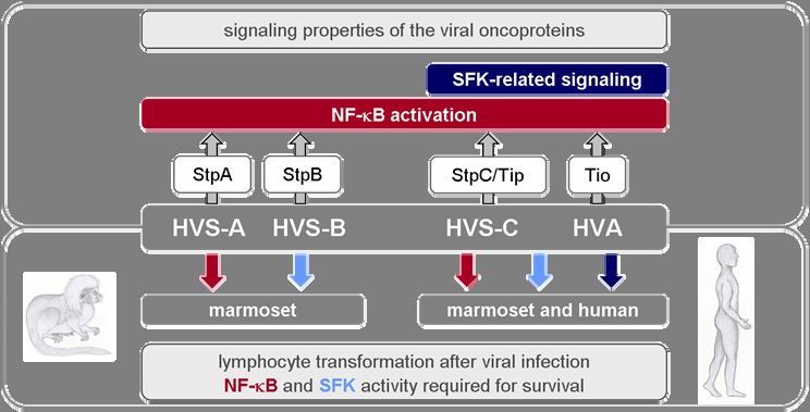
INTRODUCTION

Fig.1 Schematic representation of T-lymphotropic rhadinovirus oncoproteins. The membrane-anchored
viral oncoproteins are drawn to scale. Filled and shaded boxes indicate the position of conserved sequence motifs
mainly for SH2-, SH3-binding of Src-family kinase (SFK) and TRAF binding. Amino acid sequences are given
on the left. Arrows on the right point at interaction partners that were experimentally confirmed (continuous line)
or deduced by analogy (dashed line).

3.3 NUCLEAR FACTOR OF KAPPAB (NF-ΚB) SIGNALING

The nuclear factor kappa-light-chain-enhancer of activated B cells (NF-κB) is activated by
cellular receptors that recruit TRAFs. NF-κB regulates cell survival, differentiation,
proliferation as well as apoptosis. The family of NF-κB transcription factors consists of five
members: p65 (RelA), c-Rel, p105/p50, RelB and p100/p52. The activity of NF-κB family
members is differentially regulated via canonical or non-canonical pathways. Each pathway
involves the recruitment of distinct TRAFs, mainly TRAF2,-3 and -6. Subsequent activation
of inhibitor of κB kinases (IKKs) triggers degradation of the inhibitor of NF-κB (IκB) and
thereby induces the nuclear translocation of specific NF-κB dimers activating pro-
inflammatory and anti-apoptotic target genes (Fig.2), (Häcker et al., 2011).
TRAF binding generally maps to two classes of interaction motifs: the consensus sequence
P/S/A/T-X-Q/E-E is typical for TRAF2 and P-X-E-X-X-Ar/Ac for TRAF6 (Ye et al., 1999;
Ye et al., 2002). Related sequence motifs are present in StpA, StpB, StpC and Tio (Fig.1).
StpA triggers NF-κB activation through the TRAF6-binding PQENDE motif and, to a lesser
extent, through the TRAF2-binding PVQES motif (Jeong et al., 2007; Garcia et al., 2007).
StpB binds TRAF2, but has not been reported to induce NF-κB (Choi et al., 2000). StpC

                                                        6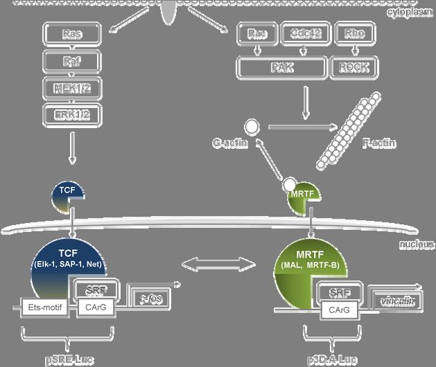
INTRODUCTION

relies on the integrity of the PIEET motif for TRAF2 binding, NF-κB activation and viral
transformation of human T cells, but not for tumor induction and in vitro transformation in
common marmosets (Lee et al., 1999; Sorokina et al., 2004). Tio recruits TRAF6 to the
PQEHEE motif to induce NF-κB activity, which in turn is required for the survival of human
T cells transformed by a Tio-recombinant HVS (Heinemann et al., 2006; de Jong et al., 2010).
While the assays usually addressed canonical NF-κB signaling, specific activation of non-
canonical NF-κB has been reported for StpA and Tio (Cho et al., 2007; de Jong et al., 2010).

3.4 SRC-FAMILY KINASES (SFK) AND RELATED SIGNALING

Cellular protein tyrosine kinases are grouped into receptor and non-receptor kinases. The
large family of SFK belongs to non-receptor kinases and essentially contributes to signaling
flux downstream of integrins, G-protein coupled-, cytokine-, antigen- and Fc-receptors. SFKs
are characterized by an N-terminal membrane localization motif, a unique region, a Src
homology 3 (SH3) domain, a Src homology 2 (SH2) domain, a kinase domain, and a
C-terminal regulatory tail. In the inactive state the kinase domain is inhibited by intra-
molecular interactions. This inactive conformation can be reversed by protein-protein
interactions, dephosphorylation of the regulatory tail and phosphorylation of a tyrosine
residue in the activation loop, resulting in increased kinase activity. This indicates multiple
mechanisms of SFK regulation, with binding of SH3 domains to proline rich sequences and
docking of SH2 domains to phosphorylated tyrosines being most the fundamental (Roskoski,
Jr., 2004; Bradshaw, 2010).
The SFKs Lck and Fyn are the main mediators of tyrosine phosphorylation after antigen
recognition by the T-cell receptor (TCR) in complex with intracellular signal transduction by
the CD3 complex (CD3) and thereby essentially contribute to T-cellular proliferation (Fig.2)
(Smith-Garvin et al., 2009). Hence, the ability of the T-lymphotropic rhadinoviruses and their
oncoproteins to target SFKs by SH2 or SH3 binding motifs may play a critical role in
stimulating their host T cells to permanent proliferation. However, the viral oncoproteins
differ in their preferred SFK partners as well as in their binding mode and downstream
effects.
Interactions of StpA and StpB with the prototypical SFK Src depend on the tyrosine-based
motif Y-A-E-I/V, which mediates binding to SH2 domains (Lee et al., 1997; Choi et al., 2000;
Hör et al., 2001). StpA elicits cellular signaling through interactions with Src, Fyn, and to a

                                              7
INTRODUCTION
lesser extent Lck (Garcia et al., 2007). Functional analyses for StpB:SFK interaction are not
available to date.
In contrast to StpA and StpB, Tip and Tio engage SFKs through more complex interactions
involving the SH3 domains of Lck, Lyn, and Hck (Jung et al., 1995a; Albrecht et al., 1999;
Hartley et al., 2000; Schweimer et al., 2002; Bauer et al., 2004). Tip:Lck interaction and also
activation of Lck requires the integrity of a motif homologous to C-terminal regulatory
sequences in the kinase domain of some SFKs (CSKH) and a proline-rich SH3 binding
sequence (SH3B) within Tip (Biesinger et al., 1995; Jung et al., 1995a; Wiese et al., 1996;
Lund et al., 1997b; Hartley et al., 1999; Kjellen et al., 2002). Three tyrosine residues, Y114,
Y127, Y155 in Tip are conserved between all HVS-C strains. Residues Y114 and Y127 are
Lck phosphorylation sites and have the potential to interact with the SH2 domains of STAT3
and Lck, respectively, (Fig.1) (Hartley & Cooper, 2000; Bauer et al., 2004; Heck et al., 2006).
Studies with HVS-C recombinants previously demonstrated that the SFK-binding sites in Tip
and Tio are indispensable for human T-cell transformation (Albrecht et al., 2005; Heck et al.,
2006), albeit the underlying mechanisms are not defined. Tip:Lck interaction activates the
kinase (Wiese et al., 1996; Lund et al., 1997a; Hartley et al., 1999; Kjellen et al., 2002;
Mitchell et al., 2007), but also inhibits TCR signaling (Jung et al., 1995b; Guo et al., 1997;
Cho et al., 2004) as well as TCR and CD4 surface expression (Park et al., 2002; Park et al.,
2003; Cho et al., 2006; Min et al., 2008).
The signal transducers and activators of transcription (STAT) are activated in response to
TCR or cytokine-receptor stimulation. This induces tyrosine phosphorylation of STATs by
non-receptor tyrosine kinases like SFK or cytokine receptor-associated Janus family tyrosine
kinases (JAKs) and thereby enables STAT dimerization (Fig.2). STAT dimers translocate to
the nucleus and bind to target genes involved in proliferation, differentiation and apoptosis
(Roskoski, Jr., 2004). Especially STAT3 is frequently activated in tumor cells and its activity
is characterized by pro-oncogenic functions (Yu et al., 2009). Phosphorylation of tyrosine
motif YRPQ in Tip (Y114) by Lck constitutes a direct binding site for STAT1 and STAT3
(Hartley & Cooper, 2000) and mediates constitutive phosphorylation of STAT3 in HVS-C
transformed marmoset and human lymphocytes (Reiss et al., 2002; Heck et al., 2005).
However, mutation of the tyrosine residue 114 essential for Tip:STAT3 interaction does not
abrogate human T-cell transformation (Hartley & Cooper, 2000; Heck et al., 2005). StpA
binds and activates STAT3 via the PTPYLP motif (Park et al., 2004; Chung et al., 2004), but
STAT3 phosphorylation has not been constantly detected in marmoset lymphocytes carrying
8INTRODUCTION
HVS-A (Reiss et al., 2002). StpB, which harbors an StpA-related PQNPYLP motif, as well as
Tio have not yet been reported to induce STATs.
Mitogen-activated protein kinases (MAPK) and their downstream transcription factors play
a key role in cell growth, survival and cancer. MAPK such as Jun N-terminal kinase (JNK)
and extracellular regulated kinase 1/2 (ERK1/2) are activated by mitogenic signaling through
SFKs and Ras (Fig.2). Classical MAPK signaling in T cells is initiated by the activation of
upstream tyrosine kinases like Lck in response to the engagement of the T-cell receptor
(TCR) and costimulatory CD28 receptor. Thereby, Lck is recruited to the cytosolic tail of the
CD3 receptor and activates a signaling platform comprising phosphorylated Zap-70, LAT and
SLP-76. Subsequently, PI3-kinase (PI3K) and phospholipase-C γ (PLCγ) are activated. PLCγ
generates diacylglycerol (DAG), an important activator of protein kinase C θ (PKCθ) and the
small G-protein Ras. Thus, TCR and CD28 receptor activation is coupled to MAPK signaling
pathways (Fig.2) (Smith-Garvin et al., 2009).
The Rac-MAPK kinase 4 and 7 (MEK-4/ 7) cascade triggers the phosphorylation of JNK and
downstream molecules like c-Jun, a subunit of the transcription factor AP-1 (Boutros et al.,
2008). AP-1 activation in turn contributes to the expression of the early activation marker
CD69 in T cells (Castellanos et al., 2002). Accordingly, StpA triggers AP-1 through its
interaction either with Src or alternatively TRAF6 (Jeong et al., 2007; Garcia et al., 2007).
However, neither StpC nor Tip activate AP-1 in human T cells (Glanz et al., 2008). Tip even
abrogates TCR-induced CD69 expression on T cells (Cho et al., 2004). Nevertheless, human
CD4+ T cells transformed by HVS subgroup C display the activation marker CD69 at their
surface (Saha et al., 1996). These cells are also characterized by the expression of IFN-γ (De
Carli et al., 1993; Bröker et al., 1993b; Saha et al., 1996), which is controlled by multiple
transcription factors including AP-1 and NF-κB. In Jurkat T cells, IFN-γ release is induced by
Tip and further enhanced by StpC (Glanz et al., 2008).
The prototypical MAPK cascade involves the Ras-effector kinase Raf, which phosphorylates
MAPK kinases 1 and 2 (MEK-1/2) and ERK-1/2, and results in activation of the transcription
factor Elk-1 (Smith-Garvin et al., 2009). Phosphorylated Elk1 belongs to the ternary complex
factor (TCF) family and is a cofactor of the serum response factor (SRF). TCF:SRF
complexes are well known to bind to the promoter of the c-fos gene (Treisman, 1994). StpC
was reported to enter this MAPK cascade by binding to cellular Ras and to activate ERK1/2
in adherent cells (Jung & Desrosiers, 1995). In the viral context, Ras substitutes for StpC in
the transformation of marmoset lymphocytes both in vitro and in vivo (Guo et al., 1998).
9INTRODUCTION
However, neither StpC nor Tip induces ERK1/2 phosphorylation in human T cells.
Nevertheless, activation of a luciferase reporter responsive to SRF was observed in
Tip-expressing cells (Glanz et al., 2008). This suggests a particular regulation of the SRF in
these cells.
Fig.2 Transformation-relevant signaling pathways in T cells. Simplified overview of pathways that promote
stimulation and proliferation of T cells. Upon engagement, the appropriate receptors recruit TNF-receptor
associated factors (TRAF) or tyrosine kinases (SFK, JAK) that induce NF-κB-, MAPK- and tyrosine
phosphorylation (P) dependent signaling pathways. Stimulus-specific activation of TRAF2,-3 or -6 triggers the
canonical or the non-canonical NF-κB pathway to entail nuclear translocation of specific NF-κB dimers and
activation of target genes like IL-8 and VCAM1. After engagement of the T-cell receptor (TCR), CD3 and co-
receptor (CD28), Src-family kinases (SFK) phosphorylate cellular proteins to recruit protein complexes that
induce e.g. MAPK signaling. Both the Ras-Raf-MEK1/2-ERK1/2-Elk1 and the Rac-MEK4/7-JNK-jun cascade
affect transcription factors that target genes like c-fos, AP-1 and CD69. Activation of the STAT-transcription
factor family also depends on tyrosine phosphorylation. In addition, multiple signals converge on the promoters
of numerous target genes such as IFN-γ. Complex crosstalk between all pathways has also been reported,
examples are indicated by dashed arrows.
10INTRODUCTION
3.5 THE MULTIFUNCTIONAL TRANSCRIPTION FACTOR SRF AND ITS
COFACTORS
SRF is a founding member of the MADS-box transcription factor family and is widely
expressed in both invertebrates and vertebrates. In vivo and in vitro studies point at an
essential role of SRF in embryogenesis (Arsenian et al., 1998), neuronal motility and heart
development (Knoll, 2010; Miano, 2010), skeletal muscle function (Li et al., 2005), cell death
(Bertolotto et al., 2000), cell morphogenesis, adhesion and migration (Schratt et al., 2002).
The MADS-box motif is a 56 amino-acid region conserved in the DNA binding site of
numerous eukaryotic transcription factors. Furthermore, it is required for protein interactions
and dimerization (Shore & Sharrocks, 1995). SRF dimers bind to single or multiple copies of
a palindromic DNA sequence known as the CArG box in the promoter of hundreds of target
genes. A relatively high number of these genes encode for actin cytoskeleton-regulating
factors, which ascribes a pivotal role to SRF in modulating the cytoskeleton. The interaction
with more than 60 cofactors turns SRF into a strong transcription factor mediating cell and
stimulus-specific signaling (Miano, 2010).
SRF cofactors – TCF and MRTF
The best characterized SRF cofactors are on the one hand TCF family members like Elk-1
that engage SRF, through MAPK regulated pathways (Shaw et al., 1989; Treisman, 1994). On
the other hand mycocardin-related transcription factors (MRTFs) bind and activate SRF in a
manner that is be controlled by Rho guanosine triphosphatases (Rho-GTPases) and
monomeric actin (Hill et al., 1995; Sotiropoulos et al., 1999; Posern et al., 2002; Miralles et
al., 2003; Posern et al., 2004).
Regulation of TCF. The well known SRF-cofactors, Ets (E-twenty six) domain TCF
proteins, like Elk-1, SAP-1 and Net, are regulated by phosphorylation via MAPK pathways,
namely Ras-Raf-MEK1/2-ERK1/2 (Fig.3). In addition to a direct binding of the SRF
MADS-box to the CArG motif, a defined DNA sequence, next to the SRF binding site, called
Ets motif, (C/A)(C/A)GGA(A/T), is important for the recruitment of TCF:SRF complexes
(Treisman, 1994). Promoter regions containing the Ets motif adjacent to the SRF-binding
CArG box are characterized as the serum response element (SRE).
11INTRODUCTION
Regulation of MRTFs. Myocardin, the founding member of the MRTF family, is enriched in
cardiac and smooth muscle cells and seems to constantly bind SRF. In contrast, MRTF-A
(MAL) and MRTF-B are widely expressed in many cell types. It has been shown that MRTF
dimers directly contact DNA near the SRF-binding CArG box for efficient complex formation
with SRF and DNA (Fig.3). However, a specific MRTF binding sequence has not yet been
found (Posern & Treisman, 2006). MAL regulation, best described in fibroblasts, depends on
an N-terminal RPEL motif, which binds monomeric, globular actin (G-actin). This interaction
prevents nuclear accumulation and, thereby, SRF-cofactor function (Miralles et al., 2003).
Recent studies demonstrate an unusual nuclear import sequence in the RPEL-motif of MAL,
which mediates an importin-dependent nuclear transport competed by globular actin
(Pawlowski et al., 2010).
RhoGTPases are considered as important regulators of actin and cytoskeleton organization.
Thereby, they influence gene transcription, cell proliferation, migration, growth and survival.
In general, this family of GTPases is activated by G-protein coupled receptors (GPCR),
integrins or antigen-receptors. GTPase activity itself is regulated by three classes of proteins.
Guanine nucleotide exchange factors (GEFs) activate GTPases by promoting the exchange of
GDP for GTP; GTPase-activating proteins (GAP) inactive them by enhancing their GTPase
activity to hydrolyse GTP; Guanine nucleotide dissociation inhibitors (GDIs) sequester
GTPases, keep them in an inactive state and prevent signaling towards their effectors. The
RhoGTPases best studied in actin regulation are RhoA, Rac1 and Cdc42, which use
serine/threonine kinases like Rho-associated coiled-coil-containing protein kinase (ROCK)
and p21-activated kinase (PAK), respectively, as effectors. They induce distinct cytoskeletal
structures like focal adhesion complexes (RhoA), lamellipodia (Rac1) and filopodia (Cdc42)
(Tybulewicz & Henderson, 2009; Lazer & Katzav, 2011). Thus, the activation of Rho-family
GTPases like RhoA, Rac1, and Cdc42 promotes actin polymerization, which enables
concomitant nuclear translocation of MRTF. The resulting SRF coactivation links the
cytoskeleton with stimulus-dependent target gene activation (Posern & Treisman, 2006;
Olson & Nordheim, 2010).
12INTRODUCTION
Fig.3 Simplified model of SRF regulation in fibroblasts and epithelial cells. Stimulus-dependent activation
of serum response factor (SRF) by Ras (left) or RhoGTPase (right) signaling. TCF proteins like Elk-1 complex
with SRF and bind to DNA with their Ets domain. SRF and Ets domain-binding DNA sequences are termed
serum response element (SRE). TCF activity is enhanced after phosphorylation induced by the Ras-MEK-ERK
cascade. The SRF cofactor and MRTF family member MAL (MRTF-A) is negatively regulated by binding to
globular actin (G-actin), which prevents its nuclear shuttling and cofactor functions. Activation of the
RhoGTPases (Rac, Cdc42 and Rho) triggers actin filament polymerization (F-actin) and thereby dissociation of
globular actin from MAL. MAL then translocates to the nucleus and binds SRF, additional unspecific DNA
contacts of MAL are likely, but not defined. TCF proteins and MAL interact with SRF by mutually exclusive
binding to a common region. Consequently, promoters of at least two classes of target genes are activated.
However, crosstalk between both signaling pathways is likely. The pSRE- and p3D.A luciferase reporters
(pSRE-Luc, p3D.A-Luc) are used to study SRF activation.
Signal-specific SRF regulation
SRF target gene specificity is dependent on the initial stimulus and subsequent activation of
signaling mediators (reviewed in (Posern & Treisman, 2006)). Such differential mechanisms
are shown for the activation of the SRF target genes c-fos, early growth reponse protein (egr1)
and vinculin, which show a distinct dependency on MEK-ERK and Rho-actin signaling
pathways in fibroblasts. A crosstalk between these two pathways is likely (Gineitis &
Treisman, 2001). The principle behind the differential regulation of SRF target genes is a
competition between TCF proteins and MRTFs for a common area in the MADS-box of SRF,
which is also required for its DNA binding (reviewed in (Posern & Treisman, 2006).
13INTRODUCTION
However, much remains unknown about the distinct pathways and target genes activated by
SRF coactivators, TCFs on the one hand and MRTFs on the other hand, especially in cell
types other than fibroblasts. Furthermore, SRF regulation in immune cells like T and B cells
has not been intensively investigated yet. Deletion of SRF in B cells results in loss of
marginal zone B-cells and reduction of CD5+ B-cell subsets. Analogous experiments with
T cells revealed an essential role of SRF in development (Fleige et al., 2007). Especially
ERK- and TCF dependent SRF activity is crucial in thymic selection of T-lymphocytes
(Mylona et al., 2011). In contrast, a specific role of MRTF:SRF complexes in T cells is
unexplored so far.
144 RATIONALE
I. The efficiency of T-cell growth transformation by Herpesvirus saimiri (HVS) and
Herpesvirus ateles (HVA) varies with the primate host species and with virus isolates, which
are classified into different subgroups. Nevertheless, comprehensive in vitro transformation
analyses of HVS subgroups and HVA in marmoset and human T cells are not available to
date. In general, the expression of the subgroup-specific oncoproteins is required for T-cell
growth transformation. Again, it remains unclear which oncoprotein features determine the
species restriction of transformation by their parental virus strains.
The subject of the first part in this thesis was to evaluate transformation efficiency by
HVS and HVA in marmoset and human lymphocytes and to search for determinants
restricting human T-cell transformation. To this end, the virus-specific transformation
potential is compared to signaling functions of the viral oncoproteins in transfected
human Jurkat T cells. Based on the known interaction motifs and functions of the
individual oncopoteins, the focus is on NF-κB and Src-family kinase (SFK) pathways.
Their requirement for the survival of the virus-transformed cell lines should be analyzed
and their activation by the oncoproteins in transfected human T cells should be
monitored using different read-out systems.
II. Human T-cell transformation by Herpesvirus saimiri subgroup-C, depends on the genomic
region coding for the oncoprotein Tip, which is also able to induce T-cell lymphoma in
transgenic mice. The transient expression of Tip in Jurkat T cells activates the cellular
transcription factor SRF without inducing MAPK activity. SRF is activated by mutually
exclusive interactions with MAPK- or actin-regulated cofactors, TCF and MRTF,
respectively.
Hence, the aim of the second part in the thesis was to shed light on the pathway and
cofactor mediating Tip-induced SRF activation in human T cells. The influence of
MAPK inhibition, TCF binding site ablation, requirement of actin polymerization as
well as the role of the MRTF member MAL in Tip-induced SRF activity should be
analyzed. To this end, the induction of two SRF luciferase reporters with different
susceptibility to the cofactors should be tested in transfected, Tip-expressing Jurkat T
cells. Furthermore, SRF activation in response to overexpression as well as inhibition of
published key regulators should be investigated in these cells.
155 RESULTS
5.1 TRANSFORMATION OF LYMPHOCYTES BY HVS AND HVA
Since the transforming potential and species restriction of HVS subgroups and HVA has
never been directly compared, T-cell transformation efficiencies were determined in a
comprehensive approach in marmoset and human T cells. First, fresh virus stocks of HVS-A,
HVS-B, HVS-C and HVA were generated by infection of permissive owl monkey kidney
(OMK) cells. Viral DNA prepared from the OMK supernatants was used for amplification of
oncoprotein-specific sequences by PCR to prove the integrity of the virus stocks (Fig.4A).
The fresh and verified virus stocks were used for infection of isolated marmoset peripheral
blood mononuclear cells (PBMC) obtained from cottontop marmosets (S. oedipus, n=5) and
common marmosets (C. jacchus, n=5). Furthermore, human cord blood lymphocytes (CBL)
of six independent donors were infected.
Fig.4 Verification of virus stocks and infection efficiency. (A) Fresh virus stocks of HVS-A (A-11), HVS-B
(SMHI), HVS-C (C-488) and HVA (At73) were generated by infection of permissive OMK cells. K-lysates
derived from supernatants of infected OMK cells were used for amplification of oncoprotein-specific sequences
(StpA, StpB, Tip+StpC, Tio) by PCR. H2O instead of K-lysate served as negative control (NC) for the
subsequent PCR amplification. Verified virus stocks were used in all subsequent infection experiments. (B,C)
Approximate infection efficiency and viral load was verified with K-lysates of OMK cells infected in parallel to
marmoset PBMC and human CBL. Cells were harvested 6 h, 24 h and 48 h post infection. Sequences specific for
the viral DNA polymerase (B) and viral oncoproteins (C) were amplified by PCR. Non-infected OMKcells (no
virus) and H2O instead of K-lysate (NC) were used as negative control for the subsequent PCR amplification.
16RESULTS
To test for the infectivity of the virus stocks, OMK cells were infected in parallel to the
marmoset and human lymphocytes. Intracellular DNA of infected OMK cells was prepared
after 6 h, 24 h and 48 h. PCR amplification of sequences coding for the viral DNA
polymerase (Fig.4B) and viral oncoproteins (Fig.4C) confirmed comparable infectivity.
5.1.1 TRANSFORMATION EFFICIENCY IN MARMOSET AND HUMAN
LYMPHOCYTES
Marmoset PBMC were cultured in the absence of IL-2 upon infection as previously described
(Fleckenstein & Ensser, 2004). Under these conditions, uninfected cells died within 3 to 9
weeks. IL-2-treated uninfected marmoset controls continued to proliferate throughout the
observation period of three months. These cells were used as controls for subsequent
experiments (Fig.5,6). Human CBL were infected and cultured in the presence of exogenous
human IL-2 as previously described (Fleckenstein & Ensser, 2004; Heck et al., 2005).
Uninfected human cells stopped proliferation after 1 to 3 weeks irrespective of IL-2
substitution. After a time period of three months the transformation efficiencies were assessed
by the number of stable lymphocyte cell lines established (Table 1).
Table 1 Outgrowth of marmoset and human cell lines after viral infection
a
cultured without exogenous IL-2
b
media supplemented with human IL-2
c
2/5 lines showed reduced proliferation rates
d
2/3 lines showed delayed onset of proliferation and were not included in the
experiments displayed in Fig. 5 and 6
e
1/6 lines showed reduced proliferation rates
f
1/4 lines showed reduced proliferation rates and was excluded from the
experiments displayed in Fig. 5 and 6
g
2/3 lines showed reduced proliferation rates
17RESULTS
Transformation by HVS-A was successful in 9 of 10 marmoset samples. Transformation with
HVS-B was least efficient as only three C. jacchus PBMC lines were generated. Furthermore
only one of these cell lines was available for further experiments (Fig.5,6), due to a delayed
proliferation onset of the other two cell lines. In contrast, HVS-C induced permanent T-cell
growth in all infected individuals of each species. HVA was also capable to transform
lymphocytes of all species tested. However, a reduced efficiency of HVA towards S. oedipus
and C. jacchus PBMC compared to human CBL was observed. Thus, HVS-C and HVA were
distinguished from HVS-A and HVS-B by their ability to transform human lymphocytes.
5.1.2 RELEVANCE OF NF-ΚB FOR THE SURVIVAL OF TRANSFORMED CELLS
T-cell growth transformation by HVS and HVA strictly depends on specific virus-encoded
oncoproteins (reviewed in (Ensser & Fleckenstein, 2005)). Sequence analyses demonstrated
that all oncoproteins except Tip exhibit a TRAF interaction motif, linking them to NF-κB
signaling. Indeed, NF-κB activation has been shown for the individual oncoproteins, albeit in
different cell and read-out systems (Lee et al., 1999; Ensser & Fleckenstein, 2005; Heinemann
et al., 2006; Jeong et al., 2007; Garcia et al., 2007; Glanz et al., 2008; de Jong et al., 2010).
Therefore, the requirement for NF-κB activity was assessed in the cell lines transformed by
HVS or HVA.
Two months after infection, marmoset PBMC and human CBL lines (Table 1) were treated
for seven days with the IKK-β inhibitor ACHP. This compound had previously proven
effective in abrogating the NF-κB-dependent survival of virus-transformed human T-cell lines
(de Jong et al., 2010). The viability of treated and untreated cells was monitored by propidium
iodide staining and subsequent flow cytometry (Fig.5). Uninfected marmoset PBMC, which
proliferate in response to exogenous human IL-2, as well as DMSO solvent-treated cells
served as controls. The average survival of the infected S. oedipus lines and of their
uninfected IL-2-treated controls ranged from 20-50% (Fig.5A). Survival of the C. jacchus
lines varied between 30-70% (Fig.5B). The human lymphocytes transformed by HVS-C and
HVA survived ACHP treatment at ratios of 20-30% (Fig.5C). In spite of considerable
variation among the cell lines infected with a specific virus and among the replicates, these
results documented that NF-κB activity is required to maintain the transformed state of the
cells irrespective of the virus strain and host species.
18RESULTS
Fig.5 Survival of HVS- and HVA-transformed
cell lines after NF-κB inhibition. Two months
after infection with HVS-A, HVS-B, HVS-C, and
HVA, PBMC of S. oedipus (A) and C. jacchus (B)
were seeded into 96-well plates. Uninfected PBMC
proliferating in the presence of human IL-2
(10 units/ml) were included as controls (no virus).
Cells were left untreated or treated with solvent
(DMSO) or the NF-κB inhibitor ACHP (10 µM).
The inhibitor was replenished every 48 h. After
seven days of treatment, cell viability was assessed
by propidium iodide staining (10 µg/ml) and flow
cytometry. (C) Infected human CBL were treated
equally, except that they were cultivated in the
presence of IL-2 and that uninfected controls were
not available.
Survival was calculated as the number viable cells
in the treated samples relative to the untreated
sample. Each bar in the graphs gives the mean value
and standard deviation for the indicated number of
cell lines (n) and summarizes two (A, B) or three
(C) successive replicates for all cell lines.
5.1.3 REQUIREMENT OF SFK FOR THE SURVIVAL OF TRANSFORMED CELLS
Another common feature of HVS and HVA oncoproteins is the capability to interact with
SFK, although the interaction mode differs dependent on the presence of an SH2 or SH3
binding motif in the oncoproteins. To assess the importance of SFK activity for their survival,
the virus-transformed cell lines (Table 1) were treated with PP2, a well established, specific
inhibitor of SFK previously used in transformed cells (Albrecht et al., 2005). Two months
after infection, marmoset PBMC as well as human CBL lines were incubated with PP2 for a
total of seven days. Viability was monitored by propidium iodide staining and subsequent
flow cytometry (Fig.6). After seven days of treatment, only 20-60% of treated cells derived
from the marmoset PBMC lines survived (Fig.6A,B). In contrast, the IL-2 treated uninfected
19RESULTS
control cells were practically unaffected by PP2 (Fig.6A,B; last columns). Similar to this
observation, PP2 treatment did not diminish the survival of the human CBL lines routinely
cultured in the presence of IL-2 (Fig.6C; white and black bars). Nevertheless, PP2 treatment
altered cell size and impaired cell-cell adhesion in these cultures as monitored by flow
cytometry and microscopy, respectively (data not shown).
To test whether the supplemented, exogenous IL-2 in the media of the cultured cell lines has
an influence on this phenomenon, the experiment was repeated after withdrawal of
conditioned, exogenous IL-2 containing medium. Culture morphology was impaired after
IL-2 depletion, as verified microscopically in comparison to IL-2-treated cells (data not
shown). Nevertheless, the data demonstrated
that PP2 treatment reduced survival to 10-
40% in the absence of IL-2 (Fig.6A-C; grey
bars). Hence, Src-family kinase activity was
required for the maintainance of the
transformed state, but was compensated by
exogenous IL-2, at least with respect to cell
survival.
Fig.6 Survival of HVS- and HVA-transformed
cell lines after Src-family kinase inibition. Two
months after infection with HVS-A, HVS-B, HVS-C,
and HVA, PBMC of S. oedipus (A) and C. jacchus
(B) were seeded into 96-well plates. Uninfected
PBMC proliferating in the presence of human IL-2
(10 units/ml) were included as controls (no virus).
Cells were left untreated or treated with solvent
(DMSO) or the SFK inhibitor, PP2 (10 µM). The
inhibitor was replenished every 48 h. After seven
days of treatment, cell viability was assessed by
propidium iodide staining (10 µg/ml) and flow
cytometry. (C) Infected human CBL cultivated in
the presence of IL-2 (white and black bars) or
depleted of the conditioned, IL-2-containing media
(grey bars) were tested for PP2 sensitivity as
described above.
Survival was calculated as the number viable cells in
the treated samples relative to the untreated sample.
Each bar in the graphs gives the mean value and
standard deviation for the indicated number of cell
lines (n) and summarizes two (A, B) or three (C)
successive replicates for all cell lines.
20RESULTS
5.2 DIFFERENTIAL SIGNALING BY THE VIRAL ONCOPROTEINS IN
HUMAN T CELLS
Expression of the viral oncoproteins in a transient transfection system. Transient
expression in Jurkat T cells was used to further elucidate the viral oncoprotein functions that
determine viral transformation of human T cells. To this end, expression plasmids coding for
StpA and StpB (AU1-tag), StpC (Myc-tag), Tip, and Tio (FLAG-tag) were transfected into
human Jurkat T cells. In general, cells were harvested after 48 h, or at the indicated time
points, and processed for the distinct, subsequent assays. Oncoprotein expression as shown in
Fig.7 is representative for all subsequent transfection experiments.
Fig.7 Transient expression of the viral oncoproteins in Jurkat
T cells. Whole cell lysates were obtained 48 h after electroporation
and analyzed for the presence of StpA and StpB (AU1-tag), StpC
(myc-tag), Tip, and Tio (FLAG-tag). Detection of Hsp90α/β served
as a loading control.
5.2.1 SURFACE EXPRESSION OF THE T-CELL ACTIVATION MARKER CD69
Proliferation of non-transformed T cells is induced by their receptor-mediated activation,
which is accompanied by upregulation of the activation marker CD69 (Sancho et al., 2005).
Thus, T-cell activating functions of the viral oncoproteins were assessed by investigating
CD69 surface expression. Jurkat T cells were analyzed by flow cytometry 24 h, 48 h (Fig.8),
and 72 h post transfection. Only Tip and Tio emerged as positive regulators of CD69 surface
expression, showing a peak after 48 h. Tio-mediated induction was strongest and already
observed after 24 h (data not shown). StpC was ineffective by itself, but enhanced Tip-
mediated CD69 upregulation. Long-term observations revealed that CD69 surface expression
was slightly downregulated after 72 h (data not shown). These results demonstrated that Tip
and Tio possess a unique potential to activate human T cells.
21RESULTS
Fig.8 Upregulation of CD69 surface expression
by the viral oncoproteins. Jurkat T cells were
stained 48 h after transient transfection of
expression plasmids coding for the viral
oncoproteins. Vector transfection was included as
negative control. Binding of the allophycocyanin
(APC)-labeled CD69 antibody was analyzed by
flow cytometry. Filled histograms represent staining
with an isotype-matched control antibody (black)
and CD69 staining of vector-transfected cells
(gray), respectively. Dotted lines depict CD69
staining of the oncoprotein-expressing cultures. A
representative expression control for the
oncoproteins is depicted in Fig.7.
5.2.2 IFN-γ SECRETION
Specific T-cell subsets respond to activation by upregulating IFN-γ production. The IFN-γ
promoter is controlled by a plentitude of transcription factors and thereby integrates different
signaling pathways (Ansel et al., 2003). Moreover, IFN-γ production is a common feature of
HVS transformed human T cells (De Carli et al., 1993). Previously, StpC has been reported to
enhance Tip-induced IFN-γ release in transfected Jurkat T cells (Glanz et al., 2008). Thus,
induction of IFN-γ production by the different viral oncoproteins was examined. Supernatants
of Jurkat T cells were taken 24, 72, 96 h after transfection and subjected to an IFN-γ ELISA
(Fig.9). StpA and StpC expression had no detectable impact on IFN-γ production. The levels
of IFN-γ secreted in response to StpB remained below the lower quantification limit of the
assay. In contrast, Tip induced IFN-γ expression 72 h after transfection, which increased with
time. Although the expression of StpC alone did not have an influence, co-expression with
Tip enhanced Tip-induced IFN-γ production as published previously (Glanz et al., 2008). In
contrast, Tio-triggered IFN-γ secretion started 24 h post transfection and, at later time points,
was 10-fold increased relative to the levels induced by Tip and StpC. These data again
distinguished Tip and Tio from the other viral oncoproteins and confirmed Tio being a highly
efficient signal inducer in T cells.
22RESULTS
Fig.9 IFN-γ secretion mediated by viral oncoproteins. Supernatants of Jurkat T cells expressing StpA, StpB,
StpC, Tip, StpC+Tip, or Tio were collected 24 h, 72 h, and 96 h after transfection. Supernatants of vector-
transfected cells (pcDNA, pEF1) served as negative control. The graphs display mean and standard deviation
calculated from two independent experiments, each performed in biological triplicates; a dashed line indicates
the lower limit of quantification; different scales are used to adjust resolution. A representative expression
control for the oncoproteins is depicted in Fig.7.
5.2.3 NF-ΚB ACTIVATION
In many cell types NF-κB activity contributes to proliferation. The presence of TRAF binding
motifs in StpA, StpB, StpC and Tio (Fig.1) and the requirement of NF-κB activity for the
survival of the virus transformed cell lines (Fig.5) indicated that this transcription factor may
be a common target of the T-lymphotropic rhadinovirus oncoproteins. Given the diversity of
NF-κB pathways and target genes, several read-out systems were used to detect differential
NF-κB signaling by the viral oncoproteins.
An NF-κB luciferase reporter was activated, to variable extent, by all oncoproteins except
Tip, which also had no influence on the effect of cotransfected StpC (Fig.10A). This data
confirmed previous studies on the NF-κB-inducing activity of StpA, StpC, and Tio. In
contrast, StpB had been reported negative, but, together with Tio, displayed the highest
NF-κB-reporter activity.
The canonical NF-κB pathway was investigated with respect to the expression of its well
established, endogenous target genes. For this purpose, transcription of VCAM1 (CD106)
(Shu et al., 1993), an adhesion molecule, and secretion of IL-8 (Mukaida et al., 1994), a
neutrophil chemokine, were analysed (Fig.10B,C). VCAM1 transcripts were readily detected
in T cells expressing StpB and Tio. A very weak signal was observed in the presence of StpA,
while both StpC and Tip were negative for VCAM1 induction (Fig.10B).
23RESULTS
Fig.10 NF-κB activation in T cells by the viral oncoproteins. Jurkat T cells were transiently transfected with
expression plasmids coding for the viral oncoproteins of HVS-A (StpA), HVS-B (StpB), HVS-C (StpC, Tip,
StpC+Tip), and HVA (Tio). Vector transfections (pcDNA, pEF1) served as negative controls. (A) Activation of
a cotransfected NF-κB luciferase reporter by the viral oncoproteins was measured 48 h post transfection with
reference to the appropriate vector control. (B) Total cellular RNA was isolated 48 h after transfection and
reversely transcribed into cDNA to perform PCR with primers specific for VCAM1 transcripts. The lower panel
shows transcriptional levels of HPRT, which served as an input control. H2O instead of cDNA served as negative
control (NC) for the subsequent PCR amplification. (C) Cell culture supernatants were collected 24 h post
transfection and secreted IL-8 was detected by ELISA. (D) Expression of p52 was detected in whole cell
lysates 48 h post transfection by immunoblot analysis. Detection of Hsp90α/β served as loading control. The
graph in (A) displays mean values and standard deviations of an assay performed in triplicates and is
representative for three independent experiments. The graph in (C) summarizes the results of three independent
experiments with a total of seven transfections per data point. Data shown in (B) and (D) are representative for
three independent experiments. A representative expression control for the oncoproteins is depicted in Fig.7.
A similar pattern of induction was observed for IL-8 secretion (Fig.10C). Chemokine release
was induced 24 h post transfection, and concentrations increased from StpA and StpB to Tio.
StpC and Tip, alone or in combination, did not trigger any IL-8 release. IL-8 values peaked
24 h post transfection and were not further enhanced after 48 h of oncoprotein expression
(data not shown).
Non-canonical NF-κB activation was addressed by investigating the processing of the NF-κB
subunit and precursor molecule p100 to its active form p52. To this end, p52 expression was
monitored by Western blot analysis. Concordant with the reporter assay, p52 was detectable
upon expression of StpA, StpB, StpC and Tio (Fig.10D). The amount of p52 was highest in
the presence of StpB and Tio, intermediate with StpA and lowest with StpC.
These findings further substantiate and extend the knowledge about the capability of the
oncoproteins to activate NF- κB. All oncoproteins except Tip activate transcription factors of
the NF-κB family in human T cells. Notably, StpC-induced reporter activity was not reflected
in an upregulation of the selected canonical NF-κB target genes. In conclusion, the level of
24RESULTS
NF-κB activation by the viral oncoproteins does not correlate with the transforming potential
of the parental virus strain. Most prominently, the oncoproteins of highly oncogenic HVS-C,
StpC and Tip, showed the weakest effects in all different read-out systems.
5.2.4 PROTEIN TYROSINE PHOSPHORYLATION
The ability of the T-lymphotropic herpesviruses and their oncoproteins to activate T cells
most likely plays a critical role in stimulating host cells to permanent proliferation. In this
context, tyrosine phosphorylation of proteins by Src-family kinases is one of the initial steps
in T-cell activation (Smith-Garvin et al., 2009). All HVS and HVA oncoproteins, except
StpC, harbor an SH2 or SH3 binding motif (Fig.1) that enables interactions with SFK.
However, the SH2-binding sites in StpA and StpB on the one hand and the SH3-binding sites
in Tip and Tio on the other hand connect these viral oncoproteins with SKFs in a distinct
manner (Fig.1), which might differentially impact protein tyrosine phosphorylation in the cell.
Hence, total cellular tyrosine phosphorylation was analyzed by Western blot upon transient
expression of the viral oncoproteins in Jurkat T cells. Variations in the cellular tyrosine
phosphorylation patterns were detectable 24 h after transfection only in Tip- and Tio-
expressing cells (Fig.11A). Bands migrating at 45-50 kDa were assigned to Tip and Tio in the
respective lanes. All other deviant bands indicate proteins whose tyrosine phosphorylation
was similarly affected by Tip and Tio, but not by StpA, StpB or StpC. Furthermore, co-
expression of StpC and Tip had no influence on Tip-mediated changes in cellular protein
tyrosine phosphorylation. The unique phosphorylation pattern induced by Tip and Tio
remained constant over time. However, band intensities were reduced after 72 h relative to
vector and StpA, StpB or StpC expressing cells.
The STAT family of transcription factors is activated by tyrosine phosphorylation e.g. in
response to cytokine-receptor mediated signals but also in response to active SFK (Roskoski,
Jr., 2004; Yu et al., 2009). The role of STAT factors in primary T-cell proliferation is
documented by its dependence on IL-2, which mainly triggers STAT5 activation (Yu et al.,
2009). Furthermore, previous studies identified STAT1 and STAT3 as downstream targets of
Tip:Lck interaction, and STAT3 activation has also been described for StpA (Fig.1) (Lund et
al., 1997b; Lund et al., 1999a; Chung et al., 2004). To address the relevance of all viral
oncoproteins in mediating phosphorylation of STAT1, 3 and 5, Western blot analysis was
performed 48 h after transfection of Jurkat T cells (Fig.11B). The expression of Tip and Tio,
but not of StpA, StpB and StpC resulted in tyrosine phosphorylation of STAT1, 3 and 5. In
25You can also read


