Enhancing Dendritic Cell Vaccination by Immune Checkpoint Blockade as Therapy in AML
←
→
Page content transcription
If your browser does not render page correctly, please read the page content below
Enhancing Dendritic Cell Vaccination by
    Immune Checkpoint Blockade as
             Therapy in AML

               Dissertation
zum Erwerb des Doctor of Philosophy (Ph.D.)
     an der Medizinischen Fakultät der
Ludwig-Maximilians-Universität zu München

               vorgelegt von
       Maurine Daniela Clara Rothe

                   aus
            Frankfurt am Main

                    am
             13. Februar 2020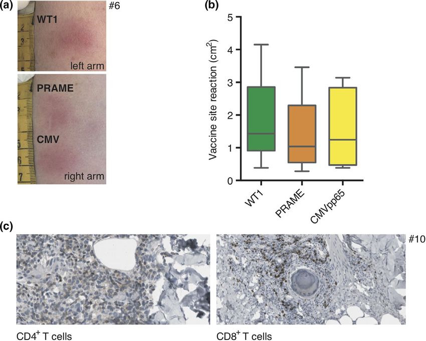
Supervisor:             Prof. Dr. med. Marion Subklewe

Second reviewer:        Prof. Dr. med. Andreas Mackensen

Dean:                   Prof. Dr. med. dent. Reinhard Hickel

Date of oral defense:   28. April 2020

                                                           ii
List of Abbreviations

AML             Acute myeloid leukemia
APC             Antigen-presenting cell
CAR             Chimeric antigen receptor
CD              Cluster of differentiation
CMV             Cytomegalovirus
CR              Complete remission
CTLA-4          Cytotoxic T lymphocyte associated protein-4
DC              Dendritic cell
FDA             Food and drug administration
FGL-1           Fibrinogen-like Protein 1
GAL-3           Galectin-3
HSCT            Hematological stem cell transplantation
IFN             Interferon
IL              Interleukin
LAA             Leukemia-associated antigen
LAG-3           Lymphocyte activation gene 3
LSECtin         Liver sinusoidal endothelial cell lectin
MHC             Major histocompatibility complex
mRNA            Messenger Ribonucleic acid
NK cell         Natural killer cell
OS              Overall survival
PBMC            Peripheral blood mononuclear cell
PD-1            Programmed cell death protein 1
PD-L1           Programmed cell death protein ligand 1
PD-L2           Programmed cell death protein ligand 2
PRAME           Preferentially expressed antigen in melanoma
RFS             Relapse-free survival
RNA             Ribonucleic acid
TAA             Tumor-associated antigen
TCR             T cell receptor
TLR             Toll-like receptor
TNF             Tumor necrosis factor

                                                               v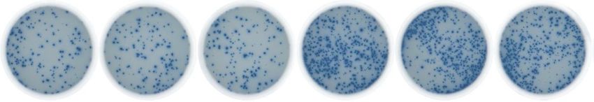
Treg   Regulatory T cell
WT1    Wilms tumor 1

                           vi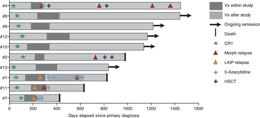
List of Publications

This thesis includes two publications which have been accepted for publication in
peer-reviewed journals:

Publication I:

„Toll-like receptor 7/8-matured RNA-transduced dendritic cells as post-
remission therapy in acute myeloid leukemia: results of a phase I trial.”

Felix S. Lichtenegger, Frauke M. Schnorfeil, Maurine Rothe, Katrin Deiser, Torben
Altmann, Veit L. Bücklein, Thomas Köhnke, Christian Augsberger, Nikola P.
Konstandin, Karsten Spiekermann, Andreas Moosmann, Stephan Boehm, Melanie
Boxberg, Mirjam H.M. Heemskerk, Dennis Goerlich, Georg Wittmann, Beate
Wagner, Wolfgang Hiddemann, Dolores J. Schendel, Gunnar Kvalheim, Iris
Bigalke, Marion Subklewe

Journal of Clinical & Translational Immunology. 2020 Feb; doi: 10.1002/cti2.1117

Publication II:

„Targeting LAG-3 and PD-1 to enhance T cell activation by antigen-Presenting
cells.”

Maurine Rothe*, Felix S. Lichtenegger*, Frauke M. Schnorfeil, Katrin Deiser,
Christina Krupka, Christian Augsberger, Miriam Schlüter, Julia Neitz and Marion
Subklewe:

Frontiers in Immunology. 2018 Feb; doi: 10.3389/ fimmu.2018.00385

*contributed equally

The proportion and distribution of work that has been contributed by the individual
authors is listed in the publication (chapter 3, page 23-24, 39 and 63).

                                                                                vii
Table of Contents

Affidavit ........................................................................................................................ iii
Conformation of Congruency....................................................................................... iv
List of Abbreviations .................................................................................................... v
List of Publications...................................................................................................... vii
Table of Contents ...................................................................................................... viii
1. Summary ................................................................................................................. 9
2. Introduction ............................................................................................................ 11
     2.1 Acute Myeloid Leukemia ................................................................................ 11
     2.2 Cancer Immunotherapy in AML ..................................................................... 12
     2.3 Dendritic Cell Vaccination in AML .................................................................. 13
     2.3.1 Vaccination Strategies ................................................................................ 13
     2.3.2 Ongoing Phase II Clinical Trials on DC-Vaccination as Therapy in AML .... 14
     2.3.3 New Generation DC Vaccine for Immunotherapy of AML........................... 15
     2.3.4 The Clinical Study Antigens WT1, PRAME, and CMVpp65 ........................ 16
     2.3.5 Boosting DC-induced T Cell Responses ..................................................... 17
     2.4 Immune Checkpoint Blockade in Cancer Therapy ......................................... 18
     2.4.1 PD-1 ............................................................................................................ 19
     2.4.2 LAG-3 .......................................................................................................... 20
     2.5 Aim of this Thesis ........................................................................................... 22
3. Publications ........................................................................................................... 23
     3.1 Author Contributions Publication I .................................................................. 23
     3.2 Author Contributions Publication II ................................................................. 24
     3.3 Publication I .................................................................................................... 25
     3.4 Publication II ................................................................................................... 53
4. Acknowledgements ............................................................................................... 73
5. References ............................................................................................................ 75
     5.1 Abstracts ........................................................................................................ 75
     5.2 Original Research Articles and Reviews ........................................................ 75

                                                                                                                                  viii
Summary

1. Summary
The success of checkpoint inhibition has changed treatment algorithms in several
tumor entities within the past years. Treatment success has mainly been observed
in cancers with an inflamed microenvironment and an immune infiltrate leading to
upregulation of checkpoint molecules on tumor cells as a means of immune
escape. Hence, in tumor entities with a low endogenous anti-tumor response, such
as acute myeloid leukemia (AML), checkpoint inhibition as monotherapy has so far
shown no clinical benefit. Therapeutic vaccination based on autologous dendritic
cells (DCs) pulsed with leukemia-associated antigens (LAA) is able to elicit anti-
leukemic immunity. The combination with checkpoint inhibitors might enable to
enhanced anti-leukemic immune responses in two ways: First, by blocking the
interaction between checkpoint molecules on anti-leukemic T cells and upregulated
checkpoint molecules on the leukemic target cells; and second, by enhancing the
initial interaction between T cells and DCs which constitutively express inhibitory
checkpoint molecules on their surface. Thus, a combinatorial therapy of DC
vaccination and checkpoint blockade, in particular for cancers with a low
endogenous anti-tumor response is a promising treatment strategy.
We have implemented a phase I/II first-in-human clinical study using monocyte-
derived toll-like receptor (TLR) 7/8-matured next-generation DCs loaded with wilms
tumor 1 (WT1), preferentially expressed antigen in melanoma (PRAME) and
cytomegalovirus (CMV)pp65 RNA as post-remission therapy of AML patients with
a non-favorable risk profile.
DC vaccination was feasible and safe and induced antigen-specific immune
responses. AML-specific T cell responses correlated with improved relapse-free
survival (RFS), especially in younger patients (≤ 65 years).
Despite a strong co-stimulatory profile, DCs also expressed co-inhibitory
checkpoint ligands. We examined those inhibitory interactions using an in vitro T
cell-DC coculture. DC-activated T cells upregulated programmed cell death protein
1 (PD-1) and lymphocyte activation gene 3 (LAG-3), while DCs expressed the
respective ligands programmed cell death protein ligand 1 (PD-L1) and major
histocompatibility complex (MHC) class-II. As hypothesized, we demonstrated that
blockade of PD-1 and particularly of LAG-3 by suitable blocking antibodies
enhanced DC-induced T cell activation.

                                                                                 9
Summary

We conclude that TLR7/8-matured next-generation DC vaccination induces
vaccine antigen-specific immune responses which may lead to delay or prevention
of relapse. Our in vitro data supports the rationale of combining DC vaccination with
PD-1 and/or LAG-3 blockade to further augment anti-leukemic immune responses
and improve clinical outcome.

                                                                                  10
Introduction
2. Introduction
2.1 Acute Myeloid Leukemia
Acute myeloid leukemia (AML) is a hematological malignancy, which is defined by
disrupted differentiation and uncontrolled proliferation of myeloid progenitor cells in
bone marrow and blood (Dohner, Weisdorf et al. 2015).This results in proneness
to infections and anemia and ultimately in multiple organ failure and death. AML is
the most frequent leukemia among adults with a median age of 68 years and an
incidence rate of 19,520 new cases and 10,670 deaths in the US in 2018
(Lichtenegger, Krupka et al. 2017, Siegel, Miller et al. 2018).
AML is a genetically and clinically heterogeneous disease with a very poor
prognosis. Despite the strong need for improvement, the treatment has barely
changed over the past decades: The standard treatment after diagnosis is a high
dose induction chemotherapy comprising three days of anthracycline and seven
days of cytarabine. This so called “3+7” regimen induces complete remission (CR)
in about 80% of the patients (Dohner, Estey et al. 2010, Burnett, Wetzler et al. 2011,
Ferrara and Schiffer 2013, Lichtenegger, Krupka et al. 2017). However, the risk of
relapse is high due to chemorefractory leukemic cells. A post-remission therapy to
eliminate residual leukemic cells is therefore mandatory (Reinisch, Chan et al.
2015).
Usually, patients with a favorable genetic risk profile get additional cycles of
chemotherapy as consolidation, whereas the method of choice for AML patients
with high relapse risk is allogeneic hematopoietic stem cell transfer (HSCT). HSCT
was the first curative immunotherapy for patients with hematological malignancies
(cure rate over 50%). Its clinical benefit relies in particular on the so-called graft-
versus-leukemia effect: allogeneic T and natural killer (NK) cells of the donor
recognize and target malignant cells of the recipient. However, the beneficial
potential of anti-leukemic responses is opposed by the individual risk of graft-
versus-host disease. In addition, the donor availability is challenging (Stelljes, Krug
et al. 2014, Kassim and Savani 2017). Especially elderly patients (< 60 years) are
often not medically fit for intensive therapies including HSCT (Klepin, Rao et al.
2014). Thus, there is a high medical need for the development and improvement
of novel therapies.
11Introduction
2.2 Cancer Immunotherapy in AML
Cancer immunotherapy aims to direct the body´s own immune system against
malignant tumor cells. It represents one of the most promising novel strategies to
cure cancer and to decrease relapse rates (Rusch, Bayry et al. 2018). Various T
cell based immunotherapeutic strategies to eliminate chemorefractory leukemic
cells are currently under preclinical- and clinical investigation (Lichtenegger,
Krupka et al. 2015, Lichtenegger, Krupka et al. 2017). The most prominent are:
• T cell engaging antibody based approaches to recruit T cells to target antigen
expressing tumor cells (Jin, Lee et al. 2009, Laszlo, Gudgeon et al. 2014).
• Adoptive T cell transfer (TCR-, or CAR T cell therapy) to augment autologous
T cells in number and tumor antigen specificity (Xue, Gao et al. 2005,
Spranger, Jeremias et al. 2012, Brenner 2013, Pizzitola, Anjos-Afonso et al.
2014, Prommersberger, Jetani et al. 2018, Gomes-Silva, Atilla et al. 2019)
• Dendritic cell vaccination to induce strong and durable tumor antigen-specific
T cell responses (Van Tendeloo, Van de Velde et al. 2010, Anguille, Willemen
et al. 2012, Khoury, Collins et al. 2017, Weinstock, Rosenblatt et al. 2017).
• Immune checkpoint blockade to enhance or reactivate preexisting anti-tumor
T cell responses (Alatrash, Daver et al. 2016).
The future will show the advantages and disadvantages of each anti-leukemic
treatment strategy in AML.
12Introduction
2.3 Dendritic Cell Vaccination in AML
2.3.1 Vaccination Strategies
The induction of tumor antigen-specific immune responses is the primary goal in
cancers with a low mutational burden and no (or only low) preexisting endogenous
anti-tumor immune responses, such as AML (Yarchoan, Hopkins et al. 2017).
Consequently, leukemia-specific neoantigens arised by mutations and restricted to
the tumor are very rare in AML.
Leukemia-associated antigens (LAAs) are endogenous antigens which are
overexpressed by leukemic cells compared to healthy tissues. Thus, LAAs can be
presented via peptide MHC-complexes (pMHC) to T cells. Despite immunological
tolerance towards self-antigens, these pMHC-T cell interactions were shown to
induce detectable anti-LAA-specific immune responses (Rosenberg 1999, Anguille,
Van Tendeloo et al. 2012). In the last decades, numerous LAAs were identified
(Greiner, Li et al. 2005). Nevertheless, the clinical outcome of LAA peptide
vaccination in AML still remains unsatisfying. A major difficulty of peptide
vaccination is to overcome T cell tolerance against self-restricted LAAs and
transformation into efficient specific T cell responses eradicating the tumor
(Schmitt, Casalegno-Garduno et al. 2009, Subklewe, Geiger et al. 2014).
As the most potent professional antigen-presenting cells (APCs) DCs are capable
to initiate both, tolerance as well as strong long-lasting (innate and adaptive)
immune responses (Banchereau and Steinman 1998, Steinman 2001). In the
context of an inflammatory response, DCs undergo a maturation process that
includes upregulation of cell surface MHC and co-stimulatory molecules and
secretion of numerous cytokines including IL-12p70 which are crucial for the
induction of primary T cell responses (Sallusto and Lanzavecchia 2002). Thus,
mature tumor-associated antigen (TAA)-presenting DCs are highly eligible as
cellular adjuvant for targeted therapeutic vaccination (Timmerman and Levy 1999).
Numerous in vivo experiments have already demonstrated the capacity of injected
TAA-loaded DCs to induce TAA-specific T cell responses and tumor regression.
The therapy relies on patient-derived DCs that are ex vivo manipulated- and TAA -
loaded. Crucial parameters for DC vaccination are inter alia the source of DC
precursors (PBMCs, primary DCs,..), DC maturation protocol (TLR agonist based
etc), target antigen (TAA/LAA), way of antigen loading, route of application (peptide
pulsing, in vitro-transcribed RNA electroporation etc), and application interval
13Introduction
(Saxena and Bhardwaj 2018). Monocyte-derived DCs have been reported to induce
the most potent immune responses. However, there are also alternative attempts
using DC-like constructs (Rosenblatt, Stone et al. 2016, Lichtenegger, Krupka et
al. 2017, Sprooten, Ceusters et al. 2019).
2.3.2 Ongoing Phase II Clinical Trials on DC-Vaccination as Therapy in AML
Different treatment strategies of high-risk AML patients are currently under
investigation. Noteworthy is a personalized DC-AML hybridoma vaccination
strategy, which relies on the fusion of autologous AML cells with autologous DCs.
This hybridoma is thought to stimulate broad anti-tumor responses and was tested
in 17 AML patients in CR. It was well tolerated and showed an augmentation of
leukemia-specific T cell responses as well as durable remissions (Rosenblatt,
Stone et al. 2016, Nahas, Stroopinsky et al. 2019). However, this study comes with
a substantial bias regarding the selection of long-term survivors, which complicates
further conclusions (Lichtenegger, Krupka et al. 2017).
Most clinical trials are based on in vitro differentiated DCs from monocytes, but also
some from CD34+ progenitors. Different ways of antigen loading are tested. A
common approach is DC pulsing with a single tumor-associated protein or a peptide
fragment. Specific immune responses were observed in vitro, whereas clinical
benefit failed in vivo (Lesterhuis, Schreibelt et al. 2011). Major issues with peptide-
pulsing are the restriction of T cell responses to the selected epitope and
unattended post-translational modifications of the tumor antigen. DC
electroporation with mRNAs circumvents this problem (Lesterhuis, De Vries et al.
2010). This approach has already been successfully tested in phase I/II studies for
post-remission therapy of AML: A vaccination trial using hTERT mRNA
electroporated DCs demonstrated vaccine antigen-specific T cell responses and
RFS after a median observation time of 52 months in 58% of the vaccinated
patients (n=19) (Khoury, Collins et al. 2017). In another trial, DCs loaded with WT1
mRNA induced anti-leukemic responses in 43% and molecular remission in 30%
of the patients (n=30). A correlation between overall survival (OS) and WT1-specific
CD8+ T cell responses was thereby demonstrated (Van Tendeloo, Van de Velde et
al. 2010, Anguille, Van de Velde et al. 2017).
In those studies DC maturation was performed using the gold standard cocktail
consisting of pro-inflammatory cytokines (TNF-α, IL-1β, IL-6) and prostaglandins
14Introduction
(PGE2) (Jonuleit, Kuhn et al. 1997). While this protocol was developed to increase
the expression of DC maturation markers as well as immunostimulatory and
migratory capacities, the resulting cells lack secretion of IL-12p70, which is crucial
to induce optimal anti-tumor immune responses. IL-12p70 leads to a Th1
polarization of CD4+ cells, which, in turn, support the activation of both, TAA-
specific CD8+ T cells as well as of NK cells (Carreno, Becker-Hapak et al. 2013).
Inflammatory cytokine secretion by DCs is typically triggered by activation of toll-
like receptors (TLR). Physiologically, TLRs signal upon recognition of pathogen
patterns. Many of those receptors exist with different functions and pathways to be
involved in (Schreibelt, Tel et al. 2010). In the case of IL-12p70, TLR7/8 and TLR3
signaling pathways need to get activated (Napolitani, Rinaldi et al. 2005).
Furthermore, the standard protocols for DC differentiation from monocytes were
based on seven days (7d) of cell culture. An accelerated production of clinical-
grade mature DCs lowers the expenses of manufacture. In addition, the faster
differentiation might reflect more appropriate the situation in vivo (Burdek, Spranger
et al. 2010, Subklewe, Geiger et al. 2014).
2.3.3 New Generation DC Vaccine for Immunotherapy of AML
Our group developed a three-day (3d) GMP compliant protocol for the generation
of DCs based on a TLR7/8 agonist (Subklewe, Geiger et al. 2014). Monocytes are
isolated and subsequently stimulated by addition of GM-CSF and IL-4. The
following day, DCs are matured by a 24h in vitro culture using a cocktail composed
of the synthetical TLR7/8 agonist R848 together with PGE2 as well as the cytokines
TNF-α, IL-1β and IFN-γ (Zobywalski, Javorovic et al. 2007, Beck, Dorfel et al. 2011,
Subklewe, Geiger et al. 2014). We tested our new cocktail in comparison to the
gold standard cocktail for the maturation of monocyte-derived 3d- and 7d-DCs,
respectively. Similar to DCs generated based on a standard 7d-protocol, 3d-DCs
displayed a mature surface phenotype and demonstrated their capacity to take up
and present antigens and were able to stimulate antigen-specific T cells. However,
they had several superiorities: Increased yields of viable DCs were obtained. The
relative surface-expression of the co-stimulatory molecules CD80 and CD86 was
higher than the one of the co-inhibitory molecule PD-L1. Dramatically higher levels
of IL-12p70 were secreted upon CD40-CD40-ligand interaction whereas IL-10
secretion, which would support an undesired Th2 polarization of CD4+ T cells was
15Introduction
low. Moreover, 3d-DCs demonstrated an enhanced migratory ability in vitro
compared to 7d-DCs. This observation was in line with a substantial expression of
the chemokine receptor CCR7 (Burdek, Spranger et al. 2010, Lichtenegger,
Mueller et al. 2012). Thus, TLR7/8-matured 3d-DCs demonstrated a significantly
improved ability for Th1 polarization and antigen-specific activation of autologous
T cells compared to DCs generated with the standard cocktail. In addition, NK cells
were highly increased. The protocol was also evaluated for the generation of
mature 3d-DCs from monocytes of AML patients in remission. (Zobywalski,
Javorovic et al. 2007, Spranger, Javorovic et al. 2010, Beck, Dorfel et al. 2011,
Lichtenegger, Mueller et al. 2012, Subklewe, Geiger et al. 2014).
Based on these DCs we conducted a phase I/II proof-of-concept clinical study,
which has been recently completed (publication I). DCs were pulsed with mRNA
encoding the LAAs WT1, PRAME, and CMVpp65 as adjuvant and control antigen
for vaccination of AML patients in CR with a non-favourable risk profile and not
eligible for allogeneic HSCT (NCT01734304).
2.3.4 The Clinical Study Antigens WT1, PRAME, and CMVpp65
A fundamental part of the DC vaccination study design is the selection of suitable
TAAs to elicit beneficial anti-tumor immunity and to prevent adverse events. WT1
and PRAME are both oncogenic LAAs with high expression on AML bulk cells
(>85% and 65%) and on leukemic stem cells. Both were shown to be immunogenic
and have already proven clinical efficacy and safety (Li, Giannopoulos et al. 2006,
Keilholz, Letsch et al. 2009, Rezvani, Yong et al. 2009, Maslak, Dao et al. 2010,
Quintarelli, Dotti et al. 2011).
WT1 is a zinc finger transcription factor and overexpressed in a wide range of
cancers (including ovarian cancer) while it is rarely found in normal adult tissue.
WT1-specific T cells were detected in both, healthy individuals and in AML patients.
AML patients treated with WT1 peptide-based vaccines demonstrated immune
responses in clinical trials (Mailander, Scheibenbogen et al. 2004, Rezvani, Yong
et al. 2008, Keilholz, Letsch et al. 2009, Subklewe, Geiger et al. 2014).
PRAME is a cancer testis antigen. It contributes to oncogenesis by impeding cell
differentiation, growth arrest, and programmed cell death/apoptosis. High mRNA
amounts of PRAME were detected in AML patients and correlated to the disease
16Introduction
(Greiner, Ringhoffer et al. 2004, Wadelin, Fulton et al. 2010, Subklewe, Geiger et
al. 2014).
Human cytomegalovirus (CMV) is a member of the herpes virus family. Its 65-kDa
phosphoprotein (pp65) has been verified as a main immunodominant and
immunogenic target antigen for CMV-specific CD8+ T cells. Hence, it was selected
as a control and helper antigen for the DC study (Grigoleit, Kapp et al. 2007,
Subklewe, Geiger et al. 2014).
2.3.5 Boosting DC-induced T Cell Responses
Besides high surface expression of co-stimulatory molecules, co-inhibitory
molecules, such as PD-L1, have also been described to be expressed on DCs
(Lichtenegger, Mueller et al. 2012). In this regard, it is noteworthy that MHC class-
II, which is highly expressed on 3d-DCs can act as an inhibitory ligand of
lymphocyte activation gene 3 (LAG-3), an immune checkpoint receptor particularly
upregulated on activated T cells (more detailed in 2.4). Blockade of those
interactions by monoclonal antibodies is a promising approach to reverse the
inhibitory effects (Pardoll 2012). Therefore, combination of DC vaccination with
checkpoint inhibition is a promising strategy to further enhance immune responses,
particularly in cancer entities with a low endogenous anti-tumor response (Hobo,
Maas et al. 2010).
17Introduction
2.4 Immune Checkpoint Blockade in Cancer Therapy
Immune checkpoint molecules are major targets in the field of cancer research.
Inhibitory checkpoint receptors are mainly upregulated on activated T cells, while
the interacting ligands were found to be constitutively expressed on the surface of
APCs and/or upregulated on inflamed tissues usually triggered by inflammatory
cytokines (such as IFN-γ). The physiological role of immune checkpoints is
silencing of immune responses to protect from collateral tissue damage and
autoimmunity. However, inhibitory immune checkpoint ligands can also be
expressed or upregulated on cancer cells in the context of inflammation. This
provides an escape mechanism for tumor cells from successful immune recognition
and elimination (Pardoll 2012, Chen and Flies 2013).
Numerous of those molecules have been discovered in the last decades. Cytotoxic
T lymphocyte associated protein-4 (CTLA-4) and programmed cell death protein-1
(PD-1) are the most prominent ones (Ishida, Agata et al. 1992, Krummel and Allison
1995, Weber 2010). The Blockade of the inhibitory interactions with their ligands
(CD80/86 and PD-L1/-L2, respectively) using monoclonal antibodies demonstrated
successful enhancement of anti-tumor immune responses in preclinical and clinical
studies (Hodi, Mihm et al. 2003, Iwai, Terawaki et al. 2005, Pardoll 2012, Kyi and
Postow 2014). This resulted in the FDA approval of antibodies against CTLA-4, PD-
1 and PD-L1 for application in advanced solid tumors but also in classical Hodgkin
lymphoma (Ansell, Lesokhin et al. 2015, Ottaviano, De Placido et al. 2019). In 2018,
James P. Allison and Tasuku Honjo won the Nobel Prize in medicine for the
discovery of these checkpoint inhibition approaches (Bazhin, Amedei et al. 2018,
Smyth and Teng 2018).
Despite the clinical success of checkpoint inhibitors in terms of raised life
expectancy, there still remains a large population of cancer patients who does not
benefit from the treatment. Responses are limited to cancer entities with a high
tumor mutational burden and side effects are still challenging (Topalian, Hodi et al.
2012, Ansell, Lesokhin et al. 2015, Sharma, Hu-Lieskovan et al. 2017, Seidel,
Otsuka et al. 2018, Ottaviano, De Placido et al. 2019). Novel targets and
combinatorial strategies for a broader application are sought after.
Studies with checkpoint inhibitors for the treatment of AML are still in early
development and mainly restricted to PD-1 so far.
18Introduction
2.4.1 PD-1
Programmed cell death protein 1 (PD-1; CD279) belongs to the B7/CD28 family.
Besides activated CD4+ and CD8+ T cells, PD-1 expression has been shown by B
cells, monocytes, DCs and NK cells (Liang, Latchman et al. 2003, Okazaki and
Honjo 2006). PD-1 interacts with PD-L1 (CD274) as well as with PD-L2 (CD273).
PD-L1 is basically little expressed on normal tissues. However, it was shown to be
upregulated by various tumor entities in inflammatory conditions (Taube, Anders et
al. 2012). High PD-L1 surface expression on tumor cells correlates with decreased
immune responses. PD-L2 is expressed on APCs and was also detected on certain
solid tumors (Hobo, Hutten et al. 2018).
Some AML mouse models demonstrated the enhancement of anti-leukemic
immune responses through blockade of the PD-1/PD-L1 pathway (Saudemont and
Quesnel 2004, Zhang, Gajewski et al. 2009). The detection of PD-L1 and PD-L2
expression on human AML cells is heterogeneously reported among different
studies (Chen, Liu et al. 2008, Zhang, Zhang et al. 2015, Annibali, Crescenzi et al.
2018). Nevertheless, PD-L1 was inducible upon stimulation with proinflammatory
cytokines (Kronig, Kremmler et al. 2014). Similarly, PD-1 expression on T cells from
AML patients was not always increased compared to healthy controls depending
on the status of the disease, origin and T cell population (Schnorfeil, Lichtenegger
et al. 2015, Tan, Chen et al. 2017, Jia, Wang et al. 2018). In a phase I clinical study
of patients with advanced hematologic malignancies, the PD-1 modulating antibody
Pidilizumab (CT-011) showed only one minimal response among the examined
AML patients (Berger, Rotem-Yehudar et al. 2008). However, recent evidence
suggests that Pidilizumab binds primary another target Delta-like 1 while the effects
on PD-1 are only secondary and restricted to non-glycosylated and
hypoglycosylated forms of this checkpoint molecule. Nevertheless, this underlines
the acknowledged findings that response to checkpoint inhibition requires
endogenous anti-tumor immune responses and correlates with the tumor
mutational burden (Rizvi, Hellmann et al. 2015, Schumacher and Schreiber 2015,
Yarchoan, Hopkins et al. 2017). As AML belongs to the cancer entities with low
mutational rates, the clinical use of checkpoint inhibitors as a monotherapy is less
encouraging in comparison to other hemato-oncological malignancies (Boddu,
Kantarjian et al. 2018, Seidel, Otsuka et al. 2018, Curran and Glisson 2019) ASH
19Introduction
abstract/Blood (2016) 128(22):764).
The combination of checkpoint inhibitors with T cell inducing strategies are
therefore of high interest. DC vaccination is a promising approach to induce T cell
responses. T cell responses can be enhanced through blockade of upregulated
checkpoint molecules (Ribas, Comin-Anduix et al. 2009, Curran and Glisson 2019).
We examined the expression of several inhibitory checkpoint molecules on TLR7/8-
matured next-generation DCs. PD-L1 and in particular MHC class-II which also acts
as an inhibitory ligand of LAG-3, were highly expressed. DC-activated T cells
upregulated corresponding receptors PD-1 and LAG-3. Therefore, we
hypothesized that blockade of those interactions with suitable blocking antibodies
further increase T cell activation by DCs (ASH abstract/Blood (2016) 128(22):764).
2.4.2 LAG-3
The inhibitory checkpoint molecule lymphocyte activation gene 3 (LAG-3; CD223)
is a member of the immunoglobulin superfamily. LAG-3 is related to CD4 and also
interacts with MHC class-II molecules on APCs, however with a higher affinity
(Triebel, Jitsukawa et al. 1990, Demeure, Wolfers et al. 2001, Li, Wang et al. 2007).
LAG-3 is expressed on activated CD4+ and CD8+ T cells and Tregs (Huang,
Workman et al. 2004), as well as on certain NK cells (Huard, Tournier et al. 1998),
B cells (Kisielow, Kisielow et al. 2005) and plasmacytoid DCs (Andreae, Buisson et
al. 2003, Workman, Wang et al. 2009).
LAG-3 is localized in endosomal compartments in resting T cells, but it gets quickly
upregulated on the T cell surface upon activation (Bae, Lee et al. 2014).
Metalloproteases control surface expression of LAG-3 via cleavage from the
membrane (Li, Wang et al. 2007). The soluble LAG-3 isoform was shown to activate
APCs but also the proliferation of tumor cells particularly with regard to chronic
lymphocytic leukemia (Shapiro, Herishanu et al. 2017). LAG-3 signaling silences
activation and expansion of CD4+ and CD8+ T cells (Workman and Vignali 2003).
Therefore, LAG-3 receptors associate with the CD3-TCR complex during formation
of the immunological synapse (Hannier and Triebel 1999). The interaction of LAG-
3 with MHC class-II molecules was shown to impede antigen-dependent activation
of CD4+ T cells (Hannier, Tournier et al. 1998, Macon-Lemaitre and Triebel 2005,
Hobo, Hutten et al. 2018) and a similar role of MHC class-II for CD8+ T cell
activation is presumed (Matsuzaki, Gnjatic et al. 2010, Andrews, Marciscano et al.
20Introduction
2017). Nevertheless, the search for other binding partners is pursued. Galectin-3
has been shown to mediate inhibition of CD8+ T cell responses via LAG-3 binding
(Kouo, Huang et al. 2015). Furthermore, a role of LSECtin has been demonstrated
in melanoma (Hemon, Jean-Louis et al. 2011) and Fibrinogen-like Protein 1 (FGL-
1) was recently discovered as a major inhibitory ligand of LAG-3 (Figure 1) (Wang,
Sanmamed et al. 2019). LAG-3 were found to be co-expressed with PD-1 by T cells
in numerous viral and tumor murine models and human ex vivo experiments
(Wherry, Ha et al. 2007, Tian, Zhang et al. 2015, Zarour 2016). Consequently,
double knockouts or double blockade showed synergy in anti-virus and anti-tumor
immune responses (Woo, Turnis et al. 2012, Huang, Eppolito et al. 2015, Foy,
Sennino et al. 2016).
Monoclonal blocking antibodies targeting LAG-3 (relatlimab/BMS-986016,
LAG525) alone or in combination with anti-PD-1 are currently assessed in clinical
trials of patients with hematologic neoplasms/malignancies (NCT02061761,
NCT03365791) (Long, Zhang et al. 2018, Andrews, Yano et al. 2019). Furthermore,
dual-affinity re-targeting (DART) proteins targeting PD-1 and LAG-3 (MGD013 and
FS118) are under current investigation. MGD013 is evaluated in phase I clinical
studies for hematologic neoplasms (NCT03219268).
Initial data from a clinical trial using relatlimab in combination with nivolumab
demonstrated efficacy in melanoma patients refractory to immunotherapeutic
pretreatments (NCT01968109). The combined therapy was safe, with a similar risk
profile to nivolumab alone. In total, the treatment resulted in responses of 11.5% of
the patients (n=68). In addition, a correlation between LAG-3 expression (≥1%) and
therapeutic success has been observed (ESMO abstract/ Ann Oncol (2017)
28(Suppl_5):v605–49. doi:10.1093).
A broad multiparameter flow cytometry analysis showed presence of LAG-3
positive T cells in bone marrow samples from AML patients. Notably, it was shown
that the frequency of PD-1/LAG-3 double-positive CD8+ and CD4+ effector T cells
was increased in bone marrow of AML patients compared to healthy donor controls
(Williams, Basu et al. 2019). However, to this day, no clinical trials are testing the
potential of LAG-3 blockade in AML.
21Introduction
Tumor cell CD4+ T cell
pMHCII TCR
LAG-3
LSECtin
GAL-3 FGL-1
Figure 1: Lymphocyte activation gene 3 (LAG-3) receptor and its ligands. Major
histocompatibility complex class II (MHCII), Fibrinogen-like Protein 1 (FGL-1), Galectin-3 (GAL-3),
Liver sinusoidal endothelial cell lectin (LSECtin)
2.5 Aim of this Thesis
In the first part of this doctoral thesis I conducted the analysis of patient data from
our DC vaccination clinical trial with respect to vaccine antigen-specific immune
responses, OS and RFS (publication I). In the second part of the thesis, my focus
was on studying the enhancement of T cell responses through the addition of
checkpoint inhibitors to T cell – DC interaction. In particular, I evaluated the
augmentation of antigen-specific T cell responses through DCs with or without
checkpoint inhibition, with focus on PD-1 and LAG-3 to assess weather blockade
of inhibitory checkpoint interactions enhances DC-induced T cell activation
(publication II).
Results support the combination of therapeutic vaccines with checkpoint inhibition
to augment antigen-specific T cell responses and reverse adaptive immune
escape. The two publications are presented in the following chapter.
22Publications
3. Publications
3.1 Author Contributions Publication I
„Toll-like receptor 7/8-matured RNA-transduced dendritic cells as post-
remission therapy in acute myeloid leukemia: results of a phase I trial.”
Felix S. Lichtenegger, Frauke M. Schnorfeil, Maurine Rothe, Katrin Deiser, Torben
Altmann, Veit L. Bücklein, Thomas Köhnke, Christian Augsberger, Nikola P.
Konstandin, Karsten Spiekermann, Andreas Moosmann, Stephan Boehm, Melanie
Boxberg, Mirjam H.M. Heemskerk, Dennis Goerlich, Georg Wittmann, Beate
Wagner, Wolfgang Hiddemann, Dolores J. Schendel, Gunnar Kvalheim, Iris
Bigalke, Marion Subklewe
Journal of Clinical & Translational Immunology. 2020 Feb; doi: 10.1002/cti2.1117
The initial project idea of the clinical trial came from Felix Lichtenegger and Marion
Subklewe. Together they planned the concept, all necessary requirements and
designed the study protocol. Certain processes were supported by Wolfgang
Hiddemann, Dolores Schendel, Gunnar Kvalheim and Iris Bigalke.
The clinical trial was performed by medical doctors at the University Hospital
Munich. Mainly by members of the Subklewe group: Felix Lichtenegger, Torben
Altmann, Veit Bücklein, Thomas Köhnke, Georg Wittmann, Beate Wagner, and
Marion Subklewe. Generation of the vaccine was performed at the Oslo University
Hospital by Gunnar Kvalheim and Iris Bigalke.
Immunomonitoring/Data acquisition was largely performed by Frauke Schnorfeil.
The establishment and performance of the qPCR for the patients´ PRAME status
on mRNA-levels, was done by me (Table 1). This included data analyses using the
corresponding software.
WT1-specific T cells (Figure 1f) were generated by Christian Augsberger and me.
The data analysis/interpretation of all other experiments (Figure 1-5) was
performed by Frauke Schnorfeil, Felix Lichtenegger, Marion Subklewe and me.
Together, we evaluated and discussed the results. In particular, I created the
swimmer plot (Figure 4) and the survival curves (Figure 5), and I analyzed and
assigned the data of the matched AML-CG cohort (Figure 5 and Table S3) which
was acquired by Felix Lichtenegger. Katrin Deiser, Chistian Augsberger, Nikola
Konstandin, Karsten Spiekermann, Andreas Moosmann, Stephan Böhm, Melanie
23Publications
Boxberg, and Mirjam Heemskerk were also involved in certain data acquisitions
and/or interpretations.
The final statistical analysis for the manuscript were performed by me. Former
statistical analyses were performed by Felix Lichtenegger and Frauke Schnorfeil,
Katrin Deiser, and Dennis Görlich.
I designed all final figures and completely created the swimmer plot and the survival
curves after consultation with Felix Lichtenegger and Marion Subklewe. Frauke
Schnorfeil and Katrin Deiser performed the initial figure design. The manuscript
was written by Felix Lichtenegger, Frauke Schnorfeil, Marion Subklewe, and me.
3.2 Author Contributions Publication II
„Targeting LAG-3 and PD-1 to enhance T cell activation by antigen-Presenting
cells.”
Maurine Rothe*, Felix S. Lichtenegger*, Frauke M. Schnorfeil, Katrin Deiser,
Christina Krupka, Christian Augsberger, Miriam Schlüter, Julia Neitz and Marion
Subklewe:
Frontiers in Immunology. 2018 Feb; doi: 10.3389/ fimmu.2018.00385
*contributed equally
Based on the vaccination trial (publication I), my supervisors, Felix Lichtenegger
Marion Subklewe, and I conceptualized this project. Together, we conceived and
designed the experiments.
All experiments (Figure 1-7) were performed either completely or mainly by me.
Frauke Schnorfeil, Katrin Deiser and Christina Krupka constructively supported this
project in terms of technical questions and data analyses.
Christian Augsberger supported and guided me by the generation of WT1-specific
T cells (Figure 7).
Miriam Schlüter and Julia Neitz performed a few of the replicates (Figure 1-3).
I performed data analysed in consultation with Felix Lichtenegger.
Based on the data Felix Lichtenegger, Marion Subklewe, and I planned the figure
design, which was independently executed by me.
Felix Lichtenegger, Marion Subklewe, and I wrote the manuscript.
24Publications
3.3 Publication I
Clinical & Translational Immunology 2020; e1117. doi: 10.1002/cti2.1117
www.wileyonlinelibrary.com/journal/cti
ORIGINAL ARTICLE
Toll-like receptor 7/8-matured RNA-transduced dendritic
cells as post-remission therapy in acute myeloid leukaemia:
results of a phase I trial
Felix S Lichtenegger1,2, Frauke M Schnorfeil1,2,3, Maurine Rothe1,2, Katrin Deiser1,2,
Torben Altmann1,2, Veit L Bu € cklein1,2, Thomas Ko€ hnke1,2, Christian Augsberger1,2,
Nikola P Konstandin1, Karsten Spiekermann1, Andreas Moosmann4, Stephan Boehm5,
Melanie Boxberg6, Mirjam HM Heemskerk7, Dennis Goerlich8, Georg Wittmann9, Beate Wagner9,
Wolfgang Hiddemann1,3, Dolores J Schendel10, Gunnar Kvalheim11, Iris Bigalke11 &
Marion Subklewe1,2,3
1
Department of Medicine III, University Hospital, LMU Munich, Munich, Germany
2
Laboratory for Translational Cancer Immunology, Gene Center, LMU Munich, Munich, Germany
3
German Cancer Consortium (DKTK) and German Cancer Research Center (DKFZ), Heidelberg, Germany
4
DZIF Research Group “Host Control of Viral Latency and Reactivation” (HOCOVLAR), Helmholtz Zentrum M€ unchen, Munich,
Germany
5
Max von Pettenkofer Institute, LMU Munich, Munich, Germany
6
Institute of Pathology, Technical University of Munich, Munich, Germany
7
Department of Hematology, Leiden University Medical Center, Leiden, The Netherlands
8
Institute of Biostatistics and Clinical Research, University of Muenster, Muenster, Germany
9
Department of Transfusion Medicine, Cellular Therapeutics and Hemostaseology, University Hospital, LMU Munich, Munich,
Germany
10
Medigene AG, Planegg, Germany
11
Department of Cellular Therapy, The Norwegian Radium Hospital, Oslo University Hospital, Oslo, Norway
Correspondence
Abstract
M Subklewe, Department of Medicine III,
University Hospital, LMU Munich, Objectives. Innovative post-remission therapies are needed to
Marchioninistr. 15, D-81377 M€unchen, eliminate residual AML cells. DC vaccination is a promising
Germany.
strategy to induce anti-leukaemic immune responses. Methods.
Email: Marion.Subklewe@med.uni-
We conducted a first-in-human phase I study using TLR7/8-
muenchen.de
matured DCs transfected with RNA encoding the two AML-
Present addresses associated antigens WT1 and PRAME as well as CMVpp65. AML
Felix S Lichtenegger, Roche Innovation patients in CR at high risk of relapse were vaccinated 109 over
Center Munich, Penzberg, Germany 26 weeks. Results. Despite heavy pretreatment, DCs of sufficient
number and quality were generated from a single leukapheresis in
Frauke M Schnorfeil, Medigene AG,
11/12 cases, and 10 patients were vaccinated. Administration was
Planegg, Germany
safe and resulted in local inflammatory responses with dense T-cell
Iris Bigalke, BioNTech IMFS, Idar-Oberstein, infiltration. In peripheral blood, increased antigen-specific CD8+ T
Germany cells were seen for WT1 (2/10), PRAME (4/10) and CMVpp65 (9/10).
For CMVpp65, increased CD4+ T cells were detected in 4/7 patients,
and an antibody response was induced in 3/7 initially seronegative
Received 25 November 2019; patients. Median OS was not reached after 1057 days; median RFS
Revised 10 February 2020; was 1084 days. A positive correlation was observed between
Accepted 10 February 2020
clinical benefit and younger age as well as mounting of antigen-
specific immune responses. Conclusions. Administration of TLR7/8-
doi: 10.1002/cti2.1117
matured DCs to AML patients in CR at high risk of relapse was
Clinical & Translational Immunology feasible and safe and resulted in induction of antigen-specific
2020; 9 : e1117 immune responses. Clinical benefit appeared to occur more likely
ª 2020 The Authors. Clinical & Translational Immunology published by John Wiley & Sons Australia, Ltd on behalf of 2020 | Vol. 9 | e1117
Australian and New Zealand Society for Immunology Inc. Page 1
25Publications
TLR-matured dendritic cells for therapy of AML FS Lichtenegger et al.
in patientsPublications
FS Lichtenegger et al. TLR-matured dendritic cells for therapy of AML
Hence, we have developed a good unexpected decrease in leucocyte count between
manufacturing practice (GMP)-compliant protocol screening and leukapheresis (from 5.9 to
for the generation of next-generation DCs, 3.0 G L!1), and as DC recovery after
combining a short, only 3-day differentiation electroporation was suboptimal. Two patients
period with a novel maturation cocktail that completed leukapheresis but were not vaccinated
includes the TLR 7/8 agonist R848.15 As accounted because of early relapse during vaccine
for in detail previously16 mRNAs encoding the production (#3) and because of characteristics of
LAAs WT1 and preferentially expressed antigen in the vaccine (#8, see below). Of the 10 patients
melanoma (PRAME) as well as the viral control who actually initiated vaccination, seven
antigen cytomegalovirus (CMV)pp65 were chosen underwent the complete regular schedule of 10
for antigen loading of three separate batches of vaccinations. Patient #2 received all six
DCs by electroporation. Here, we describe the vaccinations that were available, which was the
results of a phase I first-in-human proof-of- minimum required by the study protocol; patient
concept trial using next-generation DCs for post- #4 developed a relapse after seven vaccinations
remission therapy of 10 AML patients in first CR and received two further vaccinations in
with a high risk of relapse (non-favorable risk combination with one cycle of 5-azacytidine; and
group or MRD positivity). patient #7 also developed a relapse after seven
vaccinations and received three further
vaccinations in combination with two cycles of 5-
RESULTS
azacytidine. Two patients received further DC
vaccinations after the end of the study in
Patient characteristics
combination with 5-azacytidine in view of an
The characteristics of the 13 patients who were impending or established relapse: eight
enrolled into the study are shown in Table 1. vaccinations with five cycles of 5-azacytidine in
Twelve patients were positive for WT1 by qPCR at patient #1 and two vaccinations with one cycle of
primary diagnosis, four were positive for PRAME 5-azacytidine in patient #11. Median time from
by qPCR, and CMV serostatus was positive in four CR/CRi to first vaccination was 110 days (range 34–
patients before vaccination. Eastern Cooperative 205 days), mainly because of further cycles of
Oncology Group (ECOG) performance status was 0 consolidation therapy; median time from
in two patients, 1 in 10 patients and 2 in one leukapheresis to first vaccination was 25 days
patient. (range 18–38 days).
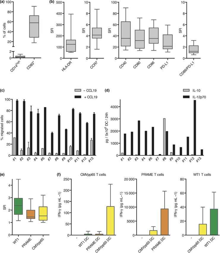
Feasibility of vaccine generation and Vaccine characterisation
administration
All 12 generated DC preparations were tested for
Twelve patients underwent leukapheresis for their phenotype, migration capacity, cytokine
production of the DC vaccine; patient #5 secretion, and processing and presentation of the
developed a leukaemia relapse in the short time three selected antigens after RNA electroporation
span between screening and planned (Figure 1 and Supplementary figure 1). For all
leukapheresis and was excluded from the study patients, the cells showed a typical DC phenotype
before leukapheresis. Key figures of the (CD14low and CD83+; Figure 1a). Expression of
leukapheresis product are presented in various costimulatory or chemokine receptor
Supplementary table 1. A median of 1.25 9 1010 molecules was measured, and the specific
10
(range 0.6–2.8 9 10 ) viable white blood cells fluorescence intensity (SFI) was calculated
was collected per patient. Median monocyte yield (Figure 1b). Median SFI was 124.6 for HLA-DR, 4.1
was 3.6 9 109 (range 1.0–7.5 9 109). Median DC for CCR7, 35.6 for CD40, 31.6 for CD80, 35.4 for
yield after electroporation was 3.65 9 108 (range CD86, and 21.5 for PD-L1. The ratio of CD86 to
1.27–5.68 9 108). After quality control and PD-L1 expression as a potential measure of
removal of retain samples, sufficient DCs for the positive costimulation was 1.25 in median. A
full schedule of 10 vaccinations (1.5 9 108 DCs) median of 74.5% (range 38.3–98.4%) of DCs
were produced for 11 of 12 patients. For patient showed migration towards a CCL19 gradient
#2, only six vaccinations were available as the (Figure 1c). Ten of 12 DC preparations secreted
monocyte yield was low because of an relatively high amounts of IL-12p70 (median of
ª 2020 The Authors. Clinical & Translational Immunology published by John Wiley & Sons Australia, Ltd on behalf of 2020 | Vol. 9 | e1117
Australian and New Zealand Society for Immunology Inc. Page 3
27Table 1. Patient characteristics
Page 4
Status of WT1 PRAME CMV Leukocytes
Age disease expr expr serostatus at
Gender (years) FAB Cytogenetics Molecular genetics ELN risk group at SV1 prim dx prim dx study start ECOG dx (G L!1) Tx prior DC vx
#1 m 72 M1 Complex karyotype NPM1 wt Adverse CR pos neg pos 1 2.6 s-HAM, TAD-9
2020 | Vol. 9 | e1117
CEBPA wt
MLL neg
#2 m 54 s-AML del(12)(p13p13)(ETV6-) NPM1 wt Intermediate II CRi pos pos neg 1 3.7 s-HAM, TAD-9
(MDS) FLT3-ITD neg
CEBPA wt
MLL neg
#3 m 62 M4 Normal karyotype NPM1 mut Intermediate I beginning pos pos neg 1 93.9 s-HAM, TAD-9,
FLT3-ITD+, FLT3-TKD+ relapse AD, Vidaza
MLL neg
TLR-matured dendritic cells for therapy of AML
#4 f 48 M0 Normal karyotype NPM1 wt Intermediate I beginning pos neg neg 1 0.9 s-HAM, TAD-9,
CEBPA wt relapse AD
MLL neg, FLT3-TKD-,
FLT3-ITD-, CEBPA wt
#5 f 44 M1 Normal karyotype NPM1 wt Intermediate I beginning r pos neg pos 1 1.6 s-HAM, TAD-9
CEBPA wt elapse
MLL neg
#6 m 65 M2 Normal karyotype NPM1 wt Intermediate II CR pos neg pos 1 13.9 7 + 3, HAM,
MLL-PTD+, FLT3-ITD, 2 9 HD-Ara-C
CEBPA wt
#7 f 74 M1 del(7q) NPM1 wt Intermediate II CR neg neg neg 1 1.2 s-HAM, TAD-9
FLT3 neg
#8 f 79 s-AML Normal karyotype n.a. Intermediate I CR pos neg neg 2 n.a. Vidaza
(MDS)
#9 m 64 s-AML Normal karyotype NPM1 wt Intermediate I CR pos neg neg 1 n.a. s-HAM, TAD-9
(MDS) MLL-PTD neg,
FLT3 neg, CEBPA wt
#10 m 50 M1 Complex karyotype NPM1wt, MLL-PTD neg, Favorable CR, MRD+ pos pos neg 1 75.1 AraC, sHAM,
with inv(16) inv16, FLT3-ITD+, TAD-9
FLT3-TKD+, CBFß-MYH11
fusion transcript
#11 m 69 M1 inv(16) NPM1 wt, FLT3-ITD neg, Favorable CRi, MRD+ pos pos neg 1 3.7 s-HAM, TAD-9
FLT3-TKD neg, MLL neg,
CBFß-MYH11 fusion
transcript, inv16
#12 m 55 M2 Normal karyotype NPM1 wt, FLT3-ITD neg, Intermediate I CR pos neg pos 0 2.8 s-HAM, TAD-9,
FLT3-TKD neg, MLL-PTD AD, AC
neg, CEBPA + mt
(Continued)
Australian and New Zealand Society for Immunology Inc.
ª 2020 The Authors. Clinical & Translational Immunology published by John Wiley & Sons Australia, Ltd on behalf of
FS Lichtenegger et al.
28
PublicationsPublications
FS Lichtenegger et al. TLR-matured dendritic cells for therapy of AML
1845 pg/5 9 106 DC/24 h; range 470–4525 pg/
classification; FLT3, fms-like tyrosine kinase 3; ITD, internal tandem duplication; MLL, mixed-lineage leukaemia; MRD, minimal residual disease; NPM1, nucleophosmin; s-HAM, double induction
AC, cytotoxic regimen consisting of cytarabine and cyclophosphamide; AD, cytotoxic regimen consisting of cytarabine and daunorubicin; CEBPA, CCAAT/enhancer-binding protein alpha; CR,
complete response; CRi, complete response with incomplete haematologic recovery; ECOG, Eastern Cooperative Oncology Group; ELN, European Leukemia Net; FAB, French–American–British
regimen consisting of sequential high-dose cytarabine and mitoxantrone; SV1, Screening Visit 1; TAD-9, cytotoxic regimen consisting of thioguanine, cytarabine and daunorubicin; TKD, tyrosine
s-HAM, 3 days
Tx prior DC vx
5 9 106 DC/24 h) and low amounts of IL-10
Fludarabin
(median of 17.3 pg/5 9 106 DC/24 h; range 0–
241 pg/5 9 106 DC/24 h), as expected from our
previous experiments.12 DCs of patient #7 showed
very low IL-12p70 production (81.5 pg/5 9 106
Leukocytes
dx (G L!1)
DC/24 h) and no IL-10 production. DCs of patient
1.6 #8 showed high IL-12p70 production (1969 pg/
at
5 9 106 DC/24 h), but even higher IL-10
ECOG
production (3031 pg/5 9 106 DC/24 h; Figure 1d).
0
Because of the unknown effects of vaccinations
with IL-10-producing DCs in the AML setting, this
study start
serostatus
patient was excluded from the study and not
CMV
neg
vaccinated, although all release criteria for the
vaccine were fulfilled. Successful translation of
prim dx
PRAME
the electroporated RNA was proven by
expr
neg
intracellular staining of the DCs for the resulting
proteins (median SFI 2.36 for WT1, 1.44 for
prim dx
PRAME, 1.53 for CMVpp65); DCs electroporated
WT1
expr
pos
with one of the other two RNA molecules served
as control (Figure 1e and Supplementary figure
Status of
2). Presentation of the antigens in the context of
disease
at SV1
HLA molecules was functionally proven by IFN-c
CR
secretion of specific T-cell clones after coculture
with the different DC batches. Each T-cell clone
ELN risk group
Intermediate I
was preferentially stimulated by the respective
DC batch (Figure 1f).
Vaccine-induced immune responses
MLL-PTD neg, CEBPA wt
NPM1 wt, FLT3-ITD neg,
For all 10 vaccinated patients, local immune
Molecular genetics
response was measured 48 h after the fifth
FLT3-TKD neg,
vaccination by size of local erythema and
induration (Figure 2a). Vaccine site reaction was
detectable for all patients and all antigens.
Variability between patients was high, but no
significant differences were found between the
three antigens (WT1: median of 1.43 cm2, range
0.38–4.15 cm2; PRAME: median of 1.04 cm2,
Normal karyotype
range 0.28–3.46 cm2; CMV: median of 1.24 cm2,
Cytogenetics
range 0.38–3.14 cm2; Figure 2b). Skin biopsies
were taken from nine patients. Dense CD4+ and
CD8+ T-cell infiltration was seen by
immunohistochemistry (Figure 2c).
Immunomonitoring was performed on PBMCs
FAB
M0
and plasma samples obtained before vaccination,
after five vaccinations and at the end of the
(years)
study. We found no major changes in the course
Table 1. Continued.
Age
47
of the therapy with respect to absolute and
kinase domain.
Gender
relative numbers of leucocytes, granulocytes,
monocytes, lymphocytes, CD3+ T cells, CD4+ T
m
cells, CD8+ T cells, CD19+ B cells or CD3-/CD16_56+
#13
NK cells (data not shown). Antigen-specific T-cell
ª 2020 The Authors. Clinical & Translational Immunology published by John Wiley & Sons Australia, Ltd on behalf of 2020 | Vol. 9 | e1117
Australian and New Zealand Society for Immunology Inc. Page 5
29Publications
TLR-matured dendritic cells for therapy of AML FS Lichtenegger et al.
Figure 1. Characterisation of DC phenotype, migration capacity, cytokine secretion and antigen processing and presentation. For all 12
generated DC preparations, surface expression of (a) the DC markers CD14 and CD83 and (b) various costimulatory or chemokine receptor
molecules was determined by flow cytometry. (c) Migration towards a CCL19 gradient was measured in a trans-well assay (2 technical replicates
per sample). (d) Secretion of IL-10 and IL-12p70 after CD40 ligation was analysed. To prove successful antigen translation and presentation after
RNA electroporation, DCs were (e) intracellularly stained for the resulting proteins and (f) used for stimulation of specific T-cell clones as
measured by IFN-c secretion (n = 3–7). For a, b and e, results are presented in box-and-whisker plots, with boxes representing the lower quartile,
the median and the upper quartile, while the whiskers show the minimal and the maximal values. For all other graphs, data shown reflect mean
and standard deviation.
2020 | Vol. 9 | e1117 ª 2020 The Authors. Clinical & Translational Immunology published by John Wiley & Sons Australia, Ltd on behalf of
Page 6 Australian and New Zealand Society for Immunology Inc.
30Publications
FS Lichtenegger et al. TLR-matured dendritic cells for therapy of AML
Figure 2. Vaccine site reaction. (a) For all 10 vaccinated patients and all antigens, erythema and induration of the vaccine sites were observed.
(b) There was high variability between patients, but no significant difference between the three antigens in size of local reaction. (c)
Immunohistochemical analysis of skin biopsies at the vaccine sites revealed dense CD4+ and CD8+ T-cell infiltration (one representative example
shown).
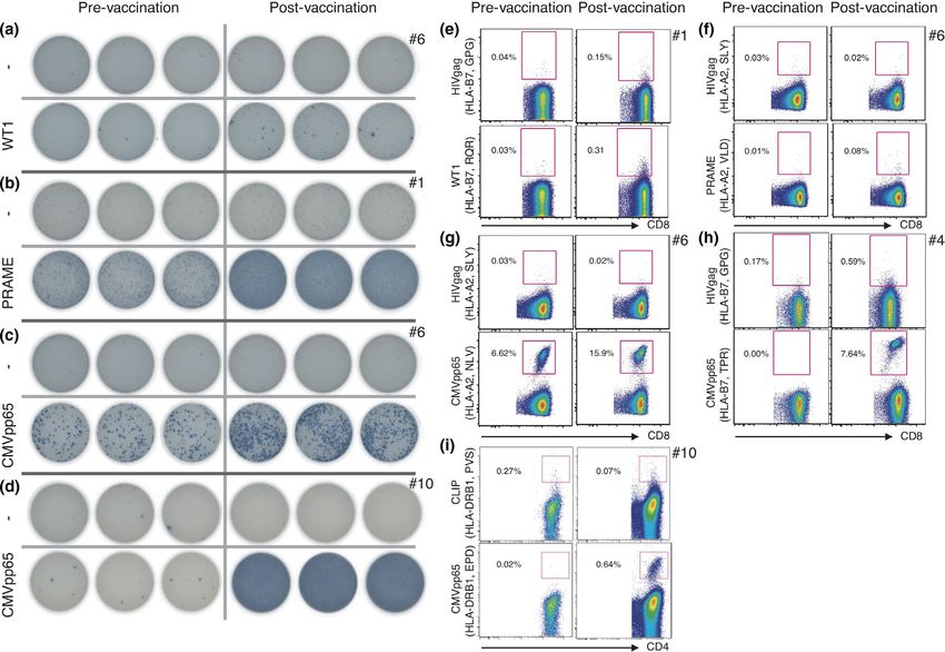
responses were measured by ELISpot and by 3). Post-vaccination LAA-specific T-cell responses
multimer staining, as shown for representative were significantly lower, but still clearly detectable
patients in Figure 3 (complete immunomonitoring in some patients (Figure 3e and f). In 4/7 patients
data of these patients is presented in where a CMV-specific multimer for HLA type II was
Supplementary figure 3). An increased ELISpot available, an increase in antigen-specific CD4+ T
response after vaccination as defined by a ≥ 1.5- cells could be detected as well (Figure 3i; Table 2).
fold increase of antigen-specific spot count was Vaccine-induced B-cell responses were measured
detected in 2/10 patients for WT1 (Figure 3a), in 4/ by detection of CMV antibodies. Of seven patients
10 patients for PRAME (Figure 3b), and in 9/10 who were CMV seronegative before vaccination,
patients for CMV (Figure 3c and d; Table 2). These antibodies against CMV were detected in three
results were largely reflected by multimer staining: patients after vaccination (#7, #10, #13), and one
an increased response as defined by a ≥ 2-fold patient had a borderline reaction after
increase of multimer-positive CD8+ T cells was vaccination (#2), while no antibodies against CMV
detected in 1/6 patients for WT1, in 0/3 patients were detectable in three patients (#4, #9, #11).
for PRAME, and in 6/8 patients for CMV, with Seroconversion as a result of primary CMV
limitations because of the availability of multimers infection was excluded by the methodology.
for the various HLA types (Table 2 and
Supplementary figure 4). CMV responses were
Clinical responses to vaccination
generally very high, with up to 15.9% of all CD8+
T cells stained with a single CMV multimer after The vaccination protocol was generally very well
vaccination in a primarily seropositive patient (#6; tolerated. All patients observed transient vaccine
Figure 3g), and up to 9.6% of all CD8+ T cells site reactions (erythema, induration, pruritus) of
stained with a single CMV multimer after grade 1 intensity. Other frequent adverse events
vaccination in a primarily seronegative patient were musculoskeletal pain (6/10), skin reactions
(#10). Of note, also decreased frequencies after outside of vaccine sites (5/10), diarrhoea (4/10)
vaccination were observed (Supplementary figure and fatigue (4/10). All potentially treatment-
ª 2020 The Authors. Clinical & Translational Immunology published by John Wiley & Sons Australia, Ltd on behalf of 2020 | Vol. 9 | e1117
Australian and New Zealand Society for Immunology Inc. Page 7
31You can also read


