Natural killer cells in cancer biology and therapy - Molecular ...
←
→
Page content transcription
If your browser does not render page correctly, please read the page content below
Wu et al. Molecular Cancer     (2020) 19:120
https://doi.org/10.1186/s12943-020-01238-x

 REVIEW                                                                                                                                            Open Access

Natural killer cells in cancer biology and
therapy
Song-Yang Wu1,2, Tong Fu1,2, Yi-Zhou Jiang1,2* and Zhi-Ming Shao1,2*

  Abstract
  The tumor microenvironment is highly complex, and immune escape is currently considered an important hallmark
  of cancer, largely contributing to tumor progression and metastasis. Named for their capability of killing target cells
  autonomously, natural killer (NK) cells serve as the main effector cells toward cancer in innate immunity and are
  highly heterogeneous in the microenvironment. Most current treatment options harnessing the tumor
  microenvironment focus on T cell-immunity, either by promoting activating signals or suppressing inhibitory ones.
  The limited success achieved by T cell immunotherapy highlights the importance of developing new-generation
  immunotherapeutics, for example utilizing previously ignored NK cells. Although tumors also evolve to resist NK
  cell-induced cytotoxicity, cytokine supplement, blockade of suppressive molecules and genetic engineering of NK
  cells may overcome such resistance with great promise in both solid and hematological malignancies. In this
  review, we summarized the fundamental characteristics and recent advances of NK cells within tumor
  immunometabolic microenvironment, and discussed potential application and limitations of emerging NK cell-
  based therapeutic strategies in the era of presicion medicine.
  Keywords: Innate immunity, Natural killer cell, Tumor progression, Immunometabolism, Immunotherapy, Precision
  treatment

Background                                                                              The immune system can be generally divided into the
The diversity of infiltrating stromal cells occurring in                              innate and adaptive immune systems, both contributing
human cancers exceeds 30 distinct subgroups, reflecting                               to the recognition and removal of foreign pathogens as
the huge complexity of the tumor microenvironment                                     well as tumors [2]. Adaptive immunity is mainly com-
(TME), thereby deeply affecting the treatment option for                              posed of cells represented by T and B lymphocytes,
each patient [1]. Attempts have been made to distill this                             which harbor an enormous repertoire of T-cell and B-
out-of-order situation into a unifying method to better                               cell receptors, respectively, that can respond specifically
describe actual composition of the TME using both                                     to different antigens in the body. Current immunothera-
multi-omics and experimental technologies, shedding                                   peutic methods mainly focus on T lymphocytes, espe-
light on cancer biology. This trend led to a transition in                            cially restoring exhausted CD8+ cytotoxic T cells
cancer treatment from only targeting tumor cells (like                                (CTLs). An example of such approach is immune-
traditional chemotherapy and radiotherapy) to a new                                   checkpoint blockade, with blocking of receptors or li-
generation of approaches emphasizing the modulation of                                gands that inhibit the activation of CTLs, including pro-
endogenous immune response toward cancer.                                             grammed cell death protein 1 (PD-1), its main ligand
                                                                                      PD-L1, cytotoxic T-lymphocyte antigen 4 (CTLA-4) and
                                                                                      lymphocyte-activation gene-3 (LAG-3), by monoclonal
* Correspondence: yizhoujiang@fudan.edu.cn; zhimingshao@yahoo.com
1
 Department of Breast Surgery, Fudan University Shanghai Cancer Center,               neutralizing antibodies [3, 4]. In recent years, the rapid
Shanghai 200032, China                                                                and potent anti-tumor function of innate immunity,
Full list of author information is available at the end of the article

                                       © The Author(s). 2020 Open Access This article is licensed under a Creative Commons Attribution 4.0 International License,
                                       which permits use, sharing, adaptation, distribution and reproduction in any medium or format, as long as you give
                                       appropriate credit to the original author(s) and the source, provide a link to the Creative Commons licence, and indicate if
                                       changes were made. The images or other third party material in this article are included in the article's Creative Commons
                                       licence, unless indicated otherwise in a credit line to the material. If material is not included in the article's Creative Commons
                                       licence and your intended use is not permitted by statutory regulation or exceeds the permitted use, you will need to obtain
                                       permission directly from the copyright holder. To view a copy of this licence, visit http://creativecommons.org/licenses/by/4.0/.
                                       The Creative Commons Public Domain Dedication waiver (http://creativecommons.org/publicdomain/zero/1.0/) applies to the
                                       data made available in this article, unless otherwise stated in a credit line to the data.
Wu et al. Molecular Cancer (2020) 19:120 Page 2 of 26
which even occurs at a very early stage of tumor pro- also play important roles in the body, e.g., eliminating
gression, has attracted increasing attention. As a subset murine cytomegalovirus (MCMV) infection and
of whole innate lymphoid cells, natural killer (NK) cells, MHC-I+ cells [15].
defined by Herberman in 1976 [5] and often considered To date, NK cell survival and development is thought
a part of type 1 innate-like cells (ILC1s), are currently to mainly rely on cytokines (especially IL-2 and IL-15)
defined as effector cells similar to CTLs, exerting natural [16–19] and transcription factors (Nfil3, Id2 and Tox for
cytotoxicity against primary tumor cells and metastasis development, and EOMES and T-bet for maturation)
by inhibiting proliferation, migration and colonization to [16, 20]. GRB2-associated binding protein 3 (GAB3) is
distant tissues [6]. Beside their cytotoxic role, NK cells essential for IL-2 and IL-15-mediated, and its deficiency
have been reported to produce a large number of cyto- leads to impaired NK cell expansion [21]. In addition,
kines, mainly interferon-γ (IFN-γ), to modulate adaptive targeting related signals is a potential option for promot-
immune responses and participate in other related path- ing NK cell-induced cytotoxicity toward cancer. As re-
ways [7, 8]. In addition, as documented in multiple ported previously, ablation of cytokine-inducible SH2-
models and experiments, NK cells could distinguish ab- containg protein (CIS), which negatively regulates IL-15
normal cells from healthy ones, leading to more specific to restrict NK cell function, could prevent metastasis
anti-tumor cytotoxicity and reduced off-target complica- and also potentiate CTLA-4 and PD-1 blockade therapy
tions [9, 10]. in vivo [22].
Considering the pivotal role of NK cells in cancer biol-
ogy, they naturally emerged as a prospective target for Identification and molecular features of NK cells
cancer therapy, and a growing number of studies and mul- Surface molecules of NK cells
tiple therapeutic agents inhibiting cancer target NK cell- Due to variable expression of surface markers on NK
related pathways. In this review, we will review the funda- cells, it is hard to use one or two simple molecules or
mental characteristics and emerging subpopulations of traditional immunohistochemistry to accurately identify
NK cells. Next, we will mainly use breast cancer (BC) to this cell type and more importantly, their functional sta-
discuss the plasticity of NK cells in cancer biology and tus. However, in humans, in both clinical and research
metabolism, as well as current therapeutic regimens, in- settings, CD3−CD56+ cells are commonly identified as
cluding ongoing clinical trials and FDA-approved therap- NK cells and can be further divided into the CD56bright
ies targeting NK cells, and future possible approaches for and CD56dim subgroups. CD56 is not only a marker but
improving cancer treatment. also plays an important role in the terminal differenti-
ation of NK cells since its blockade by monoclonal anti-
Development of NK cells bodies obviously inhibits the transition from CD56bright
NK cells possess cytotoxic abilities similar to CD8+ T to CD56dim, thus limiting the cytotoxic ability [23]. Con-
cells functioning in the adaptive immunity but lack sistently, CD3−NK1.1+ and CD3−CD49b+ cells are de-
CD3 and the T cell receptors (TCRs). Largely circu- fined as NK cells in mice. In recent studies, the notion
lating in blood and counting for about 5–10% of per- that natural cytotoxicity receptor 46 (NKp46), belonging
ipheral blood mononuclear cells (PBMCs), NK cells to natural cytotoxicity receptors (NCRs), should also be
are found in bone marrow and lymphoid tissues such included in this panel has been proposed based on the
as the spleen [11, 12]. Similar to other ILCs, NK cells consensus of adding more functional proteins rather
are originated from common lymphoid progenitor than surface molecules into the classification system of
(CLP) cells in bone marrow (Fig. 1) with an average NK cells [24, 25].
renewal cycle of about 2 weeks [12]. During develop-
ment, a process termed education, which describes Activating and inhibitory signals in NK cells
the interaction of NK cells expressing immunorecep- As the main effector cell type in innate immunity, NK
tor tyrosine-based inhibitory motifs (ITIMs) with cells are capable of killing tumor cells and virus-infected
major histocompatibility complex-I (MHC-I), helps cells at a very early stage. Due to the lack of abundant
NK cells become licensed and avoid attacking healthy production of receptors for distinguishing incalculable
normal cells [6, 9]. Interestingly, tumor cells always antigens in the body specifically, they rely on the “miss-
lack or only express low levels of MHC-I to evade ing self” and “induced self” modes to identify target cells
CD8+ T cell-mediated cytotoxicity, whereas licensed by maintaining a precise balance between activating co-
NK cells are fully activated. However, tumor cells also stimulatory and inhibitory signals (mainly by functional
express molecules that activate NK cells, e.g., MHC receptors). Those interacting signals finally decide the
class I polypeptide-related sequence A (MICA) and activation and functional status of NK cells.
MICB [13, 14], supporting the use of NK cells as Activating signals include cytokine-binding receptors,
anti-cancer agents. In addition, unlicensed NK cells integrins, killing-receptors (CD16, NKp40, NKp30 andWu et al. Molecular Cancer (2020) 19:120 Page 3 of 26 Fig. 1 Development and subgroups of NK cells. In bone marrow, NK cells develop from hematopoietic stem cells (HSCs) through common lymphoid progenitors (CLPs) and NK cell precursors (NKPs), and then migrate to peripheral blood (cNK cells) or tissue (trNK cells). The differentiation of trNK-cells occurs in distinct tissue sites, including the lung, thymus, liver, uterus, skin, subcutaneous adipose tissue, and kidney. In these sites, NK cells have different phenotypic features and functions, which constitute the circulation of NK cells at different stages of maturation. CLA, cutaneous lymphocyte-associated antigen; CCR8, C-C motif chemokine receptor 8; GATA3, GATA binding protein 3; CXCR6, C-X- C motif chemokine receptor 6; KIR, killer cell immunoglobulin-like receptor; CILCP, common innate-like cell precursor
Wu et al. Molecular Cancer           (2020) 19:120                                                                                                        Page 4 of 26

NKp44), receptors recognizing non-self-antigens (Ly49H)                                the respective ligands, including MHC-I specific, MHC-I
and other receptors (e.g., NKp80, SLAMs, CD18, CD2 and                                 related and MHC-I non-related receptors (Table 1) [13,
TLR3/9) [26, 27]. In total, the activating receptors of NK                             28–43]. To emphasize, NCRs, which belong to the third
cells can be divided into at least three types according to                            group, include three molecules (NKp30, NKp44 and

Table 1 Key mediators of NK cells
Classification                              Mediator                               Host ligand                                                                   Ref.
NK cell activator
  MHC-I specific receptor                   KIR2DS1, 2DS3, 3DS5                    MHC I                                                                         [28]
                                            Ly49c, Ly49i                           MHC I                                                                         [29]
                                            NKp80                                  MHC I                                                                         [30]
                                            NKG2C, NKG2E                           MHC I                                                                         [31]
  MHC-I related receptor                    NKG2D                                  MICA, MICB, ULBPs                                                             [13]
  MHC-I non-related receptor                DNAM1                                  Nectin-2, CD155                                                               [32]
                                            NKp46 (NCR1)                           HS GAGs, CFP                                                                  [33]
                                            NKp44 (NCR2)                           HS GAGs, MLL5, NKp44L, PCNA, BAT3, PDGF-DD, Nidogen-1                         [34]
                                            NKp30 (NCR3)                           HS GAGs, B7-H6, Galectin-3                                                    [35]
                                            Nkp65                                  Keratinocyte-associated C-type lectin                                         [36]
                                            LFA-1 (αLβ2 integrin)                  Intercellular cell adhesion molecule 1                                        [37, 38]
                                            α4 integrin                            Vascular cell adhesion molecule 1                                             [39]
                                            CD16                                   Fc-γ                                                                          [40, 41]
                                            CD2                                    CD581                                                                         [41]
                                            TLR3                                   Microbial constituents, adjuvant                                              [42]
                                            TLR9                                   CpG                                                                           [43]
NK cell inhibitor
  MHC-I specific receptor                   KIR3DL1                                MHC I                                                                         [44]
                                            KIR2DL3, 2DL1                          MHC I                                                                         [45]
                                            NKG2A                                  MHC I                                                                         [46]
                                            KLRB1, LLT1                            MHC I                                                                         [47]
                                            LILRB1, LILRB2                         MHC I                                                                         [48]
  MHC-I non-related receptor                KLRG1                                  E-, N-, and R- cadherins                                                      [49]
                                            siglec-3, siglec-7, siglec-9           Sialic acid                                                                   [50, 51]
                                            CEACAM1                                CEACAM1, CEACAM5                                                              [52]
                                            2B4 (CD244)                            CD48                                                                          [53]
                                            IRp60                                  Phosphatidylserine                                                            [54]
                                            LAIR1                                  Ep-CAM                                                                        [55]
                                            CD96                                   CD155                                                                         [56]
                                            CD73                                   Antibodies                                                                    [57]
                                            PD-1                                   PD-L1                                                                         [58]
                                            TIGIT                                  CD155                                                                         [20]
                                            NKR-P1B                                Clr-b                                                                         [59]
                                            LAG3                                   MHC-II                                                                        [60]
Abbreviations: MHC major histocompatibility complex, KIR killer cell immunoglobulin-like receptor, MIC MHC class I chain-related, ULBP UL16-binding protein 1,
DNAM1 DNAX accessory molecule 1, NCR natural cytotoxicity receptor, HS GAGs heparan sulfate glycosaminoglycans, CFP complement factor P, MLL5 mixed-
lineage leukemia protein-5, PCNA proliferating cell nuclear antigen, BAT3 HLA-B-associated transcript 3, PDGF-DD platelet-derived growth factor-DD, LFA-1
lymphocyte function-associated antigen-1, TLR toll-like receptor, KLR killer cell lectin-like receptor, LLT1 lectin-like transcript 1, LILR leukocyte immunoglobulin-like
receptor, Siglec sialic acid-binding immunoglobulin-like lectin, CEACAM carcinoembryonic antigen-related cell-adhesion molecule, IRp60 inhibitory receptor protein
60, LAIR1 leukocyte-associated immunoglobulin-like receptor 1, Ep-CAM epithelial cellular adhesion molecule, PD-1 programmed cell death protein 1, TIGIT T-cell
immunoreceptor with Ig and ITIM domains, LAG3 lymphocyte activation gene 3
Wu et al. Molecular Cancer (2020) 19:120 Page 5 of 26
NKp46), and NKp30 was shown to be capable of recogniz- CD56dimCD16+, both lacking CD3 and CD19 [8]. In
ing B7-H6 expressed on tumor cells, and could be used as addition, CD49a+CD56+CD3−CD19− NK cells have been
a novel treatment option in the future [35]. identified in liver biopsies [70]. Besides, hepatic NK cells
Inhibitory signals mainly comprise receptors recogniz- can develop memory for structurally diverse antigens,
ing MHC-I, such as Ly49s, NKG2A and LLT1, as well as dependent on the surface molecule CXCR6 [71]. In the
some MHC-I non-related receptors (Table 1) [20, 44– uterus, most NK cells are CD56brightCD16−, expressing
60]. Moreover, MHC-I specific inhibitory receptors can high levels of KIRs [72]. Decidual NK cells are also
be generally divided into three types according to struc- CD49a+. For skin NK cells, it is intriguing that only few
ture and function: killer cell immunoglobulin-like recep- CD56+CD16+ can be detected, which are common in
tors (KIRs), killer lectin-like receptors (KLRs) and peripheral blood [73]. Interestingly, trNK cells are dis-
leukocyte immunoglobulin-like receptors (LILRs). tinct between subcutaneous (CD56dim) and visceral
(CD56bright) adipose tissues, and can be generally divided
NK cell subpopulations according to site of maturation into three groups according to CD49b and Eomes, show-
Conventional NK (cNK) cells are mainly found in per- ing different expression levels of CD49a (CD49b+Eomes−
ipheral blood and migrate to a specific location to exert subgroup) and CD69 (CD49b−Eomes+ subgroup) [74,
their effects. NK cells also include tissue-resident NK 75].
(trNK) cells. The complex process of NK-cell differenti- Besides different tissue types, NK cells are also highly
ation occurs in several distinct tissues, including bone heterogeneous even in the same organ and the same tis-
marrow, liver, thymus, spleen and lymph nodes, and sue [61]. Through high-dimensional single-cell RNA-
may involve cell circulation at different stages of matur- seq, Crinier et al. revealed the heterogeneity of human
ation among these tissues [61]. In bone marrow, blood, and mouse NK cells in spleen and blood and identified
spleen and lungs, NK cells are fully differentiated, while several subpopulations of NK cells, respectively [76]. As
that in lymph nodes and intestines are immature and mentioned above, NK cells are considered a subgroup of
precursory [62]. Single-cell transcriptome ananlysis of ILC1s [6]. Although ILC1s are not detectable in many
bone marrow and blood NK cells helps to illustrate the tissues, intra-epithelial ILC1 (ieILC1)-like cells, which
changes of their characteristics during development. For highly express IFN-γ, integrins and other cytotoxic mol-
example, high expreesion of TIM-3, CX3CR1 and ZEB2 ecules similar to the ieILC1s previously described by
represents a more mature status [63]. Many hypotheses Fuchs except for different NKp44 expression, could rep-
have been proposed to describe the motivation of their resent the majority of NK cells in the mucosal tissue [61,
migration and different biological behaviors of identi- 77]. Due to their unique features, this cell type repre-
cally originated NK cells in different tissues. The first sents a subgroup of NK cells other than conventional
question could be partly explained by the multi- ILCs. Unlike other trNK cells, DX5−CD11chi liver-
direction differentiation induced by heterogeneous mi- resident NK cells participate in autoimmune cholangitis,
croenvironments in different tissues, or more straight- negatively regulating immune responses, especially by
forward, different phenotypes originated from similar inhibiting the proliferation and function of CD4+ T cells
chemokine-recruited peripheral cNK cells. in vivo, which was validated by severer biliary disease in
To conclude, NK cells in various tissues have diverse NK-depleted mice resulting from Nfil3 knockdown or
features, possessing different functions and forming a treatment with neutralizing antibodies [78].
close relationship with other stromal cells (Fig. 1). In the
lung, trNK cells show a different phenotype from that of NK cell subpopulations according to functional molecules
circulating NK cells (mainly CD56dim) and are consid- According to surface CD56 expression, NK cells can be
ered to express different levels of CD16, CD49a and divided into CD56bright and CD56dim. CD56dim NK cells
CD69, with CD56dimCD16+ cells representing the major- are mainly found in peripheral blood, and are always
ity of the whole NK family there [64, 65]. Of note, also CD16-positive, expressing high levels of KIR and
CD69+ cells are the main type of CD56brightCD16− NK LFA-1 and showing cell killing ability. CD16 is a key re-
cells. However, in the thymus, most NK cells are ceptor mediating antibody-dependent cell cytotoxicity
CD56highCD16−CD127+, highly relying on GATA3 com- (ADCC), inducing the phosphorylation of immunorecep-
pared with the CD56+CD16+ subgroup [66]. Besides, tor tyrosine-based activation motif (ITAM) [40, 79, 80].
they produce more effector molecules, including TNF-α According to a time-resolved single-cell assay, the cyto-
and IFN-γ [66, 67]. Similar to the phenotypic features in toxicity of NK cells is repressed through both necrosis
humans, skin NK cells in the mouse can be generally di- and apoptosis. As a result, FasL/FasR interaction, per-
vided into two types: CD49a+DX5− and CD49a−DX5+ forin/granzyme release and Ca2+ influx are all important
[68, 69]. Similarly, hepatic trNK cells can be classified for NK cell function [81]. However, CD56bright NK cells
into two groups, including CD56brightCD16+/− and are similar to helper cells, which mainly secreteWu et al. Molecular Cancer (2020) 19:120 Page 6 of 26
cytokines such as IFN-γ, TNF-β and GM-CSF [23]. Re- form immunological memory, termed “trained immun-
searchers even further subgroup these cells into the NK1 ity”. Once considered as a hallmark of adaptive immun-
and NK2 categories, consistent with Th1 and Th2, ity, in recent years, the phenomenon of immunological
mainly secreting IFN-γ and IL-5, respectively [82]. memory has also been found in innate immune cells, es-
Besides established cytotoxic cNK cells, it has been pecially in the myeloid lineage, e.g., monocytes and mac-
demonstrated that NK cells could differentiate into rophages. In addition, mounting evidence indicates that
antigen-presenting NK (AP-NK) cells [83], helper NK in humans, NK cells can remember previous exposure to
(NKh) cells [84] and regulatory NK (NKreg) cells, each inflammatory microenvironment, and occurrence of
defined by surface molecules and individual functions. A similar cytokines could induce trans-differentiation from
new CD8αα+MHC-II+ phenotype with professional APC normal NK cells to memory NK (NKm) cells [98–101].
capacity was considered to represent unusual AP-NK This was evidently observed in response to viral infec-
cells, recognizing and eliminating autoreactive T cells tion in humans, prompting the development of NK cell-
and finally killing them like cNK cells [85]. Human plas- based vaccines to generate potent effects toward diseases
macytoid dendritic cells (DCs) activated by the prevent- [102, 103]. A large number of NKm cells are observed in
ive vaccine FSME upregulate CD56 expression on their the tumor microenvironment, producing high levels of
surface [86]; in mice, B220+CD11cintNK1.1+ cells have IFN-γ, perforin and granzyme family molecules after re-
antigen-presenting capacity like DC, hence their name stimulation [104]. However, concerning to tumors, dys-
interferon-producing killer DC [87, 88]. function of NK and NKm cells is emerging as an indis-
Invariant natural killer T cells (iNKT) constitute a sub- pensable and undeniable event, leading to not only
group of T cells expressing NK cell-markers. Activated proliferation of tumor cells but also the formation of dis-
by CD1d-presenting antigens, NKT could secrete not tant metastases [101]. It was observed that repeated ex-
only Th1-type but also Th2-type cytokines to participate posure of NK cells to NK receptor ligand-expressing
in immunity [89, 90]. Th1-polarized iNKT cells exhibit a tumor cells (e.g. NKG2D) finally results in NK cell dys-
tumor-depletion phenotype, and Th2-polarized iNKT function, and effector responses cannot be stimulated
cells contribute to tumor progression, similar to polar- in vivo [105, 106]. These results indicate that the forma-
ized T cells [91, 92]. Recent studies also highlighted new tion of NKm cells may not just depend on target cell
functional subtypes of iNKT cells. However, in recent recognition through surface receptors, and certain cyto-
years, due to their close relationship with innate immun- kines (including IL-12, IL-15 and IL-18) could be key to
ity, iNKT cells are potentially defined as a special sub- this process.
group of ILCs. Though the half-life of normal NK cells is only for 1–
2 weeks, NKm cells can live for 3–4 weeks [107]. This
NK cells in the tumor microenvironment long-term effect truly helps researchers better modulate
Conventional roles of NK cells in immunity the function of NK cells in protecting against tumors,
Detection of aberrant cells by NK cells is determined by and emerging results suggest that NK cells not only rely
the intergradation of complex signals such as IL-12, IL- on MHC-I recognition but also depend on many other
15 and IL-18 [93, 94], and the balance between activat- signals, shedding light on the use of NK cells and related
ing and inhibitory signals interacting with MHC-I on the signaling pathways as future treatment options.
surface of target cells (Fig. 2). During infection and in-
flammation, NK cells are recruited and activated in a Infiltration of NK cells with cancer genotypes and
short period of time, proliferate quickly and contribute phenotypes
largely to both innate and adaptive immune responses In 2000, an 11-year follow-up study of the Japanese general
[8, 95]. Except for their newly proven regulatory effects, population, with rigorous use of various related biochemical
NK cells were first found to directly target infected cells and immunological markers, indicated that elevated cyto-
or foreign pathogens; therefore, deficiency in NK cells in toxic activity of peripheral NK cells is positively associated
both mice and humans results in susceptibility to many with reduced cancer risk and vice versa, suggesting the cer-
viral infections and adverse clinical outcomes, validated tain importance of natural immune response toward tu-
by clinicians and researchers. mors [108]. However, the specific role of NK cells remains
Similar to other innate immune cells that are unable controversial and largely depends on distinct cancer types
to accurately recognize target cells, NK cells rely on [109]. Even in the same type of cancer, NK cells are highly
other stromal cells, including DC, which trans-presents heterogeneous, characterized by the abundance of surface
IL-15 for NK cell activation [96], and MICA-expressing receptors and the complexity of tumor intrinsic signaling
monocytes, which bind to Fc receptor to enhance antitu- pathways [95, 110]. In the CIBERSORT analysis, NK cells
mor function [97], to fully differentiate and induce ef- were thoroughly divided into resting and activated sub-
fector responses, but surprisingly possess the ability to types, each contributing to the formation of the tumorWu et al. Molecular Cancer (2020) 19:120 Page 7 of 26 Fig. 2 The complex interaction between NK cells and the extracellular matrix. Exposure of NK cells to the adjacent cells, molecules and metabolites in the extracellular matrix affects their development, maturation, activation and functions. CXCR3, C-X-C motif chemokine receptor 3; NKG2D, nature-killer group 2, member D; IFN-γ, interferon γ; TNF-α, tumor necrosis factor α; IDO, indoleamine 2,3-dioxygenase; MICA, MHC class I polypeptide-related sequence A; PGE2, prostaglandin E2; HCC, hepatocellular carcinoma; CIS, cytokine-inducible SH2-containg protein; TGF-β, transforming growth factor-β; HMGB1, high-mobility group box 1; HIF-1α, hypoxia inducible factor-1α; 27HC, 27-hydroxycholesterol; iNKT, invariant natural killer T; GM2, β-N-acetylhexosaminidase; TCR, T cell receptor microenvironment [111]. In preclinical studies, NK cells using different surface and functional markers (Table 2) were shown to indicate survival and thus therapeutic re- [46, 56, 112–129]. Although CD3−CD16+CD56+ cells re- sponse in different types of cancer, as detected by immu- flect different clinical outcomes in different cancers, func- nohistochemistry, immunofluorescence or flow cytometry tional molecule-positive NK cells, including NKp30+ and
Wu et al. Molecular Cancer         (2020) 19:120                                                                                                                  Page 8 of 26

Table 2 Infiltration of NK cells in different cancer types and its influence on clinical outcome
Cancer type                       Sample          Detection method Marker                                                                  Clinical outcome              Ref.
Pancreatic cancer                 Blood           Flow cytometry        CD3−CD16+CD56+                                                     Adverse OS                    [112]
Colorectal cancer                 Tumor           IHC                   CD57+                                                              Favorable OF and DFS          [113]
                                                                                +
                                  Lymph node IHC                        CD56                                                               Favorable RFS                 [114]
                                  Blood           Flow cytometry        CD3−CD16+CD56+                                                     Favorable OS                  [115]
                                                                            −         +           dim
Chronic myeloid leukemia          Blood           Flow cytometry        CD3 CD16 CD56                                                      Favorable molecular RFS after [116]
                                                                                                                                           imatinib discontinuation
Chronic lymphocytic leukemia Blood                Flow cytometry        CD3−CD16+ and/or CD56+                                             Favorable OS                  [117]
Follicular lymphoma               Blood           Flow cytometry        CD3−CD56+ and/or CD16+                                             Favorable OS                  [118]
                                                                            −         +                               +
Mantle cell lymphoma              Blood           Flow cytometry        CD3 CD16 and/or CD56                                               Adverse OS and PFS            [119]
Liver cancer                      Tumor           IF                    CD56+PD1+                                                          Adverse survival              [120]
                                  Tumor           IHC                   NKG2A+                                                             Adverse OS and DFS            [46]
                                  Tumor           Flow cytometry        CD3−CD56+CD49a+                                                    Adverse OS and DFS            [121]
                                                                            −         +           +
                                  Tumor           Flow cytometry        CD3 CD56 CD96                                                      Adverse DFS                   [56]
Prostate cancer                   Blood           Flow cytometry        CD3−CD56+ NKp30+ or NKp46+                                         Favorable OS                  [122]
                                                                                dim           +               +
Lung cancer                       Blood           Flow cytometry        CD56        CD16 NKp46                                             Favorable OS                  [123]
                                  Blood           qRT-PCR               NKp30                                                              Adverse OS and PFS            [124]
                                                                                +                         +
                                  Tumor           IF                    CD56 and/or CD16                                                   Favorable OS                  [125]
Breast cancer                     Tumor           IHC                   CD3−CD56+                                                          Favorable DFS                 [126]
                                                                                +
                                  Tumor           IHC                   CD56                                                               Adverse OS                    [127]
Gastric cancer                    Tumor           IHC                   NKG2D+                                                             Favorable OS                  [128]
                                                                                +         −           −           −       −   −   bright
Bladder cancer                    Tumor           Flow cytometry        CD45 CD14 CD19 CD3 ILT3 cKIT CD56                                  Favorable OS and CSS          [129]
Abbreviations: IHC immunohistochemistry, IF immunofluorescence, qRT-PCR quantitative real time polymerase chain reaction, OS overall survival, RFS recurrence-
free survival, PFS progression-free survival, DFS disease-free survival, CSS cancer-specific survival

NKp46+, indicate favorable survival, pointing out the fact                            cells, as a newly-found subgroup of NK cells, activated
that full activation but not only infiltration density finally                        NKT cells directly detect and kill CD1d+ tumor cells in
determines NK cell-associated immune response.                                        several types of cancer [132, 133]. In addition, by ex-
  In BC, besides the ability of total NK cells to reflect fa-                         pressing high levels of CD40, NKT cells induce DC mat-
vorable survival, peritumoral abundance of NK cells also                              uration, thus activating CTL and cNK cells to enhance
correlates with elevated pCR rate of neoadjuvant chemo-                               their anti-tumor effects [134, 135].
therapy in large and locally advanced breast cancer                                     Apart from primary tumor proliferation, metastasis re-
[130], and vice versa. To address this, it is currently well                          mains lethal, accounting for the majority of cancer-
accepted that tumors are highly heterogeneous, and even                               associated deaths, and invasion-metastasis cascade is
one histologic type can be separated into several mo-                                 largely attributable to the immune escape [136]. With
lecular subtypes. BC can be thoroughly clustered into lu-                             such rapid and effective ability to target tumor cells dir-
minal, HER2-enriched and triple-negative types                                        ectly and indirectly, NK cells are suppressed by tumor-
according to the expression of surface molecules, and                                 derived molecules, tumor-educated stromal cells (Fig. 2)
both infiltration and activation status of NK cells vary by                           and tumor cells (Fig. 3), eventually contributing to the
cluster, e.g., obviously elevated NKG2D in luminal tu-                                progression and multi-step metastatic process of cancer.
mors [131]. However, due to the complex and variable                                  For example, single-cell analyses found that in lung
functional status of NK cells, their actual role in the                               adenocarcinoma, CD16+ NK cells are hardly infiltrated
TME still awaits further elucidation.                                                 and present lower granzyme B and CD57 expression
                                                                                      compared with normal lung tissue, appearing to form
Underlying mechanisms of immune escape and metastasis                                 NK cell-excluded TME [137].
of cancer                                                                               Class IA phosphatidylinositol 3 kinases (PI3Ks) are in-
As noted above, NK cells rely on the balance between                                  volved in growth and survival of normal cells, and muta-
activating and inhibitory receptors to exert their killing                            tion of the PI3KCA isoform is commonly found in the
effects, and perforin and the granzyme family of proteins                             genomic landscape of many cancers. Inhibiting abnor-
are the main effector molecules. Consistent with cNK                                  mally activating signals of PI3KCB with a tested inhibitor
Wu et al. Molecular Cancer (2020) 19:120 Page 9 of 26 Fig. 3 Interplay between cancer cells and NK cells during tumorigenesis. The interaction between tumor cells and NK cells changes continuously with NK cell development, tumor progression and metastasis. During the stage of tumorigenesis (a), NK cells recognize tumor cells through various surface molecules and switch to the active status. In the immune control stage (b), NK cells exert killing effects by ADCC, secreting cytokines and generating memory NK cells. Meanwhile, changes in the surface molecules of tumor cells also promote anti-tumor metabolic responses. However, long-term exposure of NK cells to tumor cells, tumor- derived molecules and tumor-educated stromal cells, including fibroblast, monocyte and macrophage, causes NK cells to be in an immunosuppressive state, thereby promoting tumor immune escape and metastasis (c). MHC- I, major histocompatibility complex-I; MICA, MHC class I polypeptide-related sequence A; MICB, MHC class I polypeptide- related sequence B; NCR, natural cytotoxicity receptor; Nfil3, nuclear factor interleukin-3-regulated protein; Id2, inhibitor of DNA binding 2; Tox, thymocyte selection associated high mobility group box; EOMES, eomesodermin; T-bet, T-box transcription factor 21; ADCC, antibody-dependent cell-mediated cytotoxicity; GM-CSF, granulocyte-macrophage colony stimulating factor; PRF1, perforin 1; GZMB, granzyme B; PD-L1, programmed cell death ligand 1; PGE-2, prostaglandin E2; HCC, Hepatocellular Carcinoma; IFN, interfron; TNFα, Tumor Necrosis Factor α;PI3K, Class IA phosphatidylinositol 3 kinase
Wu et al. Molecular Cancer   (2020) 19:120                                                                     Page 10 of 26

called P110β in hematologic malignancies obviously en-          membrane tetraspan molecule MS4A4A [151]. In colo-
hances susceptibility of tumors to NK cell activity in vitro,   rectal cancer, lipid accumulation is common for postop-
probably through the regulation of MHC-I [138]. Tumor-          erative patients and facilitates the formation of
derived prostaglandin E2 [139], extracellular adenosine         metastasis by impairing NK cell function, by elevating
[140], fragmented mitochondria in the cytoplasm of              CD36 expression [152]. Hence, NK cells can be rarely
tumor-infiltrating NK cells [141] and actin cytoskeleton        seen in metastatic melanoma and are mainly TIGI
remodeling [142] also lead to immunosuppression and             T−CD226− which are deprived of cytotoxicity toward
help potentially metastatic cancer cells to avoid NK cell       MHC-I-defient malignant cells [153]. Interestingly,
elimination. Interestingly, in a breast cancer cell line,       proved by convincing genetically engineered models, it is
CDC42 or WASP knockdown does not change the activa-             the absence of NK cells but not CD8+ T cells that evi-
tion status of NK cells but obviously increases the expres-     dently leads to the metastatic dissemination of small cell
sion of effective granzyme B and overcomes resistance to        lung cancer, pushing us to better define the pivotal role
NK cell-mediated attack [142]. Further investigation in         of NK cells in both initial progression and later metasta-
this field might help identify a new signaling pathway or a     sis of cancer [154].
new marker of NK cell-activation and provide new in-
sights into NK cell-therapy. Similar to the inhibitory role     Relationship between NK cell-based and CD8+ T cell-based
of CD73 in T cell-immunity, tumors can train normal NK          immunity
cells into CD73+ NK cells which express high levels of          NK cells, though belonging to innate immunity, have
checkpoint molecules, including LAG-3, PD-1, and PD-            characteristics similar to cytotoxic CD8+ T cells [95].
L1, finally resulting in immune escape [57]. As mentioned       Originally proven important in the two-signal activation
above, Th2-polarized iNKT cells in the TME contribute to        model of T cells, CD28 is also necessary for optimal
tumor progression through immunosuppressive effects             cytokine secretion and proliferation of NK cells both
[91], and continuous exposure to ligands expressed on the       in vitro and in vivo [155]. It was shown that IL-21 is im-
surface of tumor cells induces the dysfunction of NKm           portant for the maturation and activation of NK cells
cells that are engaged in long-term anti-tumor immunity         [156–158]. However, data also uncovered the double-
[106]. Moreover, absence of NKG2D is a common feature           sided function of IL-21 in the development of NK cells
of functionally suppressed NK cells, which is achieved          and surprisingly, its positive role in T-cell-based immune
through many different pathways [105, 143], thus could be       response [159, 160], for example inducing KLRG1+CD8+
used as a marker to guide NK cell-related therapies [144].      T cells during acute intracellular infection [161]. Besides
   In addition, NK cell-associated cytotoxicity can also be     IL-21, NK cells produce multiple other cytokines during
impaired by stromal cells, for example cancer-associated        their proliferation, maturation and function similar to T
fibroblasts, monocytes, macrophages and other immune            cells, including IL-2 [162], IL-7 [163] and IL-15 [164],
cells. Fibroblasts in TME suppress function of NK cells         validating the close relationship between these two types
through downregulating ligands of NK cell activating re-        of effector cells in the body’s immune system.
ceptors [145], enhancing tumor-associated macrophages              Although sharing many similarities, compared with ef-
enrichment [146], and producing extracelluar matrix             fector T cells, NK cells are more cytotoxic to tumors
components such as IDO and PGE2 [147]. In human                 and possess lower immunogenicity [10]. As mentioned
gastric cancer, tumor infiltrating monoctyes/macro-             above, NK cells respond to target cells more quickly and
phages can reduce IFNγ, TNFα, and Ki-67 expression of           do not need extra ligation of activating receptors [95].
NK cells via TGFβ1, thus impairing NK cell function             On the contrary, NK cells have the ability of suppressing
[148]. Meanwhile, the interaction of PD-1+ NK cells and         the function of CD8+ T cells via NKG2D in severe aplas-
PD-L1+ monoctyes/macrophages in Hodgkin lymphoma                tic anemia [165]. It has been underlined that during in-
results in immune evasion, which can be reversed by             fection with chronic lymphocytic choriomeningitis virus,
PD-1 blockade [149]. The monocytes from hepatocellu-            NK cell-intrinsic FcRγ signaling could inhibit the expan-
lar carcinoma express CD48, which could block 2B4 on            sion of CD8+ T cells [166]. Interestingly, tumor cells that
NK cells and induce NK cell dysfunction [150].                  develop checkpoint blockade resistance to CTLs, espe-
   Consistent with CD8+ T cells, activated PD-1+ NK             cially through suppressed MHC-I expression, are more
cells are inhibited by elevated PD-L1 expression in the         vulnerable to NK cell-based immunity; thus, combin-
TME [58]. Dysfunction of NK cell following surgery has          ation immunotherapy utilizing both NK cells and CD8+
been regarded as a risk factor of metastasis and can be         T cells can constitute a future strategy in terms of tumor
partly explained by disturbing the balance between acti-        immune escape [167]. Besides, in tumors that lack
vating and inhibitory signals. NK cell-mediated metasta-        MHC-I-related molecules, elevated amounts of HLA-E
sis control was found dependent on dectin-1-mediated            and HLA-G were observed, indicating the possibility for
activation of macrophages, especially the plasma                NK cells to harness this unclassical pathway [168]. More
Wu et al. Molecular Cancer   (2020) 19:120                                                                       Page 11 of 26

basic researches are urgently needed for better under-          activated, especially the fatty acid synthase glycolysis path-
standing of the complex relationship between these two          way, compared with paired peritumoral tissue.
major effector cells, as NK cells-based treatment is cur-          Similar to other lymphocytes, NK cells require energy
rently highly underestimated.                                   to survive, and glucose consumption is evidently in-
                                                                creased after full activation, while the competition be-
                                                                tween NK cells and tumor cells could disturb such need.
Crosstalk of NK cells and metabolic signaling in cancer
                                                                It has been shown that surface transporters, especially
Immunometabolic disorder as a hallmark of cancer
                                                                glucotransporter 1 (GLUT1), help NK cells utilize glu-
Similar to high blood pressure and diabetes, cancer is
                                                                cose to generate ATP and pyruvate, contributing to gly-
currently regarded as not only a process of pathogenesis
                                                                colysis and oxidative phosphorylation [171, 172]. Several
but also a social issue. Lipid accumulation in the liver,
                                                                studies have pointed out the importance of sufficient
abnormal glucose metabolism and irregular lifestyle all
                                                                glucose supply for NK cell activities, including prolifera-
contribute to tumorigenesis and cancer progression,
                                                                tive capacity, activation status, cytokine production and
which eventually prompt research about the underlying
                                                                direct cytotoxicity [173, 174]. Glycolysis and oxidative
mechanisms of this phenomenon. It is admitted that the
                                                                phosphorylation contribute to maintain the cytotoxic
metabolic competition between tumor and stromal cells
                                                                ability of NK cells, as their inhibition highly decreases
largely affects the process of tumorigenesis and cancer
                                                                the expression levels of IFN-γ and Fas ligand [175].
progression. In the TME, NK cell function is impaired
                                                                NKG2D is essential for the activation of NK cells, which
not only by suppressive cytokines but is also attributable
                                                                relies on glycolysis. Researchers have identified several
to inappropriate metabolic conditions, including hyp-
                                                                pathways pertaining to the interlinked metabolic activity
oxia, lack of nutrition and abnormal concentrations of
                                                                and NK cell function. Obesity-related inflammation is
tumor-derived products such as lactate, which induces
                                                                dependent on the IL-6/Stat-dependent pathway, thereby
unfavorable acidic condition, hindering the proliferation
                                                                resulting in a distinct functional status of NK cells [176].
and cytokine production of CTLs as well (Fig. 2) [169].
                                                                In addition, Assmann et al. highlighted that sterol regu-
As metabolic disorder is currently considered a hallmark
                                                                latory element-binding proteins (SREBP) transcription
of cancer, which shares a close relationship with the
                                                                factor-controlled glucose metabolism is essential for
microenvironment, the idea of harnessing immunometa-
                                                                metabolic reprogramming in activated NK cells, provid-
bolism attracts increasing attention to improve the effi-
                                                                ing new insights into this process [177]. Accordingly,
cacy of NK cell-dependent anti-tumor therapy.
                                                                SREBP inhibitors such as 27-hydroxycholesterol (27HC)
  The normal breast tissue is surrounded by adipose tis-
                                                                are accumulated in the TME, partly affecting SREBP-
sue, and obesity is considered a potential risk factor for
                                                                related glycolysis in ER-positive BC [178]. However, as
breast cancer, which is supported by population-based
                                                                most studies only focus on GLUT1, other transporters
studies, together with high mental pressure, evidently af-
                                                                and unclassical pathways should also be paid attention
fecting lipid and glucose metabolic pathways. Obesity-
                                                                to, paving the way for deeper understanding of the com-
induced inflammation in adipose tissue could result in
                                                                plex relationship between glucose metabolism and NK
the recruitment of M1-polarized macrophages, neutro-
                                                                cell function.
phils, NK cells and CD8+ T cells, and higher expression
                                                                   Hypercholesterolemia remains a risk factor for ER-
levels of pro-inflammatory cytokines, as well as obvious
                                                                positive BC. In 2013, 27HC, which negatively regulates
exclusion of Treg and invariant NKT (iNKT) cells [170].
                                                                SREBP, was also found by Nelson et al. to be a bridge
In 2011, aware of the importance of tumor-promoting
                                                                linking hypercholesterolemia and BC [179]. It was also
inflammation in the TME, Weinberg et al. included this
                                                                found that treating mice submitted to high-cholesterol
phenomenon into the hallmarks of cancer and particu-
                                                                feed with an inhibitor of CYP27A1, an enzyme import-
larly highlighted inflammation induced by the innate im-
                                                                ant in 27HC biosynthesis, obviously decreases the num-
mune response [1]. Thus, improved understanding of
                                                                ber of metastases in mice, which reverses immune
the mechanism by which metabolic activity affects the
                                                                suppressive environment [180]. Therefore, using drugs
function of tumor-infiltrating stromal cells, finally result-
                                                                designed to decrease blood cholesterol or directly inhi-
ing in cancer progression and immune escape, would
                                                                biting the formation of 27HC could be a potential strat-
provide clues for developing novel therapeutics for
                                                                egy for patients with ER-positive BC. Surprisingly, a
immunometabolic targets.
                                                                recent study suggested that high serum cholesterol and
                                                                cholesterol accumulation in NK cells increase their anti-
Metabolic disorder of conventional NK cells in the TME          tumor ability by facilitating the formation of lipid rafts
NK cell function can be altered by different components         in the liver-tumor-bearing murine model [181],
in the TME. Breast cancer metabolomics data overtly             highlighting the heterogeneous functions of lipid metab-
show that lipid and glucose metabolic pathways are highly       olism in cancer.
Wu et al. Molecular Cancer (2020) 19:120 Page 12 of 26 Hypoxia is also a common feature of cancer, and Aberrant metabolic features of iNKT cells often mentioned concurrently with low pH in the As mentioned above, iNKT cells can be polarized into TME. Previous findings indicated that besides pro- different properties, each possessing distinct functions. moting tumorigenesis and cancer progression, hypoxia The normal breast tissue is surrounded by adipose tis- also stimulates NK cell formation via HIF-1α, initiat- sue. Different from conventional T cells, iNKT cells ing a conflict between suppressing and activating this comprise large amounts of stromal cells in adipose tis- signal [182]. Studies have also shown that low O2 in sue, whose infiltration decreases apparently in high-BMI TME harms the function of NK cells by downregulat- individuals [195, 196]. ing activating signals such as NKG2D, NKp30 and With invariant TCR on the surface, iNKT cells are CD16, thereby limiting cytokine production and cyto- termed innate-like T lymphocytes and act on the toxicity and resulting in metastasis [183, 184]. In front line of the immunity battle against cancer [197, addition to regulating intracellular signals directly, the 198]. iNKT cells recognize glycolipid signals but not hypoxic microenvironment could degrade NK cell- peptides via semi-invariant TCR, and are restricted to secreted functional molecules such as granzyme B glycolipid antigens presented via CD1d-related mole- [185], together with CTL-based immunity, which is cules, which are MHC-like and highly enriched, espe- partly rescued by IL-2 [186, 187]. Considering the cially in adipocytes and hepatocytes, linking innate pivotal roles of interleukin family members in the and adaptive immune responses. This process can be maintenance of NK cells, we paid more attention to altered by metabolic activity. GM2 is a glycosphingoli- these cytokine-primed metabolic pathways and found pid that binds the CD1d molecule. Pereira et al. more clues under hypoxic conditions. pointed out that GM2 inhibits the activation of iNKT Besides, tumor cells can directly alter the metabolic cells in a dose-dependent manner, which might result status of NK cells. This can be positive as CD25 ex- from its competition with α-GalCer for binding CD1d pression on NK cells is overexpressed after interaction [199]. Compared with T lymphocytes, iNKT cells with tumor cells, inducing long-term anti-tumor show much higher capacity of glycolysis but reduced metabolic responses by promoting glycolysis and NK mitochondrial respiratory activity, resulting in particu- cell survival, supported by mTORC1/cMYC signaling lar molecular features. Under hypoxia, widespread activation. However, worsening occurs later as the RNA editing can be induced by mitochondrial re- harmful effects overcome the positive impact. For in- spiratory inhibition via APOBEC3G, an endogenous stance, glutamine addiction and high consumption of RNA editing enzyme [200]. Fu et al. demonstrated nutrients remain common in BC. In vitro studies that aerobic glycolysis in iNKT cells is highly in- showed arginine deficiency inhibits IFN-γ production creased after TCR engagement, which is essential for by primary human NK cells [188]. Additionally, the production of IFN-γ [90]. This process can also mTOR signaling within NK cells can be largely sup- be inhibited by the lack of glucose. Hence, reduced pressed under low-arginine or glutamine conditions, expression of IFN-γ in iNKT cells compared with the which also affect IL-2-related stimulation process via normal tissue was confirmed in several tumor types, cMYC [189]. Upon direct contact with tumor cells to predicting patient response to the therapy of PD-1 form an immune synapse in response to local energy blockade [201–203]. In humanized mouse models consumption, mitochondria of NK cells are depolar- undergoing PD-1 blockade and CAR-T (with different ized and lose metabolic energy [190]. Inhibiting costimulatory molecules) combination therapy, only PPARα/δ or blocking the transport of lipids into those with Δ-CD28 CAR control tumor growth, and mitochondria reverses NK cell metabolic incapability in vitro analysis showed that these cells exhibit ele- and restores cytotoxicity [191]. vated glycolysis, fatty acid oxidation and oxidative Indeed, previous studies have reported that anti-PD-L1 phosphorylation [204]. Overcoming the impaired therapy could reshape metabolic pathways in the tumor metabolic function combined with immune check- microenvironment and re-stimulate exhausted CD8+ T point blockade would be a potential strategy in future cells for cytotoxicity [192]. Interestingly, NK cells also researches and clinical practice. express the ligands of these checkpoints. Glycoengineer- However, what is currently known about iNKT cells ing of NK cells enhances their killing ability toward is just the tip of the iceberg. Compared with T CD22+ lymphoma in a CD22-dependent manner [193]. lymphocytes, it remains unclear how intracellular Blockade of monocarboxylate transporter 1, which regu- metabolic signals influence the survival and function lates cell metabolism, using AZD3965 also potentiates of iNKT cells, which deserves further investigation, as NK cell activity [194]. Studies that focus on translating this may be the next potential target of cancer mature theories into the practical use of NK cells are immunometabolic therapy after CD8+T cells and NK promising. cells.
Wu et al. Molecular Cancer (2020) 19:120 Page 13 of 26
NK cells in cancer therapy cell cytotoxicity due to the lack of MHC-I, combination of
As an important effector of innate immunity, though IL-15 and TIGIT blockade shall be effective by stimulating
suffering a resistance evolved by tumor cells, NK cells NK cell-mediated immunity [153].
show their potential to be used in clinical practice [205– As mentioned above, cytokines (especially IL-12, IL-15
207]. In the past few years, researches about NK cell- and IL-18) are critical to the formation of NKm cells.
related immunotherapy have flourished and the latest Memory-like NK cells supplemented with IL-12, IL-15
development mainly focused on cytokine supplement, and IL-18 also show enhanced responses against acute
monoclonal antibody, modification of internal signal myeloid leukemia both in vitro and in vivo [107], and
pathway, adoptive transfer and genetic engineering of are currently assessed in a first-in-human clinical trial.
NK cells. Besides, NK cell-based therapy has achieved fa- However, studies also pointed out that IL-12 could in-
vorable results used either alone or in combination with crease NKG2A expression and inhibit the activation of
other therapies, which suggests a wide and effective use NK cells [216].
in malignancies.
Monoclonal antibodies
Cytokine supplement As shown previously, in parallel with CD8+ T cells, NK
IL-15 promotes the development and cytotoxic ability of cells can also be suppressed by immune checkpoint mol-
NK cells, and several clinical trials have illustrated the ecules. After cetuximab treatment, PD-1+ NK cells are
safety profile of recombinant human IL-15 (rhIL-15) in more enriched in the TME and are correlated with fa-
multiple tumors [208, 209] as well as its agonist, ALT- vorable clinical outcome in head and neck cancer pa-
803, in metastatic lung cancer and post-transplantation tients, which was further demonstrated by in vivo
patients (Fig. 4) [210, 211]. In an open-label, phase Ib experiments and a stage III/IVA clinical trial assessing
trial, ALT-803 showed a fantastic potential when com- neoadjuvant cetuximab (NCT01218048) [58]. With PD-1
bined with anti-PD-1 monoclonal antibody (nivolumab) blockade (nivolumab), cetuximab-induced NK cell acti-
without increasing the incidence of very severe grade 4 vation and function are remarkably enhanced in PD-
or 5 adverse events [211], which evidently could be the L1high tumors.
future way to enhance rhIL-15 treatment. In addition to In addition to already-well-defined PD-1 and PD-L1,
soluble IL-15 in the microenvironment, it has been re- NK cells with reduced amounts of T-cell immuno-
vealed that in the mouse, direct contact with membrane- globulins and ITIM domain (TIGIT) show higher
bound IL-15 on adjacent stromal cells could induce levels of cytokine secretion, degranulation activity and
stronger cytotoxic effects in NK cells [212]. Heterodi- cytotoxicity [217], and blockade of TIGIT could pre-
meric IL-15 can also increase intratumoral NK cell and vent exhaustion in NK cells [20]. A recent study also
CD8+ T cell infiltration, elevating the effective rate of highlighted that TIGIT-depleted NK cells are highly
current immunotherapy [213]. sensitized [218] and develop resistance to MDSC-
Apart from IL-15, other interleukins are also synergetic mediated immunosuppression [219]. Meanwhile,
to this process (Table 3). IL-21 enhances tumor rejection in CD96, which shares the same ligand CD155 with
mice via NKG2D-dependent NK cell activity, suggesting CD226 and TIGIT, negatively controls the immune
IL-21 to be a possible target for immune escape induced by response by NK cells [220] and predicts adverse sur-
NKG2D elicitation [214]. However, IL-15-dependent ex- vival in human hepatocellular carcinoma [56]. Single
pansion of resting NK cells can be suppressed by IL-21, use of CD96 antibody promotes NK-cell-induced anti-
while on the other hand adaptive immune response is en- metastatic ability [221], and such effect is largely in-
hanced [159], providing substantial insights into this com- creased when combined with anti-CTLA-4, anti-PD-1
plex network in clinical use. Besides, blocking CIS could or doxorubicin chemotherapy [222].
promote IL-15-type cytotoxicity and thus results in in- Though prospective, unexpected biological events
creased production of IFN-γ [22]. NK cells pre-exposed to have been observed in a single-arm phase II study
IL-12, IL-15 and IL-18 accumulate in the tumor tissue and showing that intravenous infusion of 1 mg/kg
retain their anti-tumor function both in vitro and in vivo. IPH2101 (a human monoclonal antibody against
However, IL-15 alone does not exert such effects [104]. In KIRs) results in severe contraction and obvious inhib-
addition to the inner pathway, treatment with IL-2 and IL- ition of NK cells in myeloma patients [223]. Lirilu-
15 obviously enhances glycolysis and oxidative phosphoryl- mab, a 2nd generation antibody targeting KIR, had
ation of NK cells, thus promoting the killing ability. In a encouraging results in a phase I trial, which demon-
first-in-human phase I multicenter study, NKTR-214, a strated its safety [224]; however, the subsequent phase
novel IL-2 pathway agonist, was found promoting prolifera- II trial in AML patients showed no clinical effects.
tion and activation of NK cells without expansion of Treg Combination of CIS inhibition with CTLA-4 and PD-
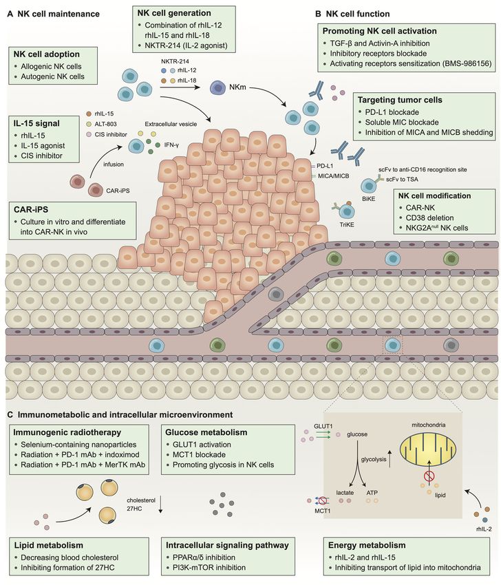
cells [215]. For metastatic melanoma refractory to CD8+ T 1 blockade exerts even greater effects in reducingWu et al. Molecular Cancer (2020) 19:120 Page 14 of 26 Fig. 4 Possible targets harnessing NK cells in cancer therapy. In order to obtain better clinical efficacy and reduced severe adverse events, the development of NK cell-based therapies that support NK cell maintenance (a), enhance NK cell function (b) and harness abnormal immunometabolic and intracellular microenvironment (c) is essential. rhIL-12/15/18, recombinant human interleukin-12/15/18; CAR-iPS, chimeric antigen receptor-induced pluripotent stem cell; MIC: MHC I chain related molecule; MICA, MHC class I polypeptide-related sequence A; MICB, MHC class I polypeptide-related sequence B; PD-L1, programmed cell death-ligand 1;scFv, single-chain variable fragment; TSA, tumor specific antigen; BiKE, bispecific killer cell engager; TriKE, trispecific killer engager; CAR-NK, chimeric antigen receptor-nature kill; PD-1, programmed cell death protein 1; MerTK, MER proto-oncogene, tyrosine kinase; 27HC, 27-hydroxycholesterol; GLUT1, glucotransporter 1; MCT1, monocarboxylate transporter 1
Wu et al. Molecular Cancer       (2020) 19:120                                                                                         Page 15 of 26

Table 3 Clinical trials for established NK cell-related therapies
Mechanism           Condition                              Intervention                                     Phase          Trial identifiers
IL-15 signal        Metastatic malignant melanoma,         Recombinant human interleukin-15(rIL-15)         I (first-in    NCT01021059
pathway             RCC                                                                                     human)
                    Advanced metastatic solid tumor        IL-15 by continuous infusion                     I              NCT01572493
                    Refractory and relapsed adult T cell   IL-15 + alemtuzumab (anti-CD52)                  I              NCT02689453
                    leukemia
                    Refractory and relapsed chronic        IL-15+ obinutuzumab (anti-CD20)                  I              NCT03759184
                    lymphocytic leukemia
                    Hematologic malignancies recurring ALT-803 (IL-15 superagonist)                         I (first-in    NCT01885897
                    after transplantation                                                                   human)
                    Metastatic NSCLC                       ALT-803 + Nivolumab (anti-PD-1 antibody)         Ib             NCT02523469
IL-21 signal        Relapse/refractory low-grade B-cell    Recombinant human interleukin-21 (rIL-21)        I              NCT00347971
pathway             LPD                                    + Rituximab (anti-CD20 antibody)
                    Metastatic malignant melanoma,         rIL-21                                           I              NCT00095108
                    RCC
                    Stage IV malignant melanoma            rIL-21                                           IIa            NCT00336986
                    without prior treatment
IL-12 signal        Metastatic solid tumors                NHS-muIL12 (two IL12 heterodimers fused          I (first-in    NCT01417546
pathway                                                    to the NHS76 antibody)                           human)
                    Murine mammary/subcutaneous            NHS-muIL12+ Avelumab (anti-PD-L1 antibody)       Preclinical    –
                    tumors                                                                                  models
IL-2 signal pathway Locally advanced or metastatic solid NKTR-214 (IL-2 pathway agonist)                    I/II           NCT02869295
                    tumors
                    Advanced Solid Tumors (Japanese)       NKTR-214 + Nivolumab                             I              NCT03745807
Anti-KIR antibody   AML in FCR                             IPH2101 (anti-KIR antibody)                      I              EUDRACT 2005–005298-
                                                                                                                           31
                    Relapsed/refractory MM                 IPH2101                                          I              NCT00552396
                    Smoldering MM                          IPH2101                                          II             NCT01248455
                    Relapsed/Refractory MM                 IPH2101+ lenalidomide (immunomodulatory          I              NCT01217203
                                                           agent)
                    AML                                    Lirilumab (2nd generation anti-KIR antibody))    II             NCT01687387
                    SCCHN                                  Lirilumab + Nivolumab                            II             NCT03341936
                    Cisplatin-ineligible muscle-invasive   Lirilumab + Nivolumab                            Ib             NCT03532451
                    bladder cancer
Anti-NKG2A          Advanced gynecologic malignancies Monalizumab (IPH2201, anti-NKG2A antibody)            I              CCGT-IND221
antibody
                    metastatic microsatellite- stable      Monalizumab + durvalumab                         First-in       NCT02671435
                    colorectal cancer                                                                       human
                    recurrent or metastatic head and       Monalizumab + cetuximab                          I              NCT02643550
                    neck cancer
TNF pathway         Advanced solid tumors                  BMS-986156 (glucocorticoid-induced TNF          I/IIa           NCT02598960
                                                           Receptor-Related Protein Agonist) +/− Nivolumab
Cell adoptive       Canine sarcomas                        Radiotherapy+ intra-tumoral autologous NK        first-in-dog   –
therapy                                                    transfer
                    Recurrent medulloblastoma and          ex-vivo-expanded NK cells                        I              NCT02271711
                    ependymoma (children)
                    Metastatic gastrointestinal            Adoptive transferred autologous NK cells +       I              NCT02845999
                    carcinoma                              cetuximab
                    HER2-positive cancers                  Adoptive transferred autologous NK cells +       I              NCT02030561
                                                           trastuzumab
                    Locally advanced colon carcinoma       Adoptive transferred autologous NK cells +       I              –
                                                           chemotherapy
                    Malignant lymphoma or advanced         Adoptive transferred allogeneic NK cells         I              NCT01212341
                    solid tumors.
You can also read