PROGRAM Natural Killer Cell Symposium 2018 - September 10th-12th, 2018 - Hamburg, Germany - NK symposium
←
→
Page content transcription
If your browser does not render page correctly, please read the page content below
PROGRAM
  Natural Killer Cell Symposium 2018

September 10th-12th, 2018 – Hamburg, Germany
GENERAL INFORMATION

Table of Contents
Sponsors ................................................................................................................................................ 1
General information ........................................................................................................................... 2
   Invited speakers and venues ......................................................................................................... 2
   Transportation ................................................................................................................................ 3
   Geographical maps ......................................................................................................................... 4
Program at a glance ........................................................................................................................... 5
Oral presentations (Talks) ................................................................................................................ 7
Poster presentations ........................................................................................................................ 41
List of participants ............................................................................................................................ 65
Notes.................................................................................................................................................... 69

Sponsors
We thank the following companies and institutions for supporting the Natural Killer Cell
Symposium 2018!

__________________________________________________________________________________
                                         1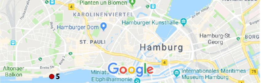
GENERAL INFORMATION
General information
NK cell symposium 10th of September – 12th of September 2018
Invited speakers and venues
Meeting venue
The meeting will take place in the Auditorium (Lecture Hall) of the Institute of
Neuroanatomy, Building N61, University Medical Center Hamburg-Eppendorf
(UKE), Martinistraße 52, 20246 Hamburg, Germany.
Invited Speakers
Zusen Fan, China
David Finlay, Ireland
Salim Khakoo, UK
Ming Li, USA
Emily Mace, USA
Ashley Moffett, UK
Joseph C. Sun, USA
Hergen Spits, Netherlands
Sophie Ugolini, France
Eric Vivier, France
Conference dinner location
La Vela
Ristorante & Wine Bar
Große Elbstraße 27, 22767 Hamburg
Germany
Internet Access
Free Wifi will be available at the conference venue using the UKE_freeWiFi network. No
password is required. Alternatively, the eduroam network can be utilized using your own
eduroam credentials.
Organizing Committee Local Organizing Committee
Marcus Altfeld Angelique Hölzemer
Andreas Diefenbach Christian Körner
Chiara Romagnani Anja Lindemann
Carsten Watzl Sebastian Lunemann
Glòria Martrus
__________________________________________________________________________________
2
GENERAL INFORMATION
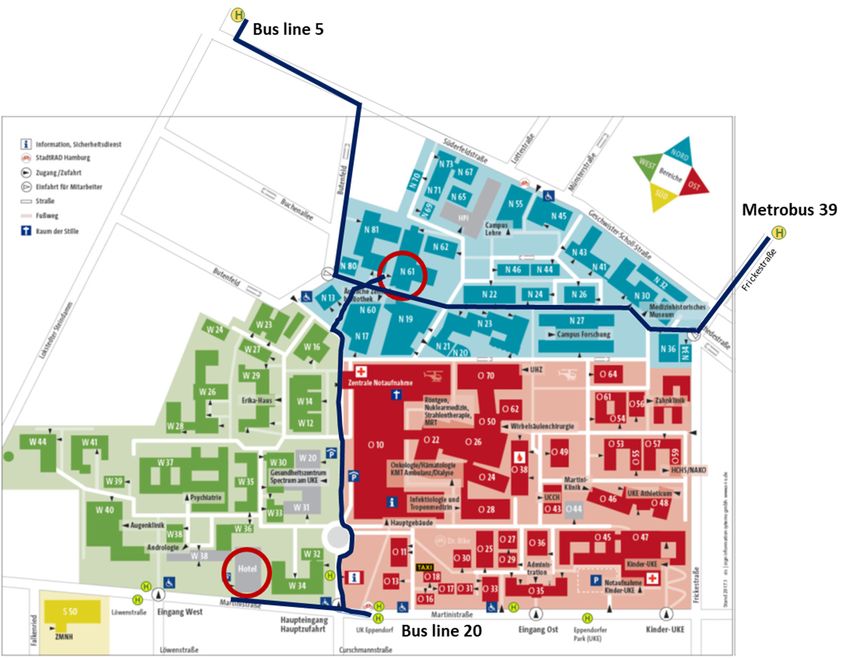
Transportation
Arrival at Hamburg airport
The city railway line (S1) and the express bus line 39 are stopping directly at the airport.
Tickets (Einzelkarten, Hamburg AB) can be bought on the way to the platforms at a DB ticket
machine or on the bus. You have several options to get to the UKE:
1. Take the S1 until Ohlsdorf (1 stop), switch platforms to take the U1 direction Ohlstedt
to Hudtwalckerstraße (4 stops) and switch there to take the bus line 20 direction to
Altona until UK Eppendorf (4 stops). The trip takes 40 min.
2. Take the express bus line 39 at the airport (direction Teufelsbrück) until Frickestraße
(11 stops) and walk through the back entrance of the UKE (off Geschwister-Scholl-
Straße) (see map). The trip takes 40 min.
3. A cab from Hamburg airport will cost app. 20 €.
Arrival at Hamburg central station or Hamburg Dammtor
1. From Central Station: Take the Metro-Bus 5 (HBF/Mönckebergstraße) direction
Burgwedel/Nedderfeld to Brunsberg (14 stops). From Dammtor: Take the Metro-Bus
5 direction Burgwedel/Nedderfeld to Brunsberg (8 stops). From there, it is a 10-15
min walk to the Anatomie Hörsaal at the building N61. Take the back entrance of the
UKE (off Butenfeld) (see map). The whole trip takes 35 min.
2. A cab from Hamburg central station will cost approx. 20 €.
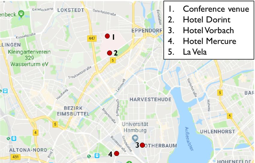
From designated accomodations
Dorint Hotel Hamburg-Eppendorf: Martinistraße 72, 20251 Hamburg. 7 min walking
distance through the UKE campus.
Hotel Vorbach: Johnsallee 63 – 67, 20146 Hamburg. Take Metro-Bus 5 (direction
Burgwedel) from Universität/Staatsbibliothek to Brunsberg. The trip takes 23 min.
Mercure Hotel Hamburg-Mitte: Schröderstiftstraße 3, 20146 Hamburg. Take Metro-Bus
5 (direction Burgwedel) from Grindelhof to Brunsberg. The trip takes 22 min.
Transportation Congress Dinner location
La Vela
Große Elbstraße 27, 22767 Hamburg
Shuttle Service
1. Bus (leaving Dorint Hotel Eppendorf, Martinistraße 71, 20251 Hamburg)
Departure Time: 7pm (11th of Sept. 2018)
2. Bus (leaving Mercure Hotel, Schroederstiftstr.3, 20146 Hamburg)
Departure Time: 7pm (11th of Sept. 2018)
Return Trip (stopping at Mercure Hotel with final destination Dorint Hotel):
1. Departure Time: 12am (12th of Sept. 2018)
2. Departure Time: 1am (12th of Sept. 2018)
__________________________________________________________________________________
3
GENERAL INFORMATION

Geographical maps

__________________________________________________________________________________
                                         4
PROGRAM AT GLANCE

Program at a glance

                                                   10th of September
   TIME                        Lecture Hall, Institute for Neuroanatomy (Building N61)
08:30-18:00    Registration opens
09:30-12:30    Young Academy Meeting
13:45-14:00    Welcome address: Marcus Altfeld
                                           Session I: NK cells and infection
14:00-15:40
                                                   Chair: Lutz Walter
               Salim Khakoo - KIR2DS2 recognizes highly conserved flaviviral peptides in the
14:00-14:40
               context of MHC class I
               Markus Uhrberg - KIR polymorphism modulates the size of the adaptive NK cell pool in
14:40-14:55
               chronic HCMV infection
               Martha Böning - Role of ADAP in NK cell-activation and the initiation of innate immunity to
14:55-15:10
               bacterial infections
15:10-15:25 Christine Thöns - NK cells from CMV-Seropositive Donors Inhibit Virus-Specific CD8 T cells
            Sebastian Lunemann - Interactions Between KIR3DS1 and HLA-F Activate Natural Killer Cells
15:25-15:40
            to Control HCV
15:40-16:15 Coffee Break
                                   Session II: NK cell subsets and memory
16:15-17:55
                                           Chair: Chiara Romagnani
16:15-16:55 Joseph Sun - Epigenetic control of NK cell memory
            Victoria Stary - Human liver- and skin-derived NK cells exhibit antigen-specific memory
16:55-17:10
            responses
            Judith Secklehner - Intravascular resident Natural Killer cells shape neutrophil dynamics in the
17:10-17:25
            pulmonary vasculature
            Stephanie Jost - Semaphorin 7A as a potential immune regulator of cytokine-induced
17:25-17:40
            memory-like responses by human natural killer cells
17:40-17:55 Beatrix Petersen - NKG2 receptors in CMV-infected rhesus macaques
            General Assembly of the NK Cell Study Group in the German Society of
18:00-18:30
            Immunology
18:30-20:00 Poster Session 1 (even numbers) - Welcome Reception - sponsored by Affimed
20:00-21:30 Meet and Greet

                                                 11th of September
   TIME                      Lecture Hall, Institute for Neuroanatomy (Building N61)
                                   Session III: NK cell function and metabolics
09:00-10:40
                                             Chair: Adelheid Cerwenka
09:00-09:40 David Finlay - What fuels Natural Killer Cell anti-tumour responses
               Cathal Harmon - Lactate mediated acidification of tumour microenvironment induces
09:40-09:55
               apoptosis of liver-resident NK cells in colorectal liver metastasis
               Hanna van Ooijen - Natural killer cells switch mechanism of cytotoxicity from Granzyme B to
09:55-10:10
               Caspase 8-induced cytolysis during serial killing
               Lutz Walter - Transcriptomic and functional analyses of human NK cells suggest a role for
10:10-10:25
               NMUR1 as specific stimulator of CD56dim NK cells
               Evelyn Ullrich -Enhancing the activation and releasing the brakes of NK cells for treatment of
10:25-10:40
               Multiple Myeloma
10:40-11:10    Coffee Break

__________________________________________________________________________________
                                         5
PROGRAM AT GLANCE
                                   Session IV: NK cell development and education
11:10-12:50
                                               Chair: Markus Uhrberg
11:10-11:50 Sophie Ugolini - Neuroendocrine regulation of NK cell functions
            Thuy Luu - SHP-1, Reactive Oxygen Species, and General Phosphorylation Status are Linked
11:50-12:05
            with Functional Education of NK cells
            Annika Niehrs - Identification of HLA-DP molecules as new ligands for the natural
12:05-12:20
            cytotoxicity receptor NKp44
            Vera Schwane - Deep immune-receptor profiling reveals significant differences in NK-cell
12:20-12:35
            receptor repertoires between individuals and functionally divergent NK-cell subsets
12:35-12:50 Kerstin Stegmann - Hepatocytes promote survival of liver-resident NK cells
12:50-14:20 Lunch Break
                                        Session V: ILCs and NK cells in tissues
14:20-16:25
                                                Chair: Jan-Eric Turner
14:20-15:00 Zusen Fan
               Ralf Dressel - The cardiac ejection fraction is preserved in mice lacking innate lymphoid cells
15:00-15:15
               (ILCs) despite pressure overload
15:15-15:30 Jan Raabe - Role of human ILC3 in the modulation of hepatic fibrogenesis
               Christine Zimmer - NK cells are robustly activated and primed for skin-homing during acute
15:30-15:45
               dengue virus infection in humans
15:45-16:25 Eric Vivier - Innate Lymphoid cells: heterogeneity and cancer immunotherapy
16:25-16:55    Coffee Break
16:55-18:15    Poster Session 2 (odd numbers)
   19:00       Departure of buses from Hotels to Dinner Location
   19:30       Dinner for all registered participants at La Vela Restaurant

                                                 12th of September
   TIME                      Lecture Hall, Institute for Neuroanatomy (Building N61)
09:00-09:40 Ashley Moffett - Interaction of uterine NK cells with placental trophoblast cells
                                 Session VI: NK cell imaging and new technologies
09:40-11:05
                                                  Chair: Carsten Watzl
               Emily Mace - Multiscale quantitative image analysis of human NK cell
09:40-10:20
               development
               Valentina Carannante - Characterization of Poliovirus Receptor in 3D cultures of Renal
10:20-10:35    Carcinoma cells and its role in shaping Natural Killer cell activity in the tumor
               microenvironment
               Veit Buchholz - Retrogenic color-barcoding for in vivo fate-mapping of single NK cells during
10:35-10:50
               infection
               Congcong Zhang - Preclinical characterization of an off-the-shelf chimeric antigen
10:50-11:05
               receptor-engineered NK cell therapeutic for adoptive cancer immunotherapy
11:05-11:35    Coffee Break
                                            Session VII: NK cells and cancer
11:35-14:00
                                                    Chair: Ingo Müller
11:35-12:15 Ming Li - Re(de)fining type 1 innate lymphocytes in the face of cancer
            Florian Babor - CD16xCD33 and CD16xIL15xCD33 Bi- and Trispecific Killer Cell Engagers
12:15-12:30 (BiKE/TriKE) as potential immunotherapy in Pediatric Patients with AML and Biphenotypic
            ALL
            Joachim Koch - Developing first-in-class immune cell engagers to redirect and activate
12:30-12:45
            immunity to fight cancer
12:45-13:00 Anais Eberhardt - Decipher the role of IL-33 as an activator of NK cells anti-tumor activity

13:00-13:40 Hergen Spits - Identification of novel transitional ILC populations in human
13:40-14:00 Final remarks/Prizes and Departure
__________________________________________________________________________________
                                         6
INVITED TALK

KIR2DS2 RECOGNIZES HIGHLY CONSERVED FLAVIVIRAL PEPTIDES IN THE
CONTEXT OF MHC CLASS I
                  1
                                                                                                                             1
Salim Khakoo

1University   of Southampton, s.i.khakoo@soton.ac.uk

Background
The activating KIR have substantial population diversity indicative of pathogen-mediated selection. Despite the observation that they
have high sequence homology in their extracellular domains with inhibitory KIR, their specificity has been difficult to determine. As KIR
bind MHC in a peptide dependent manner, we have investigated recognition of viral peptides by the activating KIR, KIR2DS2.

Results
Following a screen of hepatitis C virus peptides, we identified one peptide LNPSVAATL that induced activation of NK cells from
KIR2DS2-positive NK cells in the context of HLA-C*0102. This peptide induced binding of a KIR2DS2-tetramer to TAP-deficient
721.174 cells and, phosphorylation of VAV1 and DAP12 in KIR2DS2-positive NKL cells. LNPSVAATL was recognised by KIR2DS2
when endogenously presented and was sufficient for recognition by KIR2DS2+ NKL cells in the context of HLA-C*0102 in an in vitro
replication system. This peptide is in the viral NS3 helicase and is conserved in >98% HCV sequences. Furthermore, in a cohort of
HCV-exposed individuals, KIR2DS2 in combination with group 1 HLA-C ligands was associated with spontaneous resolution of HCV
(p
SELECTED SHORT TALK

KIR polymorphism modulates the size of the adaptive NK cell pool in chronic
HCMV infection
                    1                 1                            1                   2              3                    1
                                                                                                                                 2
Angela R. Manser , Ricarda Ising , Nadine Scherenschlich , Hartmut Hengel , Jörg Timm , Markus Uhrberg

1Institutefor Transplantation Diagnostics and Cell Therapeutics, Heinrich Heine University, Düsseldorf, Germany, 2Institute of
Virology,University Medical Center, Albert-Ludwigs-University Freiburg, Germany, 3Institute for Virology, Heinrich Heine
University,Düsseldorf, Germany, Markus.Uhrberg@med.uni-duesseldorf.de

Acute infection with human cytomegalovirus (HCMV) induces the development of NKG2C+ NK cells, which show distinct adaptive
features such as a primed state and high sensitivity to antibody-mediated activation. In about one third of chronically infected
individuals, NKG2C+ NK cells are expanded to more than 10% of the total NK cell subset. In most cases, these expansions are
accompanied by expression of a cognate inhibitory KIR with specificity for HLA-C. It is currently unclear, which role KIR gene
polymorphism plays in this process and why NKG2C expanded NK cell subsets are largely restricted to HLA-C allotypes. In the
present survey of 268 healthy donors we show that the frequency of NKG2C+ NK cells was substantially influenced by the centromeric
part of the KIR locus: individuals homozygous for the centromeric KIR haplotype A (cenAA) had larger expansions of NKG2C+ NK
cells than individuals carrying cenB haplotypes (p>0,008). Expansions beyond a certain size (>42% NKG2C+ NK cells) were invariably
associated with co-expression of cenA KIR genes, in particular the HLA-C2-specific KIR2DL1. Co-expression of KIR2DL1 was
associated with enlarged subsets of adaptive NKG2C+ NK cells (p>0,04). Next, the question was addressed if HLA-C, constituting the
major KIR ligand for NKG2C+ NK cells, is still present on the surface of HCMV-infected cells. Indeed, in two different in vitro models
involving either HCMV-infected fibroblasts or mesenchymal stem cells, HLA-C expression was maintained or even increased,
respectively whereas overall HLA class I levels were strongly downregulated. Altogether, the study suggests that the interaction of KIR
and HLA-C allotypes on HCMV-infected cells modulates adaptive NK cell subsets. The balance between cenA haplotypes promoting
and cenB haplotypes limiting the size of the adaptive NK cell subset seems to play an important role in this process.

                                                                            8
SELECTED SHORT TALK

Role of ADAP in NK cell-activation and in the initiation of innate immunity to
bacterial infections
                1                 1                    3                     2                      2                3
                                                                                                                             3
Martha Böning , Andreas Jeron , Gerald Parzmair , Annegret Reinhold , Burkhart Schraven , Dunja Bruder

1Institute
         of Medical Microbiology and Hospital Hygiene, Otto-von-Guericke University, Magdeburg, Germany, 2Institute for
Molecularand Clinical Immunology, Otto-von-Guericke University, Magdeburg, Germany, 3Institute of Medical Microbiology and
HospitalHygiene, Otto-von-Guericke University, Magdeburg, Germany and Immune Regulation Research Group, Helmholtz
Centrefor Infection Research, Braunschweig, Germany, martha.boening@med.ovgu.de

The adaptor molecule ADAP serves as a multifunctional scaffold for other proteins and is known to be involved in the formation of
immune signaling complexes in T cells. ADAP has been studied primarily in the context of T cell activation and function. Only limited
data exist regarding its overall role in immunity to pathogens. Preliminary data from our group revealed that ADAP-/- mice are highly
susceptible to L. monocytogenes (Lm) infection and fail to control bacteria growth in the early phase of infection. Our experiments
revealed that natural killer cells (NK cells) in ADAP-/- mice appear to be more effectively activated during in vivo Lm infection
compared to those in wild type animals. NK cells are known to express ADAP and ADAP has been described as an important mediator
of cytokine production following in vitro NK cell stimulation. There is growing evidence that during infections there is an intimate
crosstalk between NK cells and neutrophils and that both cell types, via secreted mediators, mutually influence each other. Together
with our observation that ADAP-deficiency does not only alter NK cell activation but as well has a significant impact on neutrophil
recruitment to spleen and liver of Lm infected animals prompted us to perform more detailed analysis of the role of ADAP in the NK
cell-neutrophil interaction during the course of Lm-infection.
Sublethal Lm infection of mice lacking ADAP in NK cells (NKp46cre/het × ADAPfl/fl) had no impact on the bacterial burden in
comparison to WT mice (NKp46cre/het × ADAPwt/wt) on day 2, 3 and 5 post infection. Significantly reduced NK cell numbers were
found in the spleen and the liver of Lm infected NKp46cre/het × ADAPfl/fl mice. Detailed analysis of NK cell subsets revealed only
marginal effects of ADAP in NK cells during Lm infection with respect to NK cell maturation. Strikingly, ADAP-deficiency in NK cells
resulted in significantly reduced numbers of neutrophils and monocytes in spleen and liver of Lm infected NKp46cre/het × ADAPfl/fl
mice on day 3 and/or day 5 p.i.
Our preliminary data suggest that during Lm infection ADAP expression in NK cells indeed affects the migration of NK cells and
subsequently the recruitment of neutrophils and monocytes to sites of infection. Ongoing experiments focus on the ADAP-dependent
expression of selected cytokines/chemokines in NK cells that are associated with neutrophil and monocyte recruitment and the
activation of antibacterial function in these cells during in vivo Lm infection.

                                                                         9
SELECTED SHORT TALK
NK Cells From CMV-Seropositive Donors Inhibit Virus-Specific CD8 T cells
Christine Thöns1, Tina Senff1, Eugen Bäcker1, Wiebke Moskorz1, Monika Lindemann2, Albert Zimmermann1,
Markus Uhrberg3, Philipp A. Lang4, Jörg Timm1
4
1Institute of Virology, Heinrich Heine University, Medical Faculty, Düsseldorf, Germany, 2Institute for Transplantation Medicine,
UniversityHospital Essen, University of Duisburg-Essen, Essen, Germany, 3Institute for Transplantation Diagnostics and Cell
Therapeutics,University Hospital Düsseldorf, Heinrich Heine University, Düsseldorf, Germany, 4Department of Molecular
MedicineII, Medical Faculty, University Hospital Düsseldorf, Heinrich Heine University, Düsseldorf, Germany,
christine.thoens@med.uni-duesseldorf.de
Cytomegalovirus (CMV) infects the majority of the human population and has been reported to influence the outcome of other
diseases. CMV reactivation is a frequent and major complication after allogenic hematopoietic stem cell transplantation and under
other immunosuppressive conditions. In infectious diseases CMV seropositivity has been associated with both superior and inferior
immune responses to heterologous infections. Given the profound impact of CMV on immune cell subsets including NK cells and CD8
T cells, we addressed the question, whether CMV-associated NK cells interact with antiviral CD8 T cells directed against CMV and
heterologous infections.
Here, we analyzed the phenotype and the interaction of NK cells and CMV- as well as Influenza A Virus (IAV)-specific CD8 T cells
from CMV seropositive and CMV seronegative individuals. In line with previous reports, we observed a strong expansion of
NKG2Cpos positive NK cells in CMV seropositive individuals. Depletion of NKG2Cpos NK cells resulted in an improved expansion of
CMV-specific CD8 T cells in vitro while simultaneous activation of NK cells inhibited the expansion of both CMV- and IAV-specific CD8
T cells in CMV-seropositive individuals. Importantly, NK cell activation did not inhibit the expansion of IAV-specific CD8 T cells in CMV
seronegative individuals.
When we analyzed the expression of the NKG2C-ligand HLA-E on CD8 T cells, CMV-specific and terminally differentiated CD8 T cells
expressed increased levels of HLA-E. Interestingly, HLA-E was also upregulated in response to IFN alpha and IFN gamma and after T
cell activation. When CMV- or IAV-specific CD8 T cells were stimulated in vitro in the presence of activated NK cells, HLA-E low CD8
T cells preferentially proliferated in CMV seropositive but not in CMV seronegative individuals, suggesting that HLA-E high CD8 T cells
are negatively regulated in CMV seropositive individuals. Finally, we show that NKG2Cpos NK cells produce IFN gamma in response
to autologous activated CD8 T cells.
In conclusion, we provide evidence that NKG2Cpos NK cells in CMV seropositive individuals regulate activated HLA-E high expressing
CD8 T cells and thereby contribute to shaping of antigen-specific CD8 T cell responses. We believe that our work provides new
insights into CMV immunity and how it impacts heterologous CD8 T cell immune responses and further highlight that it is important to
take the CMV serostatus into account when immune response to other infectious diseases are analyzed.
10SELECTED SHORT TALK
KIR3DS1/HLA-F interactions control Hepatitis C virus infection
Sebastian Lunemann1, Anja Schöbel2, Janine Kah3, Pia Fittje1, Angelique Hölzemer1, Annika L. Langeneckert1,
Leonard Hess1, Tobias Poch3, Gloria Martrus1, Wilfredo F. Garcia-Beltran4, Christian Körner1, Karl. J. Oldhafer5,
5
Julian Schulze zur Wiesch6, Christoph Schramm3, Maura Dandri3, Eva Herker2, Marcus Altfeld1
1Department of Virus Immunology, Heinrich Pette Institute, Leibniz Institute for Experimental Virology, Hamburg, Germany,
2JuniorResearch Group HCV Replication, Heinrich Pette Institute, Leibniz Institute for Experimental Virology, Hamburg,
Germany,3I. Department of Medicine, Center for Internal Medicine, University Medical Center Hamburg-Eppendorf, Hamburg,
Germany,4Ragon Institute of MGH, MIT, and Harvard, Cambridge, Massachusetts, USA, 5Department of General and
AbdominalSurgery, Asklepios Hospital Barmbek, Semmelweis University of Medicine, Asklepios Campus, Hamburg, Germany ,
6I.Department of Medicine, Section Infectious Diseases, University Medical Center Hamburg-Eppendorf, Hamburg, Germany,
7Instituteof Immunology, University Medical Center Hamburg-Eppendorf, Hamburg, Germany, 8German Center for Infection
Research(DZIF), Partner site Hamburg-Lübeck-Borstel-Riems, Hamburg, Germany, sebastian.lunemann@leibniz-hpi.de
Introduction
Natural killer (NK) cells recognize altered and infected cells through a plethora of receptors, including the killer-cell immunoglobulin-like
receptors (KIRs). KIRs have been implicated in shaping the outcome of various human diseases and are especially relevant for
hepatitis V virus (HCV) infection. The activating receptor KIR3DS1 is associated with resolution of hepatitis C virus (HCV) infection,
better treatment responses and prevention of hepatocellular carcinoma. The recent observation that open conformers of the
non-classical HLA class I molecule HLA-F serve as ligands for KIR3DS1 now allow for the first time to investigate the molecular
mechanisms mediating KIR3DS1-associated HCV disease outcome in more detail.
Methods
HLA-F surface expression was assessed by flow cytometry and immunofluoresence microscopy using Huh7.5 cells infected with a
Jc1-eGFP reporter strain, uPA/SCID/beige (USB) chimeric humanized mice infected with patient-derived HCV, and primary human
liver tissue biopsies from HCV patients. KIR3DS1-binding to HCV-infected Huh7.5 cells was determined using KIR3DS1-Fc constructs
and KIR3DS1ζ-Jurkat reporter cells. The ability of KIR3DS1+ NK cells to inhibit HCV-replication was analyzed longitudinally by
infecting Huh7 cells with a HCV Jc1-reporter virus expressing gaussia luciferase and co-culturing them with primary human NK cells
from KIR3DS1 homozygous, KIR3DS1/3DL1 heterozygous and KIR3DL1 homozygous donors as well as using sorted KIR3DS1+ and
KIR3DL1+ primary human NK cells.
Results
Huh7.5 cells exhibited an increased expression of HLA-F after infection with Jc1-eGFP (p=0.03). We observed a comparable increase
of HLA-F expression in livers of chimeric humanized mice infected with patient-derived HCV (p=0.001) and primary liver tissue
samples from HCV-infected individuals (pINVITED TALK
Epigenetic control of NK cell memory
Joseph Sun1 6
1Memorial Sloan Kettering Cancer Center, sunj@mskcc.org
Clonal expansion and immunological memory are hallmark features of the mammalian adaptive immune response and essential for
prolonged host control of pathogens. Recent work demonstrated that natural killer (NK) cells of the innate immune system also exhibit
these adaptive traits during infection. Here we demonstrate that differentiating and ‘memory’ NK cells possess distinct chromatin
accessibility states, and that their epigenetic profiles reveal a ‘poised’ regulatory program at the memory stage. Furthermore, we
elucidate how individual STAT proteins differentially control epigenetic and transcriptional states early during infection. Finally,
concurrent chromatin profiling of the canonical CD8+ T cell response against the same infection demonstrated parallel and distinct
epigenetic signatures defining NK cells and CD8+ T cells. Overall, our study reveals the dynamic nature of epigenetic modifications
during the generation of innate and adaptive lymphocyte memory.
12SELECTED SHORT TALK
Human liver- and skin-derived NK cells exhibit antigen-specific memory
responses
1 2 2 1 2
7
Stary V , Kneissl L , Strobl J , Starlinger P , Stary G
1Department of Surgery, Medical University of Vienna, Vienna, Austria, 2Department of Dermatology, Medical University of
Vienna,Vienna, Austria, victoria.stary@meduniwien.ac.at
Adaptive immunity was considered an exclusive feature of T and B cells. However, mounting evidence suggests that NK cells can
develop long-lived and highly specific memory to a variety of haptens and viral antigens in mice and in non-human primates. Despite
phenotypic analysis and description of memory-like features of long-lived human NK cells, the existence and consequences of
antigen-specific NK cell memory still needs to be proven.
We isolated NK cells of human livers and blood from individuals vaccinated against hepatitis A and/or B and characterized them
phenotypically and functionally in killing assays against antigens the patients had been vaccinated. Furthermore, we evaluated the
distribution and function of NK cells in epicutaneous patch test reactions of nickel-sensitized patients, an effector site of adaptive
immune response.
In contrast to the peripheral blood, two distinct NK cell populations were found in the liver based on their expression of CD16 and
CD49a. CD49a+CD16- liver NK cells (54.6% ± 4.2 of total NK cells) performed antigen-specific killing of hepatitis A or B-pulsed
autologous B cells matching the patients’ vaccination status. Blood-derived and CD49a-CD16+ liver NK cells did not exert
antigen-specific cytotoxicity, but recognized MHC-Ilow target cells. Although absent in healthy human skin, 57.8 ± 5.1 % of total NK
cells in nickel-induced epicutaneous patch tests were found to belong to the CD49a+CD16low NK cell subset and were capable of
specific lysis of nickel-pulsed autologous target cells.
These results suggest that antigen-specific memory NK cells in humans are present in the liver and, in case of adaptive immune
responses, as effector cells in inflamed skin. The description of underlying mechanisms for specific recognition of various viral
antigens and haptens by human memory NK cells might form the basis for novel strategies of vaccination by harnessing this NK cell
subset.
13SELECTED SHORT TALK
Intravascular resident Natural Killer cells shape neutrophil dynamics in the
pulmonary vasculature
1 2 1 2 5
8
Judith Secklehner , Katia De Filippo , John B.G. Mackey , Juho Vuononvirta , Ximena Raffo Iraolagoitia ,
Amanda McFarlane5, Matthew Neilson5, Mark B. Headley4, Max F. Krummel4, Nadia Guerra3, Leo M. Carlin1
1CRUK Beatson Institute, Glasgow/Imperial College London, NHLI, Faculty of Medicine, 2Imperial College London, NHLI,
Facultyof Medicine, 3Imperial College London, Life Sciences, 4Department of Pathology, University of California San Francisco,
5CRUKBeatson Institute, Glasgow, j.secklehner@imperial.ac.uk
The lung is both anatomically and immunologically unique in its functions. Lungs receive the right cardiac output of blood, including all
intravascular leukocytes, which, in addition to resident immune cells, comprise the pulmonary immune system. In the fine capillaries of
the lung microvasculature, the physical barrier between the blood and the external environment is exceptionally delicate. Immune cell
regulation must be closely controlled to ensure adequate protection against pathogens while limiting tissue damage which could impair
gas exchange.
Natural Killer (NK) cells are enriched in the lung compared to other tissues but the physiological implications of this accumulation are
not clear. Neutrophils are also enriched in the pulmonary vasculature making up over 50% of the lung leukocyte content. Neutrophils
can be rapidly activated and dysregulation in acute lung inflammation can be detrimental for patient outcome. Other immune cells can
play a role in controlling neutrophils and NK:neutrophil interactions have been described in a number of settings and locations. We
hypothesised that NK cells contribute to neutrophil regulation in the pulmonary vasculature.
Neutrophils are terminally differentiated and easily activated ex vivo which makes them difficult to work with. Lung intravital microscopy
allows direct visualization of immune cells in the pulmonary vasculature. This method revealed that NK cells can remain immobile
within alveolar capillaries for long periods (>60min), while neutrophils are able to transit quickly through these vessels. Moreover, we
found that most lung NK cells remain intravascular with less than 10% of NK cells found within the parenchyma. NK cells and
neutrophils frequently interacted for 5-10 minutes in the lung capillaries and occasionally material was transferred from neutrophils to
NK cells. NK depletion resulted in significant changes to neutrophil dynamics such as track length and duration, indicating a change in
cell dwell-time within pulmonary capillaries. Local stimulation with endotoxin in NK depleted mice lead to a more prominent increase in
neutrophil numbers compared to control mice. We propose that a population of intravascular resident lung NK cells directly influence
neutrophil responses in the lung which may denote a check-point for restricting neutrophilic infiltration during acute lung inflammation.
Understanding by which mechanisms these cells interact could allow us to locally influence neutrophil behaviour without affecting
systemic neutrophil functions.
14SELECTED SHORT TALK
Semaphorin 7A as a potential immune regulator of cytokine-induced memory-like
responses by human natural killer cells
1 2 3 1 1
9
Joshua Ghofrani , Haley Dugan , Marcus Altfeld , Keith Reeves , Stephanie Jost
1Center for Virology and Vaccine Research, Beth Israel Deaconess Medical Center, Harvard Medical School, Boston, MA,
USA,2Committee on Immunology, The University of Chicago, Chicago, Illinois, USA, 3Heinrich Pette Institute, Leibniz Institute
forExperimental Virology, Hamburg, Germany, 4Ragon Institute of Massachusetts General Hospital, MIT, and Harvard,
Cambridge,MA, USA, sjost@bidmc.harvard.edu
Introduction
Human cytokine-induced memory-like (CIML) NK cells are endowed with the capacity to mediate enhanced effector functions upon
cytokine or activating receptor re-stimulation for several weeks following short-term pre-activation with IL-12, IL-15 and IL-18. The
potential of CIML NK cells as adoptive immunotherapy for patients with hematologic malignancies is currently being investigated, and
preliminary findings from clinical trials have linked robust ex vivo responses to leukemia targets by CIML NK cells with remission in a
subset of acute myeloid leukemia patients. However, the mechanisms underlying CIML NK cell differentiation and increased
functionality remain incompletely understood. Semaphorin 7A (SEMA7A) is a potent immunomodulator expressed in activated
lymphocytes, including NK cells co-cultured with mitogens, and in myeloid cells. In this study, we investigated how cytokines regulate
SEMA7A expression on NK cells and evaluated the role played by SEMA7A in modulating CIML NK cell responses.
Methods
Using flow cytometry, we measured SEMA7A expression on NK cells following stimulation of PBMCs from healthy individuals with
cytokines, K562 cells, viral antigens or PHA, and assessed co-expression of key NK cell receptors as well as functionality of
SEMA7A+ NK cells in response to cytokines. Next, NK cells were pre-activated with IL-12+IL-18+IL-15 and rested for one week in the
presence or absence of antibodies against SEMA7A to compare IFN-y production upon re-stimulation with IL-12+IL-15.
Results
In unstimulated PBMCs, we found that SEMA7A was predominantly expressed on CD56bright NK cells, with 50% of this subset being
SEMA7A+ on average, and typically not co-expressed with markers of terminally differentiated or antigen-experienced NK cells.
SEMA7A was markedly upregulated on CD56bright and, to a lesser extent, on CD56dim NK cells stimulated with cytokines, with a
positive correlation between proportions of IFN-y+ and SEMA7A+ CD56bright NK cells (p=0.0006; Spearman r=0.86). Brief
pre-activation of NK cells with IL-12+IL-15+IL-18 resulted in greater than 10-fold upregulation of SEMA7A after 7 days of culture, yet
only surface expression of the ligand for SEMA7A, integrin beta-1, significantly differed between control and CIML NK cells at this
point (p=0.008). Pre-activation in the presence of antibodies targeting SEMA7A resulted in decreased IFN-y release following cytokine
re-stimulation (p=0.03).
Conclusions
These results imply a novel mechanism by which cytokine-enhanced SEMA7A/Integrin beta-1 interaction promotes CIML NK cell
differentiation and/or maintenance of increased functionality. Future investigations are warranted to assess if components targeting
SEMA7A/Integrin beta-1 signaling could enhance antitumor activity of CIML NK cells.
15SELECTED SHORT TALK
NKG2 receptors in CMV-infected rhesus macaques
Beatrix Petersen1, Ralf Dressel2, Lutz Walter1 10
1German Primate Center, Göttingen, Germany, 2University of Göttingen, Germany, bpetersen@dpz.eu
In humans, the expansion of NK cells expressing the activating receptor NKG2C has been reported in individuals infected with human
cytomegalovirus (hCMV). These cells show enhanced effector function upon re-exposure to hCMV in the context of hematopoietic
stem cell transplantation, suggesting an adapted phenotype. In order to establish a corresponding primate animal model, we have
started to analyze the NKG2/CD94 receptor system of rhesus macaques. We could demonstrate that beside single polymorphic
NKG2A and CD94 genes, rhesus macaques have expanded their NKG2C genes, with three diverse members. In particular NKG2C-2
is highly polymorphic. Besides allelic variation, NKG2C-2 shows alternative splicing resulting in a longer stalk region. This is
particularly interesting, as the stalk region of mouse Ly49H binds the mCMV m157 antigen. The individuals in our rhesus macaque
colonies are all positive for rhCMV. Some of the analyzed animals only express NKG2C-2 with the short stalk, some express both
types, and some only those with a long stalk. We are currently investigating whether these stalk length differences contribute to
functional changes of NKG2C-2. Further, we found that NKG2A as well as all three NKG2C molecules are expressed on the cell
surface in combination with CD94 upon transfection and react with the anti-human NKG2A antibody Z199. This antibody is known to
not discriminate between NKG2A and NKG2C in macaques. Therefore, we are currently establishing specific monoclonal antibodies
against all NKG2 receptors. One of the antibodies raised against NKG2C-2, clone 4A8, only binds to NKG2C-2 and the closely related
NKG2C-1, but not to NKG2A or NKG2C-3. Preliminary flow cytometry data of primary NK cells from rhCMV+ macaques show that 4A8
binds to the majority of Z199-positive cells. This suggests that rhCMV infection leads to an increase in the frequency of
NKG2C-positive NK cells in macaques similar to what is observed in CMV-infected humans.
16INVITED TALK
What fuels Natural Killer Cell anti-tumour responses
David Finlay1 11
1School of Biochemistry and Immunology, Trinity College Dublin, finlayd@tcd.ie
Cytokine activated NK cells acquire an elevated metabolic phenotype that is essential for robust NK cell anti-tumour responses. We
have characterised the metabolic changes that occur in NK cells following cytokine stimulation; within 18 hours NK cells dramatically
increase rates of glycolysis and OXPHOS and these changes are associated with an upregulation of the glycolytic machinery and
increased mitochondrial mass. This increased metabolism supports cellular biosynthesis and energy production. Interestingly, NK cells
are found to adopt a novel metabolic configuration that has not been described for any other lymphocyte subset. NK cells do not use
the TCA cycle to drive OXPHOS, as is described in textbooks, but instead use the glucose-fuelled citrate malate shuttle. Our research
has revealed the signal transduction pathways leading to this metabolic phenotype. The transcription factors Srebp and cMyc are
essential for cytokine-induced metabolic and functional responses in murine NK cells. Srebp is required for the metabolic switch to the
citrate-malate shuttle and cMyc promotes mitochondrial biogenesis and the increased expression of the glycolytic machinery. These
discoveries help us to understand why NK cells are dysfunctional in various disease states including cancer and obesity where NK cell
metabolism is found to be significantly perturbed.
17SELECTED SHORT TALK
Lactate mediated acidification of tumour microenvironment induces apoptosis of
liver-resident NK cells in colorectal liver metastasis.
1 1 1 2 2 2 2
12
Cathal Harmon , Mark W Robinson , Dalal Almuaili , Fiona Hand , Keno Mentor , Diarmaid Houlihan , Emir Hoti ,
Lydia Lynch3, Justin Geoghegan2, Cliona O’Farrelly1
11. Schoolof Biochemistry & Immunology, Trinity Biomedical Sciences Institute, Trinity College Dublin, Dublin, Ireland, 22. Liver
Unit,St. Vincent’s University Hospital, Dublin, Ireland, 33. HarvardInstitutes of Medicine, Harvard Medical School, Boston, USA,
charmon@tcd.ie
Colorectal cancer is the third most common malignancy worldwide. Metastasis to the liver is the most common cause of mortality in
these patients. We recently identified a population of liver-resident natural killer (lrNK) cells with a unique Eomeshi Tbetlo CXCR6+
phenotype. These cells rapidly degranulate and kill tumour cells. Here, we investigate changes in lrNK cells in colorectal liver
metastasis (CRLM) with a view to identifying novel immunotherapeutic targets.
Patients undergoing resection of colorectal liver metastasis (CRLM, n=18) were biopsied and the frequency and phenotype of lrNK
cells was assessed by flow cytometry. Tumour and donor tissue were cultured in vitro in order to generate conditioned media (CM) for
analysis of cytokine and metabolite production, and to assess the impact of the tumour microenvironment on lrNK cells isolated from
donor liver perfusate during transplantation (n=10).
Liver-resident NK cells are highly cytotoxic and capable of killing CRLM tumour cells. They are significantly depleted from CRLM
tumours compared to surrounding tissue (tumour: 38.1±3.4%, distal: 56.0±3.2%). Depletion of NK cells from tumour bearing tissue
correlated with tumour recurrence within 6 months. Healthy lrNK cells exposed to tumour conditioned media undergo apoptosis in vitro
but conventional NK cells do not. Tumour infiltrating lrNK cells displayed evidence of mitochondrial stress, with decreased
mitochondrial mass and increased mitochondrial ROS production. CRLM tumours are highly glycolytic and produce significantly more
lactate than healthy tissue (tumour: 11.5±2.8mM, healthy: 1.7±0.3mM). In vitro lactic acid induces apoptosis of lrNK cells by
decreasing intracellular pH, causing mitochondrial dysfunction, and this process can be abrogated by blocking mitochondrial ROS
accumulation, using the ROS scavenger MitoTempo.
Our results support a model where the accumulation of lactate from CRLM tumours decreases the pH of the tumour
microenvironment. Liver-resident NK cells migrating toward the tumour are unable to regulate intracellular pH resulting in mitochondrial
stress and apoptosis. The metabolism of CRLM tumour cells provides a novel therapeutic target to prevent tumour growth and restore
local anti-tumour NK cell immunity through regulation of the glycolysis pathway.
18SELECTED SHORT TALK
Natural killer cells switch mechanism of cytotoxicity from Granzyme B to Caspase
8-induced cytolysis during serial killing
1 2 2 3
13
Hanna van Ooijen , Isabel Prager , Carsten Watzl , Björn Önfelt
1Departmentof Applied Physics, Science for Life Laboratory, KTH Royal Institute of Technology, Stockholm, Sweden,
2DepartmentforImmunology, Leibniz Research Centre for Working Environment and Human Factors (IfADo), Dortmund,
Germany,3Department of Microbiology, Tumor and Cell Biology, Karolinska Institutet, Stockholm, Sweden, hannavo@kth.se
Natural Killer (NK) cells can induce cytolysis of target cells through release of granules containing lytic molecules such as perforin and
granzyme B, as well as by direct interaction with death receptors on the target cell leading to apoptosis through caspase 8 activation.
High amounts of perforin can also cause a rapid necrosis-like cytolysis leading to a fast leakage of the cytoplasmic content. We and
others have previously shown that single NK cells can sequentially kill multiple target cells, in a process called serial killing. In this
study we have used a target cell line expressing fluorescent reporter molecules for granzyme B and caspase 8 activity, enabling us to
determine the role of these two proteins during serial killing. Through time lapse live-cell imaging using a microwell platform, we have
followed individual NK cells and determined the temporal parameters of their cytotoxic behavior. Furthermore, we have studied the
mechanism, speed and morphological aspect of target cell death during serial killing. We show that NK cells change their mechanism
of cytotoxicity during serial killing from granzyme B-dependent for the early killing events to caspase 8-dependent for later events. The
first target cell was most commonly killed by granzyme B (75% of cells displayed granzyme B activity) for both serial killing (4 killed
target cells) and less cytotoxic NK cells. However, a significantly larger fraction of necrotic target cell death was observed for serial
killing NK cells. These NK cells also more commonly used granzyme B for later killing events compared to less cytotoxic NK cells.
Thus, our data suggests that serial killing NK cells release more perforin in their cytotoxic hits and have a larger reservoir of granzyme
B available for sequential killing.
19SELECTED SHORT TALK
Transcriptomic and functional analyses of human NK cells suggest a role for
NMUR1 as specific stimulator of CD56dim NK cells
1 1 1 1 2 3
14
Lutz Walter , Beatrix Petersen , Angela Noll , Olena Zaitseva , Gabriela Salinas , Theresa Hydes ,
Salim Khakoo3, Sandra Weinhold4, Markus Uhrberg4
1German Primate Center, Göttingen, Germany, 2University of Göttingen, Germany, 3University of Southampton, UK, 4University
ofDüsseldorf, Germany, walter@dpz.eu
The human NK cells transcriptome is not very well studied and so far largely limited to microarray analyses. Although superior to
microarrays, comprehensive RNA seq studies addressing the transcriptional identity of known human NK cell subsets are so far not
available. In order to thoroughly characterize the transcriptome of human NK cell subsets, we performed bulk RNA seq analysis of
>100 NK cell samples from different functional subsets, different stages of development, and various organs. We used B, T, ILCs, and
monocytes as outgroup. To reduce noise and increase specificity, we exclusively used cells that were purified by flow cytometrical cell
sorting. Firstly, we addressed the question of specific gene expression of blood NK cells. We identified 1335 genes that were
differentially expressed between CD56dim NK cells and the outgroup subsets. Among the novel factors was the Neuromedin U
receptor 1 (NMUR1), which is a G protein-coupled receptor for NMU, a peptide particularly well expressed by neurons in the brain and
the gastrointestinal tract and overexpressed by several different types of cancer. Prominent NMUR1 transcription was found in
CD56dim NK cells, which is in stark contrast to mouse NK cells, which do not express Nmur1. Corresponding cell surface expression
of NMUR1 was detected by flow cytometry on CD56dim but not CD56bright cells. Functional analyses demonstrated that binding of
the ligand NMU stimulates CD56dim NK cells including the NKG2C+ adaptive NK cell subset. Prolonged NK cell stimulation leads to
downregulation of the NMUR1 receptor. Notably, besides the known expression of the NMU neuropeptide in the nervous system, high
levels of expression were also found in the erythroleukemic cell line K-562 and we are currently investigating the contribution of the
NMU-NMUR1 axis to NK cell-mediated killing of K-562 as well as other NK cell-sensitive tumor targets. Together this study indicates
that NMUR1 is involved in neuronal regulation of NK cell stimulation and contributes to changes in the threshold of NK cell activation.
20SELECTED SHORT TALK
Enhancing the activation and releasing the brakes of NK cells for treatment of
Multiple Myeloma
1 1 1 2 3 2
15
Sara Tognarelli , Sebastian Wirsching , Bushra Rais , Hubert Serve , Peter Bader , Ivana von Metzler ,
Evelyn Ullrich1
1GoetheUniversity, Experimental Immunology, Childrens Hospital, Frankfurt, Germany, 2Goethe University, Hematology and
Oncology,Frankfurt, Germany, 3Goethe University, Pediatric Stem Cell Transplantation, Childrens Hospital, Frankfurt,
Germany,evelyn.ullrich@kgu.de
Natural Killer (NK) cells are innate lymphocytes with a strong anti-tumor ability. In multiple myeloma (MM) patients, an elevated
number of NK cells after stem cell transplantation (SCT) seems to correlate with a higher overall survival rate. With the aim of NK cell
use for adoptive cell therapy, we investigated the activation status and cytotoxic capacity of patient-derived NK cells at first diagnosis,
before and after autologeous SCT. At all addressed time points, we observed significant changes in NK cell phenotype as well as
cytotoxic function before and after cytokine-induced ex vivo expansion. Remarkably, after cytokine stimulation, patients NK cells
showed a highly activated phenotype and significantly enhanced killing capacity of different types of MM cells that was comparable to
healthy donors NK cells. In a small cohort of MM patients, we were able to isolate both NK cells and autlogeous MM cells from the
bone marrow (BM). Interesting, BM-NK cells were able to kill MM cells as efficiently as patients PB NK cells. In parallel, we could show
that PB-NK cells expanded from the same patient, were able to gain in lytic capacity against autologous tumor cells, suggesting a
potential use of ex vivo activated autologeous NK cells as adoptive therapy for MM patients. With the goal to further improve the NK
cell killing capacity against MM cells, we investigated the potential use of NK specific check point inhibitors with focus on NKG2A
because this inhibitory NK cell receptor was upregulated following ex vivo cytokine stimulation and MM cells showed HLA-E
expression that could even be increased by exposure to IFN-g.
Importantly, the blocking of NKG2A resulted in a significant increase in the NK cell-mediated lysis of all different MM target cells
tested, including the primary autologous MM cells.
Finally, these results let suggest that combining cytokine induced NK cell activation and the specific check point inhibition of the
NKG2A-mediated pathways can be an effective strategy to optimize NK cell therapeutic approaches against multiple myeloma.
21INVITED TALK
Neuroendocrine regulation of NK cell functions
Sophie Ugolini1 16
1Centre d\'immunologie de Marseille-Luminy (CIML), ugolini@ciml.univ-mrs.fr
Upon infection, the immune system produces inflammatory mediators important for pathogen clearance. However, inflammation can
also have deleterious effect on the host and is tightly regulated. Immune system-derived cytokines stimulate the
hypothalamic-pituitary-adrenal (HPA) axis, triggering endogenous glucocorticoid production. Through interaction with ubiquitously
expressed glucocorticoid receptors (GRs), this steroid hormone has pleiotropic effects on many cell types. Using a genetic mouse
model in which the gene encoding the GR is selectively deleted in NKp46+ innate lymphoid cells (ILCs), we demonstrated a major role
for the HPA pathway in host resistance to endotoxin-induced septic shock. GR expression in group 1 ILCs is required to limit their
IFN-γ production, thereby allowing the development of IL-10-dependent tolerance to endotoxin. The role of this
neuroendocrine-immune regulatory pathway was also investigated in the context of mouse cytomegalovirus (MCMV) infection. Our
studies suggest that neuroendocrine axes are crucial for tolerization of the innate immune system to pathogen exposure through direct
corticosterone-mediated effects on NKp46-expressing innate cells, revealing a novel strategy of host protection from
immunopathology.
22SELECTED SHORT TALK
SHP-1, Reactive Oxygen Species, and General Phosphorylation Status are Linked
with Functional Education of NK cells
1 2 3 3 1 1
17
Thuy Luu , Sunitha Vignesh , Johan Tornmalm , Jerker Widengren , Nadir Kadri , Stephan Meinke ,
Sofia Johansson2, Petter Höglund1, Arnika Kathleen Wagner1, Michael Chrobok1, Evren Alici1
1Center for Hematology and Regenerative Medicine (HERM), Department of Medicine, Huddinge, Karolinska Institute,
2Departmentof Microbiology, Tumor and Cell Biology (MTC), Karolinska Institute, 3Department of Applied Physics, Royal
Instituteof Technology, Thuy.luu.thanh@ki.se
Natural killer (NK) cells acquire functional capacity in a process called “NK cell education”, which is achieved through a presumable
continuous signal from MHC class I on surrounding cells. The molecular mechanism underlying the education process is largely
unknown. We hypothesized that the functional difference between eductaed and uneducated NK cells was manifested in a general
cellular modulation of cellular metabolism and signal transduction. Consistent with this hypothesis, we found that depressed function
was associated with a generally lower phosphorylation state of many signaling molecules in uneducated versus educated NK cells,
including ZAP70/SYK, LCK, SLP76 and PLC-gamma. Gene expression arrays revealed lower expression levels of genes involved in
the mitochondrial processes and oxidative stress response in uneducated NK cells. Consistent with these findings, the amount of
reactive oxygen species (ROS) in NK cells at steady-state was associated with their educational state. Our analysis also revealed
higher expression levels of phosphatases, mainly SHP-1, in uneducated versus educated NK cells. Furthermore, ROS (hydrogen
peroxide) inhibited tyrosine phosphatase activity in NK cells, which suggested a link between the general state of phosphorylation and
high metabolic activity. We propose a model for NK cells education in which MHC class I interactions dampens SHP-1 expression,
which leads to a decrease in tyrosine phosphatase activity, a generally higher phosphorylation status of signalling molecules, higher
OXPHOS/ROS levels and utimately more efficient functional reponses.
23SELECTED SHORT TALK
Identification of HLA-DP molecules as new ligands for the natural cytotoxicity
receptor NKp44
1 2 3 4 1 1
18
Annika Niehrs , Angelique Hölzemer , Anais Chapel , Wilfredo Garcia-Beltran , Gloria Martrus , Marcus Altfeld
1Department of Virus Immunology, Heinrich Pette Institute, Hamburg, Germany, 2I.Department of Internal Medicine, University
MedicalCenter Hamburg-Eppendorf, Hamburg, Germany; German Center for Infection Research (DZIF), Hamburg, Germany,
3Departmentof Virology, Institut Pasteur, Paris, France, 4Ragon Institute of MGH, MIT, and Harvard, Cambridge,
Massachusetts,USA, annika.niehrs@leibniz-hpi.de
Introduction: Natural killer (NK) cells represent a major component of the human innate immune system with the ability to recognize
and lyse virus-infected and tumor cells. While interactions between NK-cell receptors and HLA class I molecules are well established,
little is known about the shaping of NK-cell activity by interaction with HLA class II (HLA-II) molecules.
Objectives: The aim of this project was to identify possible receptor-ligand interactions between different NK-cell receptors and HLA-II
molecules with focus on the natural cytotoxicity receptor (NCR) group.
Material and Methods: Soluble NCR IgG1 Fc fusion constructs, namely NKp30-Fc, NKp44-Fc and NKp46-Fc, were incubated with a
panel of single HLA-II-coated Luminex beads in order to quantify binding to specific HLA-II molecules. To confirm identified
NCR–HLA-II interactions using cell-based assays, an NKp44ζ- as well as an NKp46ζ-expressing Jurkat cell line was created. Both
reporter cell lines were seeded onto wells coated with HLA-II monomers and ligand engagement was determined by CD69 expression
after 5h of co-incubation. For blocking experiments, NKp44ζ+ Jurkat cells were pre-incubated for 30 minutes with an α-NKp44 blocking
or an isotype control antibody, respectively.
Results: Remarkably, NKp44-Fc construct displayed binding to a subset of HLA-DP molecules, but not to any HLA-DR- or
HLA-DQ–coated beads tested. In contrast, neither NKp30-Fc nor NKp46-Fc constructs bound to any HLA-II–coated beads. LAG-3 was
described to be a CD4 homolog and the LAG-3 FC construct was used as positive control in this assay. In cell-based functional
assays, NKp44ζ+ Jurkat cells were activated and upregulated CD69 after co-incubation with HLA-DPB1*04:01 monomers (median
percentage of CD69+ cells: 29.3%, n=7), while less reporter cell activity was observed after co-incubation with HLA-DRB1*07:01
monomers (median percentage of CD69+ cells: 4%; n=7). In contrast, NKp46ζ+ Jurkat reporter cells did not respond to any
plate-coated HLA-II molecules and only upregulated CD69 upon incubation with plate-coated α-NKp46. These results were in line with
the previous finding that NKp44-Fc constructs bound to HLA-DP but not HLA-DR molecules. Furthermore, reporter cell activity of
NKp44ζ+ Jurkat cells in response to HLA-II molecules can be inhibited by pre-incubation of the reporter cells with an α-NKp44
blocking antibody.
Conclusion: This study demonstrates a novel interaction between a subset of HLA-DP molecules and the NCR molecule NKp44, and
uncovers a previously unknown interaction between NK cell receptors and HLA-II molecules that helps to expand our understanding of
NK-cell biology.
24SELECTED SHORT TALK
Deep immune-receptor profiling reveals significant differences in NK-cell receptor
repertoires between individuals and functionally divergent NK-cell subsets
1 2 1 1 1
19
Vera Schwane , Laura Richert , Sebastian Lunemann , Marcus Altfeld , Christian Körner
1Departmentof Virus Immunology, Heinrich Pette Institute, Leibniz Institute for Experimental Virology, Hamburg, Germany,
2Univ.Bordeaux, Inserm, Bordeaux Population Health Research Center, UMR1219 and Inria, team SISTM, Bordeaux, France,
3Departmentof Immunology, University Hospital Eppendorf (UKE), Hamburg, Germany, vera.schwane@leibniz-hpi.de
The NK-cell repertoire of an individual is defined by differential expression of a large array of receptors and influenced by
environmental factors and host genetics. Receptor expression patterns determine the activation threshold and ultimately the ability of a
given NK cell to recognize transformed and virus-infected cells. Thus, differences in the NK-cell repertoire may influence an
individual’s ability to clear viral infections and to eliminate transformed cells. In the present study, a high-throughput screening
approach was employed to assess differences in NK-cell repertoires between individuals and to identify NK-cell receptors impacting
target-cell recognition.
Expression of 371 individual surface antigens was assessed on primary human NK cells using flow cytometry, either in the absence
(n=18) or presence of target cell lines (K-562: n=18; 721.221: n=12). Antibodies against CD107a, inhibitory KIRs, NKG2A/C and CD57
were included in the experimental setup to identify NK cells undergoing degranulation and to increase dimensionality in subsequent
analyses. NK cells expressed on median 134 of the 371 antigens tested (IQR=34, 5%-positivity cutoff). Significant differences in the
distribution of receptors expressed were not only observed between individuals but also between CD56BRIGHT and CD56Dim NK
cells. Fifty-nine antigens were identified to be differently expressed between the two NK cell subsets (pYou can also read