Natural killer cells stimulated with PM21 particles expand and biodistribute in vivo: Clinical implications for cancer treatment
←
→
Page content transcription
If your browser does not render page correctly, please read the page content below
Cytotherapy, 2016; 18: 653–663

NK CELLS

Natural killer cells stimulated with PM21 particles expand and
biodistribute in vivo: Clinical implications for cancer treatment

JEREMIAH L. OYER1,a, VEETHIKA PANDEY1,a, ROBERT Y. IGARASHI1,
SRINIVAS S. SOMANCHI2, AHMED ZAKARI3, MELHEM SOLH3, DEAN A. LEE2,
DEBORAH A. ALTOMARE1 & ALICJA J. COPIK1
1
 Burnett School of Biomedical Sciences, University of Central Florida, Orlando, Florida, USA, 2Division of Pediatrics
and Cell Therapy Section,The University of Texas MD Anderson Cancer Center, Houston,Texas, USA, and 3Florida
Hospital Cancer Institute, Orlando, Florida, USA

Abstract
Background aims. Natural killer (NK) cell immunotherapy for treatment of cancer is promising, but requires methods that
expand cytotoxic NK cells that persist in circulation and home to disease site. Methods. We developed a particle-based method
that is simple, effective and specifically expands cytotoxic NK cells from peripheral blood mononuclear cells (PBMCs) both
ex vivo and in vivo. This method uses particles prepared from plasma membranes of K562-mb21-41BBL cells, expressing
41BBL and membrane bound interleukin-21 (PM21 particles). Results. Ex vivo, PM21 particles caused specific NK-cell
expansion from PBMCs from healthy donors (mean 825-fold, range 163–2216, n = 13 in 14 days) and acute myeloid leu-
kemia patients.The PM21 particles also stimulated in vivo NK cell expansion in NSG mice. Ex vivo pre-activation of PBMCs
with PM21 particles (PM21-PBMC) before intraperitoneal (i.p.) injection resulted in 66-fold higher amounts of hNK cells
in peripheral blood (PB) of mice compared with unactivated PBMCs on day 12 after injection. In vivo administration of
PM21 particles resulted in a dose-dependent increase of PB hNK cells in mice injected i.p. with 2.0 × 10⁶ PM21-PBMCs
(11% NK cells). Optimal dose of 800 μg/injection of PM21 particles (twice weekly) with low-dose interleukin 2 (1000 U/
thrice weekly) resulted in 470 ± 40 hNK/μL and 95 ± 2% of total hCD45+ cells by day 12 in PB. Furthermore, hNK cells
were found in marrow, spleen, lung, liver and brain (day 16 after i.p. PM21/PBMC injection), and mice injected with PM21
particles had higher amounts. Conclusions. The extent of NK cells observed in PB, their persistence and the biodistribution
would be relevant for cancer treatment.

Key Words: in vivo NK cell expansion, membrane bound IL-21, membrane particles, NK cells

Introduction                                                                    three general aspects must be considered: (i) a large
                                                                                enough dose of NK cells must be delivered, (ii) NK
Natural killer (NK) cells are a component of the innate                         cells must be highly cytotoxic, and (iii) NK cells must
immune system, identified by being CD56+CD3–, and                               reach, possibly localize at the site of disease, persist
can naturally recognize and lyse cells that are virally                         and specifically target tumor cells [1].
compromised or are malignant. Cell therapy with NK                                   For clinical efficacy in an AML setting, Miller
cells is promising as a cancer treatment, and multi-                            and co-workers have recommended attaining a dose
ple clinical trials have been conducted and are currently                       that would provide at least 100 NK cells per micro-
underway for treatment of various cancers (acute                                liter of peripheral blood (PB) at 2 weeks post-infusion
myeloid leukemia [AML], lymphomas, breast, ovarian,                             [2]. In some examples in which treatment with adop-
neuroblastoma, non–small cell lung carcinomas). For                             tive NK cell therapy was efficacious, more than
effective anti-cancer therapy with NK cells,                                    1000 NK cells per μL of PB were observed. These

a
These authors contributed equally to this work.
Correspondence: Alicja J. Copik, PhD, 6900 Lake Nona Boulevard, School of Biomedical Sciences, University of Central Florida, Orlando, FL 32707, USA.
E-mail: alicja.copik@ucf.edu

(Received 11 August 2015; accepted 17 February 2016)
ISSN 1465-3249 Copyright © 2016 International Society for Cellular Therapy. Published by Elsevier Inc. All rights reserved.
http://dx.doi.org/10.1016/j.jcyt.2016.02.006
654   J. L. Oyer et al.

observations highlight the importance of proficient NK       prepared from plasma membrane of K562-mb15-
cell expansion methods for delivery of a sufficient dose     41BBL cells (PM15 particles) and allowed selective
for overall treatment efficacy.                              NK cell expansion of 250-fold in 14 days and 1265-
    Currently, there are broadly three clinically used       fold after 17 days, which is comparable to the expansion
strategies for NK cell expansion for adoptive cell           efficiency using K562-mb15-41BBL feeder cells in co-
therapy. First, in vivo expansion with cytokines such        culture. PM15 particle–activated NK cells, similar to
as interleukin IL-15 and IL-2, combined with host            feeder cell–expanded NK cells, were highly cyto-
lymphodepletion/irradiation, may stimulate in vivo ex-       toxic toward CML and AML cells ex vivo. Importantly,
pansion from the relatively low amount of injected           these particles offer many advantages over the feeder
donor NK cells [3–5]. Second, ex vivo methods with           cell methods. First, they can be prepared in advance,
cytokines, mainly using IL-2 and IL-15 [6], can ac-          tested and stored for more than a year and can be used
tivate NK cells, although expansion is relatively low        as an “off-the-shelf reagent” without being con-
and variable. Also, NK cells activated ex vivo with          strained to a single Good Manufacturing Process
cytokines undergo cytokine withdrawal after infu-            facility, which greatly simplifies the clinical logistics
sion and NK cells undergo apoptosis [7,8]. Third,            of adoptive NK cell therapy. Second, use of the PM
feeder cell methods for ex vivo NK cell expansion use        particles, instead of feeder cells to stimulate NK cells,
co-cultures with other cells that are stimulatory. Feeder    eliminates steps needed for safety measures when using
cell methods for NK cell stimulation include Epstein-        tumor-derived feeder cell such as feeder cell irradia-
Barr virus lymphoblastoid cell lines [9,10], or              tion and testing their presence and proliferation in the
engineered tumor cells. Co-culture with K562 CML             final product.Third, tumor-derived feeder cells cannot
cells expressing membrane bound IL-15 (mb15) and             be injected as an adjuvant therapy, whereas the PM
4-1BB ligand (41BBL) (K562-mb15-41BBL) are able              particles can be injectable to stimulate in vivo expan-
to expand NK cells several hundred fold in about 2           sion of NK cells. The advantages offered by the PM
weeks, but the NK cells expanded by this method ex-          particle-based method for NK cell expansion would
perience senescence [11,12]. In addition, NK cells           likely allow for significant clinical benefits.
activated with IL-15 lose surface CD16 by proteo-                In this work, we tested the efficacy of PM par-
lytic activity of ADAM17 [13]. In contrast, K562 cells       ticles prepared from K562-mb21-41BBL cells for in
expressing mb21, instead of mb15, significantly im-          vivo expansion of adoptively transferred NK cells, pre-
proves NK cell expansion while avoiding telomere             activated with a relatively short and simple procedure
shortening and consequent NK cell senescence [12,14].        that could be easily implemented in a clinical setting.
Expansion of NK cells with the K562-mb21-41BBL               The method overcomes the shortcomings of previ-
is efficient, and a mean 48,000-fold expansion with          ous studies with intravenous infusion of adoptive NK
>85% enrichment is typically achieved in 3 weeks [12].       cells that only allowed minimal in vivo NK cell ex-
All of these methods are actively being investigated         pansion and limited persistence. For the current study,
in clinical trials.                                          efficacy is shown for PM21 particle stimulated ex vivo
    Although NK cell expansion methods have im-              and in vivo expansion of NK cells from unselected
proved, there are still disadvantages and challenges.        PBMCs injected into the peritoneal cavity, which is
A high, toxic dose of IL-2 is required regardless of ex-     intended to serve as an in situ site for incubation and
pansion method for survival of the infused NK cells,         stimulation by PM21 particles. This method is ex-
although the persistence of the NK cells has been            pected to be useful for the in vivo expansion of NK
limited. Although ex vivo methods with feeder cells          cells at therapeutically relevant amounts and pres-
have been effective for expansion to generate large          ents means to make NK cell–mediated immunotherapy
amounts of NK cells, concerns have been raised that          more widely accessible to patients.
long-term ex vivo culturing of NK cells causes loss of
ability to home to the site of disease, such as bone         Methods
marrow [15].Thus, there has been a debate about the
                                                             Human samples
overall benefits of in vivo versus ex vivo expansion [16].
An optimal NK cell expansion procedure would be              Primary leukemia blasts were obtained from pa-
a method that has the proliferation capability of an         tients, who signed an institutional review board–
ex vivo feeder cell based method but could be per-           approved informed consent, during active disease, and
formed either ex vivo or in vivo.                            comparable PB was collected from these patients
    Recently, our group reported a novel particle based      during remission. Leukocyte source (OneBlood) or
method for rapid and selective expansion of cyto-            fresh blood collected from healthy volunteers who
toxic NK cells starting with PB mononuclear cells            signed an institutional review board–approved in-
(PBMCs) [17]. In our earlier report, the particles cor-      formed consent were used as healthy samples. PBMCs
responding to closed plasma membrane vesicles were           were isolated using Ficoll-Paque (GE Healthcare) as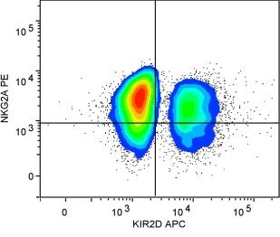
PM21 particles stimulate in vivo NK cell expansion    655

previously described [17]. All samples were de-             particles. Media with supplements was replaced rou-
identified and viably cryopreserved.                        tinely every 2–3 days after day 5.

Reagents and cell lines                                     Autologous patient NK cell cytotoxicity assays
K562 cell line was obtained from ATCC. K562-mb15-           Cytotoxicity of patient-derived NK cells against au-
41BBL cell line was kindly provided by Dr. Dario            tologous AML tumor cells was assayed with Annexin
Campana (St. Jude Children’s Research Hospital).            V (BD Bioscience). NK cells expanded for 16 days
Annexin-V fluorescein isothiocyanate (FITC) kit for         (NK cell content >90%) were stained with TFL4 dye.
cytotoxicity assays and Enumeration Flow-Count beads        Target tumor cells were co-cultured at 0.5 × 10⁶ CD34+
were purchased from Beckman Coulter. The follow-            cells/mL with NK cells at effector to target (E:T) ratios
ing dye-conjugated antibodies were used for                 of 1:1, 2:1, 5:1 and 10:1 for 2 h in 37°C, 5% CO2 at-
phenotyping: CD16-FITC, NKG2A-PE, NKp46-                    mosphere. The cells were then centrifuged and
PE, CD3-APC (Beckman Coulter); CD4-APC-                     resuspended in Annexin V labeling buffer containing
Cy7, CD8-PE, CD56-BV421, CD94-APC (BD                       Annexin V-FITC, anti-CD34-PE and anti-CD56-
Biosciences); CD3-Alexa488, NKG2D-APC, CD62L-               PC7 and incubated for 15 min at 4°C. The labeled
PE-Cy7, CD45-eFluor450, CD45-APC (eBiosciences);            cells were diluted to 250 μL and analyzed by flow
CD56-PE, KIR2D-APC (Miltenyi); NKG2C-PE,                    cytometry on an Accuri instrument (BD Bioscience).
NKp44-APC, TRAIL-PE (R&D Systems).
                                                            In vivo expansion of NK cells in NSG mice
Preparation and characterization of plasma
membrane particles                                          PBMCs, either freshly thawed or pre-activated for 2
                                                            days with 200 μg/mL PM21 and 100 U/mL IL-2, were
PM particles were prepared from K562-mb15-41BBL             washed twice and resuspended in phenol red-free
or K562-mb21-41BBL cells as previously described            RPMI media. NK cells (1 × 10⁵) in a whole PBMC
[17]. Cells were grown in RPMI-1640 media supple-           cell suspension were injected intraperitoneally (i.p.)
mented with 5% fetal bovine serum. Cells were               into NSG (NOD-scid IL-2Rgammanull) mice. PM21
harvested by centrifugation (1000 × g, 10 min), washed      particles (amounts specified in figure legends,
with Dulbecco’s phosphate buffered saline contain-          twice weekly) and IL-2 (1000 U, thrice weekly) were
ing 2 mmol/L ethylenediaminetetraacetic acid. Cells         also injected i.p., and PB was collected by cheek bleeds
were re-suspended in lysis buffer containing 50 mmol/L      or cardiac puncture. Organs were collected at nec-
HEPES, pH 7.4, 150 mmol/L NaCl, 2 mmol/L MgCl2              ropsy and were perfused to obtain single cell
and AEBSF, aprotinin, leupeptin and pepstatin A. Cells      suspensions for analysis.
were disrupted by nitrogen cavitation at 300 psi for
30 min at 4°C (Parr Instruments). Cell lysate was cen-
trifuged (1000 × g, 10 min) and the supernatant was         Results
then centrifuged (100 000 × g) to pellet the crude cell     Ex vivo and in vivo expansion of NK cells derived from
membranes. The crude membranes were further pu-             healthy donors and leukemia patients
rified by sucrose gradient centrifugation, and the
fraction that corresponds to closed plasma mem-             We recently reported that NK cells can be expanded
brane vesicles was collected. All procedures were           using particles derived from plasma membranes of
performed using aseptic techniques and sterility of the     K562-mb15-41BBL feeder cells (denoted PM15) [17].
product was tested in culture. PM particle prepara-         The PM15 particles perform similarly to K562-mb15-
tions were quantified by protein concentration by BCA       41BBL feeder cells to induce similar levels of NK cell
assay and specified as micrograms of membrane               expansion, and the expanding NK cells also have the
protein/milliliter. Presence of IL-21 and 41BBL on          same characteristics of senescing after ~3 weeks of ex-
PM particles was confirmed by enzyme-linked                 pansion. Because K562 cells engineered to express
immunosorbent assay and Western blot.                       mb21, have been reported to have better efficiency for
                                                            NK cell expansion without senescence [12], PM par-
                                                            ticles were prepared from K562-mb21-41BBL cells
Ex vivo NK cell expansion from PBMCs
                                                            and denoted PM21 particles.The PM21 particles were
NK cells from PBMCs were expanded using PM21                characterized for size distribution and the consisten-
particles as previously described [17]. Briefly, PBMCs      cy of mbIL21 content (Supplementary Figure S1) and
were seeded at 0.1 × 10⁶ NK cells/mL in stem cell           tested for their NK cell expansion capabilities.
growth medium (SCGM; CellGenix) supplemented                    PBMCs were cultured side by side with either
with 10% fetal bovine serum, 2 mmol/L Glutamax,             PM15 or PM21 particles (200 μg/mL) for 27 days.
100 U/mL IL-2 (Peprotech) and 200 μg/mL PM21                NK cells stimulated with PM21 particles expanded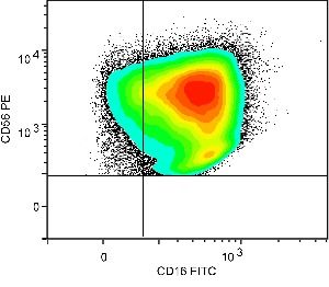
656 J. L. Oyer et al.
A B C 10 4
10 5 100% PM21
PM21 PM15 P = 0.021
PM15
10 4 80%
(fold expansion)
(fold expansion)
(% total cells)
10 3 10 3
NK cells
NK cells
NK cells
60%
10 2
40%
10 1 10 2
10 0 20%
Day 14 ± 1
10 -1 0% 10 1
0 10 20 0 10 20 PM15 PM21
Culture time (days) Culture time (days)
D E F021 M038 M050
F
10 3 F021 100% 100%
M038 Cell content (% of total)
M050 80%
80%
NK cell expansion
10 2
(Fold change)
% cytotoxicity
60% 60%
10 1
40% 40%
10 0 20% 20%
0%
10 -1 0%
0 5 10 15
1
1
1
1
:1
0 14 0 14 0 14
0:
1:
2:
5:
Culture time (days)
10
Days in culture E:T ratio
Figure 1. PM21 particles expand cytotoxic NK cells efficiently and selectively. PBMCs were isolated from leukocyte source and seeded at
0.1 × 10⁶ NK cells/mL in SCGM supplemented with 10% fetal bovine serum, 2 mM Glutamax and 50 U/mL IL-2. PBMCs were stimu-
lated with PM15 (□, black) or PM21 (○, blue) particles at 200 μg/mL for 27 days, the cell content was tested every 2–3 days and shown
are relative fold of NK cell expansion (A) and the percentage of suspension cells (B). PM21 particles (825 ± 188 fold, n = 13, 4 donors;
blue) are more efficient for NK cell expansion compared with PM15 particles (425 ± 71, n = 35, 9 donors; black) based on cumulative
analysis of day 14 data for NK cell expansion (C). PBMCs isolated from three AML patients in remission were cultured for 14 days with
PM21 particles (200 μg/mL), seeded at 0.5 × 10⁶ NK cells/mL in SCGM with 10% fetal bovine serum, 2 mmol Glutamax, 50 U/mL IL-
2. Shown are fold of NK cell expansion from the primary PBMCs (D) and lymphocyte content (E) (CD56+CD3– NK cells [●, red], CD56−CD3+
T cells [■, blue] and CD56+CD3+ NKT cells [▲, black]). PBMCs from patient F021 were cultured for 16 days as described, and au-
tologous cytotoxicity toward AML tumors from the same patient were analyzed (F). Expanded PM21-NK cells labeled with TFL4 were
co-incubated (2 h) at indicated E:T ratios with AML cells from the same patient during active disease and analyzed by flow cytometry.
The amount of spontaneous dead target cells was determined using a “Target Alone” control. Each data point was determined in
duplicate.
more rapidly compared with stimulation with PM15 with K562-mb21-41BBL feeder cells from which the
particles, and the content of NK cells reached >90% PM21 particles were derived [12]. PM21-expanded
by day 14 in PM21 particle-stimulated NK cell cul- NK cells were also cytotoxic against leukemia cell lines
tures compared with 21 days in PM15 particle- (Supplementary Figure S2).
stimulated cultures (Figure 1A,B). Cumulative analysis The NK cell expansion capabilities of PM21 par-
of NK cell expansions, at day 14 ± 1 of culture, showed ticles were further tested with PBMCs from leukemia
that PM21 particles (mean 825-fold expansion, range patients in remission. PM21 particles induced NK cell
163–2216, n = 13) are significantly (P = 0.021) more expansion relatively efficiently from all three patient-
effective compared with PM15 particles (mean 424- derived samples in 14 days of culture (113- ± 7-fold
fold, range 290–570, n = 30; Figure 1C). Furthermore, for F021, 810- ± 81-fold for M038, and 352- ± 86-
NK cells stimulated with PM21 particles expanded fold for M050, Figure 1D).The expansion was specific
exponentially during the period of 28 days, reaching for NK cells where the percentage of NK cells re-
more than 100 000 fold expansion, in contrast to the spective to total hCD45+ cells rose preferentially
NK cell expansion with PM15 particles, which stalled (Figure 1E). For sample F021, cytotoxicity of ex-
by day 22 of culture due to senescence. Thus, PM21 panded NK cells was tested in an autologous setting
particles improved NK cell expansion proficiency over against tumor blasts obtained from the same patient
the PM15 particles, and the NK cell expansion with during active disease (Figure 1F). At a relatively low
the PM21 particles was comparable to that reported E:T ratio of 1:1, 78 ± 3% of tumor cells were apoptotic.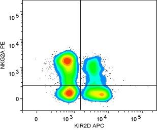
PM21 particles stimulate in vivo NK cell expansion 657
A B
not preactivated, no in vivo PM21 not preactivated, with in vivo PM21
Cells/mL of mouse blood
Cells/mL of mouse blood
50 100% 50 100%
Fraction hCD45 (%)
Fraction hCD45 (%)
40 80% 40 80%
+
+
30 60% 30 60%
20 40% 20 40%
10 20% 10 20%
0 0% 0 0%
0 5 10 15 0 5 10 15 0 5 10 15 0 5 10 15
Days from injection Days from injection Days from injection Days from injection
C D
PM21 preactivated, no in vivo PM21 PM21 preactivated, with in vivo PM21
Cells/mL of mouse blood
Cells/mL of mouse blood
50 100% 50 100%
Fraction hCD45 (%)
Fraction hCD45 (%)
40 80% 40 80%
+
+
30 60% 30 60%
20 40% 20 40%
10 20% 10 20%
0 0% 0 0%
0 5 10 15 0 5 10 15 0 5 10 15 0 5 10 15
Days from injection Days from injection Days from injection Days from injection
Figure 2. Pre-activation of unselected PBMCs with PM21 particles induces in vivo NK-cell expansion. NSG mice were injected i.p. with
2 × 10⁶ cells of either un-activated PBMCs (A and B) or PBMCs pre-activated ex vivo with PM21 particles and 100 U/mL IL-2 for 2
days (PM21-PBMCs) (C and D). Mice in all groups received 1000 U of IL-2 i.p., thrice weekly. Groups of mice were also injected with
400 μg of PM21 particles i.p., twice weekly (B and D). Peripheral blood was drawn by sequential cheek bleeds and analyzed by flow cytometry
for hCD45+ human lymphocytes twice weekly starting on day 6. NK-, T- and B-cell amounts were determined on the basis of staining for
hCD3, hCD56 and hCD19. The left plots in each experimental group shows concentration of hNK cells per microliter of PB. The right
plots show the percentage of hNK cells (○, red) and T cells (▽, black) as fraction of total hCD45+ cells.
Thus, this method could potentially be useful in an hNK cell amounts were higher compared with the mice
autologous transplant setting. with PM21-PBMCs that did not receive in vivo PM21
An unprecedented capability of the PM particles particles (Figure 2D).
would be as an injectable to spur in vivo expansion. To provide evidence that the PM21 particles induce
To test whether PM21 particles stimulate in vivo NK in vivo NK cell proliferation, analysis was performed
cell expansion and to determine if ex vivo pre-activation with CellTrace Violet labeled hNK cells expanding in
is required, NSG mice were injected i.p. with vivo at 6 days post–i.p. inoculation.The cells from mice
0.1 × 10⁶ NK cells as part of either untreated PBMCs injected with unactivated PBMCs showed no or very
or PM21-particle pre-activated PBMCs (PM21- little decrease in the CellTrace Violet fluorescence, in-
PBMCs). Mice injected with un-activated PBMCs had dicating that there was none or few cell divisions of
low amounts of human NK (hNK) cells in PB, and NK cells (Figure 3A,B).The hNK cells from mice in-
only hT cells increased as a percentage of total hCD45+ jected with PM21-PBMCs showed significant
cells over 15 days post-injection (Figure 2A,B). In sig- diminishment of the CellTrace Violet fluorescence in-
nificant contrast, PB of mice injected with PM21- tensities (Figure 3C,D). Fitting of the fluorescence
PBMCs were found to have elevated amounts of intensities showed that the intensity decrease corre-
hNK cells that peaked 12 days post i.p. injection lates with the major population, dividing seven cell
(Figure 2C,D). The NK cell content enriched to divisions in vivo within 6 days. For the hNK cells ob-
53 ± 8% of hCD45+ cells. In the same experiment, the tained from mice that received i.p. injections of PM21
efficacy was tested for in vivo i.p. application of PM21 particles, one more division can be observed.This ad-
particles to promote better in vivo NK cell expan- ditional doubling with administration of the in vivo
sion. For mice injected with regular PBMCs, additional PM21 particles correlates with the higher NK cell
in vivo PM21 particles did not stimulate hNK cell ex- amounts observed in PB with in vivo PM21 particles.
pansion. However, applying PM21 particles in vivo to To further verify whether in vivo PM21 particles
mice grafted with PM21-PBMCs had an effect where enhance in vivo NK cell expansion, a dose depen-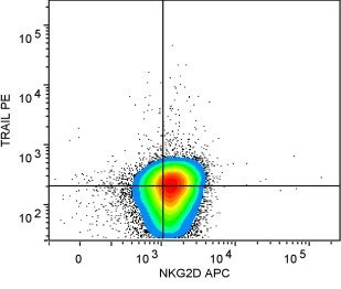
658                      J. L. Oyer et al.

                              A       no in vivo PM21     B            with in vivo PM21    organ inspected, and higher amounts of hNK cells were
                              26                              18                            found in organs from mice treated in vivo with 800 μg
                                                                                            of PM21 particles, all significantly (P < 0.05) except
not pre-activated

                              20                              14
                                                                                            in livers. Furthermore, the organs from the mice treated
                     Events

                                                         Events
                              13                                  9                         with 800 μg of PM21 particles had higher percent-
                              6                                   4
                                                                                            age of hNK cells as a fraction of total hCD45+ cells.
                                                                                                 The mice-based studies described here showed that
                                   101 102 103 104 105                101 102 103 104 105   the procedure combining ex vivo short pre-activation
                                        CT-violet                          CT-violet
                                                                                            with PM21 particles and in vivo administration of
                              C       no in vivo PM21    D              with in vivo PM21   PM21 particles induces significant in vivo NK cell ex-
                         138                                  81                            pansion, potentially in the therapeutically relevant
PM21 pre-activated

                         104                                  61
                                                                                            range. To show consistency, necessary for clinical use,
                     Events

                                                         Events

                                                                                            the procedure was applied to leukocyte sources from
                              59                              40                            three donors (different from those used in other ex-
                              34                              20                            periments; Figure 6).The average amount of hNK cells
                                                                                            in both PB and abdominal wash (AW) was relatively
                                   101 102 103 104 105                101 102 103 104 105   consistent between leukocyte sources. The percent-
                                        CT-violet                          CT-violet
                                                                                            age of hNK, hT cells and other hCD45+ cells were
Figure 3. Proliferation analysis evidences in vivo NK cell expan-                           also consistent for mice within the group injected with
sion from PM21-PBMCs. PBMCs freshly thawed or pre-activated                                 the PM21-PBMCs from a particular leukocyte source
with PM21 particles and 100 U/mL IL-2 for 2 days (PM21-                                     (n = 3) and also between leukocyte sources L8, L12
PBMCs) were labeled with CellTrace Violet. Unactivated PBMCs                                and L10.
(2 × 10⁶) (A and B) or PM21-PBMCs (C and D) were injected
i.p. to NSG mice. Mice in all groups received 1000 U IL-2 i.p.,
thrice weekly. Two of the groups of mice were also injected with                            Phenotype of NK cells expanded with PM21 particles
400 μg of PM21 particles i.p., twice weekly (B and D). Two mice
from each group were euthanized on day 6 and the peritoneal wash                            The anti-tumor cytolytic activity of NK cells are de-
was analyzed by flow cytometry for CellTrace Violet fluorescence                            termined by the balance of stimuli from activating and
of hCD45+, hCD3–, hCD56+ NK cells. Histograms of the CellTrace
Violet fluorescence were analyzed through curve fitting using the
                                                                                            inhibitory signals. Here, a detailed comparative in-
Proliferation analysis suite within FlowLogic.                                              spection was performed for the PM21 particle-
                                                                                            stimulated NK cells (i) expanded ex vivo with PM21
                                                                                            for 12 days, (ii) expanded in vivo and isolated from
dence of in vivo PM21 particles was studied (Figure 4).                                     PB, and (iii) expanded in vivo and isolated from the
A dose-dependent increase in hNK cells in PB was                                            AW. These comparisons are made using cells from a
observed from 0 to 800 μg of PM21 particles per in-                                         single donor in all of the settings and performed in
jection (Figure 4E). At a dose of 800 μg (corresponding                                     parallel (Supplementary Figure S3).
to about 100 ng of mbIL21), 470 ± 40 hNK cells per                                              Presence of CD16, the Fcγ receptor, on NK cells
microliter of PB was observed at 12 days after i.p.                                         is required for effective antibody dependent cytotox-
injection of the PM21-PBMCs. This NK cell con-                                              icity. Nearly all NK cells from in vivo expansion show
centration in PB was fivefold higher than the                                               expression of CD16 (97% and 87% for PB and AW,
concentration that is generally thought to be thera-                                        respectively). CD94 is a surface receptor that forms
peutically efficacious in an AML setting [2]. The                                           heterodimeric complexes with NKG2C or NKG2A.
dose-dependent effect for in vivo expansion was spe-                                        About half of the NK cells expanded ex vivo have
cific for hNK cells where T cell amounts did not                                            CD94 expression. For NK cells expanded in vivo, cells
increase significantly (Figure 4E). At a higher amount                                      from the AW (64 ± 9%) have higher expression than
of 1600 μg per injection, PB hNK cell amounts di-                                           NK cells from PB (38 ± 13%). Receptors of the NKG2
minished, similar to the effect observed ex vivo where                                      family both bind to CD94, inclusive of NKG2C as
~200–400 μg/mL is optimal for PM21 particles (data                                          an activating receptor and NKG2A as an inhibitory
not shown) or PM15 particles and higher amounts at-                                         receptor. The ex vivo expanded NK cells had rela-
tenuated NK-cell expansion [17].                                                            tively low expression of NKG2C, but NK cells from
     The observation of significant amounts of hNK                                          the AW were higher (53 ± 8%) and higher yet for NK
cells in PB shows that hNK cells expanding in the i.p.-                                     cells from PB (61 ± 2%).The fraction of NK cells that
injected PM21-PBMCs can migrate out from the                                                express NKG2A were higher in the AW (82 ± 8%) than
abdominal cavity to the PB. To verify that the adop-                                        PB (67 ± 12%) and those from ex vivo expansion
tively transferred hNK cells can migrate to potential                                       (74%). NKG2D is another important activating re-
sites of disease, hNK cells in various organs were quan-                                    ceptor found on NK cells and its expression was found
tified (Figure 5). Human NK cells were found in every                                       on 61 ± 6% of AW NK cells, 26 ± 3% from PB and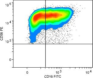
PM21 particles stimulate in vivo NK cell expansion 659
A B
0 mg in vivo PM21 400 mg in vivo PM21
Cells/mL of mouse blood
Cells/mL of mouse blood
1000 1000
% of total CD45+ cells
% of total CD45+ cells
100% 100%
800 80% 800 80%
600 60% 600 60%
400 40% 400 40%
200 200
20% 20%
0 0
0% 0%
0 5 10 15 0 5 10 15 0 5 10 15 0 5 10 15
Days from injection Days from injection Days from injection Days from injection
C D
800 mg in vivo PM21 1,600 mg in vivo PM21
Cells/mL of mouse blood
Cells/mL of mouse blood
1000 100% 1000 100%
% of total CD45+ cells
% of total CD45+ cells
800 800 80%
600 600 60%
400 50% 400
40%
200 200
20%
0 0
0% 0%
0 5 10 15 0 5 10 15 0 5 10 15 0 5 10 15
Days from injection Days from injection Days from injection Days from injection
E
600 P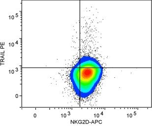
660 J. L. Oyer et al.
NK cells in bone marrow wash
Bone marrow Spleen Brain Lung Liver
NK c e lls in lung
NK cells in spleen
80,000
NK cells in brain
P=0.049 P=0.044 P=0.0003
NK cells in liver
P=0.035
4,000 20,000 60,000
10,000 60,000
40,000 40,000
5,000 2,000 10,000
20,000 20,000
0 0 0 0 0
0
0
0
0
0
0
0
0
0
0
80
80
80
80
80
PM21 (mg/injection) PM21 (mg/injection) PM21 (mg/injection) PM21 (mg/injection) PM21 (mg/injection)
% of total CD45 cells
% of total CD45 cells
% of total CD45 cells
% of total CD45 cells
% of total CD45 cells
100% 100%
120% P=0.015 120% P=0.018 120% P=0.012
100% 100% 80% 100% 80%
+
+
+
+
+
80% 80% 60% 80% 60%
60% 60% 40% 60% 40%
40% 40% 40%
20% 20% 20% 20% 20%
0% 0% 0% 0% 0%
0
0
0
0
0
0
0
0
0
80
80
80
80
PM21 (mg/injection) PM21 (mg/injection) PM21 (mg/injection) PM21 (mg/injection) PM21 (mg/injection)
Figure 5. In vivo expanded NK cells biodistribute to key physiological sites and NK cell biodistribution is increased with in vivo applica-
tion of PM21 particles. NK cells (0.2 × 10⁶ cells) as part of PM21-PBMCs, pre-activated ex vivo with PM21 particles and 100 U/mL IL-2
for 2 days, were injected i.p. to NSG mice. Mice in all groups received 1000 U of IL-2 i.p., thrice weekly. Mice were also injected with 0
or 800 μg of PM21 particles i.p., twice weekly. Mice were sacrificed 16 days after initial i.p. injection of PM21-PBMCs. On the day of
euthanasia, bone marrow (femur), spleen, lung, brain and liver were collected, organs were perfused while femur was washed to recover
cells. Cells were stained with antibodies against hCD3, hCD45, hCD56, hCD19 for flow cytometry analysis. Data for bone marrow, spleen,
brain, lung and liver (left to right) are shown with the amount of hCD45+hCD56+hCD3- NK cells (top plots for each organ) and per-
centages for hCD45+hCD56+hCD3− NK cells (○, red), hCD45+hCD3+ T cells (□, blue) and hCD45+, hCD56−hCD3– other lymphocytes
(△, black) are shown (bottom plots for each organ). The thick bar for each represents the mean.
Discussion ministration of PM21 particles. In vivo application of
the PM21 particles induces higher in vivo NK cell ex-
PM21 particles facilitate ex vivo and in vivo NK cell
pansion, dose dependent on the in vivo–applied PM21
expansion to therapeutically relevant amounts
particles. With our current optimized procedure, an
Adoptive NK cell therapy holds high promise as a average 360-fold in vivo increase of PB NK cells was
cancer therapy for initial treatment and remission main- observed between days 5 to 12 after i.p. injection of
tenance of various tumors. A requirement for PM21-PBMCs, and perhaps greater fold of expan-
therapeutic use of NK cells is a method for rapid and sion in the intraperitoneal cavity. For comparison, it
selective NK cell expansion that is safe, simple, was shown in a recent study [18] that after i.v. infu-
and overall therapeutically effective. Several cytokine sion of 1–2 × 10⁶ NK cells, only about 5 to 17 NK cells
and feeder cell based methods are currently being clin- per microliter of blood were observable on day 14 after
ically investigated and the methodology using K562- infusion. In contrast, in this study using PM21-
mb21-41BBL cell line is among the most effective for particle stimulation, we observed >400 NK cells/μL
ex vivo NK cell expansion. While feeder cell methods of blood on day 12 after i.p. infusion of 2.0 × 10⁶
are effective for providing a high initial dose and can PM21-PBMCs (11%, i.e., 0.2 × 10⁶ NK cells). Also,
allow for multiple dosing, the ability of the ex vivo ex- the former study used 5 μg (~50 000 U) per injec-
panded NK cells for homing to the bone marrow, tion (thrice weekly) of either IL-2 or IL-15, whereas
important for leukemia treatment, may be affected and a relatively low dose of IL-2 (1000 U/injection, thrice
the in vivo persistence of the infused NK cells may weekly) was used in our study. In a different study [11],
not be optimal. The combined ex and in vivo PM21- 30 × 106 NK cells, preferentially expanded ex vivo with
particle-based NK cell expansion method described K562-mb15-41BBL feeder cells, were injected i.v. fol-
here could significantly enhance the efficacy of NK lowed by tracking the injected human lymphocytes
cell adoptive therapy. using anti-CD45 antibody (not by a combination of
Importantly, PM21 particles can be used for in vivo anti-CD56 and anti-CD3). With their method, high
stimulation to promote in vivo expansion and persis- dose of i.p. injected IL-2 (25 000 U/daily) was re-
tence. The methodology developed here used a short quired for lymphocyte persistence, with the NK cell
2-day ex vivo pre-activation, followed by in vivo ad- concentrations not determined but rather implied. In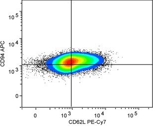
PM21 particles stimulate in vivo NK cell expansion 661
A B greater in vivo expansion compared with previous
methods that do not allow expansion or in vivo per-
% of total CD45 cells
300 100%
of mouse blood
NK cells per mL
80% sistence without the use of high dose IL-2, which has
+
200
60% been associated with clinical toxicity. For intraperi-
40%
toneal tumors, the advantages of the currently described
100 method may significantly enhance the overall anti-
20%
tumor effect. In the absence of intraperitoneal tumors,
0 0% the intraperitoneal cavity may provide a hospitable en-
vironment by confining the PM21 particle to this
0
2
0
2
L8
L8
L1
L1
L1
L1
C D volume to foster good in vivo expansion, clearly shown
% of total CD45 cells
1.5 100% by proliferation analysis with CellTrace Violet, and then
NK cells in abdominal
80% the NK cells can migrate out at significant amounts
+
wash (X 10 )
6
1.0 to the PB and organs. NK cells were not only ob-
60%
40%
served in PB but were found in organs and also were
0.5 more abundant with in vivo application of PM21 par-
20%
ticles.The NK cell amounts measured in bone marrow
0.0 0%
are comparable to those in a study [20] using NK cells
generated from CD34+ umbilical cord blood stem cells,
0
2
0
2
L8
L8
L1
L1
L1
L1
indicating that these NK cells are competent for
Figure 6. In vivo NK cells expansions from different donor sources marrow homing.
are consistent.The consistency of PM21 particle stimulated in vivo
NK-cell expansion was tested using three different PBMCs ob-
Phenotyping of NK cells expanded in parallel ex
tained from healthy donors.The PBMCs were pre-activated ex vivo vivo or in vivo (Figure S3) indicated that the result-
for 2 days with PM21 particles and 100 U/mL IL-2 for 2 days (PM21- ing cells were similar, irrespective of the approach.
PBMCs) and were injected i.p. to NSG mice. Mice in all groups Interesting differences were observed with respect to
received 1000 U of IL-2, i.p., thrice weekly. Peripheral blood was expansion of NKG2A– and NKG2C+ subpopula-
analyzed by flow cytometry for hCD45+ lymphocytes twice weekly
tions that were mostly observed with NK cells
starting on day 5 and hNK, hT and hB cell amounts were deter-
mined based on staining for hCD3, hCD56 and hCD19. Both the expanded in vivo but not in ex vivo settings [21].
concentration of hNK cells in blood 12 days after i.p. PBMC in- NKG2C+ NK cell populations have been observed
jection (A) and the amount of NK cells collected in a wash of the during viral reactivation, associated with “memory-
abdominal cavity 14 days after i.p. PBMC injection (C) were similar like” response and were recently shown to be
between the different groups injected with different NK-cell sources
dependent on monocytes for production of IL-12
(P = 0.84 for PB and P = 0.69 for AW).The corresponding cell content
of hNK cells (○, red), hT cells (□, blue) and other hCD45+ cells [21–23]. Presence of NKG2C+ NK cells in patients
(△, black) were also consistent between the groups injected with with cytomegalovirus reactivation after stem cell trans-
different PBMC sources in the peripheral blood (B) and in the plantation for AML was also associated with better
abdomen (D). The thick bar for each represents the mean. outcomes and less relapse [24]. Also, the existence of
significant population of NKG2A– NK cells that should
comparison to these previous methods, the magni- be resistant to HLA-E induced inhibition may be im-
tude of PM21 particle stimulated in vivo NK cell portant in treatment of multiple myeloma patients
expansion is unprecedented and a unique capability where cells downregulate HLA class I but express
of the PM21 particles. HLA-E to evade NK cell response [25]. Approaches
Here the route of delivery of the PM21-PBMCs aimed at downregulation of NKG2A have been pro-
to NSG mice was by i.p. injection, similar to previ- posed as means to improve NK cell cytotoxicity and
ous pre-clinical studies [11,19]. In comparison to these thus their therapeutic potential [26]. Because ex vivo–
previous studies, the PM21 particle-based method is expanded cells were mostly NKG2A+, shortening the
advantageous in several aspects. First, combined ex vivo time of ex vivo culture with subsequent in vivo ex-
pre-activation and in vivo stimulation with PM21 par- pansion may provide additional benefit in generation
ticles enables the use of a much smaller amount of of NK cells with greater phenotype diversity and po-
unselected PBMCs compared with cytokine activa- tentially good cytotoxicity against targets.
tion of isolated NK cells [19], which requires collection
of a large amount of lymphocytes by apheresis fol-
Potential clinical utility of PM21 particles
lowed by extensive laboratory processing for NK cell
enrichment. Second, the PM21 particle-based method The capabilities of PM21 particles for NK cell ex-
only requires a short 2-day pre-activation, instead of pansion may allow wider use of adoptive NK cell therapy
2-week culture-based expansion [11], that may allow for cancer treatment and potentially for other mala-
for better preservation of physiologically relevant func- dies as well.The PM21 particles can easily be substituted
tionality. Third, the current method allows for far for the feeder cells currently used in clinical trials to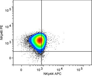
662   J. L. Oyer et al.

ease logistics and mitigate risks. For regulatory juris-       levels typically achieved only with ex vivo expansion
dictions in which the use of tumor-derived feeder cells        with feeder cells, but without the need of cell culture
are prohibited or approval is difficult to obtain, the         with feeder cells or high cytokine doses that are toxic.
PM21 particles are a ready solution for ex vivo ex-            Furthermore, PM21-PBMCs with in vivo delivery of
pansion and activation. For use of PM21 particles for          PM21 particles could be used in autologous set-
ex vivo expansion in an allogeneic setting, T cell de-         tings, to take advantage of beneficial synergistic effect
pletion can be performed before ex vivo NK cell                of other immune cells on NK cell function [34–36]
expansion. Current clinical trials of NK cells grown           and further combined with other strategies such as anti-
with K562-mbIL21-41BBL cells use T cell depletion              KIR antibodies or BiKEs to maximize NK cell
before NK cell expansion to eliminate allogeneicT cells        cytotoxicity [32,37,38]. Thus, this method meets the
that may cause graft-versus-host disease. Moreover,            criteria for generation of NK cells for potential ther-
in vivo administration of the PM21 particles can further       apeutic efficacy while being simple and more amenable
expand NK cells in vivo, an unprecedented capabil-             for clinical translation and may be impactful for treat-
ity, and possibly diminish T cell expansion to mitigate        ment of cancer or other maladies.
graft-versus-host disease. For treatment of peritoneal
cancer and other intraperitoneal tumors such as in per-        Acknowledgments
sistent ovarian epithelial cancer or desmoplastic small-       The authors thank Dr. Dario Campana (St. Jude Chil-
round-cell tumor, this NK cell expansion method could          dren’s Research Hospital, Memphis,TN) for the K562-
potentially be clinically translated. Antitumor effica-        mb15-41BBL cell line.The authors thank the Florida
cy experiments for elimination of intraperitoneal tumor        Department of Health, Bankhead-Coley Biomedical
are currently underway. Usage of PM21-PBMC and                 Research Program (3BN02 and 4BB06 to AJC) for
PM21 particles for autologous treatment is possible,           financial support.
and methodologies for incorporating T cell depletion
are being explored for application in an allogeneic            Disclosure of interest: J.L.O., R.Y.I., S.S.S., D.A.L.,
setting.                                                       D.A.A. and A.J.C. are co-owners of Cyto-Sen Ther-
     Importantly, the NK cells expanded by this method         apeutics, Inc. The other authors have no commercial,
biodistribute out from the abdominal cavity to pe-             proprietary, or financial interest in the products or com-
ripheral blood and multiple organs that are potential          panies described in this article.
sites of various other cancers. Although the i.p.
route of injection is unconventional for treatment of          References
hematological malignancies, delivery of NK cells by             [1] Klingemann HG. Cellular therapy of cancer with natural killer
this i.p. path results in PB concentration of NK cells              cells—where do we stand? Cytotherapy 2013;15:1185–94.
                                                                [2] Knorr DA, Bachanova V, Verneris MR, Miller JS. Clinical
that would be relevant for AML treatment. There-
                                                                    utility of natural killer cells in cancer therapy and
fore, future exploration and testing may be worthwhile              transplantation. Semin Immunol 2014;26:161–72.
to consider for i.p. delivery of NK cells and PM21 par-         [3] West WH, Tauer KW, Yannelli JR, Marshall GD, Orr DW,
ticles for treatment of non-i.p. cancers such as AML.               Thurman GB, et al. Constant-infusion recombinant
     The particle-based approach for NK cell specific               interleukin-2 in adoptive immunotherapy of advanced cancer.
                                                                    N Engl J Med 1987;316:898–905.
signaling could be a platform to include other signal-
                                                                [4] Miller JS, Soignier Y, Panoskaltsis-Mortari A, McNearney SA,
ing molecules or even a vehicle for packaged delivery               Yun GH, Fautsch SK, et al. Successful adoptive transfer and
of agents for further targeted stimulation of NK cells              in vivo expansion of human haploidentical NK cells in patients
to enhance homing, antitumor cytotoxicity and                       with cancer. Blood 2005;105:3051–7.
persistence. The PM21 particles can be highly com-              [5] Leclercq G, Debacker V, de Smedt M, Plum J. Differential
                                                                    effects of interleukin-15 and interleukin-2 on differentiation
plementary with all the innovative NK cell specific
                                                                    of bipotential T/natural killer progenitor cells. J Exp Med
immunotherapy methods (check point inhibitors [27],                 1996;184:325–36.
CARs [28–31], bispecific engagers (BiKE) [32], DT-              [6] Rodella L, Zamai L, Rezzani R, Artico M, Peri G, Falconi
fused IL-2 for Treg depletion [33] etc.) being developed            M, et al. Interleukin 2 and interleukin 15 differentially
and with the beneficial effects being compounded upon               predispose natural killer cells to apoptosis mediated by
                                                                    endothelial and tumour cells. Br J Haematol 2001;115:442–50.
in vivo expansion of NK cells with PM21 stimula-
                                                                [7] Ross ME, Caligiuri MA. Cytokine-induced apoptosis of human
tion. Even as a preclinical utility, the currently described        natural killer cells identifies a novel mechanism to regulate
method can allow an unprecedented ability to study                  the innate immune response. Blood 1997;89:910–18.
such combination methods. Although of course there              [8] Dijkers PF, Birkenkamp KU, Lam EW, Thomas NS,
are murine models, there are no other methods to study              Lammers JW, Koenderman L, et al. FKHR-L1 can act as a
                                                                    critical effector of cell death induced by cytokine withdrawal:
human NK cells that can be present in vivo for a sig-
                                                                    protein kinase B-enhanced cell survival through maintenance
nificant duration.                                                  of mitochondrial integrity. J Cell Biol 2002;156:531–42.
     To summarize, this procedure with PM21 par-                [9] Berg M, Lundqvist A, McCoy P Jr, Samsel L, Fan Y, Tawab
ticles allows in vivo preferential NK cell expansion at             A, et al. Clinical-grade ex vivo-expanded human natural killer
PM21 particles stimulate in vivo NK cell expansion 663
cells up-regulate activating receptors and death receptor ligands hematopoietic cell transplantation. Biol Blood Marrow
and have enhanced cytolytic activity against tumor cells. Transplant 2015;21:1653–62.
Cytotherapy 2009;11:341–55. [25] Sarkar S, van Gelder M, Noort W, Xu Y, Rouschop KM,
[10] Park KU, Jin P, Sabatino M, Feng J, Civini S, Khuu H, et al. Groen R, et al. Optimal selection of natural killer cells to kill
Gene expression analysis of ex vivo expanded and freshly myeloma: the role of HLA-E and NKG2A. Cancer Immunol
isolated NK cells from cancer patients. J Immunother Immunother 2015;64:941–63.
2010;33:945–55. [26] Carlsten M, Childs RW. Genetic manipulation of NK cells
[11] Fujisaki H, Kakuda H, Shimasaki N, Imai C, Ma J, Lockey for cancer Immunotherapy: techniques and clinical
T, et al. Expansion of highly cytotoxic human natural killer implications. Front Immunol 2015;6:266.
cells for cancer cell therapy. Cancer Res 2009;69:4010–17. [27] Phan TG, Long GV, Scolyer RA. Checkpoint inhibitors for
[12] Denman CJ, Senyukov VV, Somanchi SS, Phatarpekar PV, cancer immunotherapy. Multiple checkpoints on the long
Kopp LM, Johnson JL, et al. Membrane-bound IL-21 road towards cancer immunotherapy. Immunol Cell Biol
promotes sustained ex vivo proliferation of human natural killer 2015;93:323–5.
cells. PLoS ONE 2012;7:e30264. [28] Glienke W, Esser R, Priesner C, Suerth JD, Schambach A,
[13] Romee R, Foley B, Lenvik T, Wang Y, Zhang B, Ankarlo D, Wels WS, et al. Advantages and applications of CAR-
et al. NK cell CD16 surface expression and function is expressing natural killer cells. Front Pharmacol 2015;6:21.
regulated by a disintegrin and metalloprotease-17 (ADAM17). [29] Chang YH, Connolly J, Shimasaki N, Mimura K, Kono K,
Blood 2013;121:3599–608. Campana D. A chimeric receptor with NKG2D specificity
[14] Somanchi SS, Senyukov VV, Denman CJ, Lee DA. Expansion, enhances natural killer cell activation and killing of tumor cells.
purification, and functional assessment of human peripheral Cancer Res 2013;73:1777–86.
blood NK cells. J Vis Exp 2011;(48):doi:10.3791/2540. [30] Shimasaki N, Fujisaki H, Cho D, Masselli M, Lockey T,
[15] Childs RW, Carlsten M. Therapeutic approaches to enhance Eldridge P, et al. A clinically adaptable method to enhance
natural killer cell cytotoxicity against cancer: the force awakens. the cytotoxicity of natural killer cells against B-cell
Nat Rev Drug Discov 2015;14(7):487–98. malignancies. Cytotherapy 2012;14:830–40.
[16] Miller JS. Should natural killer cells be expanded in vivo or [31] Altvater B, Landmeier S, Pscherer S, Temme J, Schweer K,
ex vivo to maximize their therapeutic potential? Cytotherapy Kailayangiri S, et al. 2B4 (CD244) signaling by recombinant
2009;11:259–60. antigen-specific chimeric receptors costimulates natural killer
[17] Oyer JL, Igarashi RY, Kulikowski AR, Colosimo DA, Solh cell activation to leukemia and neuroblastoma cells. Clin
MM, Zakari A, et al. Generation of highly cytotoxic natural Cancer Res 2009;15:4857–66.
killer cells for treatment of acute myelogenous leukemia using [32] Gleason MK, Ross JA, Warlick ED, Lund TC, Verneris MR,
a feeder-free, particle-based approach. Biol Blood Marrow Wiernik A, et al. CD16xCD33 bispecific killer cell engager
Transplant 2015;21:632–9. (BiKE) activates NK cells against primary MDS and MDSC
[18] Miller JS, Rooney CM, Curtsinger J, McElmurry R, McCullar CD33+ targets. Blood 2014;123:3016–26.
V, Verneris MR, et al. Expansion and homing of adoptively [33] Bachanova V, Cooley S, Defor TE, Verneris MR, Zhang B,
transferred human natural killer cells in immunodeficient mice McKenna DH, et al. Clearance of acute myeloid leukemia
varies with product preparation and in vivo cytokine by haploidentical natural killer cells is improved using
administration: implications for clinical therapy. Biol Blood IL-2 diphtheria toxin fusion protein. Blood 2014;123:3855–63.
Marrow Transplant 2014;20:1252–7. [34] Zhu EF, Gai SA, Opel CF, Kwan BH, Surana R, Mihm MC,
[19] Geller MA, Knorr DA, Hermanson DA, Pribyl L, Bendzick et al. Synergistic innate and adaptive immune response to
L, McCullar V, et al. Intraperitoneal delivery of human natural combination immunotherapy with anti-tumor antigen
killer cells for treatment of ovarian cancer in a mouse xenograft antibodies and extended serum half-life IL-2. Cancer Cell
model. Cytotherapy 2013;15:1297–306. 2015;27:489–501.
[20] Cany J, van der Waart AB, Tordoir M, Franssen GM, [35] Nguyen S, Kuentz M, Vernant JP, Dhedin N, Bories D, Debre
Hangalapura BN, de Vries J, et al. Natural killer cells generated P, et al. Involvement of mature donor T cells in the NK cell
from cord blood hematopoietic progenitor cells efficiently reconstitution after haploidentical hematopoietic stem-cell
target bone marrow–residing human leukemia cells in NOD/ transplantation. Leukemia 2008;22:344–52.
SCID/IL2Rg(null) mice. PLoS ONE 2013;8:e64384. [36] Fehniger TA, Cooper MA, Nuovo GJ, Cella M, Facchetti F,
[21] Rolle A, Pollmann J, Ewen EM, Le VT, Halenius A, Hengel Colonna M, et al. CD56bright natural killer cells are present
H, et al. IL-12-producing monocytes and HLA-E control in human lymph nodes and are activated by T cell-derived
HCMV-driven NKG2C+ NK cell expansion. J Clin Invest IL-2: a potential new link between adaptive and innate
2014;124:5305–16. immunity. Blood 2003;101:3052–7.
[22] Foley B, Cooley S, Verneris MR, Pitt M, Curtsinger J, Luo [37] Kohrt HE, Thielens A, Marabelle A, Sagiv-Barfi I, Sola C,
X, et al. Cytomegalovirus reactivation after allogeneic Chanuc F, et al. Anti-KIR antibody enhancement of anti-
transplantation promotes a lasting increase in educated lymphoma activity of natural killer cells as monotherapy
NKG2C+ natural killer cells with potent function. Blood and in combination with anti-CD20 antibodies. Blood
2012;119:2665–74. 2014;123:678–86.
[23] Foley B, Cooley S, Verneris MR, Curtsinger J, Luo X, Waller [38] Romagne F, Andre P, Spee P, Zahn S, Anfossi N, Gauthier
EK, et al. Human cytomegalovirus (CMV)-induced memory- L, et al. Preclinical characterization of 1-7F9, a novel human
like NKG2C(+) NK cells are transplantable and expand in anti-KIR receptor therapeutic antibody that augments
vivo in response to recipient CMV antigen. J Immunol natural killer-mediated killing of tumor cells. Blood
2012;189:5082–8. 2009;114:2667–77.
[24] Davis ZB, Cooley SA, Cichocki F, Felices M, Wangen R, Luo
X, et al. Adaptive natural killer cell and killer cell Appendix: Supplementary material
immunoglobulin-like receptor-expressing T cell responses are
induced by cytomegalovirus and are associated with protection Supplementary data to this article can be found online
against cytomegalovirus reactivation after allogeneic donor at doi:10.1016/j.jcyt.2016.02.006.Figure S1
B
A
C
ng IL21/mg of total PM protein Concentration (particles/mL)
5/
15
/ 20
14
11
/5
/ 20
14
11
/1
3 /2
01
4
11
/1
9 /2
01
4Figure S2
K562 cells HL-60 cells KG-1 cells
100 100 100
80 80 80
60 60 60
40 40 40
20 20 20
0 0 0
1
1
1
1
1
1
1
1
1
1
1
1
1
1
1
5:
5:
5:
1:
2:
3:
5:
1:
2:
3:
5:
1:
2:
3:
5:
0.
0.
0.
E:T ratio E:T ratio E:T ratioFigure S3
ex vivo PB AWYou can also read