Differential Zika Virus Infection of Testicular Cell Lines - MDPI
←
→
Page content transcription
If your browser does not render page correctly, please read the page content below
viruses
Communication
Differential Zika Virus Infection of Testicular
Cell Lines
Luwanika Mlera † and Marshall E. Bloom *
 Biology of Vector-Borne Viruses Section, Laboratory of Virology, Rocky Mountain Laboratories, NIAID/NIH,
 Hamilton, MT 59840, USA; Luwanika.Mlera@gmail.com
 * Correspondence: mbloom@niaid.nih.gov; Tel.: +1-406-375-9707
 † Present address: BIO5 Institute, University of Arizona, Tucson, AZ 85721, USA.

 Received: 7 December 2018; Accepted: 5 January 2019; Published: 9 January 2019

 Abstract: Background: Zika virus is a mosquito-borne flavivirus responsible for recent outbreaks
 of epidemic proportions in Latin America. Sexual transmission of the virus has been reported in
 13 countries and may be an important route of infection. Sexual transmission of ZIKV has mostly
 been male-to-female, and persistence of viral RNA in semen for up to 370 days has been recorded.
 The susceptibility to ZIKV of different testicular cell types merits investigation. Methods: We infected
 primary Sertoli cells, a primary testicular fibroblast Hs1.Tes, and 2 seminoma cell lines SEM-1 and
 TCam-2 cells with ZIKV Paraiba and the prototype ZIKV MR766 to evaluate their susceptibility and
 to look for viral persistence. A human neuroblastoma cell line SK-N-SH served as a control cell type.
 Results: Both virus strains were able to replicate in all cell lines tested, but ZIKV MR766 attained
 higher titers. Initiation of viral persistence by ZIKV Paraiba was observed in Sertoli, Hs1.Tes, SEM-1
 and TCam-2 cells, but was of limited duration due to delayed cell death. ZIKV MR766 persisted
 only in Hs1.Tes and Sertoli cells, and persistence was also limited. In contrast, SK-N-SH cells were
 killed by both ZIKV MR766 and ZIKV Paraiba and persistence could not be established in these cells.
 Conclusions: ZIKV prototype strain MR766 and the clinically relevant Paraiba strain replicated in
 several testicular cell types. Persistence of ZIKV MR766 was only observed in Hs1.Tes and Sertoli cells,
 but the persistence did not last more than 3 or 4 passages, respectively. ZIKV Paraiba persisted in
 TCam-2, Hs1.Tes, Sertoli and SEM-1 cells for up to 5 passages, depending on cell type. TCam-2 cells
 appeared to clear persistent infection by ZIKV Paraiba.

 Keywords: flavivirus; Zika virus; viral persistence; testicular cells; testes

1. Introduction
     Zika virus (ZIKV) is a mosquito-borne flavivirus originally described in captive Macaca mulatta
monkeys in Uganda [1]. ZIKV recently caused an outbreak of epidemic proportions in Latin American
countries and was associated with devastating microcephaly in neonates that contracted the infection
in utero [2]. Other complications of ZIKV are varied and include Guillian Barre syndrome [3–6].
     Although ZIKV is primarily transmitted by Aedes mosquito bites, sexual transmission is now
well-documented. The first description of sexual transmission is probably that of 2 American scientists
who were bitten by Aedes mosquitoes while working in Senegal in 2008 [7]. The male transmitted ZIKV
to his wife and she presented clinical signs of disease consistent with ZIKV infection [7]. Additional
recent reports described infection in partners following travel to outbreak regions [8,9]. An interesting
example is that of an asymptomatic French couple who were only diagnosed when they sought assisted
reproductive health services after returning from the French island of Martinique [8]. Most of the
sexual transmission cases reported have been male-to-female, but a suspected female-to-male case has
been reported [10]. To date, 13 countries have documented sexual transmission of ZIKV [11]. In the US

Viruses 2019, 11, 42; doi:10.3390/v11010042                                      www.mdpi.com/journal/viruses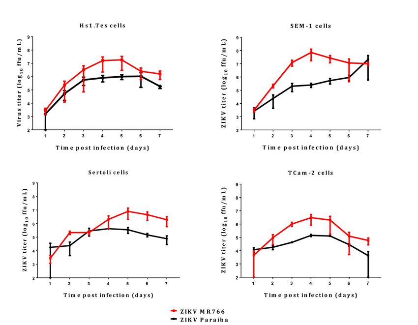
Viruses 2019, 11, 42                                                                                   2 of 11

in 2016, 47/5168 ZIKV cases were attributed to sexual transmission [12], whereas 8/451 cases could
have been sexually transmitted in 2017 [13]. Thus, sexual transmission may be an important route
of acquiring infection although it would be difficult to assess such transmission in the face of a large
vector-borne outbreak [14].
      The testes are male organs that contain germ cells which differentiate into mature spermatozoa.
Sertoli cells are interspaced between germinal epithelial cells and provide support for the germ cells. Leydig
cells are irregularly shaped interstitial cells that produce the hormone testosterone. Sexual transmission
of ZIKV by males and the presence of virus in semen suggests that cells in the male genitourinary
tract are infected [15]. Animal studies have also shown that the testes are infected with various
consequences, including testicular atrophy with implications in male fertility [16,17]. Virus was
reported to be mainly in the interstitial Leydig cells and Sertoli cells, but this varied from study
to study [16,18,19]. Govero and colleagues showed that Sertoli cells detached from the basement
membrane and that there was a decline in the germ cell population in ZIKV infected mice [17].
Thus, the different cells in the testes may play different roles in harboring virus for transmission or
pathogenesis, which leads to the destruction of organ integrity.
      In this paper, we infected several human testicular cells lines to evaluate the extent to which the
cells permitted ZIKV replication in vitro; primary Sertoli cells, a primary testicular fibroblast Hs1.Tes
and the 2 seminoma cell lines SEM-1 and TCam-2. The infection in the testicular cell lines was compared
to infection in a human neuroblastoma cell line SK-N-SH. We were also interested in determining if
ZIKV would persist in any of these cell lines. Our results showed that ZIKV differentially infected
the testicular cell lines tested and could persist in some cells in a strain-dependent manner. Delayed
apoptotic cell death was observed during viral persistence, thus limiting duration of persistence to 5
passages at most.

2. Materials and Methods

2.1. Viruses and Cells
      The Ugandan ZIKV strain MR766 were generously provided by Dr. Stephen Whitehead
(Laboratory of Infectious Disease, NIAID/NIH). The Brazilian ZIKV Paraiba was isolated by Dr. Pedro
F.C Vasconcelos, Instituto Evandro Chagas, Brazil and it was a kind gift from Dr. Stephen Whitehead
(Laboratory of Infectious Disease, NIAID/NIH). Virus stocks were prepared by infecting Vero (ATCC)
cells and harvesting the supernatants 3 days post infection. Virus in the supernatants was semi-purified
by ultracentrifugation over a 20% sucrose solution, followed by quantification using a plaque assay on
Vero cells.
      The neuroblastoma SK-N-SH cell line [20] was purchased from ATCC and maintained in
antibiotic-free Eagle’s minimum essential medium (EMEM; Gibco, Hampton, NH, USA) containing 10%
fetal bovine serum (FBS). Fibroblast Hs1.Tes cells (ATCC) and the TCam-2 seminoma cell line (a kind
gift from Dr. Constantine Stratakis, NICHD/NIH) were maintained in Dulbecco’s modified Eagle’s
medium (DMEM) supplemented with 10% FBS and 1× antibiotic-antimycotic (Gibco). Doubling time
for the TCam-2 seminoma cell line in culture is approximately 50 h. Sertoli cells (Lonza, Basel,
Switzerland) were grown in DMEM/F12 medium with 10% FBS and penicillin/streptomycin (Gibco)
and the cells grow to confluence in 7–10 days when seeded at 450–500 cells/cm2 . SEM-1 seminoma
cells were a kind gift from Dr. Alan Epstein (USC Keck School of Medicine, Los Angeles, CA, USA),
and they were maintained in RPMI 1640 (Gibco/ThermoFischer) supplemented with 10% FBS and
penicillin/streptomycin. SEM-1 cell doubling time is 50 h.

2.2. ZIKV Infection
     Two million cells were seeded in 75 cm2 flasks a day before the infection and allowed to grow
at 37 ◦ C and 5% CO2 . ZIKV infections with either MR766 or Paraiba strains were performed at
a multiplicity of infection of 0.1 with adsorption at 37 ◦ C and rocking for 1 h. All infections were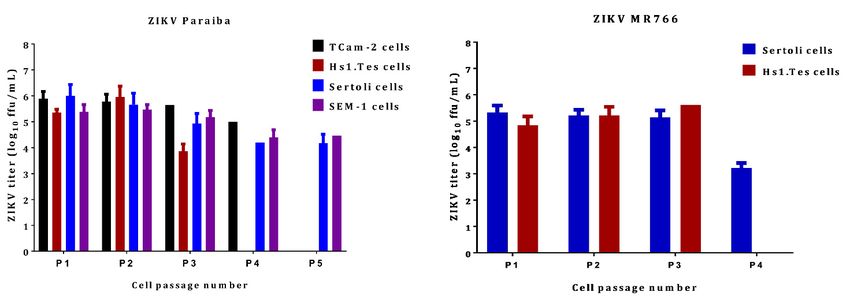
Viruses 2019, 11, 42                                                                             3 of 11

performed in triplicate (biologically independent replicates). The inoculum was removed, and cells
were washed 3 times with phosphate buffered saline (PBS; Gibco). Twenty mL of the appropriate
cell culture medium was added prior to incubation at 37 ◦ C and 5% CO2 . Supernatant aliquots were
removed daily for 7 days and stored at −80 ◦ C until virus titration. The infected cells were also
microscopically observed daily for the development of cytopathic effect (CPE). At 7 dpi, intact cell
monolayers were washed twice in PBS, trypsinized and passaged at 1:10 in new flasks with fresh
culture media.

2.3. ZIKV Titration by Immunofocus Assay
     Supernatants from infected cultures were harvested at different time points post infection. Serial
10-fold dilutions were carried out and 250 µL of each dilution was plated onto confluent Vero cells
(ATCC) in 12-well plates. Each dilution was plated in duplicate. ZIKV was adsorbed for 1 h at 37 ◦ C
with rocking, followed by washing twice with PBS. The infected monolayer was overlaid with DMEM
containing 0.8% methylcellulose, 2% FBA and antibiotics. Plates were incubated at 37 ◦ C for 3 days
after which the overlay was removed, and the cells were washed with PBS twice. The cells were fixed
with 100% methanol for 15 min, washed twice with PBS and probed with an anti-ZIKV E antibody
(BioFront Technologies, Tallahassee, FL, USA) at a 1:1000 dilution. Cells in the primary antibody were
incubated at 37 ◦ C for 1 h. Following primary antibody incubation, cells were washed twice with PBS
and an anti-mouse secondary antibody (Dako, Santa Clara, CA, USA) was added at a 1:1000 dilution
and incubated with the cells at 37 ◦ C for 1 h. Next, the cells were washed twice with PBS followed by
development of immunofoci with a diaminobenzidine/peroxide substrate (Sigma-Aldrich, St. Louis,
MO, USA) and enumeration.

2.4. Monolayer Staining with Giemsa Stain
     To visualize the cytopathic effect of ZIKV on infected cell monolayers, cells were washed twice
in PBS and fixed with 4% paraformaldehyde (PFA) for 10 min at room temperature. The PFA was
aspirated followed by washing twice with PBS. Cells were stained with a 1:5 Giemsa stain for 30 min.
The stain was removed, and cells were washed twice with PBS and imaged using an AxioVert.A1
microscope equipped with Zeiss Axiocam 503 monochromatic camera.

2.5. Immunofluorescence Microscopy
    ZIKV-infected Sertoli cells at P1 were plated into a 4-well chamber slide at 1 × 10⁴ cells/well
and allowed to attach overnight at 37 ◦ C in 5% CO2 . The medium was aspirated, and cells were
washed twice with PBS. The cells were fixed with 4% paraformaldehyde/5% sucrose in PBS for 10 min.
The fixed cells were permeabilized with 0.1% Triton X/4% PFA in PBS for 10 min with shaking.
Aldehydes were quenched using 50 mM glycine for 10 min. Blocking was done with 2% bovine
serum albumin for 1 h. Cells were probed with a mouse anti-ZIKV E monoclonal antibody (BioFront
Technologies) at 1:1000 dilution, and a rabbit anti-cleaved caspase 3 (BD Biosciences, San Jose, CA,
USA) at 1:1000 dilution. The primary antibodies were detected with anti-mouse (conjugated with Alexa
Flour 647) and Alexa Flour 488-conjugated anti-rabbit antibodies (Invitrogen, Carlsbad, CA, USA).
Images were captured using a Laser Scanning Microscope (LSM) 710 (Zeiss, Oberkochen, Germany) at
40× magnification.

3. Results

3.1. ZIKV Infection of the Fibroblast Hs1.Tes Cell Line
    The Hs1.Tes cell line is a fibroblast which represents testicular connective tissue. Both ZIKV
MR766 and Paraiba strains were able to infect this cell line in culture, and ZIKV MR766 replicated to
higher titers than ZIKV Paraiba (Figure 1). The ZIKV MR766 titer peaked to 1.8 × 10⁷ ffu/mL at 4 dpi,
whereas ZIKV Paraiba titers peaked to 1.0 × 10⁶ at 5 dpi (Figure 1). Infection of Hs1.Tes cells with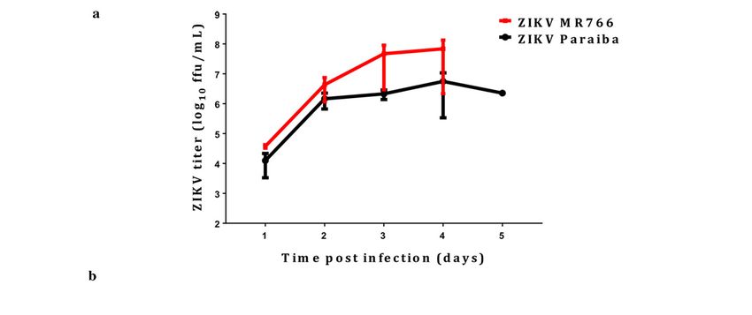
Viruses 2019, 11, 42                                                                                         4 of 11
Viruses 2019, 11, x FOR PEER REVIEW                                                                                      4 of 11

       To ZIKV
   either  determine
                  Paraibaif ZIKV
                            or ZIKVcould
                                      MR766persist
                                                did in
                                                     notHs1.Tes
                                                         result infibroblast cellcytopathic
                                                                    any obvious   line, we passaged      theby
                                                                                              effect (CPE)    ZIKV
                                                                                                                 7 dpiinfected
cells   after2 7and
  (Figures        dpi.
                    3). Infectious ZIKV Paraiba and ZIKV MR766 was recovered from the Hs1.Tes
supernatants      at eachifpassage
         To determine        ZIKV couldfor 3 persist
                                              passages,   suggesting
                                                       in Hs1.Tes       that ZIKV
                                                                    fibroblast       couldwe
                                                                               cell line,   persist  in these
                                                                                               passaged     the cells
                                                                                                                ZIKV(Figure
4).infected
     However,cellswe
                   afterobserved    a decline
                          7 dpi. Infectious      in Paraiba
                                              ZIKV   the celland
                                                              population
                                                                   ZIKV MR766uponwaspassage    each,
                                                                                       recovered  from suggesting
                                                                                                         the Hs1.Tesdelayed
andsupernatants   at each passage
      slowly progressive             for 3 passages,
                               cell death   and a 4th  suggesting  that ZIKV
                                                         passage was          could persist
                                                                        not possible        in these
                                                                                       (Figure  5a,b).cells (Figure
                                                                                                        Thus,   the 4).
                                                                                                                    duration
   However,     we observed    a  decline  in the cell  population   upon
of ZIKV persistence in Hs1.Tes cells was limited by continued cell death. passage   each, suggesting    delayed   and
   slowly progressive cell death and a 4th passage was not possible (Figure 5a,b). Thus, the duration of
   ZIKV persistence in Hs1.Tes cells was limited by continued cell death.

                    Replication kinetics
        Figure1.1. Replication
      Figure                    kinetics ofofZIKV
                                               ZIKVMR766
                                                     MR766andand
                                                               Paraiba overover
                                                                   Paraiba  the course   of 7 days.
                                                                                 the course    of 7 Error
                                                                                                    days. bars
                                                                                                           Error bars
        represent  standard  deviation (SD)  from the mean for 3 independent replicates.
      represent standard deviation (SD) from the mean for 3 independent replicates.
  3.2. ZIKV Infection of Sertoli Cells
3.2. ZIKV Infection of Sertoli Cells
        Sertoli cells are testicular cells, which support the germ cells. In culture, we observed that these
      Sertoli
  cells        cellspermissive
        were also     are testicular   cells, which
                                 to infection  by bothsupport    the germ
                                                        ZIKV Paraiba     andcells.
                                                                              MR766  In strains,
                                                                                         culture,which
                                                                                                   we observed
                                                                                                         replicatedthat
                                                                                                                    to these
cells were titers
  maximal    also permissive      to respectively
                    at 4 and 5 dpi,   infection by(Figure
                                                     both ZIKV
                                                            1). TheParaiba    and MR766
                                                                    ZIKV Paraiba              strains,
                                                                                     titers did         whichbeyond
                                                                                                 not proceed    replicated to
  106 ffu/mL
maximal         forat
           titers   7 dpi,
                      4 andbut
                             5 MR766
                               dpi, respectively      107 ffu/mL
                                        titers neared(Figure        at 5ZIKV
                                                               1). The   dpi. Similar
                                                                               Paraiba  to Hs1.Tes  cells,
                                                                                           titers did  notinfection
                                                                                                            proceedofbeyond
  Sertoli cells with  ZIKV   Paraiba   did not  cause an observable   cytopathic    effect
10 ffu/mL for 7 dpi, but MR766 titers neared 10 ffu/mL at 5 dpi. Similar to Hs1.Tes cells,
   6                                                        7                              (CPE)  by 7 dpi  (Figure 2),
                                                                                                                 infection of
  but  ZIKV   MR766    caused  minimal    CPE   which  was   observed   at 7 dpi (Figure   3).
Sertoli cells with ZIKV Paraiba did not cause an observable cytopathic effect (CPE) by 7 dpi (Figure
        After 7 dpi, we passaged Sertoli cells infected with either ZIKV Paraiba or ZIKV MR766 and
2), but ZIKV MR766 caused minimal CPE which was observed at 7 dpi (Figure 3).
  observed that ZIKV Paraiba persistently infected Sertoli cells for up to 5 passages before the delayed
      After 7 dpi, we passaged Sertoli cells infected with either ZIKV Paraiba or ZIKV MR766 and
  cell death, like that observed in Hs1.Tes cells, rendered the cells unfit for a 6th passage. Detection of
observed    that ZIKV Paraiba persistently infected Sertoli cells for up to 5 passages before the delayed
  cleaved caspase 3 by immunofluorescence (Figure 5c) suggested that cell death was via apoptosis.
cell death,  like thatthat
  We also observed       observed     in Hs1.Tes cells,
                             not all ZIKV-infected    cellsrendered    the cellsforunfit
                                                             stained positive             for acaspase
                                                                                     cleaved     6th passage.   Detection
                                                                                                        3 (Figure  5c).    of
cleaved caspase 3 by immunofluorescence (Figure 5c) suggested that cell death was via apoptosis.
We also observed that not all ZIKV-infected cells stained positive for cleaved caspase 3 (Figure 5c).
The progressive cell death was accompanied by a decline in the ZIKV Paraiba titers from 1.0 × 10⁶
ffu/mL at passage 1, to 1.5 × 10⁴ ffu/mL at passage 5 (Figure 4). ZIKV MR766 infection persisted in
Sertoli cells for only 4 passages and the viral titers were constant at ~105 ffu/mL from passage 1 to
Viruses 2019, 11, 42 5 of 11
The progressive cell death was accompanied by a decline in the ZIKV Paraiba titers from 1.0 × 10⁶
ffu/mL at passage 1, to 1.5 × 10⁴ ffu/mL at passage 5 (Figure 4). ZIKV MR766 infection persisted in
Sertoli cells for only 4 passages and the viral titers were constant at ~105 ffu/mL from passage 1 to
4
passage 3, 11,
Viruses 2019, butx the
FORtiter REVIEW to 1.6 × 10 at the 4th passage (Figure 4).
PEERdeclined 5 of 11
Figure 2.2. Microscopic
Microscopic evaluation
evaluation of testicular cell lines
lines infected
infected with
with ZIKV
ZIKV Paraiba.
Paraiba. No obvious
cytopathic effect was observed in allall testicular
testicular cell
cell lines
lines infected
infected with
with ZIKV
ZIKV Paraiba.
Paraiba. Images were
Viruses 2019, 11, x FOR
captured PEER REVIEW of 400×.
at a magnification
magnification 6 of 11
of 400×.
3.3. ZIKV Infection of Seminoma Cell Lines
TCam-2 cells are a germ cell seminoma cell line, and SEM-1 cells are a testicular cell line which
is an intermediate between a non-seminoma and a true seminoma [21,22]. Compared to all cell lines
tested, SEM-1 seminoma cells supported the highest titers of both ZIKV MR766 and ZIKV Paraiba
replication (Figure 1). The ZIKV MR766 titer peaked to 7.1 × 10⁷ at 4 dpi, whereas the peak ZIKV
Paraiba titer was attained at 7 dpi (Figure 1). Interestingly, the ZIKV replication graph in SEM-1 cells
suggested that ZIKV Paraiba was still replicating upwards of the peak attained at 7 dpi. No obvious
CPE was observed in ZIKV Paraiba-infected SEM-1 cells (Figure 2), but ZIKV MR766 caused notable
cell death by 7 dpi and the infected cells appeared smaller in comparison to uninfected controls
(Figure 3). Upon a single passage, ZIKV MR766 infection induced a lytic crisis in SEM-1 seminoma
cells. The culture of ZIKV MR766 infected cells did not recover, and thus, a persistent ZIKV MR76 6
infection of
Figure
Figure 3.SEM-1 cells
Cytopathic
3. Cytopathic could
effect
effect not
of ZIKV beMR766
of ZIKVestablished.
MR766 However,
on testicular
on testicular a persistent
cell cell
lineslines ZIKV
at 7 We
at 7 dpi. dpi. WeParaiba
noted noted infection
that
that ZIKV ZIKV
MR766- was
maintained
infected in SEM-1
MR766-infected
Sertoli cells and
andSertoli
SEM-1 up toSEM-1
cells 5appeared
passages (Figure
cells smaller
appeared 4).
smaller
than than uninfected
uninfected controls.and
controls. TCam-2 TCam-2 and
Hs1.Tes cells
Hs1.Tes
did cellsCPE
show any did show any Images
at 7 dpi. CPE at 7were
dpi. obtained
Images were
at aobtained at a magnification
magnification of 400×. of 400×.
In TCam-2 seminoma cells, ZIKV titers peaked at 4 dpi, with the MR766 strain at 3 × 10⁶ ffu/mL
and the Paraiba strain at 1.4 × 10⁵ ffu/mL (Figure 1). By 7 dpi, ZIKV MR766 and ZIKV Paraiba titers
declined to 6 × 10⁴ ffu/mL and 4 × 10³ ffu/mL, respectively. Interestingly, both ZIKV strains did not
cause any CPE in TCam-2 cells by 7 dpi (Figures 2,3). Although ZIKV Paraiba persisted in TCam-2
cells for 4 passages, ZIKV MR766 killed the cells upon passage into P1, suggesting a strain-dependentIn TCam-2 seminoma cells, ZIKV titers peaked at 4 dpi, with the MR766 strain at 3 × 10 ffu/mL
and the Paraiba strain at 1.4 × 10⁵ ffu/mL (Figure 1). By 7 dpi, ZIKV MR766 and ZIKV Paraiba titers
declined to 6 × 10⁴ ffu/mL and 4 × 10³ ffu/mL, respectively. Interestingly, both ZIKV strains did not
cause any CPE in TCam-2 cells by 7 dpi (Figures 2,3). Although ZIKV Paraiba persisted in TCam-2
cells for 4 passages, ZIKV MR766 killed the cells upon passage into P1, suggesting a strain-dependent
Viruses 2019, 11, 42 6 of 11
delayed cell death mechanism.
Figure
Figure 4.4. ZIKV
ZIKV titers
titers in
in persistently
persistently infected
infected testicular
testicular cell
cell lines.
lines. The
The clinical
clinical isolate
isolate ZIKV
ZIKV Paraiba
Paraiba was
was
able to persist in all cell lines tested for up to 5 passages (P1 through to P5),
able to persist in all cell lines tested for up to 5 passages (P1 through to P5), depending on depending on cell
cell line.
line.
ZIKV
ZIKV MR766
MR766 waswas only
only able
able to
to persist
persist in
in Sertoli
Sertoli and Hs1.Tes cells.
and Hs1.Tes cells. Virus
Virus titration
titration was
was performed
performed using
using
supernatants
supernatants collected at the end of each 7-day period and each data point represents an
collected at the end of each 7-day period and each data point represents an average
average ofof 33
biological replicates. Error bars represent standard deviation from the mean. Each
biological replicates. Error bars represent standard deviation from the mean. Each passage was done passage was done
after
after 77 days
days byby washing
washing the the monolayer
monolayer twicetwice with
with PBS,
PBS, trypsinizing
trypsinizing thethe cells
cells and
and seeding
seeding into
into new
new
Viruses
flasks 2019,
with 11, x FOR
fresh PEERmedium
culture REVIEW at 1:10. 7 of 11
flasks with fresh culture medium at 1:10.
Figure 5. Cont.Viruses 2019, 11, 42 7 of 11
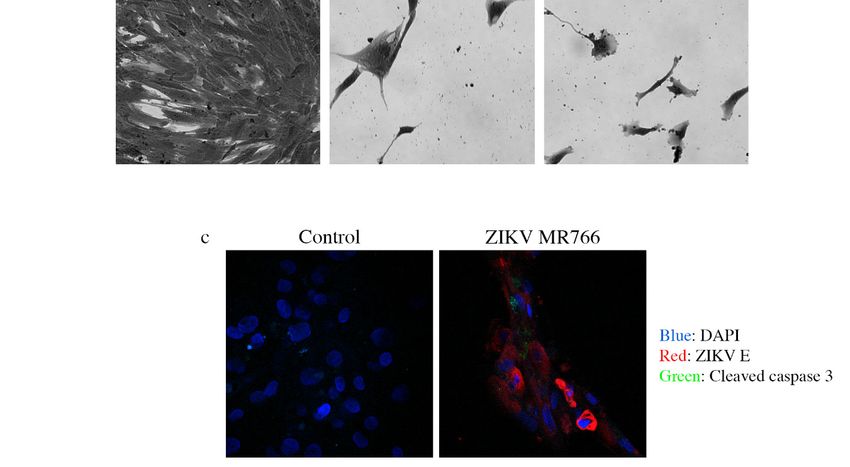
Figure 5. Analysis of continued cell death in ZIKV-infected testicular cells. (a) Cell count graph
Figure 5. Analysis of continued cell death in ZIKV-infected testicular cells. (a) Cell count graph
showing that there were 2-times more cells in the uninfected Hs1.Tes control, compared to ZIKV
showing that there were 2-times more cells in the uninfected Hs1.Tes control, compared to ZIKV
Paraiba-infected Hs1.Tes cells at P1. For both control and ZIKV-infected cells, the count was done
Paraiba-infected Hs1.Tes cells at P1. For both control and ZIKV-infected cells, the count was done
after 7 days of cell passage. ****, p < 0.0001 (unpaired t-test). (b) Microscopic images showing loss
after 7 days of cell passage.
of the Hs1.Tes****, p < 0.0001
monolayer (unpaired t-test).
in ZIKV-infected cells (b) Microscopic
at P3. images
The morphology of showing lossatofthis time
infected cells
the Hs1.Tes monolayer in ZIKV-infected
point appeared cells atand
grossly aberrant P3.pleomorphic
The morphology of infectedtocells
when compared that at
of this time point
uninfected control cells.
appeared grosslyCells
aberrant and pleomorphic when compared to that of uninfected control
were imaged at a magnification of 400×. (c) Confocal microscopy images showing cleavedcells. Cells
were imaged at acaspase
magnification
3 in someof 400×. (c) Confocal
ZIKV-infected microscopy
Sertoli cells at P1. Notimages showing
all ZIKV cleaved caspase
E protein-expressing cells3 stained
positiveSertoli
in some ZIKV-infected for cleaved
cellscaspase 3, supporting
at P1. Not all ZIKV the notion that cell death
E protein-expressing in persistently
cells infected
stained positive forcells was
progressive. Cells were imaged at a magnification of 400 × .
cleaved caspase 3, supporting the notion that cell death in persistently infected cells was progressive.
Cells were imaged
3.3. ZIKVat Infection
a magnification of 400×.
of Seminoma Cell Lines
3.4. ZIKV Infection inTCam-2 cells are a germ cell seminoma cell line, and SEM-1 cells are a testicular cell line which
a Human Neuroblastoma SK-N-SH Cell Line
is an intermediate between a non-seminoma and a true seminoma [21,22]. Compared to all cell
Our laboratory has previously
lines tested, reported cells
SEM-1 seminoma infection of the the
supported human neuroblastoma
highest titers of bothSK-N-SH
ZIKV MR766cell line
and ZIKV
× 7 at 4 dpi, whereas the peak
[23]. We used the SK-N-SH cells to compare ZIKV infection in testicular cells lines, and to determine
Paraiba replication (Figure 1). The ZIKV MR766 titer peaked to 7.1 10
if ZIKV would ZIKV Paraibaintiter
also persist was
these attained
cells. ZIKVatMR766
7 dpi (Figure 1). Interestingly,
replicated the than
to higher titers ZIKVZIKV
replication
Paraibagraph in
(Fig 6a) as expected [23]. Infection of SK-N-SH cells with either ZIKV MR766 or ZIKV Paraiba resultedat 7 dpi.
SEM-1 cells suggested that ZIKV Paraiba was still replicating upwards of the peak attained
No obvious CPE was observed in ZIKV Paraiba-infected SEM-1 cells (Figure 2), but ZIKV MR766
caused notable cell death by 7 dpi and the infected cells appeared smaller in comparison to uninfected
controls (Figure 3). Upon a single passage, ZIKV MR766 infection induced a lytic crisis in SEM-1
seminoma cells. The culture of ZIKV MR766 infected cells did not recover, and thus, a persistent ZIKV
MR76 6 infection of SEM-1 cells could not be established. However, a persistent ZIKV Paraiba infection
was maintained in SEM-1 cells up to 5 passages (Figure 4).
In TCam-2 seminoma cells, ZIKV titers peaked at 4 dpi, with the MR766 strain at 3 × 10⁶ ffu/mL
and the Paraiba strain at 1.4 × 10⁵ ffu/mL (Figure 1). By 7 dpi, ZIKV MR766 and ZIKV Paraiba titers
declined to 6 × 10⁴ ffu/mL and 4 × 10³ ffu/mL, respectively. Interestingly, both ZIKV strains did not
cause any CPE in TCam-2 cells by 7 dpi (Figures 2 and 3). Although ZIKV Paraiba persisted in TCam-2
cells for 4 passages, ZIKV MR766 killed the cells upon passage into P1, suggesting a strain-dependent
delayed cell death mechanism.
3.4. ZIKV Infection in a Human Neuroblastoma SK-N-SH Cell Line
Our laboratory has previously reported infection of the human neuroblastoma SK-N-SH cell
line [23]. We used the SK-N-SH cells to compare ZIKV infection in testicular cells lines, and to
determine if ZIKV would also persist in these cells. ZIKV MR766 replicated to higher titers than ZIKV
Paraiba (Fig 6a) as expected [23]. Infection of SK-N-SH cells with either ZIKV MR766 or ZIKV Paraiba
resulted in extensive CPE by day 4 or 5, respectively (Figure 6b). Following the lytic crises, the few
remaining cells (Figure 6b) died a few days after media replenishment. Thus, no persistent ZIKV
infection of SK-N-SH cells could be established.Viruses 2019, 11, x FOR PEER REVIEW 8 of 11
in extensive CPE by day 4 or 5, respectively (Figure 6b). Following the lytic crises, the few remaining
cells (Figure 6b) died a few days after media replenishment. Thus, no persistent ZIKV infection
Viruses 2019, 11, 42
of
8 of 11
SK-N-SH cells could be established.
ZIKV MR766
Figure6.
Figure 6. ZIKV
ZIKV infection
infection in
in a human neuroblastoma
neuroblastoma SK-N-SH
SK-N-SHcell cellline.
line. (a)
(a) Replication
Replication kinetics
kineticsof
of ZIKV
ZIKV
MR766and
MR766 andParaiba
Paraibastrains
strainsin
inSK-N-SH
SK-N-SHcells.
cells. Error
Error bars
bars represent
representSD SDfrom
fromthe themean
meanfor
for 33 independent
independent
experiments. (b)
experiments. (b) Cell
Cell death
death in
in SK-N-SH
SK-N-SH cell
cell monolayers infected with ZIKV Paraiba or ZIKV MR766.
ZIKV MR766
ZIKV MR766 waswas more
moreaggressive
aggressiveat atkilling
killingSK-N-SH
SK-N-SHcells.
cells. Images
Images ofof the
the SK-N-SH
SK-N-SH monolayer
monolayer were
were
acquiredat
acquired ataamagnification
magnificationof of400 ×.
400×.
4.
4. Discussion
Discussion
ZIKV
ZIKV has has recently
recently been
been shown
shown to to cause
cause devastating
devastating effects
effects inin neonates
neonates following
following in in utero
utero
infection
infection [2,24]. In adults, sexual transmission has been well documented and was associated with
[2,24]. In adults, sexual transmission has been well documented and was associated with
testicular
testicularatrophy
atrophyandandmale
malesterility
sterility[16,17,25,26].
[16,17,25,26]. A A few
few reports
reports have
have subsequently
subsequently reported
reported infection
infection
of
of testicular cells lines,
testicular cells lines, particularly
particularlySertoli
Sertolicells
cells[27–29].
[27–29].InIn order
order to to further
further evaluate
evaluate thesethese reports,
reports, we
we
infected 4 testicular cell lines representing connective tissue (Hs1.Tes cells) and germ cells (SEM-1
infected 4 testicular cell lines representing connective tissue (Hs1.Tes cells) and germ cells (SEM-1
and
and TCam-2
TCam-2 cells)
cells) as
as well
well as
as Sertoli
Sertoli cells
cells with
with the
the prototype
prototype ZIKV
ZIKV MR766
MR766 and and aa clinical
clinical isolate
isolate ZIKV
ZIKV
Paraiba to determine the extent to which the viruses would persist
Paraiba to determine the extent to which the viruses would persist in these cells. in these cells.
All
All the
the cells
cells we
we tested
tested were
were permissive
permissive to to both
both ZIKV
ZIKV MR766
MR766 andand ZIKV
ZIKV Paraiba,
Paraiba, butbut the
the former
former
replicated
replicated to higher titers than the clinical isolate (Figure 1). These results suggested that any of
to higher titers than the clinical isolate (Figure 1). These results suggested that any of the
the
testicular cells may be responsible for disseminating virus in the infected organ.
testicular cells may be responsible for disseminating virus in the infected organ. However, Leydig However, Leydig cells
were
cells reportedly less susceptible
were reportedly to ZIKV
less susceptible infection
to ZIKV [27], and
infection [27],their
and role
theirinrole
virus dissemination
in virus dissemination may
be limited. The higher replication levels of ZIKV MR766 could be related to
may be limited. The higher replication levels of ZIKV MR766 could be related to the fact that this the fact that this strain
has been
strain haspassaged extensively
been passaged in mouse
extensively in brains
mouse[1,30].
brainsIn[1,30].
addition, ZIKV MR766
In addition, ZIKV andMR766ZIKV Paraiba
and ZIKV
are 89% identical at nucleotide sequence level and 97% (3313/3423) identical at amino acid level
(BLAST; https://blast.ncbi.nlm.nih.gov/Blast.cgi). Thus, the ZIKV MR766 strain may have adaptedViruses 2019, 11, 42 9 of 11 to mammalian cell culture better than the ZIKV Paraiba strain which was passaged only
Viruses 2019, 11, 42                                                                                            10 of 11

persistence. Our results are consistent with reports that ZIKV persists in testicular cells and suggest
that testicular atrophy may be a result of a slow and progressive cell death.

Author Contributions: Conceptualization, L.M. and M.E.B.; Experiments, L.M.; Writing—Original Draft
Preparation, L.M.; Writing—Review & Editing, L.M. and M.E.B.
Funding: This study was supported by the Division of Intramural Research program of the National Institute of
Allergy and Infectious Diseases at the National Institutes of Health.
Acknowledgments: We thank members of the Biology of Vector-Borne Viruses section for useful discussions.
Conflicts of Interest: The authors declare no conflict of interest.

References
1.    Dick, G.W.A.; Kitchen, S.F.; Haddow, A.J. Zika Virus (I). Isolations and serological specificity. Trans. R. Soc.
      Trop. Med. Hyg. 1952, 46, 509–520. [CrossRef]
2.    Mlakar, J.; Korva, M.; Tul, N.; Popović, M.; Poljšak-Prijatelj, M.; Mraz, J.; Kolenc, M.; Resman Rus, K.;
      Vesnaver Vipotnik, T.; Fabjan Vodušek, V.; et al. Zika Virus Associated with Microcephaly. N. Engl. J. Med.
      2016, 374, 951–958. [CrossRef] [PubMed]
3.    Nascimento, O.J.M.; da Silva, I.R.F. Guillain–Barré syndrome and Zika virus outbreaks. Curr. Opin. Neurol.
      2017, 30, 500–507. [CrossRef] [PubMed]
4.    Roze, B.; Najioullah, F.; Ferge, J.L.; Apetse, K.; Brouste, Y.; Cesaire, R.; Fagour, C.; Fagour, L.; Hochedez, P.;
      Jeannin, S.; et al. Zika virus detection in urine from patients with Guillain-Barre syndrome on Martinique,
      January 2016. Euro Surveill. 2016, 21, 30154. [CrossRef] [PubMed]
5.    Rozé, B.; Najioullah, F.; Fergé, J.-L.; Dorléans, F.; Apetse, K.; Barnay, J.-L.; Daudens-Vaysse, E.; Brouste, Y.;
      Césaire, R.; Fagour, L.; et al. Guillain-Barré Syndrome Associated With Zika Virus Infection in Martinique in
      2016: A Prospective Study. Clin. Infect. Dis. 2017, 65, 1462–1468. [CrossRef] [PubMed]
6.    Read, J.S.; Torres-Velasquez, B.; Lorenzi, O.; Rivera Sanchez, A.; Torres-Torres, S.; Rivera, L.V.;
      Capre-Franceschi, S.M.; Garcia-Gubern, C.; Munoz-Jordan, J.; Santiago, G.A.; et al. Symptomatic zika
      virus infection in infants, children, and adolescents living in puerto rico. JAMA Pediatr. 2018, 172, 686–693.
      [CrossRef]
7.    Foy, B.D.; Kobylinski, K.C.; Foy, J.L.C.; Blitvich, B.J.; Travassos da Rosa, A.; Haddow, A.D.; Lanciotti, R.S.;
      Tesh, R.B. Probable Non–Vector-borne Transmission of Zika Virus, Colorado, USA. Emerg. Infect. Dis. 2011,
      17, 880–882. [CrossRef]
8.    Fréour, T.; Mirallié, S.; Hubert, B.; Splingart, C.; Barrière, P.; Maquart, M.; Leparc-Goffart, I. Sexual
      transmission of Zika virus in an entirely asymptomatic couple returning from a Zika epidemic area, France,
      April 2016. Euro Surveill. 2016, 21, 30254. [CrossRef]
9.    Nicastri, E.; Castilletti, C.; Liuzzi, G.; Iannetta, M.; Capobianchi, M.R.; Ippolito, G. Persistent detection of
      Zika virus RNA in semen for six months after symptom onset in a traveller returning from Haiti to Italy,
      February 2016. Euro Surveill. 2016, 21. [CrossRef]
10.   Davidson, A.; Slavinski, S.; Komoto, K.; Rakeman, J.; Weiss, D. Suspected Female-to-Male Sexual
      Transmission of Zika Virus—New York City, 2016. MMWR Morb. Mortal. Wkly. Rep. 2016, 65, 716–717.
      [CrossRef]
11.   Zika Situation Report. Available online: http://www.who.int/emergencies/zika-virus/situation-report/10-
      march-2017/en/ (accessed on 8 January 2019).
12.   2016 Case Counts in the US. Available online: https://www.cdc.gov/zika/reporting/2016-case-counts.html
      (accessed on 8 January 2019).
13.   2017 Case Counts in the US. Available online: https://www.cdc.gov/zika/reporting/2017-case-counts.html
      (accessed on 8 January 2019).
14.   Hastings, A.K.; Fikrig, E. Zika Virus and Sexual Transmission: A New Route of Transmission for
      Mosquito-borne Flaviviruses. Yale J. Biol. Med. 2017, 90, 325–330. [PubMed]
15.   Mead, P.S.; Duggal, N.K.; Hook, S.A.; Delorey, M.; Fischer, M.; Olzenak McGuire, D.; Becksted, H.; Max, R.J.;
      Anishchenko, M.; Schwartz, A.M.; et al. Zika Virus Shedding in Semen of Symptomatic Infected Men.
      N. Engl. J. Med. 2018, 378, 1377–1385. [CrossRef] [PubMed]
Viruses 2019, 11, 42 11 of 11
16. Uraki, R.; Hwang, J.; Jurado, K.A.; Householder, S.; Yockey, L.J.; Hastings, A.K.; Homer, R.J.; Iwasaki, A.;
Fikrig, E. Zika virus causes testicular atrophy. Sci. Adv. 2017, 3, e1602899. [CrossRef] [PubMed]
17. Govero, J.; Esakky, P.; Scheaffer, S.M.; Fernandez, E.; Drury, A.; Platt, D.J.; Gorman, M.J.; Richner, J.M.;
Caine, E.A.; Salazar, V.; et al. Zika virus infection damages the testes in mice. Nature 2016, 540, 438–442.
[CrossRef] [PubMed]
18. Sheng, Z.-Y.; Gao, N.; Wang, Z.-Y.; Cui, X.-Y.; Zhou, D.-S.; Fan, D.-Y.; Chen, H.; Wang, P.-G.; An, J. Sertoli
Cells Are Susceptible to ZIKV Infection in Mouse Testis. Front. Cell. Infect. Microbiol. 2017, 7, 272. [CrossRef]
[PubMed]
19. Müller, J.A.; Harms, M.; Krüger, F.; Groß, R.; Joas, S.; Hayn, M.; Dietz, A.N.; Lippold, S.; von Einem, J.;
Schubert, A.; et al. Semen inhibits Zika virus infection of cells and tissues from the anogenital region.
Nature Commun. 2018, 9, 2207. [CrossRef] [PubMed]
20. Biedler, J.L.; Helson, L.; Spengler, B.A. Morphology and Growth, Tumorigenicity, and Cytogenetics of Human
Neuroblastoma Cells in Continuous Culture. Cancer Res. 1973, 33, 2643–2652.
21. Mizuno, Y.; Gotoh, A.; Kamidono, S.; Kitazawa, S. Establishment and characterization of a new human
testicukar germ cell tumor cell line (TCam-2). Jpn. J. Urol. 1993, 84, 1211–1218. [CrossRef]
22. Russell, S.M.; Lechner, M.G.; Mokashi, A.; Megiel, C.; Jang, J.K.; Taylor, C.R.; Looijenga, L.H.J.; French, C.A.;
Epstein, A.L. Establishment and Characterization of a new Human Extragonadal Germ Cell Line, SEM-1,
and its Comparison With TCam-2 and JKT-1. Urology 2013, 81, 464.e1–464.e9. [CrossRef]
23. Offerdahl, D.K.; Dorward, D.W.; Hansen, B.T.; Bloom, M.E. Cytoarchitecture of Zika virus infection in human
neuroblastoma and Aedes albopictus cell lines. Virology 2017, 501, 54–62. [CrossRef]
24. Tang, H.; Hammack, C.; Ogden, S.C.; Wen, Z.; Qian, X.; Li, Y.; Yao, B.; Shin, J.; Zhang, F.; Lee, E.M.; et al.
Zika Virus Infects Human Cortical Neural Precursors and Attenuates Their Growth. Cell Stem Cell 2016, 18,
587–590. [CrossRef] [PubMed]
25. Ma, W.; Li, S.; Ma, S.; Jia, L.; Zhang, F.; Zhang, Y.; Zhang, J.; Wong, G.; Zhang, S.; Lu, X.; et al. Zika Virus
Causes Testis Damage and Leads to Male Infertility in Mice. Cell 2016, 167, 1511–1524.e10. [CrossRef]
[PubMed]
26. Stassen, L.; Armitage, C.; van der Heide, D.; Beagley, K.; Frentiu, F. Zika Virus in the Male Reproductive
Tract. Viruses 2018, 10, 198. [CrossRef]
27. Kumar, A.; Jovel, J.; Lopez-Orozco, J.; Limonta, D.; Airo, A.M.; Hou, S.; Stryapunina, I.; Fibke, C.; Moore, R.B.;
Hobman, T.C. Human Sertoli cells support high levels of Zika virus replication and persistence. Sci. Rep.
2018, 8, 5477. [CrossRef] [PubMed]
28. Strange, D.P.; Green, R.; Siemann, D.N.; Gale, M.; Verma, S. Immunoprofiles of human Sertoli cells infected
with Zika virus reveals unique insights into host-pathogen crosstalk. Sci. Rep. 2018, 8, 8702. [CrossRef]
29. Siemann, D.N.; Strange, D.P.; Maharaj, P.N.; Shi, P.-Y.; Verma, S. Zika Virus Infects Human Sertoli Cells and
Modulates the Integrity of the In Vitro Blood-Testis Barrier Model. J. Virol. 2017, 91, e00623-17. [CrossRef]
[PubMed]
30. Haddow, A.D.; Schuh, A.J.; Yasuda, C.Y.; Kasper, M.R.; Heang, V.; Huy, R.; Guzman, H.; Tesh, R.B.;
Weaver, S.C. Genetic Characterization of Zika Virus Strains: Geographic Expansion of the Asian Lineage.
PLoS Negl. Trop. Dis. 2012, 6, e1477. [CrossRef]
31. Marzi, A.; Emanuel, J.; Callison, J.; McNally, K.L.; Arndt, N.; Chadinha, S.; Martellaro, C.; Rosenke, R.;
Scott, D.P.; Safronetz, D.; Whitehead, S.S.; et al. Lethal Zika Virus Disease Models in Young and Older
Interferon α/β Receptor Knock Out Mice. Front. Cell. Infect. Microbiol. 2018, 8, 117. [CrossRef]
32. Duggal, N.K.; Ritter, J.M.; Pestorius, S.E.; Zaki, S.R.; Davis, B.S.; Chang, G.-J.J.; Bowen, R.A.; Brault, A.C.
Frequent Zika Virus Sexual Transmission and Prolonged Viral RNA Shedding in an Immunodeficient Mouse
Model. Cell Rep. 2017, 18, 1751–1760. [CrossRef]
© 2019 by the authors. Licensee MDPI, Basel, Switzerland. This article is an open access
article distributed under the terms and conditions of the Creative Commons Attribution
(CC BY) license (http://creativecommons.org/licenses/by/4.0/).You can also read