Plasticity in Colorectal Cancer: Why Cancer Cells Differentiate
←
→
Page content transcription
If your browser does not render page correctly, please read the page content below
Review

Plasticity in Colorectal Cancer: Why Cancer Cells Differentiate
Romina Judith Walter 1,†, Steffen Joachim Sonnentag 1,†, Véronique Orian‐Rousseau 1,*
and Leonel Munoz‐Sagredo 1,2,*

                                          1 Karlsruhe Institute of Technology, Institute of Biological and Chemical Systems—Functional Molecular
                                            Systems, Hermann‐von‐Helmholtz‐Platz 1, 76344 Eggenstein‐Leopoldshafen, Germany;
                                            romina.walter@kit.edu (R.J.W.); steffen.sonnentag@kit.edu (S.J.S.)
                                          2 Faculty of Medicine, University of Valparaiso, Angamos 655, Vina del Mar 2540064, Chile

                                          * Correspondence: veronique.orian‐rousseau@kit.edu (V.O.‐R.); leonel.munoz@partner.kit.edu (L.M.‐S.)
                                          † Co‐first authors of this study.

                                          Simple Summary: The cancer stem cell hypothesis postulates that tumors arise from a few cells with
                                          self‐renewal capabilities. The identification of stem cell markers, the development of mouse and
                                          human tumor organoids and their application in mouse models, allowing lineage tracing, helped to
                                          better understand the cancer stem cell model as well as the role of differentiation. This review aims
                                          at providing insights on the interplay between cancer stem cells and differentiated cells, as well as
                                          the importance of plasticity between the two states.

                                          Abstract: The cancer stem cell hypothesis poses that the bulk of differentiated cells are non‐tumor‐
                                          igenic and only a subset of cells with self‐renewal capabilities drive tumor initiation and progres‐
                                          sion. This means that differentiation could have a tumor‐suppressive effect. Accumulating evidence
                                          shows, however, that in some solid tumors, like colorectal cancer, such a hierarchical organization
                                          is necessary. The identification of Lgr5 as a reliable marker of normal intestinal epithelial stem cells,
Citation: Walter, R.J.; Sonnentag,        together with strategies to trace cell lineages within tumors and the possibility to selectively ablate
S.J.; Orian‐Rousseau, V.;                 these cells, have proven the relevance of Lgr5+ cells for cancer progression. On the contrary, the role
Munoz‐Sagredo, L. Plasticity in           of Lgr5− cells during this process remains largely unknown. In this review, we explore available
Colorectal Cancer: Why Cancer Cells       evidence pointing towards possible selective advantages of cancer cells organized hierarchically
Differentiate. Cancers 2021, 13, 918.     and its resulting cell heterogeneity. Clear evidence of plasticity between cell states, in which loss of
https://doi.org/10.3390/
                                          Lgr5+ cells can be replenished by dedifferentiation of Lgr5− cells, shows that cell hierarchies could
cancers13040918
                                          grant adaptive traits to tumors upon changing selective pressures, including those derived from
                                          anticancer therapy, as well as during tumor progression to metastasis.
Academic Editors: Cinzia Allegrucci,
Paloma Ordóñez‐Morán and Daniel
                                          Keywords: colorectal cancer; cancer stem cells; plasticity; cancer stem cell markers; tumor
Louvard
                                          heterogeneity; tumor‐organoids; lineage tracing; clonal cooperation; radio‐resistance; serial
Received: 4 January 2021                  transplantation; therapeutic resistance
Accepted: 17 February 2021
Published: 22 February 2021

Publisher’s Note: MDPI stays neu‐         1. Introduction
tral with regard to jurisdictional
                                                According to the cancer stem cell (CSC) hypothesis, only a small subset of cancer
claims in published maps and insti‐
                                          cells are capable of recapitulating the tumor of origin after serial transplantation into im‐
tutional affiliations.
                                          munocompromised mice [1,2]. This distinguishes tumorigenic from non‐tumorigenic can‐
                                          cer cells [3]. Profiling these operationally defined CSCs has enabled the identification of
                                          molecular markers that can be used to select cell populations with enhanced tumorigenic
                                          capabilities. This implies that tumors can be organized in cell hierarchies reminiscent of
Copyright: © 2021 by the author. Li‐
censee MDPI, Basel, Switzerland.
                                          the ones in normal tissues, with stem cells at the apex, that give rise to transient amplify‐
This article is an open access article
                                          ing progenitor cells that undergo processes of differentiation into several cell lineages.
distributed under the terms and con‐      This hierarchical organization of cancer cells was initially proposed for acute myeloid leu‐
ditions of the Creative Commons At‐       kemia [4], but has been extended to solid tumors, including colorectal carcinomas [5–7].
tribution (CC BY) license (http://crea‐   Initially proposed as an alternative to the model of clonal evolution of cancer cells [8] as a
tivecommons.org/licenses/by/4.0/).        source of intra‐tumoral heterogeneity, the concept of CSC and clonal evolution were

Cancers 2021, 13, 918. https://doi.org/10.3390/cancers13040918                                                       www.mdpi.com/journal/cancers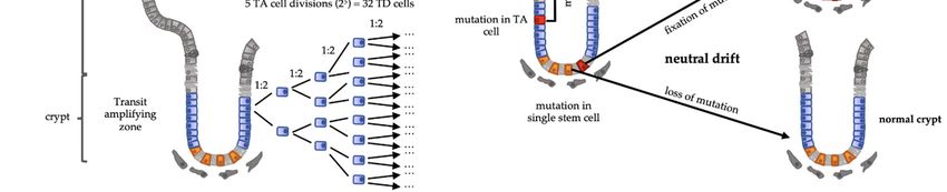
Cancers 2021, 13, 918                                                                                           2 of 18

                        found to be non‐mutually exclusive, being now integrated with other sources of pheno‐
                        typic variability like the ones influenced by distinct tumor microenvironments [2,9,10]. In
                        parallel, the discovery of leucine‐rich‐repeats containing G‐Protein‐Coupled Receptor 5
                        (Lgr5) as a bonafide marker of normal intestinal epithelial stem cells [11] enabled an im‐
                        portant step towards understanding stem cell properties and function in the intestinal ep‐
                        ithelium. Direct lineage tracing showed that colorectal adenomas retain some of the hier‐
                        archical organization of their tissue of origin [12]. This experimental strategy—together
                        with the development of ex vivo colorectal tumor organoids that are susceptible to further
                        genetic manipulation, labeling, and transplantation—enabled researchers to trace the fate
                        of mouse and human colon cancer clones in vivo during tumor progression and metastasis
                        in mice [13–16]. It also enabled the selective ablation of Lgr5+ cancer cells in vivo, demon‐
                        strating their importance for colorectal cancer progression in animal models [13–16].
                              It is interesting to note, however, that from an evolutionary perspective, the hierar‐
                        chical organization of cells in rapidly renewing tissues, like the intestinal epithelium dur‐
                        ing homeostasis, can act as a tumor suppressor mechanism, by minimizing cell divisions
                        of stem cells, in charge of self‐renewal, while depositing a higher proliferative burden on
                        transit‐amplifying cells destined to terminally differentiate into postmitotic cells that are
                        shed out from the tissue. The fact that in colorectal tumors, cells expressing stem cell mark‐
                        ers self‐renew and give rise to cells expressing different markers of differentiated lineages
                        [13–17], raises the question of the role of differentiated cancer cells along cancer progres‐
                        sion. Does cancer cell differentiation confer any advantage to the tumoral ecosystem‐pri‐
                        mary or metastatic‐subjected to Darwinian selective pressures? In this review, we explore
                        available evidence to address this question. We conclude that a hierarchical organization
                        of tumors, i.e., tumors containing cancer cells in different states along their process of dif‐
                        ferentiation, can play an important role in tumor progression. This does not refute the
                        central role of CSCs and their relevance as targets for curative cancer therapy develop‐
                        ment, given that, as in the normal intestinal epithelium, in neoplastic lesions, Lgr5+ cells
                        can be rapidly replenished by dedifferentiation of Lgr5− cells after Lgr5+ cell damage or
                        ablation [15,16]. This property, called cell plasticity, confers an adaptive trait to cancers,
                        expanding their phenotypic repertoire to overcome selective challenges along cancer pro‐
                        gression.

                        2. Cell Hierarchy Is a Tumor‐Suppressor Mechanism
                              Cells in the human intestinal epithelium, which are exposed to the potentially harm‐
                        ful environment of the microbiome and digestive processes, are continually shed and re‐
                        placed. This rapid cell renewal safeguards the physiological and structural function of this
                        tissue: actively and passively absorbing nutrients while isolating other tissues from the
                        processes of food degradation. However, high rates of cell division increase the likelihood
                        of DNA‐replication errors [18]. Many point mutations found in cancer cells arise from
                        error‐generating activities of DNA polymerases [19]. Although cells possess mechanisms
                        to avoid genomic instability, when genes involved in these mechanisms are themselves
                        the targets of mutations, a high mutagenic rate may ensue and lead to cancer. Indeed,
                        families with hereditary non‐polypous colorectal carcinoma harbor a germline mutation
                        in genes required for post‐replication mismatch repair that destabilizes their rapidly rep‐
                        licating genome [20].
                              However, if cells engaged in rapid and repeated cell division are “dismissed from the
                        task” of self‐renewal, any mutation in these cells is lost when the mutated cell is lost, pre‐
                        venting its fixation in the tissue [21]. Terminally differentiated (TD) cells are post‐mitotic
                        and are shed into the intestinal lumen after a short‐lived function. TD cells are generated
                        from transit‐amplifying (TA) cells that can undergo several cycles of symmetrical cell di‐
                        vision before becoming TD cells. Because these cells do not self‐renew, i.e., they all de‐
                        velop into TD cells, this compartment must be replenished by stem cells (SCs) which are
                        in the cell compartment responsible for long‐term self‐renewal. Therefore, an eventual
                        advantageous mutation in a cell of this compartment has a higher risk to remain within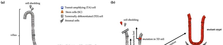
Cancers 2021, 13, 918                                                                                            3 of 18

                        the tissue and be continuously transmitted to the clonal progeny thereafter. Conversely,
                        since TA or TD cells do not self‐renew, mutations occurring in them are likely to be lost
                        upon cell shedding. If the cell division rate is directly proportional to mutation rate [18,19],
                        it is beneficial to minimize the division rate of the self‐renewing compartment and deposit
                        cell number expansion in compartments of cells destined to be lost from the tissue. This
                        three‐compartment hierarchical structure of tissues—SCs, TA cells, and TD cells—enables
                        to lower the division‐rate self‐renewal while rendering high numbers of TD cells, the
                        product of an exponential expansion of TA cells. In homeostatic conditions, TA cells un‐
                        dergo 4–5 rounds of cell division [22], giving rise to 32 TD cells from a single SC division
                        (Figure 1a). Using computational modelling of this system, Pepper and collaborators stud‐
                        ied the impact of advantageous mutations—either increasing proliferation rates or de‐
                        creasing cell death rates—arising in one of the three cell compartments. Unique mutations
                        arising in the TA or TD compartments were always lost [21]. Thus, the location of the
                        major proliferative function in a compartment devoid of self‐renewal capacity allows high
                        rates of cell turnover with low replicative mutagenic risk, resulting in a tumor‐suppressor
                        system.
                               However, although the SC compartment has a lower proliferative rate, most SCs di‐
                        vide daily [22]. If every SC would be constantly self‐renewed—either by symmetric divi‐
                        sion into two daughter SCs or asymmetric division into a SC and TA cell—along the life
                        span of an individual, they would undergo a number of divisions that would render a
                        very high risk of mutations and they would be inevitably fixated in the tissue. This is
                        indeed not the case at the level of individual SCs. Lineage tracing clearly revealed that the
                        SCs compartment also undergoes a turnover following neutral competition dynamics.
                        Without any intrinsic advantage, some SCs divide mostly symmetrically, displacing
                        neighbor SCs out of the niche [22–24]. This means that mutations in the SC compartment
                        can still be lost by neutral drift (Figure 1b).
                               Since differentiation can act as a tumor suppression mechanism by impeding self‐
                        renewal when a cell divides but does not further differentiate, it constitutes a self‐renew‐
                        ing clone [25,26]. Indeed, when Pepper and collaborators introduced mutations that
                        blocked cell differentiation to their model, these mutations were fixed in the tissue regard‐
                        less of the compartment from which they arose [21]. This can explain the existence of un‐
                        differentiated tumors. However, it does not show how differentiated tumors arise within
                        the constraints of this model.
                               Although transcriptomic analysis of in vivo colorectal cancer models shows that dis‐
                        tinguishable cell compartments in CRCs are less defined—probably because of the loss of
                        niche factor gradients that maintain them in non‐neoplastic tissues—cells expressing stem
                        cell markers are still distinguishable from populations expressing differentiation markers
                        of diverse lineages reminiscent of normal tissues [13,15–17]. This means that differentiated
                        tumors require that at least some cell‐differentiation capabilities are retained. Therefore,
                        if differentiation into non‐tumorigenic cells is tumor‐suppressive, why is this type of cell
                        organization selected under the Darwinian mechanisms of cancer progression? One hy‐
                        pothesis is that for some tumors, such as differentiated colorectal cancers, establishment
                        of different cell hierarchies through differentiation can be advantageous for the tumor. If
                        this is the case, what are the selective advantages of such tumor organization? To assess
                        this question, we will begin by analyzing the function of the putative CSCs as a baseline
                        to distinguish the relevance of differentiated cells along tumor progression.
Cancers 2021, 13, 918 4 of 18
Figure 1. (a) Intestinal crypt‐villus structure representing the stem cell (SC) function. If a stem cell generates a transit‐
amplifying (TA) cell and the latter divides 5 times, it will generate 32 (25) terminally differentiated cells (TD) from a single
stem cell division. (b) Intestinal cells harboring a mutation in a TA cell or a TD cell are shed and lost. Mutation in a SC
gets fixed if the mutated SC dominates TD: Terminally differentiated; SC: stem cells; TA: Transit amplifying.
3. Cancer Stem Cells in Colorectal Tumors
3.1. Serial Transplantation and CSC Markers
CSCs have customarily been defined operationally: the ability to recapitulate tumors
upon serial transplantation of cells from human cancers into immunocompromised mice.
Recapitulation of the organization implies self‐renewal capacities as well as potency of
differentiation, reflecting the function of normal stem cells of adult tissues. The proportion
of injected mice that developed disease was used to quantify the efficiency of tumor for‐
mation. Serial dilution of cancer cells decreased this efficiency, leading to the hypothesis
that only a small subset of cells, from a heterogeneous bulk, possess these tumor‐initiating
capabilities. These findings set the conditions to search for cell surface markers to identify
these tumorigenic cells. Fluorescent‐activated cell sorting (FACS) enabled the enrichment
of cell populations with cells possessing these markers, increasing tumorigenic efficiency
with reduced numbers of injected cells. In the field‐foundational study, cells from acute
myeloid leukemia (AML) patients were injected into non‐obese diabetic mice with severe
combined immunodeficiency disease (NOD/SCID). The leukemia‐initiating cells could be
selected with the same markers (CD34+/CD38−) as normal hematopoietic cells that are able
to repopulate NOD/SCID mice upon bone marrow transplantation [4]. This approach was
subsequently extended to solid tumors like breast cancer [27], brain tumors [28], colorectal
cancer [5–7], pancreatic cancer [29], and ovarian cancer [30].
CSCs of solid malignancies, as defined above, have been controversial [3,9,31]. Re‐
sidual immune response to xenografts in immunodeficient mice might affect tumor initi‐
ation capabilities of cells. For example, although the melanoma tumor‐initiating capacity
was estimated to be present in only one of every million cells in NOD/SCID mice, trans‐
plantation into NOD/SCID IL‐2Rγ‐null (NSG) mice enabled tumor initiation from single
cells in up to 30% of the animals [32]. This suggested that the assay was actually selecting
cells with immune‐evasive capabilities rather than reflecting the existence of a small stem
cell pool. Instead, residual immune response could underestimate the putative CSC fre‐
quency and bias their marker profile. In addition, removal of cancer cells from their mi‐
croenvironment in solid tumors by enzymatic dissociation and sorting could affect their
fitness and tumor‐initiation capabilities. This could also be the case for microenviron‐
ments that do not reflect the conditions in the tissue of origin, especially for heterotopic
transplantation, thus compromising the tumorigenicity of clones that would otherwise
possess it. Therefore, transplantation assays show the ability to form tumors despite theseCancers 2021, 13, 918 5 of 18
barriers and do not necessarily reflect the fates of cells in their tumor of origin. However,
syngeneic serial transplantation assays have supported the hierarchical organization for
mouse tumors, rejecting the possibility that these assays only reflect the action of clonal
selective pressures, unrelated to a putative CSC function [3].
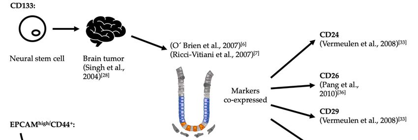
In spite of the limitations of serial transplantation, this method yielded several mark‐
ers with the potential to identify cells with self‐renewal and differentiation capabilities in
vivo. The first marker identified for colorectal CSCs was CD133 [6,7]. In parallel, cells ex‐
pressing high levels of EpCAM (epithelial cell adhesion molecule) while also expressing
CD44 were also shown to enrich for tumor‐initiating capabilities [5]. This discrepancy
epitomizes the historical development of CSC markers, which were initially extrapolated
either from markers known for normal stem cells or from the experience with models of
other cancer types (Figure 2). However, further technical development supported their
utility. Vermeulen and collaborators used single‐cell cloning of CD133+ cells from patient
samples to show that these cells could give rise to multilineage differentiated tumors upon
implantation [33]. However, frequent loss of the epitope of CD133 during immunohisto‐
chemistry of patient histological samples prevented further clinical validation [34,35].
Figure 2. Historical development of CSC markers for colorectal cancer. In papers published back‐to‐back, O’Brien as well
as Ricci‐Vitiani and colleagues identified CD133+ colorectal cells as possessing tumor initiation capabilities upon hetero‐
topic xenografts [6,7]. The criterion to probe this marker was derived from a previous experience with brain tumors, which
in turn had derived from its expression in neural stem cells [28]. In parallel, Dalebra and collaborators had similar results,
using the combination of the markers EpCAMhigh/CD44+ in subcutaneous injections [5,36]. In their case, the criterion to
probe these markers derived from their previous work identifying putative CSCs from breast tumors [27]. Since none of
these markers are expressed solely in putative CSCs, other associated markers were identified in an attempt to increase
the specificity of markers for CSCs upon use in combination CSC: Cancer stem cell.
Refinement of the specificity of putative CSC markers can potentially be achieved by
considering alternative splicing [37]. This can be specifically triggered by pathological
processes like inflammation and cancer [38]. This may be the case for CD44. Notably, this
adhesion molecule has been shown to be a putative CSC marker not only for colon cancer,
but also for cancers of the bladder, breast, gut, head and neck, liver, lung, ovary, pancreas,
prostate, and other organs [39]. However, CD44 is subject to complex alternative splicing
that generates a myriad of isoforms in the extracellular domain. Consequently, multiple
combinations of 9 variant exons in humans (10 in mice) and additional post‐Cancers 2021, 13, 918 6 of 18
transcriptional modifications lead to high heterogeneity. Relevant to colorectal cancer, To‐
daro and collaborators showed that CD44v6—the CD44 family member most associated
with malignancies—marks a population of cells that is highly tumorigenic and has meta‐
static potential [40].
Subsequent development of surface markers focused on those that reflect the func‐
tion of signaling pathways known to be essential for stemness. In terms of activated sig‐
naling pathways, strong parallels were shown to exist between physiological and patho‐
logical conditions in the intestine [17,41]. Wnt signaling plays a fundamental role in ho‐
meostasis of the intestine, and malignant transformation is initiated by mutations in pro‐
teins of this pathway [42–44]. Wnt pathway mutations were detected in 92% of all colon
cancer patients, 80% of which had a mutation in the APC gene [45–47]. This mutation leads
to permanent activation of the canonical Wnt signaling pathway due to constant β‐catenin
stabilization followed by its nuclear translocation [43]. However, even if the majority of
colorectal tumors have a hyperactivated Wnt signaling pathway, immunohistochemical
studies showed that not every cell in a tumor exhibits this high Wnt activity [48]. Colorec‐
tal cancer cells with tumorigenic capacities were shown to exhibit high Wnt activity [33].
These cells upregulated stem cell markers, like Lgr5 and Ascl2, and recapitulated the Wnt
activity heterogeneity upon tumor growth [49]. The advent of methods to trace the fate of
stem cells in vivo [11] enabled the robust validation of one of these Wnt target‐gene mark‐
ers, Lgr5, as a marker of normal intestinal stem cells. The application of these methods to
colorectal cancer, together with methods to precisely ablate Lgr5+ cells in vivo [50], initi‐
ated a new era for the CSCs concept, releasing it from its dependency on serial transplan‐
tation as an operational definition, and leading to deep insights into the function of these
cells in cancer progression.
Although Lgr5 has been proven to be a bona fide marker of CRC‐CSC and can be
reliably used in available CRC in in vivo and ex vivo models, there is evidence of CRC not
expressing Lgr5 [15,17]. This could be due to epigenetic silencing, while keeping high Wnt
signaling [51] or by emergence of cells that are recognizable by alternative markers. Recent
evidence from the Stappenbeck group identified Hopx as the marker of colitis‐associated
regenerative stem cells [52]. With chronic inflammation being a risk factor for the devel‐
opment of CRC, these findings could be relevant for some neoplastic processes.
3.2. Lineage Tracing and Organoids: Evidence for Cancer Stem Cells
Groundbreaking experiments with genetic lineage tracing were key to show that the
crypt base columnar (CBC) cells at the bottom of the intestinal crypts function as bona fide
stem cells [11]. This method allowed the identification of the Wnt target gene Lgr5, which
is specifically expressed in the CBC cells, as a reliable intestinal stem cell (ISC) marker
[11]. Barker and colleagues used the Cre‐loxP genetic recombination systems to perform
lineage tracing. In the Lgr5‐EGFP‐IRES‐CreERT2 mouse model, the Cre recombinase is ex‐
pressed under the control of the cell‐specific Lgr5 promoter. This mouse line was crossed
with R26R–lacZ reporter mice containing a loxP‐STOP‐loxP sequence in front of the re‐
porter gene lacZ. The activated recombinase specifically activates the reporter gene ex‐
pression in cells expressing Lgr5 by excising the STOP sequence. After the STOP sequence
is removed, future descendent cells of the LacZ+ stem cells continue to express the reporter
LacZ [11]. Both stem cell requirements were thus met by Lgr5+ CBC cells: the generation
of multiple lineages and long‐term self‐renewal. Although this tracing cannot be per‐
formed in humans, stem cell dynamics have been successfully studied in the human colon
when observing the spread of somatic mutations [53–55].Cancers 2021, 13, 918 7 of 18
3.3. Tumor Organoids
The ex vivo organoids culture was an important development that enabled the fur‐
ther investigation of stem cell functionality [56]. Upon incorporation into a three‐dimen‐
sional (3D) matrix, it was possible to grow single Lgr5+ adult stem cells, or crypts contain‐
ing the stem cells isolated from the intestine, into three‐dimensional multicellular tissue
models. These organoids are maintained by the self‐renewal of the intestinal stem cells,
thus mimicking the in vivo properties of the intestine and remaining genetically and phys‐
ically stable. The essential role of Lgr5+ stem cells in forming organoids was demonstrated
using the Lgr5DTR‐eGFP mouse model. In this model, Lgr5+ stem cells express eGFP and
the diphtheria toxin receptor (DTR), which enabled only the stem cells to be marked and
ablated. Lgr5DTR‐eGFP tumor organoids treated with the toxin collapsed after losing
their stem cells and were unable to regrow upon toxin treatment maintenance, thus
demonstrating the necessity of Lgr5+ stem cells [16,50].
Methods developed to culture organoids originating from human adult colonic tis‐
sue [57] or human pluripotent stem cells [58] created the foundations to expand this tech‐
nique to patient‐derived tumor tissue. Lgr5+ stem cells were shown to be required for the
formation of normal tissue organoids as well as tumor organoids [50]. The tumor organ‐
oids, however, differ phenotypically from the organoids originated from the healthy tis‐
sue. Relevantly, transcriptomic profiling of colorectal tumor biopsies and organoids de‐
rived from them showed that they conserve gene expression signatures from the tumor of
origin [14].
Cultivation over long periods of time led to the establishment of biobanks containing
a large number of differently characterized tumor organoids [59–61]. Furthermore, proto‐
cols to manipulate organoids by means of CRISPR/Cas9 [62,63] enabled modelling of com‐
mon genetic mutations of the adenoma‐carcinoma sequence of colorectal cancer in mouse
organoids [13,16] and in organoids from human intestinal epithelium [64]. The same ap‐
proach allows labelling of human tumor organoids from patient samples while simulta‐
neously introducing transgenes that enable inducible and selective ablation of Lgr5+ cells
[14,15]. Subsequent implantation of these engineered tumor organoids into mice led to
lineage tracing and ablation experiments to study the role of Lgr5+ cells or other cell types
of interest during tumor progression and metastasis.
The influence of stem cells and the result of their elimination in tumor tissue was also
demonstrated with the Lgr5‐eGFP‐DTR transgenic mouse model [13]. In heterotopic tu‐
mors generated by subcutaneous injection of tumor organoids driven by APC, Kras,
Trp53, and Smad4 mutations and carrying the Lgr5‐eGFP‐DTR allele, addition of diph‐
theria toxin (DT) efficiently eliminated Lgr5+ cells and resulted in restriction of tumor
growth, although not in tumor regression. After the withdrawal of DT, the tumor was
repopulated by Lgr5+ cells, reinitiating tumor growth. These experiments resulted in two
important findings: (1) tumor development in this model of colorectal cancer, which is
genetically relevant to the most common human colon cancer subtype, was driven by
Lgr5+ cancer stem cells, and (2) tumors could be maintained by proliferative Lgr5− cells
that were able to replenish the eliminated Lgr5+ cells. In a spontaneous metastasis model
derived from orthotopic tumors, the ablation of Lgr5+ cells did not provoke regression of
the primary tumor but abrogated the formation of metastases in the liver. In contrast, me‐
tastasis developed rapidly in control animals. Ablation of Lgr5+ cells in experimental liver
metastasis after injection of tumor organoids into the portal vein also ablated the meta‐
static tumor burden to barely detectable levels [13].
The lack of reliable antibodies against Lgr5 had previously hampered the study of
Lgr5+ cells in human samples [14]. To engineer a human model to trace the role of Lgr5+
cells, Shimokawa and colleagues used CRISPR/Cas9‐mediated homologous recombina‐
tion to establish Lgr5‐GFP clones or KRT20‐GFP clones—marking differentiated cells—of
colorectal cancer organoids derived from human colorectal tumors. Transcriptional anal‐
ysis showed that Lgr5+ cells were enriched for intestinal stem cell markers, while Lgr5−
cells had upregulation of differentiation markers. Xenotransplantation of these organoidsCancers 2021, 13, 918 8 of 18
on the sub‐renal capsule of NOG mice showed that the histological structure recapitulated
the original tumor tissue structures. They found Lgr5‐GFP+ cells in the outer area of the
tumors. Complementarily, the KRT20‐GFP+ differentiated cells were located in the inner
area [15]. They established a lineage tracing strategy, using a multi‐color rainbow reporter
under control of CreERT2 recombinase inserted into the Lgr5 allele. Tamoxifen treatment
at 1 month after xenotransplantation into the sub‐renal capsule and strict temporal follow‐
up for 31 more days enabled them to reconstruct the clonal trajectories. Shortly after ta‐
moxifen injection, single Lgr5+ clones appeared colored in the outer regions, followed by
structure formation demarcated by the color of the initial clone. Within these structures,
Lgr5+ cells gave rise to Lgr5− cells. This clearly indicated self‐renewal and differentiation
capacities of Lgr5+ CSCs. Re‐xenotransplantation showed long‐term CSC function [15]. To
test the need for Lgr5+ CSCs in these processes, they inserted a transgene expressing
iCaspase9 into the Lgr5 allele, and upon injection of a Caspase 9 dimerizer compound,
triggered apoptosis exclusively in Lgr5+ cells. This led to a reduction in tumor size. How‐
ever, a few days after treatment, Lgr5+ cells reappeared and tumor growth resumed, indi‐
cating a plastic process [15].
A similar approach using organoids established from a panel of human colorectal
tumors was undertaken in parallel by Cortina and colleagues [14]. Lgr5‐eGFP patient‐de‐
rived organoids (PDOs) were implanted into NOD/SCID mice. The xenografts displayed
glandular organization and stromal recruitment. Subsequent sorting of cells from the xen‐
ograft tumors showed that Lgr5‐eGFP+ cells from xenografts exhibited a significantly
higher clonogenic potential than Lgr5‐eGFP− in vitro, and were more tumorigenic upon
transplantation in vivo, mimicking the conventional approach to cancer stemness. An el‐
egant color switch strategy allowed them to trace the fate of Lgr5+ cells in xenotransplants.
They observed heterogeneous growth dynamics: some clones expanded constantly, while
others divided slowly or even remained as individual cells over prolonged periods of time
[14]. Cortina and colleagues also developed a strategy to evaluate proliferation rates of
clones with different differentiation status. They generated Lgr5‐eGFP (L) patient tumor
organoids that expressed TagRFP fused to the endogenous Ki67 protein (K). When they
analyzed the xenografts, Lgr5‐eGFP+/Ki67‐TagRFP− (L+K−) ranged from 20% to 50% of the
clones. FACS analysis after xenograft dissociation showed that L‐/K‐cells displayed
downregulation of proliferation genes, upregulation of the cell cycle inhibitor CDKN1A,
and expression of markers of terminal differentiation, suggesting that they were termi‐
nally differentiated postmitotic cells. L−/K+ cells displayed low levels of stem cell markers
and upregulated genes characteristic of early absorptive differentiation, suggesting that
they could be in a state analogous to transit‐amplifying cells. L+/K− cells showed down‐
regulation of proliferative genes and retained elevated levels of intestinal stem cell mark‐
ers [14]. Interestingly, these results resemble those of Kreso and colleagues, whereby ge‐
netically homogeneous clones unbiasedly labeled by lentiviral transduction showed dif‐
ferent proliferation rates along serial transplantation into NSG mice and after chemother‐
apy [65] (see below in Section 4.2.1).
In summary, lineage tracing methods combined with tumor organoid technologies
have not only demonstrated a hierarchical cell organization in relevant colorectal cancer
mouse models and in human samples but have also shown that gene expression profiles
of CSCs are shared with normal intestinal stem cells. However, in spite of its immense
utility, this approach is not exempt from limitations, given mainly by the lack of mesen‐
chymal tissue or tumor stroma and its common culture on MatrigelTM, which can poten‐
tially introduce uncontrolled variables [66,67]. Although tumor organoids have been suc‐
cessfully kept in minimal culture medium, addition of growth factors boost their devel‐
opment, underscoring the importance of studying the stromal contribution to stem cell
function. Nevertheless, these technologies have set a baseline to understand stem cell
functions during CRC progression. On this ground, the contribution of differentiated cells
to the CSC function and tumor progression can be empirically studied.Cancers 2021, 13, 918 9 of 18
4. Contribution of Differentiated Cancer Cells to Tumor Progression
4.1. Cancer Cell Plasticity
Plasticity is the ability of cells to transition from a differentiated state into an undif‐
ferentiated stemness state. After damage to Lgr5+ stem cells, the intestinal epithelium ex‐
hibits great plasticity [68], and several Lgr5− cells, including fully differentiated cells of
secretory lineages, can contribute to replenishing the stem cell compartment after it has
been damaged [50,69–74] (Figure 3). Below, we assess the importance of this phenomenon
for cancer development based on cell hierarchies within tumors.
(a) CRC tumor organoid
Ablation Plasticity LGR5+ cancer stem cell
Differentiated cancer cell
Collapse
(b) Colorectal cancer
Ablation Plasticity
Differentiated cancer cell
LGR5+ cancer stem cell
Tumor stasis
Figure 3. Cancer stem cells (CSCs) are a minority of the tumor mass. (a) The ablation of CSCs ex vivo results in a collapse
of the tumor organoid. Plasticity enables differentiated cancer cells to become CSCs. (b) In an in vivo setting, the ablation
of CSCs led to tumor stasis and the reoccurrence of CSCs derived from differentiated cancer cells.
4.1.1. Signaling Pathways Involved in Cancer Cell Plasticity
How the plastic process is orchestrated in colorectal tumors and which factors are
involved is currently unknown. Accumulating evidence shows that extracellular signals
from microenvironments within the tumor can exert a spatiotemporal regulation on the
cell states and their contribution to tumor growth [75,76]. Lenos and collaborators showed
that the invasive front of the tumor, which is in close contact to the surrounding microen‐
vironment, generates a large number of functional stem cells. Through lineage tracing in
xenograft tumors from patient‐derived engineered colorectal cancer organoids, they
showed that clonogenic activity of CSCs takes place at the periphery of tumors, while the
central area remains quiescent. Based on a mathematical model, they concluded that this
is due to microenvironmental influence in areas of contact between cancer cells and the
recruited host stroma, and that osteopontin (OPN) is a key signal coming from surround‐
ing cancer‐associated fibroblasts (CAFs) [75]. Using multicolor lineage tracing, van der
Heijden and collaborators reported similar spatiotemporal clonogenic dynamics, with
small clones in the center of the tumors and expanding clones in the periphery, being the
major contributors to tumor growth in response to microenvironmental influences [76].
Relevant to stem cell functionality, the activity of the Wnt signaling pathway itself was
shown to be highly dependent on the surrounding environment and to display a similar
pattern: high activity at the invasive front of tumors and low activity in the central tumor
areas, independent of constitutive Wnt‐activating mutations [41,49]. The latter authors
identified hepatocyte growth factor (HGF) as a key secreted factor from tumor‐stromal
myofibroblasts.Cancers 2021, 13, 918 10 of 18
Interestingly, OPN is a ligand of CD44, which is one of the first most commonly used
markers of colorectal CSCs [5]. Besides functioning as a cell adhesion molecule, CD44 also
functions as a pleiotropic co‐receptor, regulating the signal transduction of several signal‐
ing pathways simultaneously [77]. Relevant for colorectal cancer, CD44 not only acts as a
positive regulator of the Wnt pathway at the level of the signalosome, but is also a Wnt
target gene ([78]. Furthermore, effective signal transduction upon HGF binding to its cog‐
nate receptor tyrosine‐kinase MET requires the co‐receptor function of CD44v6 [79–81].
Todaro and collaborators showed that CD44v6 is expressed by colorectal CSCs, thus de‐
marcating a clonogenic population, and that OPN, HGF, and CXCL12 secreted by associ‐
ated colorectal tumor stroma induce expression of CD44v6 through Wnt, priming cells
with metastatic potential. Moreover, low expression of CD44v6 in tumor biopsies was sig‐
nificantly associated with prolonged overall survival of patients with stage III and stage
IV colorectal cancer [40].
CAFs also induce the Notch signaling pathway, which is involved in regulating stem‐
ness in both normal intestinal tissue and in colorectal cancer. Notch inhibition decreases
stemness and enhances goblet‐like differentiation at all stages of colorectal cancer [82]. Of
note, overexpression of active Notch 1 increases the EMT/stemness‐associated markers
CD44, Slug, and Smad‐3 and induces Jagged expression [83]. Notch was also found to be
important for colorectal cancer metastasis [84,85]. Recent evidence links Notch signaling
with the crypt formation in intestinal organoids. The Liberali group thoroughly dissected
cell decision‐making during organoid formation from single cells. The signaling involved
an uneven nuclear YAP (yes‐associated protein) localization in cells differentiating into
Paneth cells. These cells expressed DLL1 (Delta‐like protein 1) to produce lateral inhibi‐
tion through Notch, enabling symmetry breaking, crypt formation, and induction of LGR5
expression [86,87].This evidence underscores the potential role of the Notch pathway in
cell plasticity. Interestingly, Yap overexpression inhibited Wnt, induced loss of cancer
stem cells, and provoked tumor regression in a CRC model [88]. How this relates to Notch
signaling is still to be explored.
The RAS/MAPK pathway, which is affected in the adenoma‐carcinoma sequence of
colorectal cancer, also influences the differentiation state and proliferation of cancer cells
and seems to be involved in plasticity. In a screening study of small molecules, Zhan et al.
discovered that Mek1 inhibitors are potential activators of the Wnt signaling pathway [89].
Inhibition of the Ras signaling pathway resulted in strong reduction of the transcription
factor EGR1 (early growth response protein 1), which is known to regulate the expression
of Axin1 by binding to its enhancer region. However, blocking the Ras pathway not only
led to upregulation of the Wnt signaling pathway, but also to increased stemness in hu‐
man tumor organoids, thus indicating reprogramming of differentiated cell types towards
a stem cell state. The co‐suppression of Wnt and Ras signaling resulted in an increase in
differentiated cells and reduced expression of stem cell markers, suggesting that Wnt and
Ras signaling have a role in plasticity of colorectal cancer.
4.1.2. Plasticity to Overcome Metastatic Selective Bottlenecks: Differentiation States as an
Adaptive Trait
Along their journey from the primary tumor to the formation of metastases, cancer
cells pass through diverse challenging environments in a multistep process called the in‐
vasion‐metastasis cascade [90]. The vast majority of bloodborne cancer cells do not suc‐
ceed in establishing distant organ metastases [90–93]. Successive challenges like anoikis,
stress by shear forces in circulation, immune surveillance by natural killer cells, ability to
arrest and adhere to the endothelium, and migration into the parenchyma of target organs
impose high selective pressures on migrating cancer cells. Colorectal cancer models have
demonstrated the need for Lgr5+ CSCs to initiate and maintain full‐blown metastasis [13].
However, these observations shed no light upon the previous steps undergone by the col‐
onizing cancer cells. To this end, Fumagalli and collaborators observed these processes
through multiphoton intravital microscopy in a spontaneous metastasis model afterCancers 2021, 13, 918 11 of 18
tumor organoid orthotopic implantation. They found that both Lgr5+ and Lgr5− cells es‐
caped from the primary tumor, but that a significant majority were Lgr5− cells. These cells
appeared to achieve slightly higher displacement velocities compared to Lgr5+ cells. When
they analyzed bloodborne cancer cells from primary tumors or pre‐established metasta‐
ses, they found that nearly all circulating tumor cells were Lgr5− cells. All disseminated
cells found in the liver as single cells were also Lgr5− cells. Only after the seeded Lgr5−
cells developed into lesions over a certain size threshold did they observe appearance of
Lgr5+ stem cells in all metastatic lesions [16]. Although these experiments do not exclude
the possibility that Lgr5+ bloodborne cells lost their Lgr5 expression in the absence of a
supportive niche, they still show that the metastases were seeded by Lgr5− cells. Comple‐
mented by ex vivo tumor organoid experiments, this study show that Lgr5− colorectal
cancer cells have a basal ability to give rise to Lgr5+ cells, and that this process can be
boosted by supportive‐niche factors like HGF and FGF [16]. In summary, differentiation
and dedifferentiation processes both appear necessary to overcome the challenges faced
by colorectal cancer cells during progression to metastatic disease.
4.2. Selective Advantages of Cancer Cell Phenotypic Heterogeneity
4.2.1. Variable Selection of Clones with Distinct Phenotypic Traits
Genetic diversity between cancer cellular clones is source of heterogeneity within tu‐
mors [94]. However, there are also non‐genetic sources of variability within clones. These
correspond either to epigenetic patterns or phenotypes induced by microenvironmental
influence [2,10]. To determine the extent to which this variability influences tumorigenic‐
ity and its implications in response to cancer therapy, Kreso and collaborators used lenti‐
virally transduced cellular labels to unbiasedly trace the dynamics of different clones from
42 human primary colorectal cancers. They prospectively followed the clonal composition
during serial transplantation into immunocompromised mice [65]. Deep sequencing of
mutational hotspots of single cells revealed a certain genetic homogeneity within the xen‐
ografts and stability along the transplants. However, they noted important differences in
cell behavior. Some clones were persistently observed during all passages, while others
that were initially apparent became undetectable for a number of passages but reappeared
at later points [65]. In a similar approach, Dieter and collaborators found three distinct
tumor initiating cell populations in xenografts of lentivirally labeled patient‐derived col‐
orectal tumor cells: An extensively self‐renewing long‐term population able to recapitu‐
late tumor formation upon serial transplantation, a transient amplifying with scarce self‐
renewal capacity, contributing to tumor formation only in primary mice, and a rare de‐
layed contributing population, only active in secondary and tertiary mice [95]. These
marker‐free experiences, though not proving that the phenotypical cell heterogeneity
found was derived from cell hierarchies due to differentiation, argue in favor of a survival
advantage of tumors composed of distinct populations with varying cell behaviors.
4.2.2. Clonal Cooperation
Phenotypic heterogeneity does not only confer selective advantages to specific clones
but can also establish cooperative relationships between cells of distinct differentiation
fates. Challenging the assumption that carcinogenesis involves a breakdown in cell–cell
cooperation and that different clones in a tumor become self‐interested competitors,
Cleary and collaborators explored the possibility of clonal cooperation within tumors [96].
They used a breast cancer mouse model overexpressing Wnt1, which induces tumor ini‐
tiation by mammary epithelial progenitor cells. These cells can differentiate into luminal
and basal tumor cell types. Since only luminal cells produce Wnt1, the basal cells depend
on them to proliferate. Both subclones were required for efficient tumor propagation [96].
In an approach specifically designed to assess intra‐tumoral clonal cooperation, un‐
der microenvironmental constraints, Marusyk and collaborators analyzed a panel of 18
subclones of the human breast cancer cell line MDA‐MB‐468 implanted into the mammaryCancers 2021, 13, 918 12 of 18
fat pad of immunodeficient Foxn1nu mice. Using pools of transduced cells rather than
clones derived from single cells, they compared every subclone competing with all other
subclones (polyclonal tumor). They found that tumor growth could be driven by a pro‐
portionally minor subpopulation, which secreted factors that enhanced the proliferation
of other subclones. Breakage of this cooperative equilibrium due to a predominant, “ex‐
cessively fit” clone that outcompeted the cooperative minority provoked tumor collapse
and regression. Using a mathematical model applied to non‐cell autonomous‐driven tu‐
mors, they reported that clonal interference, which restrains the excessive predominance
of a highly proliferative clone, can stabilize sub‐clonal heterogeneity, thus maintaining the
sub‐clonal interactions necessary for tumor development and evolution of new traits [97].
These experimental models show that non‐cell autonomous relationships—through
direct paracrine action or through microenvironmental regulation—between cancer cell
subclones may be essential for tumor development. Although they do not show that the
involved cells are derived from differentiating hierarchies, they suggest that tumorigen‐
icity is not exclusively driven by a certain cell type. Whether the phenotypic heterogeneity
derived from cell hierarchies in colorectal cancers can entail selective advantages of sub‐
clones or cooperative relationships is still to be explored.
4.2.3. The Role of Hierarchical Cancer Cells’ Organization in Therapeutic Resistance
Recapitulation of tumors’ structures upon recurrence after chemotherapeutic treat‐
ment have placed CSCs at the core of drug resistance. Even when chemotherapy has
reached a high level of efficiency, a small population of cancer cells may still be resistant,
which is often referred to as minimal residual disease [98]. This is believed to be due to
CSCs’ similarities with normal stem cells such as quiescence, efficient DNA repair, and
multidrug resistance (MDR) due to high expression of ABC transporters [99,100]. Lgr5+
cancer stem cells not only showed the typical stem cell signature, but also a reduction in
cell‐cycle‐related signatures, supporting their potential function in chemotherapy re‐
sistance [101]. Putative CSCs had been shown to express high levels of ABC transporters
and ROS (reactive oxygen species) decrease by ALDH (aldehyde dehydrogenase), making
them less sensitive to cytotoxic chemotherapeutic drugs [102,103].
Soon after the extension of the hypothesis of CSCs into carcinomas, and the develop‐
ment of panels of markers to identify this cell subpopulation in various cancers, an em‐
pirical link was established between this phenotype and the induction of epithelial‐to‐
mesenchymal transitions (EMT) in breast cancer cells [104]. EMT, a process known from
the cell rearrangements during embryonic development, can be induced either by ectopic
expression of one of several transcription factors (EMT‐TFs: Snail, Slug, Zeb1‐2, Twist 1‐
2, and TCF3) or by extracellular signals like TGF‐β1 [105]. This leads to the induction of a
stem‐cell‐like phenotype based on surface markers, an enhanced ability to form mam‐
mospheres in vitro, and tumor initiation in vivo [104]. The role of this cellular program,
however, remains controversial during tumor progression in vivo, being only partially
induced with non‐redundant effects of the various EMT‐TFs [106]. Mesenchymal states
confer several survival traits, like decreased apoptosis signaling, increased drug efflux,
proliferative quiescence, increasing resistance to conventional cytotoxic or cytostatic ther‐
apies, while circumventing molecular‐targeted inhibition and desensitizing to immuno‐
therapies based on dendritic cells or immune checkpoints [105].
The notion that CSCs confer resistance to anticancer therapies is supported by the
observation that tumors that have been treated with chemotherapy can develop an in‐
creased population of CSCs. This could be due to the enhanced survival of the compart‐
ment of CSCs. However, the idea that EMTs induce stem‐cell‐like phenotypes from epi‐
thelial cells in a differentiated state also implies a mechanism of cancer cell plasticity. Since
cytotoxic chemotherapeutic agents can induce EMTs in cancer cells, the accumulation of
CSCs after therapy could be due to differentiated cells constantly feeding this compart‐
ment through EMTs [107].Cancers 2021, 13, 918 13 of 18
Nevertheless, therapeutic resistance is not necessarily restricted to CSCs. Asfaha and
collaborators showed that in the intestinal crypt, Lgr5−/keratin‐19+ (Krt19+) progenitor cells
mark radio‐resistant cells above the crypt. These cells were able to dedifferentiate into
Lgr5+ intestinal stem cells, replenishing the pool of intestinal stem cells damaged by radi‐
ation. When they specifically targeted Lgr5−/Krt19+ cells with an inducible APC homozy‐
gous mutation, these cells initiated autochthonous malignant tumors containing mutated
Lgr5+ cells—therefore generated through cell dedifferentiation—that drove tumor devel‐
opment. Irradiation of these mice 24 h after induction of this mutation did not prevent
tumor development and rapid mortality. In contrast, tumors initiated by specifically in‐
ducing the same APC mutation in Lgr5+/Krt19− intestinal stem cells did not provoke mor‐
tality in mice irradiated 24 h after the mutation induction [71]. This meant that mutated
intestinal stem cells (Lgr5+) were radiosensitive, while non‐stem cell tumor‐initiating
clones were radio‐resistant. Part of the progeny of the latter subsequently dedifferentiated
into Lgr5+ cells and reestablished a hierarchical organization to drive tumor development.
In tumors such as colorectal cancers with clear cellular hierarchies, independently of
which cellular state is more prone to resist therapy, the possibility of phenotypic switches
from differentiated cells to stem cells appears fundamental to tumor relapse. This plastic‐
ity seems therefore an essential consideration for therapeutic development.
5. Conclusions
From a Darwinian‐selection perspective of cancer progression, cell hierarchies add a
third source of cancer cell fitness to the one derived from genetically fixed and epigenet‐
ically stable acquired traits. Plasticity between cell differentiation states ensures adapta‐
bility to selective pressures. In this scenario, selective advantages of cancer cells derive not
only from genetic or epigenetic makeups that deploy specific hallmarks of cancer, but also
from the ability to dynamically deploy different hallmarks that ensure better fitness in
changing environments. Cancer cells can self‐renew in microenvironments that are per‐
missive enough to drive tumor progression and give rise to differentiated cells endorsed
with specific properties. In turn, differentiated cells can also dedifferentiate, giving rise to
CSC when required. This enables cancer cells to either contribute to shaping these micro‐
environments—in accordance with tumor sub‐clonal cooperation—or to overcome a
plethora of diverse and dynamic selective pressures.
In conclusion, the notion of plasticity between cellular states not only supports the
CSC hypothesis in colorectal cancers, but also underscores a role for the differentiated
states of tumor cells during cancer progression. This expands the scope of therapeutic de‐
velopment beyond stemness to include other cellular functions, amongst which cell plas‐
ticity itself is beginning to emerge as the next target.
Funding: This work was supported by the Deutsche Forschungsgemeinschaft (Grant Numbers
OR124/12‐2 and OR124/15‐1).
Conflicts of Interest: The authors declare no competing interests.
References
1. O’Brien, C.W.; Moorey, S. Outlook and adaptation in advanced cancer: A systematic review. Psychooncology 2010, 19, 1239–1249,
doi:10.1002/pon.1704.
2. Kreso, A.; Dick, J.E. Evolution of the Cancer Stem Cell Model. Cell Stem Cell. 2014, 14, 275–291, doi:10.1016/j.stem.2014.02.006.
3. Meacham, C.E.; Morrison, S.J. Tumour heterogeneity and cancer cell plasticity. Nature 2013, 501, 328–337, doi:10.1038/na‐
ture12624.
4. Bonnet, D.; Dick, J.E. Human acute myeloid leukemia is organized as a hierarchy that originates from a primitive hematopoietic
cell. Nat. Med. 1997, 3, 730–737, doi:10.1038/nm0797‐730.
5. Dalerba, P.; Dylla, S.J.; Park, I.‐K.; Liu, R.; Wang, X.; Cho, R.W.; Hoey, T.; Gurney, A.; Huang, E.H.; Simeone, D.M.; et al. Phe‐
notypic characterization of human colorectal cancer stem cells. Proc. Natl. Acad. Sci. USA 2007, 104, 10158–10163,
doi:10.1073/pnas.0703478104.
6. O’Brien, C.A.; Pollett, A.; Gallinger, S.; Dick, J.E. A human colon cancer cell capable of initiating tumour growth in immunode‐
ficient mice. Nature 2006, 445, 106–110, doi:10.1038/nature05372.Cancers 2021, 13, 918 14 of 18
7. Ricci‐Vitiani, L.; Lombardi, D.G.; Pilozzi, E.; Biffoni, M.; Todaro, M.; Peschle, C.; De Maria, R. Identification and expansion of
human colon‐cancer‐initiating cells. Nature 2007, 445, 111–115, doi:10.1038/nature05384.
8. Nowell, P.C. The clonal evolution of tumor cell populations. Science 1976, 194, 23–28, doi:10.1126/science.959840.
9. Magee, J.A.; Piskounova, E.; Morrison, S.J. Cancer Stem Cells: Impact, Heterogeneity, and Uncertainty. Cancer Cell 2012, 21, 283–
296, doi:10.1016/j.ccr.2012.03.003.
10. Almendro, V.; Marusyk, A.; Polyak, K. Cellular Heterogeneity and Molecular Evolution in Cancer. Annu. Rev. Pathol. 2013, 8,
277–302, doi:10.1146/annurev‐pathol‐020712‐163923.
11. Barker, N.; Van Es, J.H.; Kuipers, J.; Kujala, P.; van den Born, M.; Cozijnsen, M.; Haegebarth, A.; Korving, J.; Begthel, H.; Peters,
P.J.; et al. Identification of stem cells in small intestine and colon by marker gene Lgr5. Nature 2007, 449, 1003–1007,
doi:10.1038/nature06196.
12. Schepers, A.G.; Snippert, H.J.; Stange, D.E.; van den Born, M.; van Es, J.H.; van de Wetering, M.; Clevers, H. Lineage Tracing
Reveals Lgr5+ Stem Cell Activity in Mouse Intestinal Adenomas. Science 2012, 337, 730–735, doi:10.1126/science.1224676.
13. De Sousa e Melo, F.; Kurtova, A.V.; Harnoss, J.M.; Kljavin, N.; Hoeck, J.D.; Hung, J.; Anderson, J.E.; Storm, E.E.; Modrusan, Z.;
Koeppen, H.; et al. A distinct role for Lgr5(+) stem cells in primary and metastatic colon cancer. Nature 2017, 543, 676–680,
doi:10.1038/nature21713.
14. Cortina, C.; Turon, G.; Stork, D.; Hernando‐Momblona, X.; Sevillano, M.; Aguilera, M.; Tosi, S.; Merlos‐Suárez, A.; Attolini, C.S.‐
O.; Sancho, E.; et al. A genome editing approach to study cancer stem cells in human tumors. EMBO Mol. Med. 2017, 9, 869–879,
doi:10.15252/emmm.201707550.
15. Shimokawa, M.; Ohta, Y.; Nishikori, S.; Matano, M.; Takano, A.; Fujii, M.; Date, S.N.S.; Sugimoto, S.; Kanai, T.; Sato, T. Visuali‐
zation and targeting of LGR5(+) human colon cancer stem cells. Nature 2017, 545, 187–192, doi:10.1038/nature22081.
16. Fumagalli, A.; Oost, K.C.; Kester, L.; Morgner, J.; Bornes, L.; Bruens, L.; Spaargaren, L.; Azkanaz, M.; Schelfhorst, T.; Beerling,
E.; et al. Plasticity of Lgr5‐Negative Cancer Cells Drives Metastasis in Colorectal Cancer. Cell Stem Cell. 2020, 26, 569–578.e7,
doi:10.1016/j.stem.2020.02.008.
17. Merlos‐Suárez, A.; Barriga, F.M.; Jung, P.; Iglesias, M.; Céspedes, M.V.; Rossell, D.; Sevillano, M.; Hernando‐Momblona, X.; da
Silva‐Diz, V.; Muñoz, P.; et al. The Intestinal Stem Cell Signature Identifies Colorectal Cancer Stem Cells and Predicts Disease
Relapse. Cell Stem Cell. 2011, 8, 511–524, doi:10.1016/j.stem.2011.02.020.
18. Kitao, H.; Iimori, M.; Kataoka, Y.; Wakasa, T.; Tokunaga, E.; Saeki, H.; Oki, E.; Maehara, Y. DNA replication stress and cancer
chemotherapy. Cancer Sci. 2018, 109, 264–271, doi:10.1111/cas.13455.
19. Lange, S.S.; Takata, K.‐I.; Wood, R.D. DNA polymerases and cancer. Nat. Rev. Cancer 2011, 11, 96–110, doi:10.1038/nrc2998.
20. Umar, A.; Kunkel, T.A. DNA‐replication Fidelity, Mismatch Repair and Genome Instability in Cancer Cells. Eur. J. Biochem.
1996, 238, 297–307, doi:10.1111/j.1432‐1033.1996.0297z.x.
21. Pepper, J.W.; Sprouffske, K.; Maley, C.C. Animal Cell Differentiation Patterns Suppress Somatic Evolution. PLoS Comput. Biol.
2007, 3, e250, doi:10.1371/journal.pcbi.0030250.
22. Snippert, H.J.; Van Der Flier, L.G.; Sato, T.; Van Es, J.H.; Born, M.V.D.; Kroon‐Veenboer, C.; Barker, N.; Klein, A.M.; Van
Rheenen, J.; Simons, B.D.; et al. Intestinal Crypt Homeostasis Results from Neutral Competition between Symmetrically Divid‐
ing Lgr5 Stem Cells. Cell 2010, 143, 134–144, doi:10.1016/j.cell.2010.09.016.
23. Lopez‐Garcia, C.; Klein, A.M.; Simons, B.D.; Winton, D.J. Intestinal Stem Cell Replacement Follows a Pattern of Neutral Drift.
Science 2010, 330, 822–825, doi:10.1126/science.1196236.
24. Klein, A.M.; Simons, B.D. Universal patterns of stem cell fate in cycling adult tissues. Development 2011, 138, 3103–3111,
doi:10.1242/dev.060103.
25. O’Brien, C.A.; Kreso, A.; Jamieson, C.H. Cancer Stem Cells and Self‐renewal. Clin. Cancer Res. 2010, 16, 3113–3120,
doi:10.1158/1078‐0432.ccr‐09‐2824.
26. Sprouffske, K.; Pepper, J.W.; Maley, C.C. Accurate Reconstruction of the Temporal Order of Mutations in Neoplastic Progres‐
sion. Cancer Prev. Res. 2011, 4, 1135–1144, doi:10.1158/1940‐6207.capr‐10‐0374.
27. Al‐Hajj, M.; Wicha, M.S.; Benito‐Hernandez, A.; Morrison, S.J.; Clarke, M.F. Prospective identification of tumorigenic breast
cancer cells. Proc. Natl. Acad. Sci. USA 2003, 100, 3983–3988.
28. Singh, S.K.; Hawkins, C.; Clarke, I.D.; Squire, J.A.; Bayani, J.; Hide, T.; Henkelman, R.M.; Cusimano, M.D.; Dirks, P.B. Identifi‐
cation of human brain tumour initiating cells. Nature 2004, 432, 396–401, doi:10.1038/nature03128.
29. Li, C.; Heidt, D.G.; Dalerba, P.; Burant, C.F.; Zhang, L.; Adsay, V.; Wicha, M.S.; Clarke, M.F.; Simeone, D.M. Identification of
Pancreatic Cancer Stem Cells. Cancer Res. 2007, 67, 1030–1037, doi:10.1158/0008‐5472.can‐06‐2030.
30. Zhang, S.; Balch, C.; Chan, M.W.; Lai, H.‐C.; Matei, D.; Schilder, J.M.; Yan, P.S.; Huang, T.H.‐M.; Nephew, K.P. Identification
and Characterization of Ovarian Cancer‐Initiating Cells from Primary Human Tumors. Cancer Res. 2008, 68, 4311–4320,
doi:10.1158/0008‐5472.can‐08‐0364.
31. Clarke, M.F.; Dick, J.E.; Dirks, P.B.; Eaves, C.J.; Jamieson, C.H.; Jones, D.L.; Visvader, J.; Weissman, I.L.; Wahl, G.M. Cancer Stem
Cells—Perspectives on Current Status and Future Directions: AACR Workshop on Cancer Stem Cells. Cancer Res. 2006, 66,
9339–9344, doi:10.1158/0008‐5472.can‐06‐3126.
32. Quintana, E.; Shackleton, M.; Foster, H.R.; Fullen, D.R.; Sabel, M.S.; Johnson, T.M.; Morrison, S.J. Phenotypic Heterogeneity
among Tumorigenic Melanoma Cells from Patients that Is Reversible and Not Hierarchically Organized. Cancer Cell 2010, 18,
510–523, doi:10.1016/j.ccr.2010.10.012.You can also read