Measures of cardiovascular autonomic activity in insomnia disorder : a systematic review
←
→
Page content transcription
If your browser does not render page correctly, please read the page content below
Measures of cardiovascular autonomic activity in insomnia
disorder : a systematic review
Citation for published version (APA):
Nano, M., Fonseca, P., Vullings, R., & Aarts, R. M. (2017). Measures of cardiovascular autonomic activity in
insomnia disorder : a systematic review. PLoS ONE, 12(10), 1-31. [0186716].
https://doi.org/10.1371/journal.pone.0186716

DOI:
10.1371/journal.pone.0186716

Document status and date:
Published: 23/10/2017

Document Version:
Publisher’s PDF, also known as Version of Record (includes final page, issue and volume numbers)

Please check the document version of this publication:
• A submitted manuscript is the version of the article upon submission and before peer-review. There can be
important differences between the submitted version and the official published version of record. People
interested in the research are advised to contact the author for the final version of the publication, or visit the
DOI to the publisher's website.
• The final author version and the galley proof are versions of the publication after peer review.
• The final published version features the final layout of the paper including the volume, issue and page
numbers.
Link to publication

General rights
Copyright and moral rights for the publications made accessible in the public portal are retained by the authors and/or other copyright owners
and it is a condition of accessing publications that users recognise and abide by the legal requirements associated with these rights.

       • Users may download and print one copy of any publication from the public portal for the purpose of private study or research.
       • You may not further distribute the material or use it for any profit-making activity or commercial gain
       • You may freely distribute the URL identifying the publication in the public portal.

If the publication is distributed under the terms of Article 25fa of the Dutch Copyright Act, indicated by the “Taverne” license above, please
follow below link for the End User Agreement:
www.tue.nl/taverne

Take down policy
If you believe that this document breaches copyright please contact us at:
openaccess@tue.nl
providing details and we will investigate your claim.

Download date: 15. Oct. 2021
RESEARCH ARTICLE
Measures of cardiovascular autonomic
activity in insomnia disorder: A systematic
review
Marina-Marinela Nano1,2,3*, Pedro Fonseca1,3, Rik Vullings1, Ronald M. Aarts1,3
1 Department of Electrical Engineering, Eindhoven University of Technology, Eindhoven, The Netherlands,
2 Sleep Medicine Centre Kempenhaeghe, Heeze, The Netherlands, 3 Philips Research, High Tech Campus,
Eindhoven, The Netherlands
a1111111111 * M.M.Nano@tue.nl
a1111111111
a1111111111
a1111111111
a1111111111 Abstract
Background
Insomnia disorder is a widespread sleep disorder with a prevalence of approximately 10%.
OPEN ACCESS
Even though the link between insomnia and cardiovascular activity is not exactly clear, it is
Citation: Nano M-M, Fonseca P, Vullings R, Aarts
generally assumed that cardiovascular autonomic modifications could occur as a result of
RM (2017) Measures of cardiovascular autonomic
activity in insomnia disorder: A systematic review. sleeplessness, or, alternatively, that autonomic alterations could be an expression of a
PLoS ONE 12(10): e0186716. https://doi.org/ hyper-arousal state. This review investigates whether cardiovascular measures are different
10.1371/journal.pone.0186716 between insomniacs and controls.
Editor: Jacobus P. van Wouwe, TNO,
NETHERLANDS Methods
Received: June 16, 2017 Electronic databases were systematically searched, and 34 studies were identified. Heart
Accepted: October 8, 2017 rate variability features, the association of cardiac and EEG activity, physiologic complexity
Published: October 23, 2017 measures, and cardiovascular activity, assessed by measures such as pre-ejection time,
blood pressure, and heart rate dynamics were studied. Given the heterogeneity of the stud-
Copyright: © 2017 Nano et al. This is an open
access article distributed under the terms of the ies, a narrative synthesis of the findings was performed.
Creative Commons Attribution License, which
permits unrestricted use, distribution, and
Results
reproduction in any medium, provided the original
author and source are credited. This review study found overall differences in cardiovascular activity between insomniacs
Data Availability Statement: All relevant data are and controls in most of the observational studies (21/26), while the expression of cardiovas-
within the paper and its Supporting Information cular regulation varied between the examined insomniac groups. All the studies that investi-
files. gated the association of cardiac activity and EEG power reported an altered relation
Funding: This work has been done in the IMPULS between autonomic activity and EEG parameters in insomniacs.
framework (Eindhoven University of Technology,
Philips Research, Sleep Medicine Centre
Kempenhaeghe). The funders had no role in the
Conclusion
study design, decision to publish, or preparation of Autonomic regulation tends to be consistent between insomniacs, as long as they are
the manuscript. Philips Research provided support
grouped according to their respective phenotype, as shown in the insomnia subgroup with
in the form of salaries for authors PF and RMA, but
did not have any additional role in the study design, objectively short sleep duration. Our hypothesis is that these differences in the expression
data collection and analysis, decision to publish, or of cardiovascular activity could be explained by the heterogeneity of the disorder. Therefore,
PLOS ONE | https://doi.org/10.1371/journal.pone.0186716 October 23, 2017 1 / 31Cardiovascular autonomic activity in insomnia disorder
preparation of the manuscript. The specific roles of the determination of insomnia phenotypes, and the study of cardiovascular measures,
these authors are articulated in the ‘author rather than heart rate variability alone, will give more insight into the link between insomnia
contributions’ section.
and cardiovascular regulation. This study suggests that cardiovascular activity differs
Competing interests: M-MN, PF and RMA declare between insomniacs and controls. These new findings are of interest to clinicians and
to be affiliated with Philips Research. RV reports
other from Nemo Healthcare, outside his submitted
researchers for a more accurate insomnia assessment, and the development of personal-
work. This does not alter our adherence to all PLOS ized technological solutions in insomnia.
ONE policies on sharing data and materials.
Introduction
Difficulties initiating or maintaining sleep are very prevalent sleep complaints in the general
population [1, 2]. If sleeplessness meet specific diagnostic criteria the term insomnia disorder
is used. Multinational studies that used the Diagnostic and Statistical Manual of Mental Disor-
ders fourth edition (DSM-IV) criteria reported prevalence rates of insomnia disorder that
range from 3.9% to 22.1%, with an average of approximately 10% [3]. This is a broad range
that reflects different modalities of investigation and the population under study [1]. Informa-
tion from new studies on the prevalence of insomnia disorder using DSM-V criteria, is cur-
rently limited.
On the latest update of DSM, insomnia disorder is defined as a predominant complaint of
dissatisfaction with sleep quality or duration and is accompanied by difficulties in initiating
sleep at bedtime, frequent or prolonged awakenings, or early-morning awakening, with an
inability to return to sleep [4]. This sleep disturbance causes clinically significant social, occu-
pational, educational, academic, and behavioral distress or impairment. These difficulties
occur despite adequate opportunity for sleep. Diagnosis of insomnia is made when sleep diffi-
culties are present for 3 or more nights per week, and last for more than 3 months [4]. Thus,
insomnia is a condition characterized by both nocturnal and diurnal symptoms.
The cardiovascular autonomic nervous system (ANS) appears to be closely linked to sleep
and circadian physiology, as demonstrated by the disrupted autonomic control that accompa-
nies sleep loss [5]. Additionally, autonomic activity is integrated with cognition and emotion,
among others [6]. Sleep loss or deficiency can usually occurs as a result of sleep deprivation,
sleep fragmentation, or difficulty of falling asleep. In insomnia disorder sleep loss is usually
caused by difficulties of maintaining (fragmented sleep) or initiating sleep. In addition to sleep
loss, insomnia disorder is frequently accompanied by various changes, such as cognitive
arousal/stress, degraded mood, depression or anxiety and fatigue [7]. To date, two major
hypotheses have been made about the link between autonomic function and insomnia [8].
According to the first hypothesis, autonomic modifications could occur as a result of sleep
fragmentation [9]. This hypothesis is supported by studies showing that autonomic arousals
without cortical involvement are an epiphenomenon of sleep fragmentation and altered sleep
continuity [9]. Furthermore, autonomic sleep fragmentation has been linked to diurnal
increase in sympathetic activity and elevated blood pressure (BP) in healthy elderly [10].
According to the second hypothesis, autonomic alterations could be an expression of a hyper-
arousal state [8]. Evidence of an increase in heart rate (HR) and the absence of a normal drop
in autonomic activity during falling asleep, along with alterations of other physiologic parame-
ters (e.g. body temperatures, stress hormones), could be considered indicators of a state of
arousal that predisposes the individual to poor sleep [11–13]. Therefore, relevant physiology
data obtained by cardiovascular ANS measures may provide new insight into the link between
insomnia disorder and cardiovascular autonomic activity.
PLOS ONE | https://doi.org/10.1371/journal.pone.0186716 October 23, 2017 2 / 31Cardiovascular autonomic activity in insomnia disorder
While two reviews [14, 15] examining heart rate variability (HRV) and one review [16]
investigating cardiovascular dysfunction between normal sleep and sleep disorders have been
published, their primary focus was not insomnia disorder, so findings regarding insomnia
were not methodically incorporated. One review [17] focusing exclusively on HRV and insom-
nia was recently published. Cardiovascular activity, compared to HRV alone, provides a more
complete overview of the autonomic activity. For instance, studies have shown that the use of
HRV for the estimation of autonomic regulation has limitations [18] and additional diagnostic
value can be obtained from measures such as pre-ejection period (PEP) [19]. In this study, we
do not restrict the review to HRV, as Dodds et al. [17] did, but also incorporate other cardio-
vascular measures of autonomic activity, such as PEP, cardiopulmonary coupling (CPC), left
ventricular ejection time, BP and HR slope for the analysis of HR dynamics in order to investi-
gate whether cardiovascular activity measures are different between insomniacs and controls.
In addition, we aim to examine how interventions influence cardiovascular activity.
Methods
Search strategy
For this review, the PubMed and Scopus electronic databases were systematically searched for
articles published until 9th of October 2016, using keywords to identify all studies specifically
designed to define cardiovascular differences between insomniacs and healthy controls. A
search of publications was conducted using the following medical subject headings or key
words: “heart rate”, “cardiac”, “cardio”, “blood pressure”, “autonomic”, “sympathetic”, “para-
sympathetic”, “arterial”, “vascular”, “baroreflex” and “insomnia”. Based on search options pro-
vided by the two electronic databases, the search approach was adjusted as shown in S1
Appendix. To ensure literature saturation, we examined the reference lists of the included
papers and of the relevant reviews which were identified by the search. The literature search
was limited to studies conducted with human participants, published in the English language.
Study selection
In order to identify relevant publications, the following criteria were applied in the initial
stages of the scrutiny process: (1) participants were adult ( 18 years old), (2) studies include
adults participants diagnosed with insomnia (observational) or treated for insomnia (interven-
tional), (3) comparison of insomniacs with control group (observational studies) or same
group of insomniacs before and after intervention (interventional studies), and (4) observa-
tional studies include non-invasive techniques, but not in vitro tests, such as saliva test. Articles
meeting these criteria were collected and data was extracted for analysis by the first author.
After duplicate removal studies were reviewed for eligibility using title, abstract and full text
when it was required.
Cardiovascular measures used to explore autonomic changes and their
physiological significance interpretation
In this section, cardiovascular measures used in literature to investigate and study autonomic
changes are introduced. Additionally, their physiological interpretation is presented.
Over the past years, different methods of cardiovascular autonomic activity and HRV quan-
tification have been developed, such as frequency, time-frequency, temporal, geometrical, and
nonlinear analysis [20]. Autonomic cardiovascular measures can be examined traditionally
through the quantification of average HR and BP [5] and more recently through non-linear
approaches by using detrended fluctuation analysis (DFA), [21] entropy derived [15, 22–24],
PLOS ONE | https://doi.org/10.1371/journal.pone.0186716 October 23, 2017 3 / 31Cardiovascular autonomic activity in insomnia disorder
Table 1. Summary of time domain cardiovascular measures and their physiological interpretation.
Feature Description ANS interpretation Study
PEP the time from the onset of the ECG Q-wave to the opening of the a marker of beta-adrenergic sympathetic activity [12, 59–64]
aortic valve
RPP the product of HR and SBP index of the overall cardiac workload [63]
SDNN standard deviation of RR or NN intervals for a desired period and both sympathetic and parasympathetic activity and [8, 13, 47, 60,
is measured in ms therefore provides an index of overall HRV 64–67]
RMSSD square root of the mean squared differences of successive NN parasympathetic activity [8, 13, 47, 60,
intervals for a desired period, measured in ms 61, 64–67]
pNN50 percentage of successive NN intervals that differ more than 50 parasympathetic activity [8, 13, 60, 64–
ms 66]
Abbreviations— ANS: Autonomic nervous system, ECG: electrocardiogram, HRV: heart rate variability, ms: milliseconds, PEP: pre-ejection time, RPP: rate
pressure product, RR time series: the time elapsed between two successive R-waves of the QRS complex on the electrocardiogram, study: represents the
studies that the feature was used, Note: Abbreviation not mentioned here are described in the “Description” column of the table.
https://doi.org/10.1371/journal.pone.0186716.t001
Poincaré Plot [25], and Lempel-Ziv [26, 27] measures. As described in detail previously [5],
HR and BP variations can be expressed by the standard deviation around the mean, or by their
rhythmic and non-rhythmic components. RR time series (the time elapsed between two suc-
cessive R-waves of the QRS complex on the electrocardiogram (ECG)) and BP also show
short-term oscillations in a frequency range between 0 and 0.5 Hz. Traditionally, HRV and
cardiovascular parameters are measured in the time and frequency domain.
Standard HRV analysis has been well summarized by the task force of the European society
of cardiology [28]. The most commonly used time domain measures are described in Table 1.
Rate pressure product (RPP) is an index of the overall cardiac workload [29], and is calculated
as follows: HR systolic BP/100 [30]. HR and BP physiologically decrease at night, compared
to during the day. Systolic BP reduction at least 10% during sleep, compared to daytime, is
commonly referred to as “dipping”. The PEP is influenced by sympathetic activity [5].
In the frequency domain, HRV is evaluated by spectral analysis. As described in detail pre-
viously [5, 28, 31], spectral analysis of RR intervals and BP variability gives information on
how power of the signal is distributed as a function of the frequency. Kay and Marple pre-
sented an extensive summary of several techniques used for spectral analysis [32]. Methods for
power spectral density estimation can be generally classified as non-parametric and parametric
[28]. The two most common approaches [33, 34] used for spectral analysis of RR time series
are Fourier transform (FFT) [31] and autoregressive model (AR) [35]. The high frequency
power (HF) components (0.15-0.4 Hz) reflect the respiration-driven modulation of sinus
rhythm, and have been used as an index of tonic vagal drive [5, 28, 31, 34, 36, 37]. The physio-
logical significance of the very low frequency (VLF) component is still unclear, and limited
data suggest that it might reflect vagal and rein-angiotensin system effects on HR [14, 28, 31,
34]. The physiological interpretation of the low frequency (LF) power components (0.04-0.15
Hz) is controversial. Some studies [28, 31, 38] support the conclusion that LF power is consid-
ered to reflect both sympathetic and vagal modulation of the heart, while other studies [5, 39]
indicate that it might be an index of the baroreflex sensitivity (BRS) for control of HR. More-
over, for some researchers [28, 37, 40, 41], LF is seen as a marker of sympathetic modulation,
particularly when it is expressed in normalized units. LF rhythm can also be modulated by
irregular breathing patterns [5]. Consequently, the LF/HF ratio is considered by some
researchers to express sympatho-vagal balance, and by others, to reflect only sympathetic
PLOS ONE | https://doi.org/10.1371/journal.pone.0186716 October 23, 2017 4 / 31Cardiovascular autonomic activity in insomnia disorder
modulations [28]. It should be noted that Eckberg et al. [42] questioned the use of the LF as an
indicator of sympatho-vagal tone balance. As described previously [28], these disagreements
in the interpretation of LF can be attributed to the fact that several conditions associated with
sympathetic activation, can cause a decrease in the absolute power of the LF component. For
example, during sympathetic activation, tachycardia follows, and is usually characterized by a
reduction in total power, while the opposite happens during vagal activation [28]. In this way,
when the LF is measured in milliseconds squared, the variations in total power affect LF and
HF in the same direction(for details see [28]). Due to the reduction in total power, LF could
remain unaltered if it is measured in milliseconds squared. Nevertheless, if normalization is
performed, an increase in LF becomes more evident [28]. Other reasons that could explain this
discrepancy include the fact that respiration parameters and behavior are influenced by age
and activity, among others. For instance, during tasks, individual differences might exhibit a
wide range of spontaneous breathing rates, which may result in a contribution to the HF band
by individuals with faster breathing frequencies, and a contribution to the LF band by individ-
uals with slower breathing frequencies [43].
Regarding BP variability, LFBP components in systolic BP variability are considered an
index of efferent sympathetic vascular modulation, whereas the HFBP components express
mechanical effects of respiration on blood pressure changes [5]. BRS regulates BP in order to
preserve stability. BRS can be measured by either provocation of the carotid baroreceptors
with phenylephrine or by the spectral technique which quantifies spontaneous fluctuations of
the systolic blood pressure spectral power and the corresponding RR time series spectral
power in different frequency bands [44–46]. The latter approach was first introduced by
Robbe et al. [45] and is used by the authors of the reviewed studies [47]. The αBRS is computed
using the square root of the ratio of RR time series and systolic BP power spectra in the LF and
HF bands (αLF and αHF) [47–49]. The α-index is computed only when the squared coherence
function (k2) of the systolic BP and RR time series exceeded 0.56 [47–49]. αTotal is defined as
the mean of αLF and αHF. TF-BRS (the evaluation of the transfer function between time series
of systolic BP and RR time series) is computed by averaging the gain function in the LF band
regardless of a given coherence between systolic BP and RR time series [47, 48, 50]. The αLF
describes the gain of the relation between the BP and RR time series power spectra in the LF
band [47, 48, 50]. The αLF component describes the gain in the spectral band of the respiration
frequency. αTotal gives an assessment of the overall baroreceptor gain [51].
Recently, new methods have been used for the analysis of HRV, in order to consider the
non-stationary characteristics of the ECG signal and the non-linear fluctuations in HR. These
techniques attempt to characterize cardiovascular ANS in terms of regularity and complexity,
based on information carried by RR time series through the use of entropy derived and Lem-
pel-Ziv measures [22, 23, 26, 27]. Sample entropy is the negative logarithm of conditional
probability of the sequences of RR time series. High sample entropy shows that there is a low
probability of repeated sequences in the RR time series, which means lower regularity and
more complexity in the RR time series [22, 52]. Multiscale entropy is estimated based on the
computation of the sample entropy over a range of temporal scales [23] (For more details
about sample and multiscale entropy see [22, 23]). The Lempel-Ziv complexity algorithm pro-
vides information regarding the complexity of RR time series [27]. Complexity is related to the
number of distinct patterns along the RR time series and the rate of their occurrence within a
given sequence [26]. (For more details regarding the Lempel-Ziv complexity algorithm and
the coding procedure used in the reviewed studies see [53, 54]). Detrended fluctuation analysis
(DFA) examines the fractal scaling properties of HR fluctuations in the non-stationary RR
time series on different time scales for the detection of long-range correlation between the RR
intervals [21]. CPC was introduced by Thomas et al. [55] as the product of the coherence and
PLOS ONE | https://doi.org/10.1371/journal.pone.0186716 October 23, 2017 5 / 31Cardiovascular autonomic activity in insomnia disorder
Table 2. Summary of nonlinear cardiovascular measures.
Feature Description Study
entropy of RR time series a non-linear measure which examines the regularity of RR time [53,
series and it increases with greater degree of irregularity reaching a 68]
maximum at completely random system
Lempel-Ziv complexity of a non-linear measure that estimates the complexity of RR time [53]
RR time series series and quantifies the rate of new patterns along the sequence
DFA of RR time series a non-linear measure that characterizes the pattern of variation and [53]
long-range correlations of RR time series across multiple time
scales
Abbreviations— DFA: detrended fluctuation analysis, RR time series: the time elapsed between two
successive R-waves of the QRS complex on the electrocardiogram, study: represents the studies that the
feature was used, Note: Abbreviation not mentioned here are described in the “Description” column of the
table.
https://doi.org/10.1371/journal.pone.0186716.t002
cross-spectral power of the RR or NN time series and the ECG-derived respiratory time series.
ECG-spectrographic variables were found to correlate strongly with EEG measures of sleep
stability, suggesting that the resulting sleep spectrogram can classify sleep as “stable” (high-fre-
quency coupling band (0.1—0.4 Hz) (HFC)) and “unstable” (low-frequency coupling band
(0.1—0.4 Hz) (LFC)) [55–57]. The cardiovascular measures and their interpretation that are
used in the reviewed studies are presented in Tables 1, 2 and 3. For details about the
Table 3. Summary of the frequency cardiovascular measures and their physiological interpretation.
Feature Description ANS interpretation Study
2
Total variance of all RR or NN intervals measured in ms [59, 60]
power
VLF Low frequency power (0.003—0.04 Hz) measured in ms2 parasympathetic activity and renin- [68]
angiotensin system effects on HR
LF very low frequency power (0.04—0.15 Hz) measured in ms2 measure that includes both sympathetic [8, 11–13, 47, 59, 60,
and vagal influence* 64–67, 69–78]
LFnorm low frequency power (0.04—0.15 Hz) normalized using total power marker of sympathetic modulation* [8, 11–13, 47, 59, 60,
64–67, 69–78]
HF high frequency power (0.15—0.4 Hz) measured in ms2 marker of parasympathetic/vagal activity [8, 13, 47, 60, 64–67]
HFnorm high frequency power (0.15—0.4 Hz) normalized using total power marker of parasympathetic/vagal activity [8, 13, 47, 60, 64–67]
LF/HF ratio of LF to HF reflects sympatho/vagal balance or [8, 11, 13, 59, 60, 64–
sympathetic modulations* 67, 69–71, 73–77]
1/f slope of the power-low regression line of RR time series fitted to the [53]
power spectrum for f < 0.01 Hz
CPC the product of the coherence and cross-spectral power of the RR or NN [57]
time series and the ECG-derived respiratory time series
αLF the squared root of the ratio of RR time series and systolic BP power [47]
spectra in the (0.04-0.15 Hz) frequency band measured in ms/mmHg
αHF the squared root of the ratio of RR time series and systolic BP power [47]
spectra in the (0.15-0.4 Hz) frequency band measured in ms/mmHg
αTotal mean of αLF and αHF measured in ms/mmHg [47]
Abbreviations— ANS: autonomic nervous system, CPC: cardiopulmonary coupling, HRV: heart rate variability, mmHg: millimeter of mercury, ms:
milliseconds, Total power: variance of all NN or RR intervals, study: represents the studies that the feature was used
*: the interpretation is controversial, Note: Abbreviation not mentioned here are described in the “Description” column of the table.
https://doi.org/10.1371/journal.pone.0186716.t003
PLOS ONE | https://doi.org/10.1371/journal.pone.0186716 October 23, 2017 6 / 31Cardiovascular autonomic activity in insomnia disorder
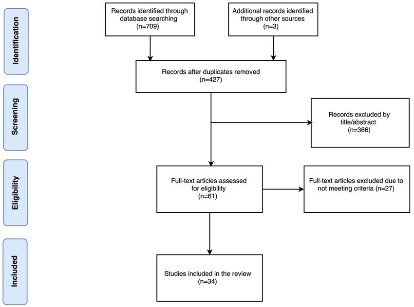
Fig 1. Study search and selection for measures of cardiovascular activity in insomniacs and controls.
Modified PRISMA 2009 flow diagram [89].
https://doi.org/10.1371/journal.pone.0186716.g001
description and ANS interpretation columns of the Tables 1, 2, and 3 please see [5, 14, 21–23,
28, 47–51, 53–55, 58].
Results
Reviewed studies
The initial combined database search generated 709 records. After duplicate removal and
English language restriction, 427 electronic records were identified and screened for eligibility.
The manual search of reference lists of relevant papers and reviews identified three papers
[53, 64, 72]. Three additional studies [79–81] were added to explain the transition from “poor”
sleepers to clinically defined insomniacs. The vast majority of these articles (n = 366) were
excluded by title or abstract alone (see Fig 1) based on criteria mentioned earlier. Full text arti-
cles were obtained for the 61 remaining articles. Ultimately, 34 studies were identified that met
the inclusion criteria for this review. Twenty six observational studies ([8, 11–13, 47, 53, 57,
59–63, 65, 66, 68–73, 82–87]) and eight interventional studies ([64, 67, 74–78, 88]). Study
details such as diagnostic criteria, demographics, number of participants, insomnia severity,
and type of intervention are presented in Tables 4 and 5.
Observational studies
Studies without specific diagnostic criteria for insomnia disorder. In studies without
specific diagnostic criteria for insomnia disorder, physiological differences during sleep were
investigated between the “poor” and “good” sleeper groups [79], as well as subjects with sleep-
PLOS ONE | https://doi.org/10.1371/journal.pone.0186716 October 23, 2017 7 / 31Cardiovascular autonomic activity in insomnia disorder
Table 4. Characteristics of the 26 observational studies.
Authors Sample, Sex Age Diagnostic criteria Insomnia Severity Sleep Efficiency Monitoring and analysis
(mean± std, (Insomniacs- details
range) Controls)
Stepanski Insomniacs: DIMS (psychophysiological or ND < 85%—> 90% Monitoring time: during sleep
et al. [82] 24 M idiopathic insomnia) and SE < 85% (PSG of 1 night) and a morning task, ECG
34.5 ± 8.3, ND on one PSG night sampling rate: ND, Period of
analysis: averaged over an
Controls: hour, Spectral analysis
25 M technique: Not applicable
34.0 ± 7.6, ND
Bonnet et al. Insomniacs: Subjectively reported insomnia Insomniacs subjectively 82±11%—93 Monitoring time: during
[11] 12 (M&F) for 4 nights per week for a year reported: i) a sleep problem; ±3.2% sleep, ECG sampling rate:
31.2 ± 6.8, ND AND (SOL>30 min or SE 1 year Program
29.1 ± 5.2, ND
de Zambotti Insomniacs: DSM-IV criteria for primary PSQI: 9.67 ± 1.67, AIS: 91(±5)%—96 Monitoring time: during
et al. [59] 9 (4 M) insomnia and subjectively reported 10.00 ±3.16, HS: 44.56± (±3)% sleep, ECG sampling rate:
23.0 ± 2.4, 20-26 insomnia history for 1 year 4.03 500 Hz, Period of analysis: 2
min, Spectral analysis
Controls: technique: FFT
9 (4 M)
23.6 ± 3.2, 19-28
de Zambotti Insomniacs: Research Diagnostic Criteria for PSQI: 10.0 ± 2.0, AIS: 15.8 87.1(±8.4)%— Monitoring time: during
et al. [60] 13 (8 F) primary insomnia and (PSQI 5, ±3.3, Length of insomnia 94.0(±3.9)% sleep, ECG sampling rate:
23.0 ± 2.4, 20-28 ISI 11) (months): 58.5± 39.9 512 Hz, Period of analysis: 2
min in frequency domain and
Controls: 5 min in time domain,
14 (7 F) Spectral analysis technique:
23.6 ± 3.2, 23-28 FFT
Spiegelhalder Insomniacs: DSM-IV. For second analysis: PSQI: 11.2 ± 2.8, BDI: 9.7 85.8(±9.8)%— Monitoring time: during
et al. [13] 58 (36 F) INSD SE 85% vs ISSD ±6.6, Length of insomnia 89.0 (±9.2)% sleep, ECG sampling rate:
39.5 ± 11.8, ND SE < 85% based on PSG (years): 10.9± 9.9 (2nd PSG night) 400 Hz, Period of analysis: 5
min, Spectral analysis
Controls: technique: FFT
48 (27 F)
37.3 ± 11.4, ND
Bianchi et al. Insomniacs: ND—subjects underwent ND ND Monitoring time: during
[53] 11 (M&F) polysomnographic evaluation sleep, ECG sampling rate:
39.5 ± 11.8, 35-50 128 Hz, Period of analysis:
ND, Spectral analysis
Controls: technique: ND
17 (M&F)
37.3 ± 11.4, 40-50
Jurysta al. [69] Insomniacs: DSM IV and ICSD-R (revised ND clearly. Available sleep 74(±9)%—90 Monitoring time: during
14 M edition) for chronic primary characteristics for revision (±3)% sleep, ECG sampling rate:
42 ± 12, 16-63 insomnia, subjective complaints 200 Hz, Period of analysis: 2
for 1 month, SE < 85% min, Spectral analysis
Controls: technique: FFT
14 M
41 ± 10, 16-55
Farina al. [8] Insomniacs: ICSD-2 for primary insomnia Mean duration of 82.4(±22)%— Monitoring time: during
85 (38 M) insomnia > 2 years 86.9(±14.2)% sleep, ECG sampling rate:
53.2 ± 13.6, 27-81 256 Hz, Period of analysis: 5
min, Spectral analysis
Controls: technique: Autoregressive
55 (23 M) Model
54.2 ± 13.9, 27-76
(Continued)
PLOS ONE | https://doi.org/10.1371/journal.pone.0186716 October 23, 2017 8 / 31Cardiovascular autonomic activity in insomnia disorder
Table 4. (Continued)
Authors Sample, Sex Age Diagnostic criteria Insomnia Severity Sleep Efficiency Monitoring and analysis
(mean± std, (Insomniacs- details
range) Controls)
Mazza et al. Insomniacs: Diagnosis of chronic Validated Italian version of 86.2(±8.5)%— Monitoring time: during
[70] 6 (2 F) benzodiazepine abuse according the PSQI>5, ESS>6 92.0(±5.3)% sleep, ECG sampling rate:
53.3 ± 14.9, 34-70 to the DSM-IV-TR. In all cases ND, Period of analysis: ND,
patients were initially prescribed Spectral analysis technique:
Controls: for the treatment of chronic Autoregressive Model
55 (32 F) insomnia
54.2 ± 13.0, 27-76
Lanfranchi Insomniacs: DSM-IV-R for chronic insomnia All subjects suffered from 65(±12)%—92 Monitoring time: during
et al. [83] 13 (9 F) and meet the following criteria: mixed (difficulties initiating (±7)% daytime and sleep, ECG
42 ± 9, 30-60 Subjectively reported SOL and/or and maintaining sleep) sampling rate: 256 Hz,
WASO > 30 min, TST < 6.5 h, insomnia, ISI: 18.2 ± 2.1, Period of analysis: averaged
Controls: SE < 85% ii) presence of BDI: 9.5 ± 2.1 over one hour, Spectral
13 (9 F) insomnia 3 nights per week analysis technique: NA
42 ± 7, 30-60 for 6 months iii) ISI 15
Fang et al. [65] Insomniacs: DSM-IV for primary insomnia and Subjectively reported SOL 90.2(±4.8)%— Monitoring time: during
18 (6 M) at least one of the following criteria and/or WASO > 30 min, 96.9(±2.1)% daytime, ECG sampling rate:
34.2 ± 14.5, 20-60 on both actigraphy and diary TST < 6.5, SE < 85% b) (based on 500 Hz, Period of analysis: 5
measures: i) WASO >30 min, ii) presence of insomnia 3 actigraphy) min, Spectral analysis
Controls: TST 6.5 h, ii) SE 85% nights per week for 6 technique: FFT
21 (7 M) months (c) ISI 15.23
27.8 ± 8.7, 20-50
Varkevisser Insomniacs: ICSD-R for chronic Available characteristics 73.3 (±3.6)%— Monitoring time: during
et al. [61] 11 (6 M) psychophysiologic insomnia (with (based on actigraphy) for 82.3 (±6.7)% daytime, ECG sampling rate:
43.8 ± 8.9, 31-54 ambulatory PSG) revision (based on 1000 Hz, Period of analysis:
actigraphy) averaged over 30 sec
Controls: segments, Spectral analysis
13 (7 M) technique: NA
44.9 ± 7.7, 33-53
Yang et al. [68] Insomniacs: DSM-IV for primary insomnia PSQI: 10.7 ± 3.9, BDI: ND Monitoring time: during
47 (4 M) 10.9 ± 6.5. Available daytime and sleep, ECG
41.6 ± 11.7, 23-63 characteristics for revision sampling rate: ND, Period of
analysis: ND, Spectral
Controls: analysis technique: ND
88 (4 M)
24.8 ± 2.7, 22-64
Floam et al. Insomniacs: DSM-V for primary insomnia PSQI: 11.2 ± 0.7, insomnia ND Monitoring time: during
[84] 29 (19 F) disorder duration (years): 6.4 ± 0.8, daytime, ECG sampling rate:
25.3 ± 1.6, 18-55 actigraphy based SOL: NA, Period of analysis:
27 ± 4, WASO: 30 ± 3, SFI: average of the 5
Controls: 15.5 ±1.4 measurements over a
19 (13 F) 15-min period Spectral
25.4 ± 1.4, ND analysis technique: NA
Covassin et al. Insomniacs: DSM-IV for primary insomnia for at Insomnia for at least 1 year 90.6(±5.1)%— Monitoring time: during
[62] 8 (4 M) least 1 year and PSQI: 6, and PSQI: 9.6 ± 1.3, AIS 96.3(±2.4)% daytime, ECG sampling rate:
22.9 ± 2.4, ND AIS 6, ISI: 11 10.8 ± 2.4, ISI: 14.0 ± 2.7, 500 Hz, Period of analysis:
SOL: 16.9 ± 14.7, WASO: averaged over 30 sec
Controls: 27.1 ± 19.1 segments, Spectral analysis
8 (4 M) technique: NA
24.8 ± 2.7, ND
Jiang et al. [66] Insomniacs: DSM-IV for primary insomnia PSQI: 8.6 ± 2.3 ND Monitoring time: during
55 (25 M) daytime, ECG sampling rate:
30.4 ± 8.4, 22-38 ND, Period of analysis: ND,
Spectral analysis technique:
Controls: ND
63 (29 M)
31.3 ± 7.7, 23-39
(Continued)
PLOS ONE | https://doi.org/10.1371/journal.pone.0186716 October 23, 2017 9 / 31Cardiovascular autonomic activity in insomnia disorder
Table 4. (Continued)
Authors Sample, Sex Age Diagnostic criteria Insomnia Severity Sleep Efficiency Monitoring and analysis
(mean± std, (Insomniacs- details
range) Controls)
Cellini et al. Insomniacs: Research Diagnostic Criteria for PSQI: 10.0 ± 2.0, AIS: 87(±8)%—95 Monitoring time: during
[63] 13 (8 F) primary insomnia, subjectively 15.77 ± 3.27, SOL: (±3)% daytime, ECG sampling rate:
24.4 ± 1.6, 20-28 reported symptoms ± 6 months 16.19 ± 16.42, WASO: 512 Hz, Period of analysis: 3
and (PSQI 5, ISI 11) 45.88 ± 30.17 min, Spectral analysis
Controls: technique: FFT
14 (6 F)
23.3 ± 2.5, 20-28
Baglioni et al. Insomniacs: DSM-IV ISI: 12.09 ± 3.32 ND Monitoring time: during
[85] 21 (19 F) daytime, ECG sampling rate:
22.8 ± 3.31, 18-30 ND, Period of analysis: ND
clearly, Spectral analysis
Controls: technique: NA
18 (12 F)
22.0 ± 2.64, 18-30
Peter et al. [47] Insomniacs: DSM-IV and PSQI 5 PSQI range: 10-17 and ND Monitoring time: during
21 (18 F) sleep characteristics daytime, ECG and BP
48.2 ± 10.4, 18-75 available for revision sampling rate: 200, Period of
analysis: 5-7 min, Spectral
Controls: analysis technique: FFT
21 (18 F)
48.5 ± 11.1, 18-75
Nelson et al. Insomniacs: DSM-IV and PSQI 5 PSQI: 9.5 ± 3.0, BDI: ND Monitoring time: during
[86] 34 (18 F) 10.3 ± 6.0. Available sleep, ECG sampling rate:
21.1 ± 4.9, 18-45 characteristics for revision ND, Period of analysis: ND,
Spectral analysis technique:
Controls: NA
38 (26 F)
20.5 ± 2.2, 18-45
de Zambotti Insomniacs: DSM-IV for primary insomnia and PSQI: 9.6 ± 1.3, AIS: 90.0(±4)%—95.0 Monitoring time: during sleep
et al. [12] 8 (4 F) PSQI: 6, AIS 6, ISI: 11 9.6 ± 3.2, ISI: 13.4± 3.3 (±2)% onset, ECG sampling rate:
23.3 ± 2.4, 20-26 Available characteristics for 500 Hz, Period of analysis:
revision 2.5 min, Spectral analysis
Controls: technique: FFT
8 (5 F)
23.3 ± 3.2, 19-28
Tsai et al. [87] Insomniacs: Subjectively reported difficulty of PSQI: 11.3 ± 2.2, SOL: 90.1(±8.7)%— Monitoring time: during sleep
19 (13 F) falling asleep 20-30 min for 3 25.9 ± 40.9. Available sleep 94.8(±2.8)% onset, ECG sampling rate:
22.9 ± 1.6, 20-25 days per week over 6 months and characteristics for revision 500 Hz, Period of analysis:
daytime consequences and PSQI: NA, Spectral analysis
Controls: > 5. Defined as technique: NA
14 (5 F) psychophysiological insomnia
22.4 ± 1.1, 20-25 based on the objective finding
Maes et al. [71] Insomniacs: DSM-IV-TR and subjective SOL: 33.8 ± 17.1, Mean 79.1(±10.1)%— Monitoring time: during sleep
17 F SOL > 30 min, at least 5 nights per duration of insomnia 89.0(±6.8)% onset and first NREM cycle,
36.2 ± 9.6, 19-53 week 10.8 ± 10.9 years. Available ECG sampling rate: 1000
sleep characteristics for Hz, Period of analysis: 5 min,
Controls: revision Spectral analysis technique:
11 F FFT
37.6 ± 12.6, 21-59
Rothenberger Insomniacs: Self-report Insomnia Symptom ND ND Monitoring time: during
et al. [72] 19 F Questionnaire (Incorporated to sleep, ECG sampling rate:
52.1 ± 2.2, ND define DSM-IV criteria for insomnia 1024 Hz, Period of analysis:
and Research Diagnostic Criteria) 2 min, Spectral analysis
Controls: technique: FFT
146 F
52.1 ± 2.2, ND
(Continued)
PLOS ONE | https://doi.org/10.1371/journal.pone.0186716 October 23, 2017 10 / 31Cardiovascular autonomic activity in insomnia disorder
Table 4. (Continued)
Authors Sample, Sex Age Diagnostic criteria Insomnia Severity Sleep Efficiency Monitoring and analysis
(mean± std, (Insomniacs- details
range) Controls)
Schramm et al. Insomniacs: DSM-IV for primary insomnia PSQI: 12.11 ± 2.69 83.2(±10.1)%— Monitoring time: during
[57] 50 (27 M) 89.1(±6.7)% sleep, ECG sampling rate:
46.4 ± 8.6, 30-63 (from the 2nd 200 Hz, Period of analysis:
night) 8.5 min, Spectral analysis
Controls: technique: FFT
36 (17 M)
44.5 ± 8.7, 30-63
Israel et al. [73] Insomniacs: DSM-IV for primary insomnia SOL: 26.1 ± 18.3, WASO: 85.6(±8.0)%— Monitoring time: during
54 (30 F) 41.4 ± 33.5 90.5(±5.5)% sleep, ECG sampling rate:
34.6 ± 9.7, ND 1024 Hz, Period of analysis:
2 min, Spectral analysis
Controls: technique: Autoregressive
22 (19 M) Model
26.5 ± 7.3, ND
Abbreviations— AIS: Athens Insomnia Scale, BDI: Beck depression inventory, CBT: Cognitive behavioral therapy, DSM: Diagnostic and Statistical Manual
of Mental Disorders, ECG: electrocardiogram, ESS: Epworth Sleepiness Scale, F: female, FFT: fast Fourier transform, ICSD: International Classification of
Sleep Disorders, INSD: Insomniacs with normal sleep duration, ISI: Insomnia Severity Index, ISSD: Insomniacs with short sleep duration, IV: 4th edition, M:
male, min: minutes, NA: not applicable, ND: not defined, NREM: Non-rapid eye movement sleep, PSG: polysomnography, PSQI: Pittsburgh Sleep Quality
Index, R: revised, SE: sleep efficiency, sec: second, SOL: Sleep onset latency, TR: text revised, TST: Total sleep time, WASO: Wake after sleep onset.
https://doi.org/10.1371/journal.pone.0186716.t004
onset insomnia [80, 81]. Monroe [79] reported significantly higher HR 30 minutes before
sleep in “poor” sleepers as compared to “good” sleepers. During sleep, HR of the “poor” sleep-
ers was slightly, but not significantly, higher. Freedman et al. [81] found that sleep-onset
insomniacs had increased HR prior to sleep, but not during sleep. Similarly, Haynes et al. [80]
reported that sleep-onset insomniacs showed a mean HR 4.6 beats per minute, significantly
higher than that of non-insomniacs, whilst the effects of pre-sleep cognitive stress were exam-
ined. Haynes et al. [80], concluded that insomniacs show higher levels of physiological arousal
compared to non-insomniac subjects.
Nocturnal cardiovascular activity. In 1994, Stepanski et al. [82] assessed physiological
activity in patients with objectively documented insomnia using specific American Sleep Dis-
orders Association diagnostic criteria [90]. Subjects with chronic insomnia and normal sleep-
ers slept in the laboratory overnight and were given a stressful performance task in the
morning. HR was assessed for all participants before sleep, during sleep, and in response to
acute stress. Nocturnal HR was significantly higher in insomniacs. The morning after, no dif-
ference was found in HR between the two groups, but HR was significantly higher during the
stressful performance task in insomniacs. These results, regarding increased nocturnal HR,
were confirmed by Bonnet et al. [11]. In particular, sleep and ECG measures were evaluated in
insomnia patients and matched controls. In this study, spectral analysis of HRV revealed sig-
nificant increases in LF and LF/HF and a decrease in HF in insomniacs compared to controls.
Those changes were present across all sleep stages.
Recently, de Zambotti et al. [59] investigated nocturnal cardiovascular modifications in pri-
mary insomniacs compared to healthy controls, focusing on cardiac autonomic functioning.
They found a significant, constant shorter PEP during all sleep stages of the insomniac group
compared to good sleepers. In addition, they found the presence of short PEP to be directly
associated with low quality of sleep assessed by the Pittsburgh Sleep Quality Index (PSQI) and
PLOS ONE | https://doi.org/10.1371/journal.pone.0186716 October 23, 2017 11 / 31Cardiovascular autonomic activity in insomnia disorder
Table 5. Characteristics of the eight interventional studies.
Authors Sample, Sex Age Diagnostic criteria Follow-up period Intervention Insomnia Severity Monitoring and
(mean±std, range) Study type Sleep Efficiency analysis details
Jobert 16 (4 M) ICSD for chronic or ND placebo- bezodiazepin ND Monitoring time: during
et al. [88] (66.7 ± 5.8, ND) subcronic controlled, lormetazepam (1mg) sleep, ECG sampling
psychophysiological randomized, 3-fold cyclopyrrolone rate: 200 Hz, Period of
insomnia crossover zopiclone (7.5mg) analysis: averaged
placebo over 30 sec segments,
Spectral analysis
technique: NA
Lo et al. 18 (7 M) Subjective complaints of 4 weeks after the Gabapentin Mean Before: Monitoring time: during
[74] (43.2 ± 15.4, ND) difficulty initiating sleep completion of dose dose 540 mg Range PSQI: 13.54 sleep, ECG sampling
and/or maintaining sleep titration open-label 200-900 mg 80.00% After: rate: 400 Hz, Period of
for 3 months PSQI: 7.67 analysis: 10 min,
87.17% Spectral analysis
technique: ND
Chung Responders: ICSD-2 8-week period after CBT of 4 sessions, Responders- Monitoring time: during
et al. [64] 16 (6 M) the beginning of one every other week Before: daytime, ECG
(57.9 ± 10.9, ND) CBT open-label over an eight-week ISI: 19.5 ± 4.0 sampling rate: ND,
period 77.7 ±18.9 Period of analysis: 5
Non-responders: After: min, Spectral analysis
10 (4 M) ISI: < 8 technique: ND
(59.4 ± 7.4, ND) ND
Non-responders-
Before:
ISI:18.1 ± 4.9
70.8 ± 26.5
After:
ISI: > 8 ND
Jarrin 65 (22 M) DSM-IV-TR and ICSD-2 6-week period after CBT, frequency of Before: Monitoring time: during
et al. [75] (51.8 ± 10.0, ND) for chronic insomnia the beginning of sessions was not ISI: 17.0±4.0 sleep, ECG sampling
CBT open-label specified Subjective: rate: ND, Period of
69.4±15.5% analysis: 2 min,
Objective: 80.1 Spectral analysis
±12.1% technique: FFT
After:
8.8±3.5
Subjective:
83.8±10
Objective:
88.6±8.4
Litscher 28 (5 M) Self-presentation at the 10 min before, 20 Acupuncture by using Before: Monitoring time: during
et al. [76] (41.9 ± 14.6, 22-82) hospital due to insomnia. min during, and 10 the Shenmen AIS: daytime, ECG
AIS ranged from 6-21 min after acupuncture point on 12.4±3.6 sampling rate: 4096
stimulation of the the left wrist ND Hz, Period of analysis:
Shenmen acupoint 5 min, Spectral
open-label After: analysis technique: ND
ND
ND
Wang 31 (6 M) Self-presentation at the 10 min before, 20 Acupuncture by using Before: Monitoring time: during
et al. [77] (54.3 ± 10.6, 39-82) hospital due to insomnia min during, and 10 the Shenmen AIS: 14.7±4.4 daytime, ECG
and AIS 7 and a range min after acupuncture point on ND sampling rate: 4096
of 7-22 stimulation of the the left ear Hz, Period of analysis:
Shenmen acupoint After: 5 min, Spectral
open-label ND analysis technique: ND
ND
(Continued)
PLOS ONE | https://doi.org/10.1371/journal.pone.0186716 October 23, 2017 12 / 31Cardiovascular autonomic activity in insomnia disorder
Table 5. (Continued)
Authors Sample, Sex Age Diagnostic criteria Follow-up period Intervention Insomnia Severity Monitoring and
(mean±std, range) Study type Sleep Efficiency analysis details
Chien Experimental group: Primary insomnia at the 4th week lavender Experimental Monitoring time: during
et al. [67] 34 F (51.09 ± 3.73, (definition not specified during treatment, aromatherapy group- daytime, ECG
45-55) and Chinese version of and after 12 weeks Before: sampling rate: ND,
Control group: PSQI (CPSQI) > 5 of treatment cohort CPSQI: 9 (8,12)† Period of analysis: 3
33 F (50.85 ± 3.73, study ND After: min, Spectral analysis
45-55) CPSQI: ND technique: ND
ND
Control group-
Before:
CPSQI: 11 (9,13)†
ND
After:
CPSQI: ND
ND
Tsai Patient group: Self-reported insomniacs 2 days later after Controlled respiration Patient group: Monitoring time: during
et al. [78] 14 ND who met the DSM-IV intervention (with a at a slow frequency Before: daytime and sleep,
(22.50 ± 1.22, 20-25) criteria for primary 1 week difference rate of 0.1 Hz, and a PSQI: 11.21±1.97 ECG sampling rate:
Control group: insomnia and PSQI > 6 of each other) forced rate of 0.2 Hz 89.55±9.89 500 Hz, Period of
14 ND (23.07 ± 1.64, open-label during daytime rest After: analysis: 64 secs,
20-25) PSQI: ND 94.38 Spectral analysis
±3.76* & technique: FFT
87.92±12.77**
Control group:
Before:
PSQI: 2.93±1.33
94.99±2.88
After:
PSQI: ND
94.47±2.80* &
93.48±3.24**
Abbreviations— AIS: Athens Insomnia Scale, CBT: Cognitive behavioral therapy, CPSQI: Chinese Pittsburgh Sleep Quality Index, DSM: Diagnostic and
Statistical Manual of Mental Disorders, F: female, FFT: fast Fourier transform, ICSD: International Classification of Sleep Disorders, ISI: Insomnia Severity
Index, M: male, min: minutes, NA: not applicable, ND: Not defined, PSQI: Pittsburgh Sleep Quality Index, sec: second
*: Paced breathing at 0.1 Hz
**: Paced breathing at 0.2 Hz
: global score
†
: Median (Interquartile Range) instead of mean value and standard deviation
https://doi.org/10.1371/journal.pone.0186716.t005
by the Athens insomnia scale (AIS). In another study by de Zambotti et al. [60], the authors
further studied ANS functioning in insomniacs and confirmed their previous results [59]
regarding shorter PEP during all sleep stages in insomniacs. However, no significant differ-
ences in vagal activity (expressed by: the standard deviation of RR or NN intervals for a desired
period (SDNN); the square root of the mean squared differences of successive NN intervals for
a desired period (RMSSD); and the percentage of successive NN intervals that differ more than
50 ms (pNN50)) were reported between insomniacs and controls. Moreover, pre-sleep RR
intervals duration (over a 5-min window) was positively associated with sleep efficiency (SE)
and negatively associated with wake after sleep onset (WASO) in insomniacs. Spiegelhalder
et al. [13] aimed at investigating the association between insomnia and alterations in polysom-
nographically determined nocturnal HR and HRV. In the insomnia group, results showed a
PLOS ONE | https://doi.org/10.1371/journal.pone.0186716 October 23, 2017 13 / 31Cardiovascular autonomic activity in insomnia disorder
lower wake-to-sleep HR difference compared to controls. The SDNN, was also significantly
lower in the insomnia group. The authors [13] characterized the initial group of patients as
subjectively reported insomniacs and they split up the insomnia group according to SE values.
Thus, when restricting their analysis to insomnia patients with objectively determined short
sleep duration (SECardiovascular autonomic activity in insomnia disorder
significant differences were observed in the HR and PSG-based profiles (with the exception of
fewer periodic limb movements observed in the insomnia group) between insomniacs and
good sleepers.
Cardiovascular activity during daytime. Several studies have been focused on the diurnal
cardiovascular activity in insomnia population [47, 61, 62, 65, 66, 84]. Fang et al. examined the
differences in HRV and daytime functioning. Five-minute recordings of ECG under paced
breathing were obtained from all participants who were resting in a supine position. Authors
found an increasing trend in LF/HF ratio in insomniacs compared to controls, but the differ-
ences between the two groups did not reach statistical significance [65]. Varkevisser et al. [61]
measured cardiovascular parameters (average HR, RMSSD, PEP) in a group of subjects with
chronic insomnia under strictly controlled constant-routine conditions with continuous wake-
fulness. Although physiologic indexes of arousal were slightly elevated in the insomnia group
relative to the controls, the differences between the groups were not statistically significant.
Floam et al. [84] investigated whether BP differs between individuals with insomnia disorder
and healthy sleepers. Standard oscillometric BP measurements were collected in a seated posi-
tion five times over a 15-min period during the day. Floam et al. [84] did not find any differ-
ences in BP measurements between individuals with insomnia disorder and healthy sleepers
during daytime.
On the other hand, when Yang et al. [68] investigated the long-term diurnal profile of
HRV, differences between insomniacs and healthy controls were seen. In this study, HRV
measures between awake state and bed-time were compared. Compared to controls, insomni-
acs exhibited significant reductions in SDNN, HF, RMSSD, pNN50 during awake period.
Alterations in LF/HF (increase), RMSSD (decrease), HF (decrease) were correlated with per-
ceived sleep questionnaire score, suggesting that according to the authors, altered cardiac auto-
nomic control and physiologic complexity is associated with poor sleep in patients with
insomnia.
Cardiovascular activity during daytime while performing tasks. Several studies exam-
ined HRV during daytime while subjects performed a task [62, 63, 66, 85]. Covassin et al. [62]
focused on cardiovascular re-activity to the task in primary insomnia. Cardiovascular re-activ-
ity is defined as the responsiveness of the cardiovascular system to react to a stressful task. The
task was administered in two sessions, before and after a night of polysomnographic recording.
Results of cardiovascular parameters showed higher HR and lower left ventricular ejection
time values in insomniacs, as compared to controls in the evening. PEP was continuously
reduced in insomniacs. Jiang et al. [66] aimed to examine HRV response to a postural change
manoeuvre, in primary insomniacs and controls. HRV features were computed at the follow-
ing times: seated rest and 0-5 min, 5-10 min and 10-15 min in the standing position. Jiang
et al. [66] reported an attenuated or absent HRV response to postural change in primary
insomnia subjects. Specifically, the increase in LF/HF ratio and the decrease in HF, reflected
that parasympathetic predominance at rest shifted to sympathetic control while standing.
However, this shift was much slower than in the normal controls, according to the authors.
Furthermore, researchers [66] reported a significantly lower LF, SDNN, RMSSD, and HF in
the primary insomnia group than in the normal control group. Two memory tasks and their
association with cardiovascular activity were examined by Cellini et al. [63]. Compared to
healthy controls, insomniacs exhibited shorter PEP, reduced HF and increased RPP at rest.
Similarly, in another study [85] where psychophysiological reactivity to emotional stimuli
using pictures (both related and unrelated to sleep) was examined, cardiac changes were
observed in insomniacs compared to controls. According to the authors [85], enhanced car-
diac vagal tone (defined as pulse-synchronized phase shifts in consecutive cardiac cycles) in
response to all pictures, by people with insomnia compared to good sleepers was found, even
PLOS ONE | https://doi.org/10.1371/journal.pone.0186716 October 23, 2017 15 / 31Cardiovascular autonomic activity in insomnia disorder
though no significant effects were evidenced for HR responses between the two groups [85]. In
another study by Nelson et al. [86], where the pre-sleep differential content of imagery and ver-
bal thought was investigated, significant differences in HR were found between insomniacs
and controls.
One study failed to report significant cardiovascular changes between insomniacs and
healthy controls during tasks. Peter et al. [47] examined BRS using an exploratory protocol
with paced breathing. The authors found no significant differences in BRS between insomnia
patients and controls.
Sleep onset. Some studies investigated the autonomic changes that characterize wake-to-
sleep transition. De Zambotti et al. [12] examined the cardiovascular activity during the switch
from wakefulness to sleep in insomniacs. The cardiac activity was studied in baseline, as well
as pre- and in post-sleep onset. Results showed higher initial HR (an index primarily modu-
lated by parasympathetic activity at rest) in baseline in the insomniac group, but no differences
between groups in pre- and post- sleep onset. In fact, HR showed a decrease in both groups
during the transition from pre- to post-sleep onset. No significant differences were found
between the two groups regarding HFnorm. The most important result of this study, according
to the authors, is the non significant changes in PEP values across sleep onset in the insomniac
group. This result was interpreted as continuous, unchanged, sympathetic hyperactivation of
insomniacs during sleep onset. In contrast, good sleepers showed the expected trend of
increased PEP values during the transition from wakefulness to sleep. PEP was also signifi-
cantly lower in insomniacs than in normal sleepers in both conditions (pre and post-sleep
onset). These results agree with recent results from Farina et al. [8]. In this study, patients
showed increased HR and LFnorm, while awake before sleep and during early-stage-N2. In
addition, Spiegelhalder et al. [13] reported that insomniacs have a lower wake-to-sleep HR
reduction compared to controls. Sleep onset was also investigated on self-reported insomniacs.
Tsai et al. [87] examined HR dynamics during the sleep onset period, between young self-
reported insomniacs with long sleep latency, and normal controls. Linear regression and non-
linear Hilbert-Huang transform of the HR slope were performed in order to analyze HR
dynamics. Results indicated that a slower drop in HR dynamics during the sleep onset period
seems to be a feature of sleep initiation difficulty. In addition, authors suggest that the magni-
tude of the change in HR during the sleep onset period is associated with the lengths of objec-
tive and subjective sleep-onset latency. In contrast to these studies, Maes et al. [71] failed to
report any significant differences between insomnia female patients and controls preceding
sleep onset, in either HR or HRV (LF, HF, and LF/HF) variables.
Cardiac activity and EEG. Some studies have evaluated cardiac autonomic tone in rela-
tion to sleep. For instance, Jurysta et al. [69] tried to determine the relation if chronic insomnia
alters the relation between HRV and delta sleep EEG power. Results showed that the coherence
between HFnorm and delta EEG was decreased significantly in insomniacs compared to healthy
men. The authors suggested that the decreased coherence between relative vagal cardiac activ-
ity and delta sleep observed in patients with insomnia, in comparison to normal controls,
could suggest a loss of control between brainstem structures, implied in cardiovascular and
sleep controls [69]. Rothenberger et al. [72] examined whether EEG-HRV relationships in
midlife women differ as a function of insomnia. They reported that time-varying correlations
between delta EEG power and HF were stronger in women with self-reported insomnia, com-
pared to healthy controls. Another study by Maes et al. [71] found an association between K-
alpha (K-complex within one, second followed by 8–12 Hz EEG activity) in Stage2 sleep and a
lower HF in SWS in female insomniacs. The authors interpreted the strong association found
between K-alpha in Stage2 sleep and the lower HF as a state of hyperarousal continuing
through sleep.
PLOS ONE | https://doi.org/10.1371/journal.pone.0186716 October 23, 2017 16 / 31You can also read