A systematic review of neuroimaging and acute cannabis exposure in age-of-risk for psychosis
←
→
Page content transcription
If your browser does not render page correctly, please read the page content below
Cupo et al. Translational Psychiatry (2021)11:217
                                    https://doi.org/10.1038/s41398-021-01295-w                                                                                                     Translational Psychiatry

                                     REVIEW ARTICLE                                                                                                                                             Open Access

                                    A systematic review of neuroimaging and acute
                                    cannabis exposure in age-of-risk for psychosis
                                                        1,2
                                    Lani Cupo               , Eric Plitman2,3, Elisa Guma1,2 and M. Mallar Chakravarty1,2,3,4

                                      Abstract
                                      Acute exposure to cannabis has been associated with an array of cognitive alterations, increased risk for
                                      neuropsychiatric illness, and other neuropsychiatric sequelae including the emergence of acute psychotic symptoms.
                                      However, the brain alterations associating cannabis use and these behavioral and clinical phenotypes remains
                                      disputed. To this end, neuroimaging can be a powerful technique to non-invasively study the impact of cannabis
                                      exposure on brain structure and function in both humans and animal models. While chronic exposure studies provide
                                      insight into how use may be related to long-term outcomes, acute exposure may reveal interesting information
                                      regarding the immediate impact of use and abuse on brain circuits. Understanding these alterations could reveal the
                                      connection with symptom dimensions in neuropsychiatric disorders and, more specifically with psychosis. The
                                      purpose of the present review is to: 1) provide an update on the findings of pharmacological neuroimaging studies
                                      examining the effects of administered cannabinoids and 2) focus the discussion on studies that examine the sensitive
                                      window for the emergence of psychosis. Current literature indicates that cannabis exposure has varied effects on the
                                      brain, with the principal compounds in cannabis (delta-9-tetrahydrocannabinol and cannabidiol) altering activity
                                      across different brain regions. Importantly, we also discovered critical gaps in the literature, particularly regarding sex-
                  1234567890():,;
1234567890():,;
                  1234567890():,;
1234567890():,;

                                      dependent responses and long-term effects of chronic exposure. Certain networks often characterized as dysregulated
                                      in psychosis, like the default mode network and limbic system, were also impacted by THC exposure, identifying areas
                                      of particular interest for future work investigating the potential relationship between the two.

                                    Introduction                                                                                        depressive6 and anxiety disorders7, and, central to this
                                       In recent years there has been a surge in public policy                                          review, psychosis spectrum disorders8. Cannabis use
                                    decriminalizing or legalizing recreational cannabis use                                             initiated during early adolescence confers the greatest risk
                                    worldwide1,2. In spite of these changing norms, our                                                 for adult psychosis9, and dose-dependent cannabis use has
                                    understanding of the mental health consequences of                                                  been associated with an increased likelihood of developing
                                    cannabis exposure remain inconclusive. From a clinical                                              psychosis and schizophrenia8 while short-term cannabis
                                    standpoint, there is an emerging consensus on how can-                                              use has been associated with increases in psychotic-like
                                    nabis may confer some therapeutic benefits (treatments                                               symptoms, such as altered perception and anxiety10. Risk
                                    for chronic pain and glaucoma)3,4, and may also increase                                            during adolescence could in part be conferred from
                                    risk for adverse mental health outcomes (major mental                                               critical periods of development in neurotransmitters.
                                    illnesses and associated symptomatology)5. Specifically,                                             Development of the GABA-ergic (γ-aminobutyric acid)
                                    cannabis use has been associated with increased risk for                                            system during adolescence has been associated with
                                                                                                                                        response inhibition and working memory11. During the
                                                                                                                                        same time period, there occurs pruning of glutamatergic
                                    Correspondence: Lani Cupo (lani.cupo@mail.mcgill.ca) or                                             neurons, and reductions in innervation in the dopami-
                                    M. Mallar Chakravarty (mallar@cobralab.ca)
                                    1
                                                                                                                                        nergic system during typical development11.
                                     Integrated Program in Neuroscience, McGill University, Montreal, QC, Canada
                                    2                                                                                                     While cannabis contains many compounds responsible
                                     Computational Brain Anatomy (CoBrA) Laboratory, Cerebral Imaging Center,
                                    Douglas Research Centre Verdun, Montreal, QC, Canada                                                for various physiological effects, tetrahydrocannabinol
                                    Full list of author information is available at the end of the article

                                    © The Author(s) 2021
                                                        Open Access This article is licensed under a Creative Commons Attribution 4.0 International License, which permits use, sharing, adaptation, distribution and reproduction
                                                        in any medium or format, as long as you give appropriate credit to the original author(s) and the source, provide a link to the Creative Commons license, and indicate if
                                    changes were made. The images or other third party material in this article are included in the article’s Creative Commons license, unless indicated otherwise in a credit line to the material. If
                                    material is not included in the article’s Creative Commons license and your intended use is not permitted by statutory regulation or exceeds the permitted use, you will need to obtain
                                    permission directly from the copyright holder. To view a copy of this license, visit http://creativecommons.org/licenses/by/4.0/.
Cupo et al. Translational Psychiatry (2021)11:217                                                              Page 2 of 19

(THC) is the psychoactive component most associated           predominate at older onset24. Although the cause of the
with psychotomimetic effects12. THC binds native can-         discrepancy is unknown, it has been suggested that sex
nabinoid receptors, such as G-protein coupled receptors       hormones, such as estrogen and testosterone, may con-
like CB1, which acts as a receptor for endocannabinoids       tribute to the sex differences24. Given that females are
like anandamide12. CB1 receptors are distributed in var-      typically more sensitive to the effects of cannabis use as
ious brain regions, and expressed on the presynaptic          they relate to psychosis25, it is important to examine sex
axon terminals of different types of neurons including        differences in cannabis response as a means to better
GABAergic and glutamatergic neurons13. As an inhibitory       understand this differential susceptibility
neurotransmitter, active GABA-ergic synapses reduce the          In this review, we examine studies that administer can-
likelihood that postsynaptic neurons will fire. When THC       nabinoids to better understand how mechanisms of acute
or endocannabinoids bind to CB1, however, they prevent        exposure during adolescence and young adulthood may be
the release of GABA, permitting the postsynaptic cell to      implicated in changing of brain circuitry, thereby increasing
fire. An example of this prevention is dopamine, where         risk for the emergence of psychoses. While understanding
GABA-ergic synapses control the release of dopamine           the impact of chronic use is critical, habituation makes it
into the system. Therefore, in the presence of THC,           difficult to tease apart how cannabis alters specific brain
dopaminergic neurons are not prevented from firing,            circuits. Studies investigating chronic use are limited by
leading to an overabundance of dopamine. CB1 receptors        confounding variables, such as concomitant tobacco26,
are present in a high density in GABAergic axon terminals     alcohol27, and polydrug use28, as well as shared genetic risk
from the striatum14, potentially relating to excess dopa-     for psychosis and cannabis use29. By focusing on acute
mine in the striatum.                                         studies, this review reduces the confounding effects asso-
   Increased dopamine in the striatum coincides with the      ciated with repeated cannabis use. To mitigate the chance
dopamine hypothesis of schizophrenia as individuals with      that genetic background may increase psychosis-proneness
schizophrenia display excess levels of dopamine in the        and cannabis use, we examine studies that use neuroima-
striatum, thought to be related to positive symptoms like     ging techniques to investigate how brain circuits and
hallucinations15. According to the dopamine hypothesis,       behavioural responses are altered following acute cannabis
patients with schizophrenia have reduced levels of            exposure. The alterations may reflect underlying alterations
dopamine in the prefrontal cortex (PFC) associated            to the GABAergic, glutamatergic, and dopaminergic sys-
with cognitive impairments and negative symptoms like         tems that undergo refinement during adolescence11. To
anhedonia15. The excitatory neurotransmitter, glutamate       capture the state of cannabis research, this review includes
is additionally dysregulated in schizophrenia16,17, notable   THC, CBD, as well as homologues of these molecules,
as glutamatergic synapses also express CB1 in the pre-        such as tetrahydrocannabivarin (THCv). We synthesize the
synaptic cell. When THC binds CB1, less glutamate is          neuroimaging studies in humans and animal models that
released into the system, relevant to the effects seen in     examine the effects of cannabinoid administration, both
psychosis17,18.                                               cross-sectionally and longitudinally in an age group coin-
   In addition to THC, other compounds in cannabis, such      cident with the typical age-of-onset of psychosis (20–22;
as cannabidiol (CBD) have a host of differential pharma-      however there are additional spikes reported around 40 for
cological effects on the brain with demonstrably different    women, and even some accounts of a third spike for women
impacts from THC. Like the endocannabinoid 2-Arachi-          around 80)30,31 to better understand the impact of canna-
donoylglycerol, CBD binds CB2, a receptor that has not        binoids on the brain during these sensitive periods23.
been as well characterized as CB1 but is largely present in      The translational neuroimaging focus of this review
the immune system19. CBD has been posited to have             aims to demonstrate how whole-brain investigations of
neuroprotective effects, reducing the effects of THC20.       the effects of cannabis on brain function, the activity of
Previous research also suggests that exposure to cannabis     specific receptor families, and neurochemistry can be
with a high THC concentration increases risk compared         contextualized across species. Ultimately this review seeks
with low-potency cannabis21. Both THC content and             to reveal the state of understanding the effects of acute
THC:CBD ratio in recreational cannabis seized by Cali-        cannabis exposure and how this relates to the etiology of
fornia law enforcement increased significantly between         psychosis. We provide it as a reference for researchers
1996 and 200822.                                              planning projects to identify gaps in the literature and
   Psychoses generally emerge earlier for men (mean age       opportunities for further investigation.
of first episode: 24.2, mean age of first negative symptom:
26.5) than for women (mean age of first episode: 27.4,         Methods
mean age of first negative symptom: 41.6)23. There is a        Literature search
higher incidence of schizophrenia among men (1.4:1);            Neuroimaging techniques, such as functional magnetic
however, prevalence rates are similar, and women              resonance imaging (fMRI) and positron emission
Cupo et al. Translational Psychiatry (2021)11:217                                                                   Page 3 of 19

tomography (PET) are ideal for detecting the acute effects      Human studies
of cannabis exposure on brain function. Additionally,             The majority of human studies reviewed (n = 22)
they permit translational approaches to research questions,     administered THC alone;37–58; methods of administra-
including studies in both humans and non-human animals,         tion varied from vaporized (n = 11)39–42,46–48,53–55,57, to
the latter of which represents an opportunity for further       smoked (n = 4)50–52,59,60, and orally in gelatin capsules
research as few studies to date utilize neuroimaging tech-      (n = 7; Table 1)37,38,43,44,49,56,58. The second most
niques to study the effects of cannabinoids on non-human        commonly administered cannabinoid was Dronabinol, a
animal brains. We used this premise to guide our Ovid           synthetic THC often prescribed medically and reported
search of Medline, Embase, and PsycINFO (1980-June Week         as Marinol (n = 6), administered orally [n = 5]61–65 and
2, 2019) to identify articles that used neuroimaging to assay   intravenously [n = 1])66. Studies that compared THC
brain function in populations within an age-range relevant      and CBD used gelatin capsules (n = 5)67–71. Remaining
to the development of psychosis-like symptoms (see below)       studies examined the THC homologue tetra-
and with acute exposure to cannabinoids. Search terms           hydrocannabivarin (n = 2)72,73, Bedrobinol (a strain of
included: (magnetic resonance imaging or MRI or functional      cannabis with 13.5% THC < 1% CBD) (n = 1)59, CBD
magnetic resonance imaging or fMRI or positron emission         alone (n = 1)74, or smoked cannabis without reporting
tomography or PET or diffusion tensor imaging or DTI or         CBD and THC concentrations (n = 1)60. This last study
computed tomography or CT or magnetic resonance spec-           was the only one to include cannabis in its full form,
troscopy or MRS) and (cannab* or tetrahydrocannabinol or        while the others employed a dichotomy between THC
THC or marijuana) and (adolescen* or develop* or teenage*       and CBD. This work relates the human studies to
or matur* or youth or young). Additionally, reference sec-      relevant results from the psychosis spectrum literature
tions of major relevant reviews32–34, were reviewed for         (CHR [n = 3]74–76, first-episode psychosis [n = 1]77, and
applicable articles that were potentially missed. Included      schizophrenia [n = 2]76,78).
studies and reviewed articles are reflected in the PRISMA
flow chart (Fig. 1).                                             Preclinical models
                                                                  All rodent studies administered the pharmacological
Inclusion criteria                                              intervention via intraperitoneal injection. These studies
  Inclusion criteria were full-length, English-language         examined the effect of THC (1 mg/kg/day for 3 weeks)79
articles that employed in vivo neuroimaging (using MRI,         or CB1 receptor agonists Hebrew University 210 (HU
MRS, PET, CT, and DTI) in humans aged 14–40 (>90%               210)80 (single injection, 1 mL/kg)81, and CP 55,940 (PND
of the sample) or adolescent aged non-human animals             28–38, 2 mL/kg)82. Finally, one study examined the effects
(mouse: postnatal day [PND] ~23–5035, rat: PND                  of acute and chronic HU 210 exposure on rats aged PND
~28–60)36 as well as administration of synthetic or nat-        35 and 7083. Both HU 210 and CP 55,940 have been
ural cannabinoids.                                              demonstrated to be significantly more potent than THC,
                                                                potentially limiting their comparison to cannabis use in
Exclusion criteria                                              humans80,84. One additional study in the search admi-
  Exclusion criteria for the systematic review included         nistered THC to Rhesus monkeys, however it falls outside
comorbid psychiatric disorders, administration of syn-          of the inclusion criteria for age85.
thetic cannabinoid receptor agonists, or case-studies.
                                                                Imaging modalities
Results                                                           The majority of human studies used fMRI to investigate
  After deduplication, the Ovid search yielded 2811             the acute effects of cannabis exposure using resting-state
results. All titles and abstracts were reviewed by L.C., and    fMRI (rs fMRI; n = 5)48,53,54,65,72 or event-related fMRI
either E.G. or E.P. (each reviewed half). Forty-four articles   (er fMRI; n = 27)37–44,46,47,49,55,56,58,59,61–64,67–71,73,74,86,
(40 human and four preclinical studies) met the inclusion       (see Table 1 for classification by task-type). Arterial spin
criteria and underwent full-text assessment for eligibility     labeling (ASL; n = 1)55 and MRS (n = 1)45 were also used.
(Table 1). In the following section, we provide an overview     Radioligand studies included PET and single-photon
of experimental methodology and summarize behavioral            emission tomography (SPET/SPECT) (n = 6)66 (see
results before synthesizing neuroimaging findings across         Table 1 for summary of tracers).
studies. In order to compare networks affected by can-            The three rat studies used PET to examine either glu-
nabis exposure and those altered across the spectrum of         cose metabolism using [18F]-2-fluoro-deoxyglucose ([18F]-
psychosis, studies from clinical high risk (CHR), first          FDG) (n = 2)81,82,87 or dopamine receptor activity with
episode psychosis (FEP), and schizophrenia are included         [18F]-Fallypride79. No preclinical studies used fMRI, ASL,
at the end of results sections by modality where available.     or 1H-MRS.
Cupo et al. Translational Psychiatry (2021)11:217 Page 4 of 19 Fig. 1 PRISMA flow diagram. PRISMA flowchart illustrating process of systematic review inclusion and explanation for excluded studies. Behavioral results Comparison of THC and CBD administration. There Twenty-three studies reported the impact of cannabis was evidence for increased intoxication, anxiety, sedation, on behavioral and psychometric assays in humans. and psychotic symptoms over time in response to THC, THC studies. The Visual Analogue Mood Scale but not to CBD70,86. Additionally, one study with a small (VAMS) was commonly used to index experiences sample (six participants) reported that three of their related to “highness”/”being high”, “alertness”, “external participants experienced acute psychotic symptoms after perception”, “internal perception”, “contentedness”, THC, but these symptoms were ameliorated by pre- and “calmness” to verify the effects of THC adminis- treatment with CBD68. Interpretation of the results of tration39–44,50,52,54,55,59,63,64,71. Rated with VAMS, CBD exposure should be considered in the context of THC exposure increased “drowsiness”, “nausea”, and small, homogenous participant samples. “euphoria”56,58, but it reduced “alertness”39,40,55, “con- Taken together, these studies provide evidence that tentedness”40,47, “tranquility”37, and “calmness”41,42. THC increases psychotic symptoms, anxiety, confusion, THC administration also increased reports of and sedation, while simultaneously reducing alertness, anxiety37,43–45,48,50,71, internal and external percep- calmness, and contentedness. By contrast, CBD may be tion40–42,47,48, tension and anger51, sedation43,45,71, and protective against these behavioral features. confusion59. Assessments also revealed increased psy- chotic symptoms on the three Positive and Negative Biometric results Syndrome Scale subscales (positive, negative, and gen- Studies examining biometric effects of acute cannabis eral psychopathology)37,43–45,69,71,88. exposure observed that THC exposure increased heart
Table 1         Study information and summary of detailed results.
Author (year) Method                   Species   Age: Mean (SD)   N(females)       Drug          Dose, Route            Multiple      Detailed results
                                                                                                                        comparison
                                                                                                                        corrections

Atakan37         e-r fMRI tasks:       Human     26.76(5)         21(0)            THC           10 mg, Oral            NC            During no-go compared to oddball: THC increased activation in the HC, tail of the
                 response                        range = 20–42                                                                        caudate nucleus, right In. THC increased activation in the right MTG in the transiently
                 inhibition                                                                                                           psychotic group and attenuated activation in the non-psychotic group
Barkus66         [123I]IBZM SPET       Human     26.3(4.2)        9(0)             Dronabinol    2.5 mg, IV            NC             No difference in striatal dopamine release
Battistella      e-r fMRI tasks:       Human     24(3)            31(0)            Bedrobinol,   0.7 g CB, ~42 mg THC, MCC            THC increased BOLD in a cluster covering the ACC and vmPFC. THC decreased BOLD in
et al.59         tracking                        range = 18–30                     11% THC,      inhaled                              anterior In, dorsomedial Thal, left middle frontal gyrus. THC induced relative decrease in
                                                                                    CBD > CT: right inferior frontal and mid-frontal gyri and In, left
                                                                                                                                      In and putamen, precentral gyri, right fusiform gyrus, left cerebellum
Borgwardt        e-r fMRI tasks:       Human     26.7(5.7),       15(0)            THC           THC: 10 mg; CBD:       MCC           THC: no-go relative to oddball: activation in right HC, right postcentral gyrus, lingual
et al.70         verbal memory                   range = 20–42                     and CBD       600 mg, Oral                         gyrus bilaterally. CBD: activation in superior and middle temporal gyri and In bilaterally
                                                                                                                                      and in right posterior cingulate gyrus. Overall: THC reduced activation in right inferior
                                                                                                                                      frontal gyrus, ACC, bilateral precuneus. THC increased activation in right HC/
                                                                                                                                      parahippocampal gyrus, right superior and transverse temporal gyri, right fusiform gyrus,
                                                                                                                                      right caudate and Thal, left posterior cingulate and precuneus. CBD: reduced activation in
                                                                                                                                      left In and left superior and transverse temporal gyri
Bossong          [11C]-Raclopride PET Human      21.9(2.7)        7(0)             THC           8 mg, vaporized        NC            THC reduced dopamine receptor availability in ventral Stri and precommissural dorsal
et al.57                                         range = 20–27                                                                        putamen
Bossong          e-r fMRI tasks:       Human     21.4(2.1)        17(0)            THC           6 mg, followed by 3    MCC           THC reduced load-dependent increase in activity associated with task. Linear interaction
et al.39 a       working memory                  range = 18–27                                   maintenance doses of                 between drug and load. The harder the task, the more THC impacts activity. Significant
                                                                                                 1 mg, vaporized                      linear difference in load between PCBO and THC in left dlPFC, left inferior temporal gyrus,
                                                                                                                                      left inferior parietal gyrus, and cerebellum
Bossong          e-r fMRI Tasks:    Human        21.6(2.1),       14(0)            THC           6 mg, followed by 3    MCC           During encoding: THC decreased activity in right In, right inferior frontal gyrus, left middle
et al.40 a       associative memory              range = 18–27                                   maintenance doses of                 occipital gyrus. recall: THC increased activity in left and right precuneus
                                                                                                 1 mg, vaporized
Bossong          e-r fMRI tasks:       Human     22.0(4.9),       23(0)            THC           6 mg, followed by 3    NC            Task-induced deactivation (TID) in ROIs: activity increased after THC. TID regions were
et al.41 a       continuous                      range = 18–40                                   maintenance doses of                 more sensitive to the effects of THC than task-induced activation networks. After THC,
                 performance                                                                     1 mg, vaporized                      negative correlation with TID activity and task performance
                                                                                                                                                                                                                                           Page 5 of 19
Table 1 continued
Author (year) Method Species Age: Mean (SD) N(females) Drug Dose, Route Multiple Detailed results
comparison
corrections
Bossong e-r fMRI tasks: Human 21.5(2.5) 11(0) THC 6 mg, followed by 3 NC THC had a different effect on happy and fearful face (FF) viewing. THC decreased activity
et al.42 a emotional range = 18–26 maintenance doses of in FF condition. Interaction between drug and condition in vermis, left occipital cortex,
processing 1 mg, vaporized right occipital cortex, left HC, right PFC, right superior parietal gyrus, right sMA
Colizzi et al.43 e-r fMRI tasks: Human 26.0(5.6) 24(0) THC 10 mg, Oral MCC THC induced greater activity in the left medial frontal gyrus and left inferior frontal gyrus.
verbal memory, Decreased activity in left cingulate gyrus and in the culmen and cerebellar lingual
response inhibition bilaterally. Left medial frontal gyrus deactivated in nonusers (NUs)s in PCBO condition,
but activated by NUs in THC and cannabis users (CU)s in PCBO. Parahippocampal gyrus
deactivated in THC. Facial expressions: THC reduced activity in right inferior frontal and
middle frontal gyrus, declive, uvula, fusiform gyrus. Left brain areas found interaction
between drug and lifetime use: NUs in placebo activated left fusiform gyrus and
deactivated left precuneus, cuneus, left posterior cingulate
Colizzi et al.44 e-r fMRI, tasks: Human 26.0(5.6) 24(0) THC 10 mg, Oral NC No significant effect of THC during encoding for verbal memory, but there was an
attention, fearful interaction between drug and previous cannabis exposure: encoding + PCBO, activation
face viewing in right superior temporal gyrus in NUs, encoding + THC activation here decreased in
NUs. NUs: THC changed activation in left parahippocampal positively correlated with
Cupo et al. Translational Psychiatry (2021)11:217
severity of psychotic symptoms. during response inhibition: THC increased activation in
the right anterior cingulate and reduced it in left In. Involvement of left inferior parietal
lobule during inhibition control, THC had different effects for cannabis users and NU
Colizzi et al.45 MRS Human 24.4(4.29) 16(9) THC 1.19 mg/2 ml, IV NC THC increased Glutamate+Glutamine (Glx) in the left caudate head, positive correlation
between previous cannabis exposure and increase in Glx, Glx levels were lower in
subjects who were sensitive to THC-induced psychotomimetic effects
Dalton and PET: [11C]- Rats PND 35 or 70 54 PND 35, HU 210 25, 50, or 100 mg NC After 14 days of HU 210 in adults, dose-dependent increase in D1 receptors in lateral
Zavitsanou83 Raclopride, [3H] (Wistar) 45 PND 70 caudate putamen and olfactory tubercle. After single injection in PND 35, overall effect
SCH 23390 on D2 receptors
de Sousa e-r fMRI, tasks: Human 22.5(2.3) 62(26) THC 300 mg/kg NC Main effect of group in left HC and right precuneus. After intoxication (cannabis or
Fernandes alcohol vs. cannabis bodyweight in 2 doses alcohol), there was a main effect of marketing on BOLD response in postcentral cluster,
Perna et al.46 marketing cingulum, temporal, parietal, frontal, and occipital cortices. Main effect of intoxication on
bold in right supplementary motor area (reduction)
Fusar-Poli e-r fMRI, tasks: Human 26.67(5.7) 15(0) THC THC: 10 mg; CBD: NC For 50% fearful faces, CBD decreased activation in a region in posterior lobe of
et al.71 emotional range = 18–35 and CBD 600 mg, Oral cerebellum bilaterally. 100% fearful faces: CBD attenuated bold signal in left medial
processing temporal region (Amyg) and anterior and posterior cingulate gyri, left middle occipital
gyrus, right posterior lobe of cerebellum. Neutral faces: THC increased activation in
posterior-middle temporal gyrus, left inferior parietal lobule. 50% fearful faces: THC
increased activation in right inferior parietal lobule, but decreased activation in left
medial frontal gyrus. 100% fearful faces: THC increased activation in left precuneus and in
primary sensorimotor cortex bilaterally; decreased activation in middle frontal gyrus
bilaterally and in posterior cingulate gyrus
Ginovart PET: [18F]fallypride Rats 4–15(0) THC in 1 mg/kg/day IP NC THC increased binding potential of 18Ffallypride in dorsolateral Stri
et al.79 and 3H-(+)-PHNO (Sprague- saline/
Dawley) ethanol/
cremophor
Gorka et al.61 b e-r fMRI, tasks: Human 20.8(2.6) 16(8) Marinol in 7.5 mg, Oral MCC Altered functional coupling between left basolateral Amyg and rostral ACC/medial PFC
emotional range = 18–28 dextrose as well as left superficial Amyg and rACC/mPFC. THC increased left basolateral Amyg to
processing ACC/mPFC connectivity
Gorka et al.62 c e-r fMRI, tasks: Human 25.43 (5.33) 78(44) Marinol in 7.5 mg, Oral NC Group by instruction interaction in left Amyg. Within THC group, left Amyg activation
emotional dextrose increased during “maintain” compared with “look”. Group by condition interaction
processing between both Amygdalae and dlPFC. Compared with PCBO, THC decreased Amyg-dlPFC
coupling during reappraise and maintain, and during look, it increased left Amyg-dlPFC
coupling
Higuera-Matas PET: [18F]-FDG Rats P28–P38 Saline: 16(9), CP 55, 940 0.4 mg/kg/day, IP NC Increased activation in frontal cortex in CP 55 females. No changes in males
et al.82 (Wistar) CP: 18(12)
Page 6 of 19Table 1 continued
Author (year) Method Species Age: Mean (SD) N(females) Drug Dose, Route Multiple Detailed results
comparison
corrections
Jansma et al.47 a e-r fMRI: monetary Human 21.2(0.8) 21(0) THC 6 mg + 1 mg/30 min, NC (2 ROIs) NAcc during anticipation: After THC, lower response in nicotine addicts (NAD) than CT.
incentive delay range = 18–26 Vaporized CPu during anticipation: CT increase in CPu brain activity with increased reward. THC,
smaller effect of reward in NAD than in CT
Klumpers rs fMRI Human 22.17(2.95) 12(3) THC 3 doses, 2, 6, and 6 mg MCC THC altered connectivity in sensorimotor, left and right dorsal visual stream networks.
et al.48 range = 18–45 at 1.5 h intervals, After THC, increases in right dorsal visual stream connection with left and bilateral frontal
vaporized pole as well as dorsomedial PFC and left superior PFC. Connectivity decreased in right
dorsal visual stream (superior frontal pole, middle and inferior frontal gyrus, dlPFC).
Increase of connectivity found between cerebellum and sensorimotor network (occipital
pole, lateral occipital cortex) and the dorsal visual stream network
Lee et al.49 e-r fMRI, tasks: pain Human R = 24–34 12(0) THC 15 mg, Oral MCC Interaction between capsaicin and THC in ACC: THC decreased activity in response to
response capsaicin. THC increased activity in right Amyg in response to noxious stimulation.
Significant correlation between effect of THC on right Amyg (increase) and analgesic
effect of THC. During pain state, THC reduced connectivity between right Amyg and
primary sensory cortex
Mathew SPECT: 133Xenon Human 25.3(6.4) 20(0) THC 3.55, 1.75, 0%, smoked MCC Cerebral blood flow increased following both low and high-doses of cannabis, especially
Cupo et al. Translational Psychiatry (2021)11:217
et al.51 d inhalation in anterior regions of hemispheres. Changes in right hemisphere persisted longer
Mathew SPECT:133Xenon Human 21.7(8) 35(0) THC 3.55, 1.75, 0%, smoked NC Drug by time interaction: increase of global cerebral blood flow following low and high
et al.50 d inhalation cannabis doses, especially in anterior parts of each hemisphere
Nguyen PET: [18F]-FDG Rats 10–11 weeks of age 12(0) HU-210 (n = 100 mg/kg, IP NC Interaction between time and treatment: HU-210 increased [18F]-FDG uptake on day 1
et al.81 (Wistar) 7) or vehicle
(n = 5)
O’Leary et al.52 PET: [15O] water Human 21.6(1.6) 12(6) THC 20 mg, inhalation MCC In both groups, THC increased regional cerebral blood flow (rCBF) in anterior cingulate,
mesial, and orbital frontal lobes, In, temporal poles, and cerebellum. THC reduced rCBF in
auditory and visual cortices
O’Leary et al.60 e-r fMRI, tasks: Human 23.5(4.3) 12(6) THC 20 mg, inhalation MCC THC increased regional blood flow in ventral forebrain: bilateral, orbital frontal lobe,
sensory processing anterior temporal lobe, In, subgenual anterior cingulate. THC increased blood flow in
superior ACC, mesial frontal lobe, right and left cerebellar regions. THC decreased rCBF in
mesial occipital lobe and precuneus. Additional interaction results
b
Phan et al.63 e-r fMRI, tasks: Human 20.8(2.6) 16(8) Marinol in 7.5 mg, Oral MCC THC attenuated Amyg activation to threatening faces. No effect on primary visual and
emotional range = 18–28 dextrose motor activation. Threat conditions: Right Amyg more activated in PCBO conditions than
processing THC. THC increased Amyg activity in response to happy faces. Extent of attenuation of
right Amyg activity related to extent of increase in “feel drug”(trend)
Rabinak et al.64 b e-r fMRI, Tasks: Human R = 18–28 16(8) Marinol in 7.5 mg, Oral MCC THC reduced subgenual ACC activity
Emotional dextrose
processing
Rabinak et al.65 c rs fMRI Human 25.43(5.05) 77(43) Marinol in 7.5 mg, Oral NC THC associated with less static connectivity between Amyg and HC; greater dynamic
dextrose connectivity between Amyg and vmPFC; low static connectivity between Amyg-HC after
extinction learning associated with higher HC activation to conditioned stimulus during
recall of extinction
Ramaekers rs fMRI Human 22.8(3.7) 122(26) THC 450 mg/kg in two MCC Cannabis decreased functional connectivity between NAcc and left ACC, frontal lobe, left
et al.53 doses, 300 followed by Thal,left Insula, temporal lobe, cerebellum, occipital lobe, In
150, vaporized
Rzepa et al.72 rs fMRI Human R = 20–36 19(9) THCv 10 mg, Oral MCC Left Amyg seed: THC reduced connectivity with the left precuneus and left posterior
cingulate area (default mode network). Right dmPFC: increased connectivity with inferior
frontal gyrus/medial frontal gyrus (dorsal visual stream)
Stokes et al.88 PET: [11C]- Human 33 (7) 13(6) Marinol in 10 mg, Oral MCC Increase in psychotomimetic symptoms
Raclopride dextrose
Page 7 of 19Table 1 continued
Author (year) Method Species Age: Mean (SD) N(females) Drug Dose, Route Multiple Detailed results
comparison
corrections
Tudge et al.73 e-r fMRI, tasks: Human 25.4(4.5) 20(10) THCv 10 mg, Unreported MCC THCv effect on chocolate sight: increased activation in putamen, ACC, caudate, mid-
sensory processing brain, cingulate gyrus. THCv effect on chocolate sight and taste: mid cingulate gyrus.
Strawberry sight: In, mid orbital frontal cortex, superior temporal gyrus, putamen.
Strawberry sight and taste: putamen, Amyg, In, mid orbital frontal cortex, superior
temporal gyrus, Thal, caudate
van Hell rs fMRI and ASL Human 21.1(2.1) 26(0) THC 6 mg, followed by 3 NC Arterial spin labelling: THC increased perfusion in ACC, left superior frontal cortex, left and
et al.54 a range = 18–27 maintenance doses of right In. Decreased perfusion in right post-central gyrus, left and right occipital gyri.
1 mg, vaporized Feeling high was negatively correlated with activity in superior frontal cortex and
moderately positive with left anterior In. rs fMRI: THC reduced temporal signal to noise
ratio in right In, left cerebellum, left substantia nigra
van Hell e-r fMRI, tasks: Human 21.7(2.3) 11(0) THC 6 mg, followed by 3 NC THC during reward trials reduced reward-related brain activity. No ROI effects survived
et al.55 a reward processing range:18–27 maintenance doses of correction for multiple comparisons
1 mg, vaporized
e
Walter et al.56 e-r fMRI, tasks: Human 28(2.7) 15(7) THC 10 mg, Oral MCC THC reduced activation in the right anterior In, HC, and cerebellum. THC decreased
sensory processing, connectivity for ventral Thal and S2. THC influenced forward connections–THC decreased
Cupo et al. Translational Psychiatry (2021)11:217
pain response strength between Thal and S2, S2 and anterior In or HC
e
Walter et al.58 e-r fMRI, tasks: Human 26.6(2.9) 15(8) THC 20 mg, Oral MCC THC reduced pleasantness of vanillin, correlated with reduced activation in the left Amyg,
sensory processing HC, and superior temporal pole
Winton-Brown e-r fMRI, tasks: Human 26.7(5.7), 14(0) THC THC: 10 mg; CBD: MCC Auditory stim: THC reduced activation in temporal cortex bilaterally in the anterior and
et al.86 sensory processing range = 20–42 and CBD 600 mg, Oral posterior superior temporal gyrus and medial temporal gyrus and bilateral In, the
supramarginal gyri, and in the right inferior frontal gyrus and left cerebellum. Correlation
between reduction of activity in the right temporal cluster and increase in positive and
negative symptom scale (PANSS) total. CBD increased activation in temporal cortex
bilaterally, medially to the Insulae and caudally to the parahippocampal gyri and bilateral
HC. CBD reduced activation relative to PCBO in a posterior-lateral region of the left
superior temporal gyrus, incorporating parts of In, posterior middle temporal gyrus, and
supramarginal gyrus. THCv CBD: CBD increased activation in right superior and middle
temporal gyri. Visual stimuli: THC reduced activation in secondary visual cortex. Increased
activation in right lingual and middle occipital gyri and in left hemisphere: increased
activation anterior to lingual and fusiform gyri. Change correlated with increase in PANSS
positive. CBD: increased activation relative to placebo in right occipital lobe. THCv CBD:
THC augmented activation in left lingual and middle occipital gyri. THC attenuated
activation in occipital regions bilaterally
Superscript letters indicate papers with overlapping samples.
er fMRI event-related fMRI, HC hippocampus, In Insula, MTG medial temporal gyrus, STG superior temporal gyrus, ACC anterior cingulate cortex, vmPFC ventromedial prefrontal cortex, dlPFC dorsolateral prefrontal cortex, Thal
thalamus, Stri striatum, PCBO placebo, CT control, sMA supplementary motor area, ROI region of interest, Glx glutamate+glutamine, NAcc nucleus accumbens, CPu caudate putamen, BOLD blood oxygen level dependent
signal, PND postnatal day.
Page 8 of 19Cupo et al. Translational Psychiatry (2021)11:217 Page 9 of 19
rate39,40,45,48,52,54,55 and blood pressure41,42. Further, Resting-state fMRI
reports of increased cortisol levels complement self- Five studies assessing rs fMRI observed divergent find-
reports of increased levels of anxiety and tension48. ings. See Table 1 for specific regions.
Meanwhile, prolactin levels were reduced, possibly related Reward pathways. A study examined the effects of
to increased dopamine activity48,89. 450 mg/kg vaporized THC on impulse control in cannabis
users with bilateral nucleus accumbens seeds53. Cannabis
Neuroimaging studies decreased resting state functional connectivity (rs fc)
First, we report PET, rs and er fMRI, ASL, and MRS between the accumbens and left anterior cingulate cortex
studies in humans; we further organize er fMRI studies by (ACC), cortex, thalamus, and cerebellum.
task type: emotional processing, memory, response inhi- Fronto-Limbic pathways. The impact of 10 mg THCv
bition, and sensory processing and examine those that do exposure was examined using a seed in the left amyg-
not cleanly fit into these categories. The final section dala72. Decreased connectivity with important “hub”
investigates the preclinical studies together. Figure 2 regions such as the left precuneus and left posterior cin-
provides a visualization of results from rs fMRI and key er gulate (key-default mode network [DMN] regions) was
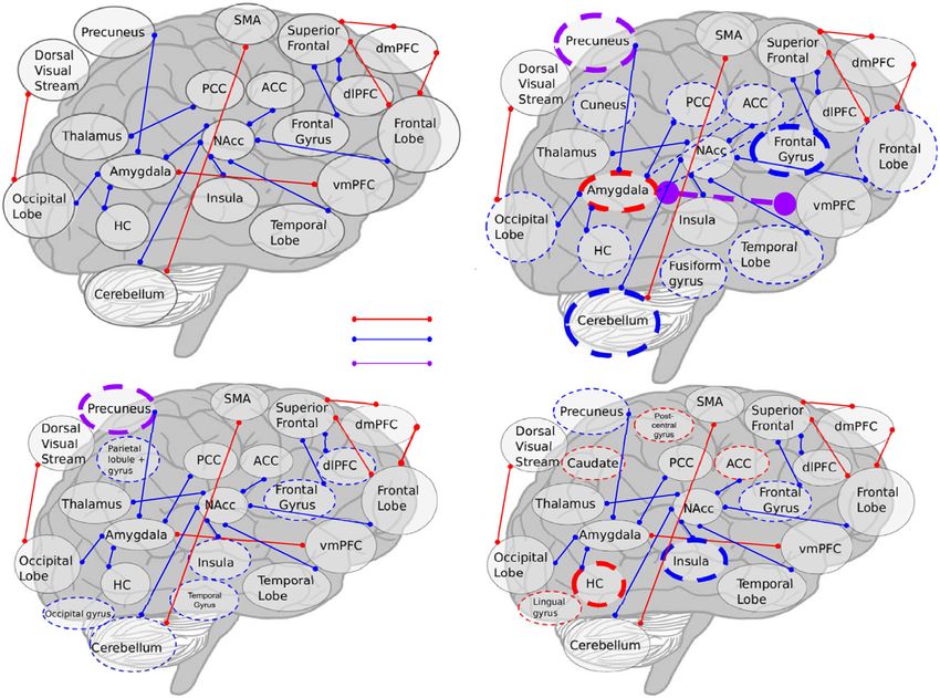
fMRI studies following THC administration. Figure 3 observed. THCv increased connectivity between a seed in
provides a comparison with the er fMRI studies super- the right dorsomedial PFC and the inferior frontal/medial
imposed on the rs fMRI study results. Figure 4 provides a frontal gyrus.
visual representation of Risk of Bias. One study orally-administering 7.5 mg Marinol used
specific regions of interest (ROIs: the amygdala, hippo-
campus [HC], and ventromedial PFC [vmPFC]) correla-
Radioligand studies tions to examine static and dynamic rs fc65. Their results
Three studies employed PET to examine striatal dopa- indicated decreased static rs fc between the amygdala and
mine receptor availability57 and regional cerebral blood HC, but increased dynamic rs fc between the amygdala
flow52,60. Additionally, SPET was used to examine dopa- and vmPFC.
mine release in the striatum66. One study also combined Whole brain analysis. Using networks of interest48 and a
data from two previously published studies, and since voxel-wise technique48,54, rs fc was most altered in the
both of the prior studies were included57,88, the third was right dorsal visual stream network following administra-
excluded. tion of 14 mg vaporized THC48. Increased connectivity
Eight milligram of vaporized THC reduced the binding with this region was localized in the frontal lobe. In the
potential of [11C]raclopride in the functionally limbic part right hemisphere, THC decreased rs fc in the right
of the ventral striatum57. However, in another study hemisphere in other regions in the frontal lobe. Finally,
10 mg did not alter binding of [11C]raclopride in the THC increased rs fc between the cerebellum and sen-
striatum88. sorimotor network, and between the left dorsal visual
Twenty milligrams inhaled THC increased regional stream and the occipital cortex. The second study
cerebral blood flow (rCBF) measured with [15O] water reported the results of nine cumulative mg THC on
PET in cortical regions, and the cerebellum (see Table 1) temporal signal-to-noise ratio (tSNR; calculated by
and decreased rCBF in auditory and visual cortices52. dividing mean blood-oxygen level dependent [BOLD]
One study administered a single dose of 2.5 mg THC via signal by its standard deviation over a time period; a
intravenous injection and compared uptake of the tracer measure thought to reflect greater spontaneous fluctua-
123I-iodobenzamide in the basal ganglia. Following THC tions and brain activity)54. THC reduced tSNR, in the
exposure, scores in the striatum ranged from a decrease right insula, left cerebellum, and substantia nigra, as
by 16% to an increase by 34% and no results were sig- hypothesized by the authors54. It is critical to note that
nificant, even though the dosages were large enough to results between the whole brain studies were markedly
elicit psychotic symptoms66. different, potentially due in part to the analytical techni-
Radioligand studies in psychosis. Increased striatal ques employed.
dopamine synthesis assessed with PET was associated rs fMRI in psychosis. Rs fMRI studies in participants
with transition from prodrome to FEP in human partici- with a FEP reveal reduced connectivity in the DMN
pants90. Additional research suggests higher baseline (dorsomedial PFC and posterior cingulate cortex (PCC)/
striatal dopamine levels in patients with schizophrenia precuneus) as well as weaker negative correlations
than healthy controls91. Following amphetamine admin- between the lateral temporal cortex and the medial
istration, there is increased dopamine release in partici- occipital lobe77. In patients with chronic schizophrenia,
pants with psychosis than healthy controls92. These functional connectivity exhibits similar patterns, with
findings are in accordance with results suggesting THC decreased strengths of connectivity in the PFC, insula, and
exposure may increase striatal dopamine release57. precuneus93.Cupo et al. Translational Psychiatry (2021)11:217 Page 10 of 19
Fig. 2 Visualization of main fMRI results across studies. a rs fMRI results, b er fMRI results, emotional processing tasks, c er fMRI results, memory
tasks, d er fMRI results, no-go trials from response inhibition. Thin lines indicate results from one study. Thick lines indicate results from two. Solid lines
indicate rs fMRI, dashed indicate resting state. Colored circles demarcate “activity” lines indicate “connectivity”.
The dorsomedial PFC was implicated in both THC The second study investigated rs fc between amygdala
exposure, where increased connectivity was observed with subfields and the cortex, revealing THC increased con-
several regions48,72, and psychosis, where decreased con- nectivity between both the amygdala and rostral ACC/
nectivity was observed77,93. Both THC exposure and medial PFC62, but was limited to viewing threatening faces.
psychosis decreased connectivity in the precuneus72,77,93, These findings suggest that the connection between these
as well as the occipital lobe53,77, insula53,93. While this two regions may be especially integral to social threat
may indicate regions for future investigation, the varia- processing and that THC exposure increases this connec-
bility in results may also reflect statistical noise. tion, of special interest as previous research associates
perception of social threat and symptoms of paranoia94. The
Event-related fMRI final study examined limbic circuitry (amygdala and ACC)
Event-related fMRI experiments used emotional pro- engagement in response to differing valence of stimuli, and
cessing, memory, sensory perception, and response inhi- observed that THC exposure reduced activity in the
bition tasks (Table 1). subgenual ACC and did not impact amygdala activity64.
These results support the view that THC decreases activity
Emotional processing tasks The amygdala is well-studied in the limbic circuit; however, the lack of effect in the
in the context of both THC exposure and emotional amygdala provides a point of contrast to the authors’
processing. A series of three studies assessed the effects of previous findings, which raises significant concerns about
7.5 mg orally-administered Marinol on emotional processing reproducibility and replicability.
in sixteen participants61–64 and found that THC attenuated In another task, participants were required to imagine
amygdala activation when viewing threatening faces63. positive contexts for negative images (e.g., reimagining aCupo et al. Translational Psychiatry (2021)11:217 Page 11 of 19 Fig. 3 Visualization of fMRI results with er fMRI superimposed on rs fMRI. a rs fMRI results, b rs and er fMRI results; emotional processing tasks, c rs and er fMRI results; memory tasks, d rs and er fMRI results, no-go trials from response inhibition. Thin lines indicate results from one study. Thick lines indicate results from two. Solid lines indicate rs fMRI, dashed indicate resting state. Colored circles demarcate “activity” lines indicate “connectivity”. woman crying outside of a church as attending her wedding; Finally, two publications from the same study population a cognitive reappraisal task)61. An increase in left amygdala and experiment examined the differential effects of THC activity and decrease in bilateral amygdala-dorsolateral PFC and CBD on emotional processing68,71. When viewing coupling was observed during the reappraisal condition fearful faces compared with neutral faces, 600 mg CBD following THC administration (7.5 mg) compared with reduced BOLD response in the left amygdala, left ACC, placebo. When matching emotional faces, 9 mg vaporized right PCC, and right cerebellum71. Ten milligram THC THC decreased activity during the fearful face condition in exposure during fearful face viewing increased activation the cerebellum. While the decrease in activity during in the left precuneus, but decreased it in frontal and negative-expression-viewing is consistent with previous temporal regions. During fearful face viewing, THC and studies, the affected areas are inconsistent62–64. CBD had opposite effects, with THC and placebo To examine the impact of long-term cannabis use on increasing amygdalar activation while CBD decreased emotional processing, one study examined fear processing it68. The authors also reported opposite effects in the in cannabis-users and nonusers (
Cupo et al. Translational Psychiatry (2021)11:217                                                                                            Page 12 of 19

                                       Green = present, red = absent, yellow = unclear, orange = not applicable

                                                                                                                         Crossover/Counter
                                        Author             Double-blind? Randomized? Placebo-controlled? Within-subject? -balanced
                                     Atakan (2013)                          Pseudo
                                     Barkus (2011)

                                   Battistella (2013)

                                 Bhattacharyya (2009)

                                 Bhattacharyya (2010)                       Pseudo

                                 Bhattacharyya (2012)                       Pseudo

                                 Bhattacharyya (2014)                       Pseudo

                                 Bhattacharyya (2018)

                                 Borgwardt et al (2008)                     Pseudo

                                    Bossong (2009)

                                   Bossong (2012a)

                                   Bossong (2012b)

                                   Bossong (2013a)

                                   Bossong (2013b)

                                    Colizzi (2018a)

                                    Colizzi (2018b)
                                    Colizzi (2019)
                                                           Researchers
                                     Dalton (2010)           blind?

                                  de Sousa Fernandes
                                      Perna(2017)                                                        mixed-factorial

                                 Fusar-Poli et al (2009)
                                                           Researchers
                                    Ginovart (2012)          blind?
                                     Gorka (2015)
                                     Gorka (2016)
                                                           Researchers
                                 Higuera-Matas (2008)        blind?

                                    Jansma (2013)                                                        mixed-factorial

                                    Klumpers (2012)
                                       Lee (2013)

                                     Mathew (1992)

                                     Mathew (1993)
                                                            Researchers
                                     Nguyen (2012)            blind?

                                     O'Leary (2003)

                                     O'Leary (2007)
                                      Phan (2008)

                                    Rabinak (2011)

                                    Rabinak (2018)

                                   Ramaekers (2016)
                                     Rzepa (2015)
                                     Stokes (2009)
                                     Tudge (2014)

                                     van Hell (2011)

                                     van Hell (2012)
                                      Walter (2016)
                                      Walter (2017)

                                  Winton-Brown (2011)                       Pseudo

 Fig. 4 Risk of bias. Assesses likelihood of bias in each paper examining for double-blind, randomized, placebo-controlled, within-subject, and
 crossover/counter-balanced. Green = present, red = absent, yellow = unclear, orange = not applicable.

 Emotional processing in psychosis. Participants at risk for                              Unlike controls, the high-risk group showed a relative
psychosis demonstrated altered activation in response to                                  increase in activation in response to neutral rather
valenced faces when compared to control groups75.                                         than sad faces in the amygdala-hippocampal complex,
Cupo et al. Translational Psychiatry (2021)11:217 Page 13 of 19
thalamus, and cuneus. The amygdala61–64,68,71 and Memory tasks in psychosis. In a verbal memory task,
cuneus43 were implicated in emotional processing during during encoding, participants at risk for psychosis showed
cannabis exposure as well. Negatively valenced stimuli did decreased activation in the frontal and parahippocampal
not elicit as strong of a response in individuals with gyri compared to healthy controls74. Surprisingly, these
psychosis compared to neutral faces, the directionality results align with findings that CBD decreased activation
consistent with response to CBD68, but not THC62,68. in the parahippocampal gyrus during recall in participants
at CHR for psychosis74.
Memory tasks Previous evidence suggests chronic can-
nabis use can impair memory95. Six studies investigated Response inhibition tasks Response inhibition was
the impact of THC on memory39,40,44,67,68,74. operationalized in a go/no-go test paradigm. In the no-
Verbal memory. One study demonstrated that cannabis go trials, 10 mg THC administration attenuated activation
users and nonuser controls both during 10 mg orally- in the left inferior frontal gyrus, adjacent insula, and
administered THC and placebo, deactivated the right precuneus, which were all activated following placebo
superior temporal gyrus during the task44. administration38 conversely THC increased engagement
Another study found that following 10 mg THC admin- from the right hippocampus and caudate nucleus.
istration, recall was associated with increased activity in One study examined the impact of previous cannabis use
the left dorsal ACC and medial PFC and decreased activity on response to acute exposure during response inhibi-
in the bilateral striatum and left rostral anterior cingulate tion44. Ten milligram THC increased activation in the
gyrus, but found no influence of the administration of right ACC and, similar to the above study, reduced
600 mg of CBD67. Contradictory results are published in activation in the left insula.
another study reporting on the same experiment in the In a study examining the contrasting effects of 10 mg
same participant group, where the authors reported that THC and 600 mg CBD, no-go trials following THC
THC and CBD had opposite effects in the striatum, ACC, exposure were associated with greater activation in the
and medial and lateral PFC during retrieval, with THC right hippocampus, right postcentral gyrus, and bilateral
decreasing activity and CBD increasing it68. The same lingual gyrus70. No-go trials in the CBD condition were
group also studied individuals at CHR for psychosis and associated with greater activation in the temporal gyri,
found that 600 mg CBD decreased activation in the left insula, and PCC. While the drugs had distinct effects,
parahippocampal gyrus during recall, but increased they did not exhibit the same oppositional pattern
activation in the left cingulate gyrus, right precentral present in the emotional processing studies. The findings
gyrus, and medial frontal gyrus74. There was a step-wise of the go/no-go task employed in the aforementioned
difference in activation across the three groups with the THC and CBD experiment were reported again in a
CHR group in the middle. These results provide paper highlighting the different effects of THC and
intriguing evidence that CBD may normalize memory- CBD68. The authors reported finding opposite effects
task impairment for CHR populations. during the go/no-go in the bilateral parahippocampal
Additional memory tasks. Two additional studies gyrus, left insula, and caudate, with THC reducing
conducted with the same participants used the Stern- activation and CBD increasing it. While the methods are
berg item recognition paradigm40 and a pictorial reported as the same, the results differ between papers.
memory task39. Difficulty of the Sternberg task can be The latter68 claims CBD and THC have opposite effects,
scaled to allow for assessment of load-dependent while activation was varied in the initial paper70.
increases in brain activity. Nine milligram of THC Visualization of the impact of THC exposure on no-go
reduced load-dependent activity in the cortex and trials is provided in Fig. 2d.
cerebellum40. In the pictorial memory task, THC Response inhibition in psychosis. Comparing healthy
reduced activity in the right insula, right inferior frontal controls to participants at CHR for psychosis and early
gyrus, left middle occipital gyrus during encoding of schizophrenia during a go/no-go task, the right inferior
images, and increased activity in the precuneus frontal gyrus and bilateral dorsal ACC showed decreased
bilaterally during recall39. While the results differed in activation during no-go relative to go in comparison with
areas impacted by THC, both studies indicate that healthy controls, this pattern arising primarily from
during encoding, THC reduces activity. Differing areas reduced no-go response activity76. THC also attenuated
of impact could be due to the respective brain-areas activation during no-go in the inferior frontal gyrus38, but
employed in the tasks, however without replication it is increased activity in the ACC44.
also possible that the reported results reflect properties
of the methodology, rather than the drug or task. Sensory processing Five studies examined the effects of
Visualization of the impact of THC on memory tasks is cannabis on sensory perceptions, examining gustation73,
provided in Fig. 2c. visual and auditory stimuli68,86, and pain49,56.Cupo et al. Translational Psychiatry (2021)11:217 Page 14 of 19 Gustation. The sole study examined how THCv however unlike individuals with psychosis, THC exposure impacted appetite depending on pleasant or aversive decreased activity in S149. flavor and visual stimuli73. While 10 mg THCv did not change subjective stimuli ratings, it increased activity in Remaining tasks The remaining studies examined the response to the chocolate stimuli (paired visual and taste) effects of THC on monetary incentive delay47,55, cannabis in the caudate, midbrain, and cingulate gyrus. In response marketing46, executive functioning41, attention43,69, and to a picture of moldy strawberries, THCv increased visuo-motor tracking59. activation in the insula, frontal cortex, temporal gyrus, Monetary incentive delay (reward processing55). Nine and putamen. microgram of THC reduced reward-related activity in the Audition. A study involving listening to neutral words parietal cortex and temporal gyrus. These results indicate read aloud demonstrated that THC reduced activity THC reduces responsivity to reward anticipation and primarily in the temporal cortex, whereas CBD increased presentation. activity in the same region86. CBD also increased activity Marketing. THC (300 mg/kg) reduced BOLD signal in the in the temporal gyri relative to THC. These results were right supplementary motor area in response to cannabis replicated in a paper discussing the opposing effects of marketing46. Additionally, THC treatment overall reduced THC and CBD, where authors observe opposite directions BOLD in the bilateral pallidum, striatum, and right caudate. of activation in the bilateral lateral temporal cortex68. Executive functioning. Task-induced deactivation in a Vision. The same study investigating audition also continuous performance task with identical pairs was examined the effects of cannabinoids on visual processing observed in a network comprising the cortical regions and of checkerboard stimuli68,86. Relative to placebo, 10 mg THC the cerebellum, which was more sensitive to the effects of reduced activity in the secondary visual cortex, and 9 mg THC than other networks41. These findings indicate increased activity in the lingual, occipital, and fusiform gyri THC may dysregulate the DMN by increasing activity whereas 600 mg CBD increased activation in the right during tasks. occipital lobe. THC increased activity in the left lingual and Visual oddball detection. Two studies used the visual middle occipital gyri, also decreasing it in scattered areas of oddball detection task, where participants respond to the occipital cortex and cerebellum relative to CBD. The presentation of visual stimuli, to assess attention69. opposite results in the occipital lobe are also reported in the Relative to placebo, 10 mg THC increased activity in the larger study comparing THC and CBD activation68. right frontal gyri and frontal pole; THC also decreased Pain perception. Two studies examined the effect of activity in the right subcortical areas. CBD (600 mg) THC on pain perception supporting the use of cannabis reduced activity in the left medial PFC and increased as an analgesic49,56. One study demonstrated that 10 mg activity in similar subcortical areas. The second study THC reduced activation in the right anterior insula, examined the impact of previous cannabis use and found hippocampus, and cerebellum after inducing pain by that after 10 mg THC exposure ingested orally, nonusers activating trigeminal nociceptors with CO256. An ROI activated the left medial frontal gyrus, as did cannabis analysis further revealed that THC decreased connectivity users after placebo43. Cannabis users in the THC between the thalamus and secondary somatosensory condition deactivated the same area, as did nonusers in cortex, which agreed with lower ratings of pain perception the placebo condition. following THC exposure. Motor control. One study examined the impact of 42 mg Fifteen microgram of THC decreased activity in the ACC inhaled THC on psychomotor control with a visuo-motor in response to a topical application of capsaicin and tracking test to assess the impact of THC exposure on lowered pain perception, but increased activity in the right driving ability59. THC increased BOLD response in the amygdala in response to painful stimuli was correlated ACC and ventromedial PFC, however it decreased activity with the analgesic effects49. THC also reduced functional in the thalamus and cortical regions. Combined with connectivity between the right amygdala and the primary results that indicate impaired tracking of the target in the sensorimotor cortex (S1) during ongoing pain, and task, these findings shed light on the urgent need for more decreased both subjective ratings of pain and limbic research of the effects of cannabis on psychomotor activity in response to painful stimuli. activity in relation to safe driving. Pain perception in psychosis. Patients with schizophrenia demonstrate reduced pain perception in comparison with healthy control, along with increased BOLD response in Arterial spin labeling S1, but relatively reduced responsivity in the PCC, insula, Examining ASL, 9 mg THC increased perfusion com- and brainstem78. The analgesia reported in psychosis pared to placebo in the ACC, left superior frontal cortex corresponds with that reported following cannabis and bilateral insula, and decreased perfusion in the exposure, as did reports of reduced activity in the insula56, postcentral and occipital gyri54. The increased perfusion
You can also read