New developments in our understanding of acne pathogenesis and treatment
←
→
Page content transcription
If your browser does not render page correctly, please read the page content below

DOI:10.1111/j.1600-0625.2009.00890.x
    www.blackwellpublishing.com/EXD
                                                                                                                                   Viewpoint

New developments in our understanding of acne
pathogenesis and treatment
Ichiro Kurokawa1, F. William Danby2, Qiang Ju3, Xiuli Wang3, Leihong Flora Xiang4, Longqing Xia5,
WenChieh Chen6,7, István Nagy8, Mauro Picardo9, Dae Hun Suh10, Ruta Ganceviciene11,
Silke Schagen12,13,14, Fragkiski Tsatsou15 and Christos C. Zouboulis15,16
1
 Department of Dermatology, Mie Universtity Graduate School of Medicine, Tsu, Mie, Japan;
2
 Division of Dermatology, Dartmouth Medical School, Hanover, NH, USA;
3
 Department of Dermatology, Hospital of Dermatology and Venereology of Shanghai, Shanghai, China;
4
 Department of Dermatology, Hua Shan Hospital, Shanghai Medical College, Fu Dan University, Shanghai, China;
5
 Institute of Dermatology, Chinese Academy of Medical Sciences & Peking Union Medical College, Nagqing, China;
6
 Department of Dermatology and Allergy, Technische Universitaet Muenchen, Munich, Germany;
7
 Department of Dermatology, Chang Gung Memorial Hospital, Kaohsiung Medical Center, Kaohsiung, Taiwan;
8
 Institute for Plant Genomics, Human Biotechnology and Bioenergy, Bay Zoltán Foundation for Applied Research, Szeged, Hungary;
9
 San Gallicano Dermatological Institute, Rome, Italy;
10
  Department of Dermatology, Seoul National University College of Medicine, Seoul, South Korea;
11
  Centre of Dermatovenereology, Vilnius University Hospital, Vilnius, Lithuania;
12
  Pentapharm, Aesch, Switzerland;
13
  University of Basel, Basel, Switzerland;
14
  s&kGrey, Freiburg, Germany;
15
  Departments of Dermatology, Venereology, Allergology and Immunology, Dessau Medical Center, Dessau, Germany;
16
  Laboratory for Biogerontology, Dermato-Pharmacology and Dermato-Endocrinology, Institute of Clinical Pharmacology and Toxicology,
Charité Universitaetsmedizin Berlin, Campus Benjamin Franklin, Berlin, Germany
Correspondence: Ichiro Kurokawa, MD, Department of Dermatology, Mie University Graduate School of Medicine, 2-174, Edobashi Tsu,
Mie 514-8507, Japan, Tel.: +81 59 232 1111 (ext. 6421), Fax: +81 59 231 5206, e-mail: kuroichi@clin.medic.mie-u.ac.jp

Accepted for publication 18 March 2009

Abstract: Interest in sebaceous gland physiology and its diseases is          stimulate the secretion of cytokines, such as interleukin (IL)-6 and
rapidly increasing. We provide a summarized update of the current             IL-8 by follicular keratinocytes and IL-8 and -12 in macrophages,
knowledge of the pathobiology of acne vulgaris and new treatment              giving rise to inflammation. Certain P. acnes species may induce an
concepts that have emerged in the last 3 years (2005–2008).                   immunological reaction by stimulating the production of sebocyte
We have tried to answer questions arising from the exploration of             and keratinocyte antimicrobial peptides, which play an important
sebaceous gland biology, hormonal factors, hyperkeratinization,               role in the innate immunity of the follicle. Qualitative changes of
role of bacteria, sebum, nutrition, cytokines and toll-like receptors         sebum lipids induce alteration of keratinocyte differentiation and
(TLRs). Sebaceous glands play an important role as active                     induce IL-1 secretion, contributing to the development of follicular
participants in the innate immunity of the skin. They produce                 hyperkeratosis. High glycemic load food and milk may induce
neuropeptides, excrete antimicrobial peptides and exhibit                     increased tissue levels of 5a-dihydrotestosterone. These new aspects
characteristics of stem cells. Androgens affect sebocytes and                 of acne pathogenesis lead to the considerations of possible
infundibular keratinocytes in a complex manner influencing cellular           customized therapeutic regimens. Current research is expected to
differentiation, proliferation, lipogenesis and comedogenesis.                lead to innovative treatments in the near future.
Retention hyperkeratosis in closed comedones and inflammatory
                                                                              Key words: sebaceous gland – acne – cytokine – Toll-like receptor
papules is attributable to a disorder of terminal keratinocyte
                                                                              – PPAR – hyperkeratinization
differentiation. Propionibacterium acnes, by acting on TLR-2, may

Please cite this paper as: New developments in our understanding of acne pathogenesis and treatment. Experimental Dermatology 2009; 18: 821–832.

                                                                              glands (1), and increased sebum excretion is a major con-
Biology of sebaceous glands
                                                                              current event that parallels the development of acne lesions.
The sebaceous gland is a holocrine gland, and its secretion                   With the development of human sebaceous gland experi-
is formed by the complete disintegration of the glandular                     mental models for in vitro studies (2–5), considerable
cells. Excreting sebum is the major function of sebaceous                     progress has been made in our understanding of many new

ª 2009 John Wiley & Sons A/S, Experimental Dermatology, 18, 821–832                                                                                821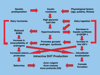
Kurokawa et al.

aspects of the gland’s function and control (6–8). These        dent pathway (22). CRH was also found to enhance mRNA
studies have illustrated why the view of the human seba-        expression of D5-3b-hydroxysteroid dehydrogenase in
ceous gland has turned from a ‘living fossil of the skin’ (9)   human sebocytes in vitro, an enzyme that is responsible for
to become the ‘brain of the skin’ (10), and are providing       androgen activation through the conversion of dehydroepi-
many new insights into the pathogenesis and treatment of        androsterone to testosterone (20). Antalarmin, a CRH-R1
sebaceous gland-associated diseases, such as acne vulgaris.     specific CRH inhibitor, reduced sebaceous neutral lipid
                                                                synthesis (21). These in vitro data may be compatible with
                                                                the significant increase in CRH expression in acne-involved
Neuropeptides and sebaceous glands
                                                                compared with sebaceous glands not involved with acne
Neuropeptides (NPs) are a heterogeneous group of biologi-       (10). The interaction between androgen signalling and
cally active peptides that are present in neurons of both the   CRH-dependent signalling mechanisms, especially the dif-
central and peripheral nervous systems (11). The human          ferential regulation of CRH-R1 and CRH-R2, needs further
sebaceous gland has been shown to express functional            study to provide elucidation in more detail.
receptors for NPs, such as corticotropin-releasing hormone         The proopiomelanocortin (POMC) system also plays an
(CRH), melanocortins, b-endorphin, vasoactive intestinal        important role, as a neuromediator system in controlling
polypeptide, NP Y and calcitonin gene-related peptide.          the sebaceous gland. The a-melanocyte-stimulating hor-
These receptors modulate the production of inflammatory         mone (a-MSH) can stimulate sebocyte differentiation and
cytokines, proliferation, differentiation, lipogenesis and      lipogenesis (23,24). Human sebocytes express MC-1Rs and
androgen metabolism in human sebocytes (6,12).                  MC-5Rs in vitro and in vivo (22,25,26). While MC-1Rs was
   The NP substance P (SP) was found to express in dermal       expressed in both undifferentiated and differentiated
nerves around the sebaceous glands of acne patients (13).       sebocytes, MC-5Rs was expressed only in differentiated
SP promotes both the proliferation and the differentiation      sebocytes. Activation of MC5R apparently stimulates lipo-
of sebaceous glands in vitro, and increases immunoreactiv-      genesis, which indicates that MC-1R expression is not
ity and RNA expression of proinflammatory factors (14). It      obligatorily associated with the sebaceous cell differentia-
induces the expression of neutral endopeptidase (CD10) in       tion process and lipogenesis (26). In contrast, MC-5Rs is a
sebaceous germinative cells and of E-selectin in periseba-      marker of sebocyte differentiation and responsible for the
ceous venules (15). Recently, ectopeptidases dipeptidyl pep-    lipogenesis of sebocytes. Studies also showed that acne-
tidase IV (DP IV or CD 26) and aminopeptidase N (APN            involved sebaceous glands express higher levels of MC-1R
or CD13), which have been shown to be involved in the           than sebocytes of healthy glands (27), but no studies have
degradation of several NPs, especially SP (16), have been       been performed on MC-5R expression in acne. The latter
found to be highly expressed in human sebocytes in vivo         data indicate that further research is required to establish
and in vitro (17). Further studies showed unexpectedly that     the role of melanocortin receptors in the physiology and
inhibitors of DP IV and APN can suppress proliferation          pathology of the sebaceous glands. Recent findings indicate
and slightly decrease neutral lipids, but can also enhance      that b-endorphin suppresses cell proliferation and induces
terminal differentiation in SZ95 sebocytes. This suggests       lipid formation in SZ95 sebocytes in vitro, which may be
that ectopeptidases may be new targets to modulate certain      mediated by the l-opioid receptor, which is expressed in
sebocyte functions, and that ectopeptidase inhibitors may       human sebocytes in vivo and in vitro (6).
have potential therapeutic roles in acne pathogenesis
(6,17).
                                                                Sebaceous gland and innate immunity
   The hypothalamic-pituitary-adrenal axis is traditionally
regarded to be responsible for neuroendocrine responses of      The pilosebaceous unit is an immunocompetent organ.
sebaceous gland to stress (18). The presence of CRH, its        Keratinocytes and sebocytes may act as immune cells capa-
binding protein (CRHBP) and its receptors CRH-R1 and            ble of pathogen recognition and abnormal lipid presenta-
R2 in human sebaceous glands in vivo and SZ95 sebocytes         tion (28). Innate immunity molecules such as toll-like
in vitro has been confirmed (10,19–21). CRH can inhibit         receptor (TLR) 2 and TLR4 (29), CD1d (28) and CD14 (6)
proliferation and induce synthesis of neutral lipids in SZ95    are expressed in SZ95 sebocytes. Keratinocytes and sebo-
sebocytes, and testosterone antagonizes CRH by downregu-        cytes, as major components of the pilosebaceous unit, may
lating CRH-R expression in human sebocytes in vitro. In         act as immune cells and may be activated by P. acnes via
addition, growth hormone, which also enhances sebaceous         TLRs and CD14 and through CD1 molecules and also may
lipid synthesis, modifies CRH-R expression by reducing          recognize altered lipid content in sebum, followed by the
mRNA levels of CRH-R1 and by enhancing CRH-R2                   production of inflammatory cytokines. In addition, anti-
mRNA levels, enhancing the release of interleukin (IL)-6        microbial peptides, such as defensin-1, defensin-2 and
and IL-8 in SZ95 sebocytes in vitro by an IL-1b-indepen-        cathelicidin, showed expression and immunoreactivity in

 822                                                               ª 2009 John Wiley & Sons A/S, Experimental Dermatology, 18, 821–832
Acne pathogenesis and treatment
the sebaceous gland (13,30). Human b-defensin-2 (hBD-2) LXRb, inhibit cell proliferation and stimulate lipid synthe-
is also expressed upon exposure to lipopolysaccharides sis (14,45). They also decrease the expression of cyclooxy-
(LPS) and P. acnes (31). genase-2 and inducible nitric oxide synthase that was
The monounsaturated fatty acids (MUFA), mainly pal- induced by LPS treatment, a function that indicates the
mitic acid (C16:1) and oleic acid (C18:1), both of which important roles of LXRa in differentiation and inflamma-
are bactericidal against Gram-positive organisms (32), are tory signalling in sebaceous glands (14).
produced by the sebaceous gland, as is sapienic acid, an The physiological role of non-neuronal acetylcholine
important antimicrobial lipid. Stearoyl coenzyme A desat- (ACh) and its receptors (AChR) in epidermal physiology
urase (SCD) 1, an enzyme responsible for the biosynthesis has been studied recently (46). The undifferentiated and
of MUFA, is also expressed by the sebaceous gland (33). mature sebocytes express different AchR subunits (47),
The TLR-2 ligand macrophage-activating lipopeptide-2 which imply that sebocyte differentiation, sebum produc-
stimulates both SCD and fatty acid desaturase-2 mRNA tion or sebum composition may be altered by endoge-
expression in SZ95 sebocytes (32). nously produced ACh acting in a paracrine manner or
stimulated exogenously by nicotine. Presence of AChR and
nicotinic activity are also found in the infundibulum of the
Sebaceous gland and stem cells
pilosebaceous unit and can promote infundibular epithelial
Sebaceous gland cells derive from the basal progenitor cells hyperplasia and follicular plugging, suggesting an important
of the sebaceous gland alveolus (34,35) and were until role for the cholinergic system in acne vulgaris (48), and a
recently regarded to be updated by the reservoir of stem possible etiological role for nicotine uptake by smoking in
cells in the hair follicle bulge (36). This interdependence is, acne and especially hidradenitis suppurativa (47).
however, not obligatory (37): sebaceous glands are present
in some mouse mutants that lack hair follicles (38,39), they
Hormones
can be maintained independently of the hair follicle bulge
(40) and they can be induced in footpad epidermis, an Androgens play an essential role in acne pathogenesis. As
anatomic region normally devoid of hair follicles and seba- most of the patients with acne have normal circulating
ceous glands. Sebaceous glands have also been found to androgen levels, acne severity does not correlate with serum
express skin stem cell marker bone morphogenetic protein- androgen levels. It is postulated that androgens may play
1 (41). The human sebocyte lines SZ95 and SebE6-E7 have only a permissive role in priming or initiating acne develop-
the ability to differentiate into both sebocytes and interfol- ment, or it may be the local overproduction of androgens
licular epidermal cells (8). This suggests that human sebo- in the skin and/or the high expression and responsiveness
cytes may represent a bipotential stem cell. Furthermore, of androgen receptors that determines the formation of
the interaction between b-catenin and Indian hedgehog acne lesions.
stimulates the proliferation of sebocyte precursors (42). The sebaceous gland has been shown to express all the
Overexpression of Myc stimulates sebocyte differentiation, necessary enzymes for the biosynthesis of testosterone
whereas overexpression of b-catenin stimulates interfollicu- de novo from cholesterol, from 5a-reduced substances
lar epidermal differentiation in vitro (8). ingested in dairy products (49), or in a shortcut from
circulating dehydroepiandrosterone (50).
The main influence of androgen on acne pathogenesis
Other sebocyte properties
concerns the proliferation/differentiation of sebocytes and
Histamine-1 receptor is expressed in SZ95 sebocytes and in infrainfundibular keratinocytes. The human models useful
human sebaceous glands, and diphenhydramine, a hista- for in vitro studies include sebaceous gland organ culture,
mine-1 receptor antagonist, significantly decreases squalene primary culture of sebocytes and immortalized human
levels, indicating that histamines and anti-histaminics could sebocyte cell lines (4,5,51).
potentially directly modulate sebocyte function (43). Perox- 1 Sebocyte proliferation: The stimulatory effect of tes-
idated squalene induced the production of inflammatory tosterone and 5a-dihydrotestosterone (DHT) on sebocyte
mediators in HaCaT keratinocytes and induced upregula- proliferation was observed in primary culture of human
tion of PPARc, mRNA and protein expression in a dose- sebocytes and hamster sebaceous gland cells (52,53). In
dependent manner (44). SZ95 sebocytes, testosterone and DHT may showed a
Liver X receptors (LXRs) are members of the nuclear stimulatory effect or no effect on cell proliferation (4,54).
receptor superfamily, which plays a critical role in choles- In cultured rat preputial cells, DHT suppressed cell pro-
terol homeostasis and lipid metabolism. Expression of liferation (55). Of note, androgens are effective on
LXRa and LXRb was detected in SZ95 sebocytes (45), and human sebocytes in vitro in concentrations above the
LXR ligands enhance the expression of LXRa, but not of physiological ones.
ª 2009 John Wiley & Sons A/S, Experimental Dermatology, 18, 821–832 823Kurokawa et al.
2 Sebocyte differentiation and lipogenesis: In the seba-
ceous gland organ culture, testosterone and DHT at nearly
physiological concentrations demonstrated no effect or
inhibitory effect on cell division rates or lipogenesis (2). In
SZ95 sebocytes, the combination of testosterone and lino-
leic acid exhibited a synergistic effect on sebaceous lipids
(56). In hamster sebocytes, DHT augmented the formation
of intracellular lipid droplets along with an increase in the
accumulation of triglycerides (5). Interestingly, DHT treat-
ment of hamster ear sebaceous glands in vivo had a pro-
found effect on sebocyte proliferation, differentiation and
induction of lipogenesis involving the upregulation of the
sterol response-element-binding protein (SREBP) pathway,
a key regulator of lipogenesis (54,57). SREBP expression
has also been detected in human sebocytes (33).
Figure 1. Pathogenesis of acne. The biology of sebaceous gland (I) has
3 Comedogenesis: Formation of microcomedones is
been elucidated recently. Hormonal factors (II) are involved in sebum
caused by hyperproliferation/hyperkeratinization of the excretion (VI) and hyperkeratinization in the infundibulum (III). Acne
infrainfundibulum of the follicular canal. It remains to be starts as microcomedones, which are generated by hyperkeratinization
determined if higher activity of the type I 5a-reductase in the infundibulum, with increased in size and in number of granular
detected in the follicular infrainfundibulum is related to layers. Microcomedones evolve into open or closed comedones. The
follicular channel is colonized by Propionibacterium acnes (IV), develops
the abnormal differentiation of keratinocytes (58). In addi-
and stimulates cytokine production (VII) via toll-like receptor (VIII),
tion to the isolated infundibulum culture, various animal resulting in inflammatory lesions. Nutrition (V) may be involved in acne
models have been used for studies on comedogenesis, such pathogenesis. IL-8, a neutrophil chemotactic factor, attracts neutrophils
as the rabbit ear assay, the Mexican hairless dog and the into the follicular walls. Once the follicular walls rupture,
Rhino mouse. It is unclear if these models can truly reflect granulomatous lesions with subcutaneous induration, scarring and
keloids are generated.
the human sebaceous follicles.
Taken together, despite the clinical evidence that andro-
gens stimulate sebaceous lipids, the in vitro effect of andro- expression of the hyperproliferative markers keratin (K) 6
gens on proliferation and differentiation of sebocytes varies and K16 (61). IL-1a activates basal keratinocyte by auto-
in different experiments. Further studies are needed to crine production inducing K16 expression in suprabasal
determine which cofactors are required for the display of cells in the active state. This finding indicates that keratin
androgen actions on sebocytes. and keratinocyte activation cycle are related to hyperprolif-
eration of the keratinocytes lining the infundibulum (62).
Regarding the hormonal response, increased DHT may
Hyperkeratinization
act on infundibular keratinocytes leading to abnormal
One of the most crucial initial events in the development hyperkeratinization (58). Follicular keratinization may be
of acne lesions is hyperkeratinization in the follicular triggered by relative deficiency of linoleic acid and perox-
infundibulum and sebaceous duct resulting in microcome- ides in sebum (32). Recently, P. acnes extracts have been
dones (Fig. 1). Electronmicroscopically, the pattern of implicated in the formation of the microcomedo (63).
hyperkeratinization demonstrates retention hyperkeratosis However, the pathogenesis of closed and open comedo
with increased number and size of keratohyaline granules formation remains ambiguous. Studies on nevus comedo-
and accumulation of lipid droplets and folding of the nicus indicated that the disturbance of terminal differenti-
retained squames on themselves as a result of pressure ation in the follicular infundibulum may play a role in
effects (59). closed comedo formation (64), and fibroblast growth
The pathogenesis of follicular hyperkeratinization is still factor receptor (FGFR) 2 signalling also seems to be
unclear. IL-1a has been reported to induce hyperkeratiniza- involved. Acneiform nevus, which is a variant of nevus
tion in follicular infundibulum in vitro and in vivo (51). In comedonicus, has been shown to be associated with
addition, abnormal infundibular keratinization has been Ser252Trp-gain-of-function mutation of FGFR2, which
associated with a disorder in terminal differentiation of also explains acne in Apert syndrome (65,66). Androgen-
infundibular keratinocytes, which is related to increased fil- dependent FGFR2b-signalling has been proposed in the
aggrin (filament aggregating protein) expression (60). Not pathogenesis of acne (67). The Ser252Trp-FGFR2-muta-
only hyperkeratinization but also hyperproliferation can be tion with increased FGFR2-signalling is associated with
observed within infundibular keratinocytes, with the increased expression of IL-1a (68). In addition, cyst
824 ª 2009 John Wiley & Sons A/S, Experimental Dermatology, 18, 821–832Acne pathogenesis and treatment
formation in acne usually occurs following closed comedo Sebaceous gland growth and the consequent increased
development. Terminal differentiation in filaggrin expres- sebum excretion are phenomena experienced by all adoles-
sion may be involved in cyst formation (69). Moreover, cents, but only in some cases does it seem to be associated
there is a debate on whether influx of inflammatory cells with the incorrect regulation of lipid metabolism. Several
precedes hyperkeratinzation (70). variations in lipid metabolism have been described in acne
patients, including a decreased amount of linoleic acid
(77). Moreover, recent studies suggest that the desaturation
Bacteria
of sebaceous fatty acids may contribute to acne develop-
The significance of the involvement of P. acnes in acne ment: an increase in the saturated/monounsaturated ratio
pathogenesis is still controversial, mainly due to the fact together with a reduction in the enzymatic desaturation of
that it belongs to the resident microbiota. The recently C16:0 seems to correlate with the lesion counts and, there-
decoded genome of P. acnes again raised the question of fore, with the clinical improvement (78). A hallmark of
the pathogenic potential of this bacterium (71). This possi- sebum in acne patients is the presence of lipoperoxides,
bility is further supported by the observation that P. acnes mainly due to the peroxidation of squalene and a decrease
induces the expression of antimicrobial peptides and proin- in the level of vitamin E, the major sebum antioxidant
flammatory cytokines/chemokines from various cell types (44). The generation of an inflammatory reaction seems to
(14,31,72,73). Recent description of phylogenetically initiate the hyperkeratinization of the acroinfundibulum
distinct P. acnes clusters (74) challenges our overall current and the manifestation of acne lesions. In this context, both
understanding of the pathogenic nature of bacteria lipoperoxides and monounsaturated fatty acid (MUFAs)
involved in acne pathogenesis and raises the possibility that are capable of inducing alteration in keratinocyte prolifera-
certain P. acnes strains may cause an opportunistic infec- tion and differentiation, whereas peroxides are capable of
tion worsening acne lesions. Indeed, phylogenetic clusters inducing production of pro-inflammatory cytokines and
of P. acnes differ not only in the production of secreted activation of peroxisome proliferator-activated receptors
proteins (74), but also in their ability to induce different (PPARs) (44,78). Seborrhoea per se is not responsible for
immune responses in keratinocytes and sebocytes; the the development of acne, as demonstrated by the success of
major difference being the ability to induce hBD-2 expres- treatment with agents with no effect on sebum secretion
sion (31,73). Although hBD-2 has no direct antimicrobial rate that can inhibit the inflammatory process, such as
effect on P. acnes (73), it does have synergistic activity with antibiotics, topical retinoids, azelaic acid and benzoyl per-
cathelicidin (14). Thus, the total antimicrobial activity in oxide. Moreover, the occurrence of seborrheic dermatitis is
the pilosebaceous unit is likely due to several antimicrobial associated with a change in the quality of sebum lipids, i.e.
peptides – and additionally antibacterial lipids (32) – acting a decreased level of polyunsaturated fatty acids and vitamin
together. E (79). Considering all these data, it is apparent that sebor-
Various antimicrobial peptides are expressed in healthy rhoea is not necessarily associated with the alteration of
skin without any visible signs of inflammation, suggesting lipid composition and the oxidant/antioxidant ratio charac-
that (i) antimicrobial peptides may be induced in the teristic of the skin surface lipids of acne patients.
absence of proinflammatory cytokines/chemokines and
(ii) resident skin microbiota may facilitate antimicrobial
Nutrition
peptide induction without inflammation. It was recently
proposed that the beneficial effects of resident microbiota Acne is driven by hormones and growth factors [particu-
may come from their ability to induce antimicrobial larly insulin-like growth factor (IGF-1)] acting on the seba-
peptide expression (75). The identification of P. acnes ceous glands and the keratinocytes lining the pilary canal.
proteins, inducing solely antimicrobial peptide and not Dairy products (and perhaps some other foods) contain
proinflammatory cytokine/chemokine expression, would 5a-reduced steroid hormones and other steroid precursors
promote stimulation of maintenance levels of antimicrobial (49) of DHT that drive sebaceous gland (and likely pilar
peptides. Consequently, increased resistance to abnormal keratinocyte) function. Dairy also contains about 60 other
P. acnes colonization could be facilitated. growth factors and micronutrients (80). Phytoestrogens in
food seem to have no impact on acne. Drinking milk
causes a direct rise in IGF-1 through a disproportionate
Sebum
elevation in blood sugar and serum insulin levels (81).
Increased sebum production is a characteristic of acne High glycemic load foods also cause IGF-1-mediated eleva-
patients even if it does not strictly correlate with the devel- tions in DHT (82). IGF-1 levels during teenage years clo-
opment of the lesions (76). Seborrhoea is, indeed, not a sely parallel acne activity and are likely synergistic with the
sufficient condition for the development of the pathology. steroid hormones (Fig. 2).
ª 2009 John Wiley & Sons A/S, Experimental Dermatology, 18, 821–832 825Kurokawa et al.
Figure 2. The acnegenic cascade: interactions among the numerous Figure 3. The formation of the anoxic duct. The expanding
hormones, growth factors, enzymes and their targets. keratinocytic mass is constricted by the glassy membrane, with resulting
anoxia represented as the bluish hue of deoxygenated haemoglobin
Vitamin A is needed for normal follicular function and (available at: http://www.acnemilk.com).
is often deficient in teens. Dietary fatty acids influence
inflammation, some pro-inflammatory, some anti-inflam- Cytokines
matory, underlining the need for careful dietary selection Cytokines are present in normal sebaceous glands, and they
for optimal control (83). Linoleic acid likewise has an are affected by many factors (87) (Table 1). IL-1a, tumor
ambivalent role in acne (84). Iodine, although not comedo- necrosis factor (TNF)-a, IL-6 and IL-8 are released into
genic, may enhance inflammation (85). supernatant in unstressed sebocyte culture (88). In a stressed
Acne can be improved by controlling hormones and environment, the amounts of released cytokines increase sig-
inflammation, both of which are influenced by diet; so full nificantly. The treatment of cultured sebocytes with P. acnes
acne control requires dietary control. Concurrent with and LPS significantly upregulated the expression of proin-
standard anti-acne therapy, all dairy products and all high flammatory cytokines (31). While LPS stimulated CXCL8,
glycemic foods should be stopped for at least 6 months to TNF-a and IL-1a, P. acnes stimulated CXCL8 and TNF-a
evaluate the effect (86). Vitamin A supplementation may only. Propionibacterium acnes had no effect on IL-1a. There
help reduce plugging of pores in deficient individuals. was also a difference in the cytokine production curve over
Foods containing x-3 essential fatty acids (EFAs) and EFA time after treatment between P. acnes and LPS. Arachidonic
supplements may help to control inflammation (83). acid and calcium ionophore enhanced the level of IL-6 and
The pilary canal is plugged by what is best viewed as a IL-8, but that of IL-1b and TNF-a was not affected (88).
straightforward mechanical effect. As the hormone-driven
keratinocytes multiply, they are propelled towards the cen-
Table 1. Current aspects of cytokine production in normal skin,
tre of the duct, which expands to accommodate the
acne lesions and associated factors
increasing bulk of the microcomedo until a point is
reached beyond which the inelastic ‘glassy membrane’ that
IL-1a IL-6 IL-8 CXCL8 TNF-a
encloses the pilosebaceous duct can expand no further.
Further production of keratinocytes into this closed system
Normal person, healthy skin ) ++
causes an increase in intraluminal pressure and this causes Acne patient, uninvolved skin + ++
hypoxia centrally in the duct (Fig. 3). This produces an Acne patient, involved skin ++ +++
Propionibacterium acnes No effect › ›
anoxic environment that favours development of intraduc- stimulation
tal P. acnes colonies, leads to rupture of the duct walls, LPS treatment › › ›
Increased PAF-R expression ›
release of the luminal antigens and the ultimate production ectopeptidase › 1
or worsening of the inflammatory acne papule. CRH › ›
a-MSH fl
The inflammatory cascade that follows is produced by a
Pandora’s boxful of mediators, cytokines and chemokines IL, interleukin; TNF, tumor necrosis factor; LPS, lipopolysaccharide;
causing the chemical and biological epiphenomena of acne; PAF-R, platelet-activating factor receptor; CRH, corticotropin-releas-
but no matter what occurs when the inflammation com- ing hormone; a-MSH, a-melanocyte-stimulating hormone.
1
mences, the initiating factors are hormonal. Thus, no acne IL-1 receptor antagonist is significantly upregulated in cultured
therapy is complete without a dietary and hormonal history sebocytes in the presence of dipeptidyl peptidase IV and aminopep-
tidase N inhibitors.
and appropriate dietary and hormonal advice.
826 ª 2009 John Wiley & Sons A/S, Experimental Dermatology, 18, 821–832Acne pathogenesis and treatment
In in vivo studies, IL-6 was barely detectable in the seba- The sebaceous gland has usually been thought to simply
ceous gland of healthy skin (88). In acne patients, a weak provide physical protection to the external skin surface, but
expression was found in uninvolved skin and a stronger its numerous functions are gradually becoming understood
one in acne-involved skin. IL-8 exhibited a stronger expres- (54). Human SZ95 sebocytes were found to express constitu-
sion in sebocytes of acne patients’ skin than in those of tively TLR2, TLR4, CD14, IL-1a, IL-1b, IL-6 and IL-8. The
healthy controls (88). latter, augmented by exposure to components of Gram-nega-
Corticotropin-releasing hormone enhances the release of tive (LPS) and Gram-positive (lipoteichonic acid) bacteria
IL-6 and IL-8 from sebocytes in vitro by an IL-1b-indepen- (29). In addition, human sebocytes produce antimicrobial
dent pathway (10). In the POMC system, a-MSH peptide lipids (32), antimicrobial peptides (hBD-2, psoriasin and
suppressed IL-1b-induced release of IL-8, a central cathelicidin) (12,31,92), which exhibit synergistic activities
pro-inflammatory mediator in the pathogenesis of acne and induce proinflammatory cytokines/chemokines via TLR-
inflammation (22). and CD14-dependent mechanisms. Certain P. acnes species
Cultured sebocytes express functional platelet-activating can stimulate SZ95 sebocytes to produce the endogenous
factor receptors (PAF-R), and PAF-R are involved in regu- TLR4 agonist hBD-2 (31), and sebaceous cathelicidin can
lating the expression of inflammatory mediators (26), even kill P. acnes (14). These findings indicate a direct func-
including cyclooxygenase-2, prostaglandin E and IL-8 (10). tional induction of innate immunity in human epithelial
Ectopeptidases found in sebocytes also modulate cytokine cells, and especially in sebocytes, without the involvement of
regulation. Inhibition of these ectopeptidases leads to an inflammatory cells, while recruitment of the latter to the
upregulation of endogenous IL-1 receptor antagonist (15). involved sites can potentiate the inflammatory events in acne
It is very important that cytokine regulation of sebocytes, (54). This prominent new and previously unsuspected pat-
which play a key role in acne pathophysiology (70), is tern recognition by the sebaceous glands establishes a func-
modulated by various factors mentioned above. Targeting tional link between lipids, lipid metabolism, antimicrobial
these cytokine-regulating factors may have a potential in proteins and epithelial innate immunity that can be impor-
the future effective treatment of acne. tant in acne pathogenesis. Therefore, the pharmacological
regulation of TLR and CD14 expression may provide a novel
target for the treatment of inflammatory acne lesions.
Toll-like receptors
Toll-like receptors are transmembrane proteins that are
Overview of acne pathogenesis
crucial players in the innate immune response to microbial
and other invaders. Ten TLRs have recently been described The pathogenesis of acne, the most common skin disease,
in humans (72). TLRs are mainly expressed on immune which manifests in the pilosebaceous follicle, is currently
cells, such as monocytes, macrophages, dendritic cells and attributed to multiple factors such as increased sebum pro-
granulocytes. TLR stimulation mimics the action of IL-1a duction, alteration of the quality of sebum lipids, regula-
and promotes the production of proinflammatory cyto- tion of cutaneous steroidogenesis, androgen activity,
kines, prostaglandins, leukotrienes (LT) and chemokines interaction with NPs, exhibition of pro- and anti-inflam-
(72). Intriguingly, selected IL-1 receptor associated kinases matory properties, follicular hyperkeratinization and the
(IRAK-1, 2, M and 4) are bifunctional. They can be proliferation of P. acnes within the follicle (6,32).
recruited to the TLR complex and thus mediate TLR The increased sebum excretion is a major concurrent
signalling but can also associate with protein partners event associated with the development of acne. Neutral and
involved in T- and B-cell receptor-mediated signalling polar lipids produced by sebaceous glands serve a variety of
pathways and so can be critical mediators for both innate roles in signal transduction and are involved in biological
and adaptive immune responses (89–91). Conceivably, pathways (6). Additionally, fatty acids act as ligands of
these molecules may be viable targets for designing new nuclear receptors such as the PPARs. Sebaceous gland lip-
therapeutic strategies for various human inflammatory ids exhibit direct pro- and anti-inflammatory properties,
diseases. whereas the induction of 5-lipoxygenase and cycloxygen-
Chemokine/cytokine synthesis in human monocytes is ase-2 pathways in sebocytes leads to the production of
induced through activation of TLR2 by P. acnes (72), but proinflammatory lipids (88).
the expression of active TLR2 and 4 and of CD14 in Furthermore, hormones like androgens control the seba-
human keratinocytes (28) has also implicated P. acnes and ceous gland size and sebum secretion. In cell culture,
TLRs in the development of acne inflammation. It is androgens only promote sebocyte proliferation, whereas
suggested that, by using this pathway, the innate immune PPAR ligands are required for induction of differentiation
system is able to recognize microbial components and then and lipogenic activity (56). On the other hand, keratino-
induce cytokine/chemokine synthesis in acne (32). cytes and sebocytes may be activated by P. acnes via TLR,
ª 2009 John Wiley & Sons A/S, Experimental Dermatology, 18, 821–832 827Kurokawa et al.
CD14 and CD1 molecules (31). Pilosebaceous follicles in undeniable effectiveness, isotretinoin is not a curative drug
acne lesions are surrounded with macrophages expressing (88). Its discontinuation may be followed by recurrence in
TLR2 on their surface. TLR2 activation leads to transcrip- the absence of appropriate maintenance treatment. Identify-
tion factor nuclear factor triggering and thus production of ing the appropriate acne patient for isotretinoin treatment is
cytokines/chemokines, phenomena observed in acne lesions. nowadays important, because the compound has already
Furthermore, P. acnes induces IL-8 and IL-12 release from drawn the attention of the European Committee on Proprie-
TLR2 positive monocytes (72). tary Medicinal Products, which released a European directive
Acne research continues to deliver new pieces to the to ensure harmonization of isotretinoin treatment through-
puzzle, helps us to understand acne pathogenesis and out the European Union (95). Both the European guidelines
assists the development of new drugs against acne. New and the iPLEDGE isotretinoin distribution programme of
compounds which are able to inhibit LTB4 synthesis, the (Food and Drug Administration) are aimed at preventing
antagonize PPAR or inhibit ectopeptidases (10) offer new the use of the drug in women during pregnancy.
ways to treat acne. PPAR regulation may be a pathway to In addition to our better understanding of its activity
modify sebaceous lipogenesis. (88), other developments in the application of this potent
compound include the lower risk of side-effects using
low-dose long-term regimens (0.1–0.3 mg/kg/day daily or
Future prospective drugs based on acne
intermittent use) and a micronised formulation, which
pathogenesis
exhibits similar efficacy and is associated with a lower risk
Rational use of available treatment options based on the of adverse events (54).
type and severity of acne lesions is presently a key compo-
nent of successful acne therapy (93). However, increasing Retinoic acid metabolism blocking agents
understanding of acne pathophysiology slowly gives rise to Retinoic acid metabolism blocking agent (RAMBA) are
the anti-acne therapeutic agents and regimens of the future compounds which block the catabolism of endogenous
(32) (Fig. 4). vitamin A. Oral talarozol, a potent retinoic acid 4-hydro-
lase inhibitor, was effective on comedones and inflamma-
Isotretinoin tory lesions in a pilot study with 17 acne patients after
Oral isotretinoin is the most effective drug available for the 12 weeks of treatment (96). Topical RAMBA modulates
treatment of acne. It directly suppresses sebaceous gland keratinization in the mouse and can induce a dose-depen-
activity leading to significant reduction in sebaceous lipogen- dent reduction in IL-1a mRNA in human skin.
esis, normalizes the pattern of keratinization within the seba-
ceous gland follicle, inhibits inflammation, and – in a Ectopeptidase inhibitors
secondary manner – reduces growth of P. acnes (27). In addi- Inhibitors of dipeptidylpeptidase IV and AP N stimulate
tion, it normalizes the expression of tissue matrix the expression of IL-1 receptor antagonist, thus they could
metalloproteinases and their inhibitors (94). It is most active be expected to reduce primarily comedogenesis and,
in the treatment of severe recalcitrant nodulocystic acne and secondarily, inflammation (17). In the SZ95 sebocyte cell
in the prevention of acne scarring. However, despite its line, the DP IV inhibitors Lys[Z(NO2)]-thiazolidide and
Lys[Z(NO2)]-pyrrolidide and the APN inhibitors actinonin
and bestatin suppressed proliferation, enhanced terminal
differentiation and slightly decreased total neutral lipid
production. The antiinflammatory cytokine IL-1 receptor
antagonist, which also restores epithelial cell differentiation,
was significantly upregulated in SZ95 sebocytes and HaCaT
keratinocytes in the presence of IL-1 receptor inhibitors.
Furthermore, the inhibitors suppressed proliferation and
IL-2 production of P. acnes-stimulated T cells ex vivo and
enhanced the expression of the immunosuppressive cyto-
kine transforming growth factor-beta 1. Therefore, the
inhibitors of dipeptidylpeptidase IV and AP N may be able
to reduce both comedogenesis and inflammation (17).
Antibiotics/anti-inflammatory agents
Figure 4. Future prospective drugs targeting elements of acne Oral antibiotics have been suggested to improve inflamma-
pathogenesis. tory acne by inhibiting the growth of P. acnes. However,
828 ª 2009 John Wiley & Sons A/S, Experimental Dermatology, 18, 821–832Acne pathogenesis and treatment
several antibiotics exhibit para-antibiotic anti-inflammatory (drospirenone 3 mg) with reduced concentrations of ethi-
properties, assisting the improvement of acne through nyl estradiol (20 and 30 lg) have been shown effective in
decreased leucocyte chemotaxis and alteration of cytokine acne vulgaris (108) and may replace the classic CPA/ethinyl
production (32). Tetracyclines, erythromycin and nadifloxa- estradiol and chlormadinone acetate/ethinyl estradiol oral
cin reduce reactive oxygen species formation by neutrophils contraceptives, thanks to comparatively less side-effects.
and, therefore, acne inflammation (97). New antibiotics for
the treatment of acne include limecycline, a second-genera- Insulin-sensitizing agents
tion tetracycline and roxithromycin, a macrolide that exhib- Insulin sensitizer treatment has been associated with a
its anti-inflammatory and anti-androgenic activities (97). reduction in serum androgen levels and an improvement
Ribosomally synthesized antimicrobial peptides have very in serum lipids. Insulin resistance and compensatory hyper-
wide bacteriotoxic spectra, and bacterial resistance to these insulinaemia may play a crucial role in the pathophysiology
peptides seems to be a rare phenomenon. Among them, of peripheral hyperandrogenism, including the development
indolicidin served as a template to omiganan (98). Omiga- of acne, as elevated serum insulin is thought to induce
nan is the most advanced molecule in the front line of clin- hyperandrogenism (54). Metformin and thiazolidinediones
ical applications of antimicrobial peptides. Its interaction (rosiglitazone, pioglitazone) can decrease both fasting
with membranes has been shown to play a fundamental and stimulated plasma insulin levels and reduce insulin
role. resistance through interaction with PPARc. Troglitazone,
Products that form peroxide radicals appear to induce a another member of the thiazolidinedione family, has been
significant alteration to the microenvironment of the withdrawn from use because of liver toxicity. Although
pilosebaceous unit. A new 5% solubilized BPO-formulation pioglitazone and rosiglitazone have an improved safety pro-
consisting of small-size particles exhibits enhanced follicular file in terms of liver toxicity, reports of increased cardiovas-
penetration of benzoyl peroxide and improved clinical effi- cular morbidity should exclude these agents from anti-acne
cacy (99). A stabilized hydrogen peroxide cream seems to treatment. In addition, they may induce hypoglycemia in
exhibit effects similar to benzoyl peroxide, but shows a bet- normoglycemic subjects (109). This leaves only metformin
ter tolerability (100). A similar effect is released by combin- as an anti-acne drug with clinical potential (110), for
ing two individually inactive compounds, triethyl citrate improvement of both hirsutism and acne in females with
and ethyl linoleate, directly on the skin (82). Dapsone gel polycystic ovary syndrome (111).
5% was effective on inflammatory lesions with minimal
systemic absorption (101). Nanoparticular sphingosine-1- 5a-reductase inhibitors
phosphate inhibits Langerhans cell migration and the cellu- It was hoped that the 5a-reductase inhibitors finasteride
lar release of pro-inflammatory cytokines, so is a candidate and dutasteride would be useful for the treatment of
for anti-inflammatory treatment of mild acne (102). androgen-dependent female acne. Unfortunately, in women
The plant extracts from Azadirachta indica, Sphaeranthus with normal serum-free testosterone levels, no clinical
indicus, Hemidesmus indicus, Rubia cordifolia and Curcuma improvement can be achieved using these molecules. It was
longa have shown anti-inflammatory activity in vitro by hypothesized that some of these women might have exces-
suppressing the activity of P. acnes-induced reactive oxygen sive activity of the enzyme 5a-reductase in peripheral tis-
species and pro-inflammatory cytokines (103). sue. However, the inhibition of 5a-reductase activity alone
Treatments that target specific components of the P. acnes has been shown to be insufficient to reduce overall sebocyte
biofilm, e.g. recombinant human DNAse I, which can inhibit activity and improve acne lesions (112). In addition,
biofilm formation (104) may have a role as future anti-acne dutasteride carries a contraindication warning for women
drugs. The development of vaccines targeting microbial in the USA.
products of P. acnes could also represent an alternative strat-
egy to conventional antibiotic therapy (105). 5-lipoxygenase inhibitor
5-lipoxygenase controls the synthesis of LTB4, a natural
Antiandrogens PPARa ligand (113). Zileuton, an oral 5-lipoxygenase
Topical therapy with cyproterone acetate (CPA) in a new inhibitor, was shown to improve inflammatory acne (54)
vehicle, solid lipid nanoparticles, which facilitates the pene- and to directly inhibit sebum synthesis in a transient man-
tration of the compound into the follicular canal, repre- ner with a potency similar to that of low-dose isotretinoin
sents an additional therapeutic opportunity (106). Such a (32).
non-systemic treatment with CPA would be available for
both men and women (107). Diet
Two new non-androgenic-progestin-containing cyclical Dairy intake is clearly associated with acne in several
oral contraceptives with strong anti-androgenic activity studies, and clinical improvement has been demonstrated
ª 2009 John Wiley & Sons A/S, Experimental Dermatology, 18, 821–832 829Kurokawa et al.
in a small study using a low glycemic load diet in young and possibly the manipulation of Langerhans cell migration
males. Further studies are needed to elucidate the role of and the expression and activity of TNFa, integrin and
dietary interventions in acne therapy (114) and are ham- TLR2. Nanotechnology may facilitate follicular targeting of
pered by the lack of availability of blind dietary substitutes such treatments.
for dairy.
References
Antisense oligonucleotides 1 Thody A J, Shuster S. Control and function of sebaceous glands. Physiol Rev
The selective inhibition of androgen receptor by antisense 1989: 69: 383–416.
2 Xia L Q, Zouboulis C, Detmar M, Mayer-da-Silva A, Stadler R, Orfanos C E.
siRNA oligonucleotide molecules, in combination with for- Isolation of human sebaceous glands and cultivation of sebaceous gland-
derived cells as an in vitro model. J Invest Dermatol 1989: 93: 315–321.
mulations that may improve compound penetration (115), 3 Zouboulis C C, Xia L, Akamatsu H et al. The human sebocyte culture model
could be a novel strategy for the blockade of the androgen provides new insights into development and management of seborrhoea and
acne. Dermatology 1998: 196: 21–31.
receptor, and could open innovative, specific therapeutic 4 Zouboulis C C, Seltmann H, Neitzel H, Orfanos C E. Establishment and charac-
possibilities in androgen-associated acne with much terization of an immortalized human sebaceous gland cell line (SZ95). J Invest
Dermatol 1999: 113: 1011–1020.
reduced risk of systemic side-effects. Inhibition of the 5 Thiboutot D, Jabara S, McAllister J M et al. Human skin is a steroidogenic tis-
sue: steroidogenic enzymes and cofactors are expressed in epidermis, normal
expression of androgen receptor by antisense oligonucleo- sebocytes, and an immortalized sebocyte cell line (SEB-1). J Invest Dermatol
tides reduces in vitro the enhanced proliferation of sebo- 2003: 120: 905–914.
6 Zouboulis C C. Acne and sebaceous gland function. Clin Dermatol 2004: 22:
cytes challenged by testosterone and DHT (116). 360–366.
7 Zouboulis C C, Baron J M, Bohm M et al. Frontiers in sebaceous gland biology
and pathology. Exp Dermatol 2008: 17: 542–551.
8 Lo Celso C, Berta M A, Braun K M et al. Characterization of bipotential epi-
Conclusions and perspective dermal progenitors derived from human sebaceous gland: contrasting roles of
c-Myc and beta-catenin. Stem Cells 2008: 26: 1241–1252.
Acne is a chronic obstructive and inflammatory disorder 9 Kligman A M, Wheatley V R, Mills O H. Comedogenicity of human sebum.
Arch Dermatol 1970: 102: 267–275.
affecting the pilosebaceous follicles mainly in adolescents. 10 Ganceviciene R, Graziene V, Fimmel S, Zouboulis C C. Involvement of the cor-
Acne pathogenesis is gradually being elucidated. Sebaceous ticotropin-releasing hormone system in the pathogenesis of acne vulgaris. Br J
Dermatol 2008: 160: 345–352.
glands and their ductal infundibula are not merely the 11 Scholzen T, Armstrong C A, Bunnett N W, Luger T A, Olerud J E, Ansel J C.
Neuropeptides in the skin: interactions between the neuroendocrine and the
cutaneous appendageal tissues supplying sebum to retain skin immune systems. Exp Dermatol 1998: 7: 81–96.
humidity in the epidermis, but also serve as the stage for 12 Lee D Y, Yamasaki K, Rudsil J et al. Sebocytes express functional cathelicidin
antimicrobial peptides and can act to kill Propionibacterium acnes. J Invest
important immunological phenomena, including innate Dermatol 2008: 128: 1863–1866.
immunity, NP production, synthesis of antimicrobial 13 Toyoda M, Nakamura M, Morohashi M. Neuropeptides and sebaceous glands.
Eur J Dermatol 2002: 12: 422–427.
peptides and expression of stem cell characteristics. 14 Hong I, Lee M H, Na T Y, Zouboulis C C, Lee M O. LXRalpha enhances lipid
synthesis in SZ95 sebocytes. J Invest Dermatol 2008: 128: 1266–1272.
A current hypothesis of acne pathogenesis in genetically 15 Toyoda M, Nakamura M, Makino T, Kagoura M, Morohashi M. Sebaceous
predisposed adolescents consists of the combined action of glands in acne patients express high levels of neutral endopeptidase. Exp Der-
matol 2002: 11: 241–247.
androgens and PPAR ligands on the pilosebaceous unit, 16 Ansorge S, Reinhold D, Lendeckel U. Propolis and some of its constituents
which results in increased sebocyte proliferation and down-regulate DNA synthesis and inflammatory cytokine production but
induce TGF-beta1 production of human immune cells. Z Naturforsch [C]
enhanced sebum excretion and quantitative sebum altera- 2003: 58: 580–589.
17 Thielitz A, Reinhold D, Vetter R et al. Inhibitors of dipeptidyl peptidase IV and
tions. The androgenic stimulation, potentiated by synergis- aminopeptidase N target major pathogenetic steps in acne initiation. J Invest
tic growth factors, NPs and IL-1a, leads to abnormal ductal Dermatol 2007: 127: 1042–1051.
18 Ziegler C G, Krug A W, Zouboulis C C, Bornstein S R. Corticotropin releasing
and infundibular hyperkeratinization. Ectopeptidases and hormone and its function in the skin. Horm Metab Res 2007: 39: 106–109.
P. acnes proliferation and the resulting increase in bacterial 19 Kono M, Nagata H, Umemura S, Kawana S, Osamura R Y. In situ expression
of corticotropin-releasing hormone (CRH) and proopiomelanocortin (POMC)
TLR2 ligands in the follicular canal may increase IL-1a genes in human skin. FASEB J 2001: 15: 2297–2299.
20 Zouboulis C C, Seltmann H, Hiroi N et al. Corticotropin-releasing hormone: an
production and IL-1b secretion, which induces IL-6, IL-8 autocrine hormone that promotes lipogenesis in human sebocytes. Proc Natl
and IL-12 production in infundibular keratinocytes and Acad Sci U S A 2002: 99: 7148–7153.
21 Krause K, Schnitger A, Fimmel S, Glass E, Zouboulis C C. Corticotropin-releas-
macrophages, resulting in inflammation and rupture of ing hormone skin signaling is receptor-mediated and is predominant in the
follicular walls and the induction of tissue matrix metallo- sebaceous glands. Horm Metab Res 2007: 39: 166–170.
22 Bohm M, Schiller M, Stander S et al. Evidence for expression of melanocortin-
proteinases that induce scar formation. 1 receptor in human sebocytes in vitro and in situ. J Invest Dermatol 2002:
118: 533–539.
Prospective new acne treatments may – in the future – 23 Huang Q, Tatro J B. Alpha-melanocyte stimulating hormone suppresses intrace-
address the normalization of abnormal keratinization in rebral tumor necrosis factor-alpha and interleukin-1beta gene expression fol-
lowing transient cerebral ischemia in mice. Neurosci Lett 2002: 334: 186–190.
the follicular infundibulum, the inhibition of IL-1a, IL-1a 24 Zhang L, Anthonavage M, Huang Q, Li W H, Eisinger M. Proopiomelanocortin
receptor antagonism, the inhibition of inflammatory medi- peptides and sebogenesis. Ann N Y Acad Sci 2003: 994: 154–161.
25 Thiboutot D, Sivarajah A, Gilliland K, Cong Z, Clawson G. The melanocortin 5
ators such as 5-lipoxygenase, the inhibition of leukocyte receptor is expressed in human sebaceous glands and rat preputial cells.
J Invest Dermatol 2000: 115: 614–619.
chemotaxis, the antagonism of pro-inflammatory cytokines, 26 Zhang L, Li W H, Anthonavage M, Eisinger M. Melanocortin-5 receptor: a
the inhibition of the production of reactive oxygen species, marker of human sebocyte differentiation. Peptides 2006: 27: 413–420.
27 Ganceviciene R, Graziene V, Bohm M, Zouboulis C C. Increased in situ expres-
the improvement of anti-androgenic effectiveness, the sion of melanocortin-1 receptor in sebaceous glands of lesional skin of
enhanced production of endogenous antimicrobial peptides patients with acne vulgaris. Exp Dermatol 2007: 16: 547–552.
830 ª 2009 John Wiley & Sons A/S, Experimental Dermatology, 18, 821–832Acne pathogenesis and treatment
28 Koreck A, Pivarcsi A, Dobozy A, Kemeny L. The role of innate immunity in the some proliferator-activated receptor ligand linoleic acid in human sebocytes.
pathogenesis of acne. Dermatology 2003: 206: 96–105. Br J Dermatol 2007: 156: 428–432.
29 Oeff M K, Seltmann H, Hiroi N et al. Differential regulation of toll-like recep- 57 Rosignoli C, Nicolas J C, Jomard A, Michel S. Involvement of the SREBP path-
tor and CD14 pathways by retinoids and corticosteroids in human sebocytes. way in the mode of action of androgens in sebaceous glands in vivo. Exp Der-
Dermatology 2006: 213: 266. matol 2003: 12: 480–489.
30 Chronnell C M, Ghali L R, Ali R S et al. Human beta defensin-1 and -2 expres- 58 Thiboutot D M, Knaggs H, Gilliland K, Hagari S. Activity of type 1 5 alpha-
sion in human pilosebaceous units: upregulation in acne vulgaris lesions. reductase is greater in the follicular infrainfundibulum compared with the epi-
J Invest Dermatol 2001: 117: 1120–1125. dermis. Br J Dermatol 1997: 136: 166–171.
31 Nagy I, Pivarcsi A, Kis K et al. Propionibacterium acnes and lipopolysaccharide 59 Knutson D D. Ultrastructural observations in acne vulgaris: the normal seba-
induce the expression of antimicrobial peptides and proinflammatory cyto- ceous follicle and acne lesions. J Invest Dermatol 1974: 62: 288–307.
kines/chemokines in human sebocytes. Microbes Infect 2006: 8: 2195–2205. 60 Kurokawa I, Mayer-da-Silva, Gollnick H, Orfanos C E. Monoclonal antibody
32 Georgel P, Crozat K, Lauth X et al. A toll-like receptor 2-responsive lipid effec- labeling for cytokeratins and filaggrin in the human pilosebaceous unit of nor-
tor pathway protects mammals against skin infections with Gram-positive bac- mal, seborrhoeic and acne skin. J Invest Dermatol 1988: 91: 566–571.
teria. Infect Immun 2005: 73: 4512–4521. 61 Hughes B R, Morris C, Cunliffe W J, Leigh I M. Keratin expression in pilose-
33 Harrison W J, Bull J J, Seltmann H, Zouboulis C C, Philpott M P. Expression of baceous epithelia in truncal skin of acne patients. Br J Dermatol 1999: 134:
lipogenic factors galectin-12, resistin, SREBP-1, and SCD in human sebaceous 247–256.
glands and cultured sebocytes. J Invest Dermatol 2007: 127: 1309–1317. 62 Freedberg I M, Tomic-Canic M, Komine M, Blumenberg M. Keratins and the
34 Zouboulis C C, Xia L Q, Detmar M et al. Culture of human sebocytes and keratinocyte activation cycle. J Invest Dermatol 2001: 116: 633–640.
markers of sebocytic differentiation in vitro. Skin Pharmacol 1991: 4: 74–83. 63 Jarrousse V, Castex-Rizzi N, Khammari A, Charveron M, Dreno B. Modulation
35 Zouboulis C C, Krieter A, Gollnick H, Mischke D, Orfanos C E. Progressive of integrins and filaggrin expression by Propionibacterium acnes extracts on
differentiation of human sebocytes in vitro is characterized by increasing cell keratinocytes. Arch Dermatol Res 2007: 299: 441–447.
size and altering antigen expression and is regulated by culture duration and 64 Kurokawa I, Nakai Y, Nishimura K et al. Cytokeratin and filaggrin expression
retinoids. Exp Dermatol 1994: 3: 151–160. in nevus comedonicus. J Cutan Pathol 2007: 34: 338–341.
36 Taylor G, Lehrer M S, Jensen P J, Sun T T, Lavker R M. Involvement of follicu- 65 Munro C S, Wilkie A O. Epidermal mosaicism producing localised acne:
lar stem cells in forming not only the follicle but also the epidermis. Cell somatic mutation in FGFR2. Lancet 1998: 352: 704–705.
2000: 102: 451–461. 66 Melnik B C, Vakilzadeh F, Aslanidis C, Schmitz G. Unilateral segmental acne-
37 Zouboulis C C, Adjaye J, Akamatsu H, Moe-Behrens G, Niemann C. Human iform naevus: a model disorder towards understanding fibroblast growth fac-
skin stem cells and the ageing process. Exp Gerontol 2008: 43: 986–997. tor receptor 2 function in acne? Br J Dermatol 2008: 158: 1397–1399.
38 Bernerd F, Schweizer J, Demarchez M. Dermal cysts of the rhino mouse develop 67 Melnik B, Schmitz G. FGFR2 signaling and the pathogenesis of acne. J Dtsch
into unopened sebaceous glands. Arch Dermatol Res 1996: 288: 586–595. Dermatol Ges 2008: 6: 721–728.
39 Nakamura M, Sundberg J P, Paus R. Mutant laboratory mice with abnormali- 68 Lomri A, Lemonnier J, Delannoy P, Marie P J. Increased expression of protein
ties in hair follicle morphogenesis, cycling, and/or structure: annotated tables. kinase Calpha, interleukin-1alpha, and RhoA guanosine 5’-triphosphatase in
Exp Dermatol 2001: 10: 369–390. osteoblasts expressing the Ser252Trp fibroblast growth factor 2 receptor
40 Ghazizadeh S, Taichman L B. Multiple classes of stem cells in cutaneous Apert mutation: identification by analysis of complementary DNA microarray.
epithelium: a lineage analysis of adult mouse skin. EMBO J 2001: 20: 1215– J Bone Miner Res 2001: 16: 705–712.
1222. 69 Kurokawa I, Umeda K, Nishimura K et al. Filaggrin expression and the patho-
41 Fuchs E, Horsley V. More than one way to skin. Genes Dev 2008: 22: 976– genesis of epidermal cysts. Br J Dermatol 2007: 157: 415–416.
985. 70 Trivedi N R, Cong Z, Nelson A M et al. Peroxisome proliferator-activated
42 Niemann C, Unden A B, Lyle S, Zouboulis Ch C, Toftgard R, Watt F M. Indian receptors increase human sebum production. J Invest Dermatol 2006: 126:
hedgehog and beta-catenin signaling: role in the sebaceous lineage of normal 2002–2009.
and neoplastic mammalian epidermis. Proc Natl Acad Sci U S A 2003: 100 71 Bruggemann H, Henne A, Hoster F et al. The complete genome sequence of
(Suppl. 1): 11873–11880. Propionibacterium acnes, a commensal of human skin. Science 2004: 305:
43 Pelle E, McCarthy J, Seltmann H et al. Identification of histamine receptors 671–673.
and reduction of squalene levels by an antihistamine in sebocytes. J Invest 72 Kim J, Ochoa M T, Krutzik S R et al. Activation of toll-like receptor 2 in acne
Dermatol 2008: 128: 1280–1285. triggers inflammatory cytokine responses. J Immunol 2002: 169: 1535–1541.
44 Ottaviani M, Alestas T, Flori E, Mastrofrancesco A, Zouboulis C C, Picardo M. 73 Nagy I, Pivarcsi A, Koreck A, Szell M, Urban E, Kemeny L. Distinct strains of
Peroxidated squalene induces the production of inflammatory mediators in Propionibacterium acnes induce selective human beta-defensin-2 and interleu-
HaCaT keratinocytes: a possible role in acne vulgaris. J Invest Dermatol 2006: kin-8 expression in human keratinocytes through toll-like receptors. J Invest
126: 2430–2437. Dermatol 2005: 124: 931–938.
45 Russell L E, Harrison W J, Bahta A W, Zouboulis C C, Burrin J M, Philpott M P. 74 McDowell A, Valanne S, Ramage G et al. Propionibacterium acnes types I and
Characterization of liver X receptor expression and function in human skin II represent phylogenetically distinct groups. J Clin Microbiol 2005: 43: 326–
and the pilosebaceous unit. Exp Dermatol 2007: 16: 844–852. 334.
46 Grando S A, Pittelkow M R, Schallreuter K U. Adrenergic and cholinergic con- 75 Schroder J M, Harder J. Antimicrobial skin peptides and proteins. Cell Mol Life
trol in the biology of epidermis: physiological and clinical significance. J Invest Sci 2006: 63: 469–486.
Dermatol 2006: 126: 1948–1965. 76 Youn S W, Park E S, Lee D H, Huh C H, Park K C. Does facial sebum excretion
47 Kurzen H, Berger H, Jager C et al. Phenotypical and molecular profiling of the really affect the development of acne? Br J Dermatol 2005: 153: 919–924.
extraneuronal cholinergic system of the skin. J Invest Dermatol 2004: 123: 77 Downing D T, Stewart M E, Wertz P W, Strauss J S. Essential fatty acids and
937–949. acne. J Am Acad Dermatol 1986: 14: 221–225.
48 Hana A, Booken D, Henrich C et al. Functional significance of non-neuronal 78 Smith R N, Braue A, Varigos G A, Mann N J. The effect of a low glycemic
acetylcholine in skin epithelia. Life Sci 2007: 80: 2214–2220. load diet on acne vulgaris and the fatty acid composition of skin surface trigly-
49 Darling J A, Laing A H, Harkness R A. A survey of the steroids in cows’ milk. cerides. J Dermatol Sci 2008: 50: 41–52.
J Endocrinol 1974: 62: 291–297. 79 Passi S, Picardo M, Morrone A, De Luca C, Ippolito F. Skin surface lipids in
50 Chen W, Tsai S J, Liao C Y et al. Higher levels of steroidogenic acute regula- HIV sero-positive and HIV sero-negative patients affected with seborrheic der-
tory protein and type I 3beta-hydroxysteroid dehydrogenase in the scalp of matitis. J Dermatol Sci 1991: 2: 84–91.
men with androgenetic alopecia. J Invest Dermatol 2006: 126: 2332–2335. 80 Koldovsky O. Hormones in milk. Vitam Horm 1995: 50: 77–149.
51 Guy R, Ridden C, Kealey T. The improved organ maintenance of the human 81 Hoyt G, Hickey M S, Cordain L. Dissociation of the glycaemic and insulinaemic
sebaceous gland: modeling in vitro the effects of epidermal growth factor, responses to whole and skimmed milk. Br J Nutr 2005: 93: 175–177.
androgens, estrogens, 13-cis retinoic acid, and phenol red. J Invest Dermatol 82 Charakida A, Charakida M, Chu A C. Double-blind, randomized, placebo-con-
1996: 106: 454–460. trolled study of a lotion containing triethyl citrate and ethyl linoleate in the
52 Akamatsu H, Zouboulis C C, Orfanos C E. Control of human sebocyte prolifer- treatment of acne vulgaris. Br J Dermatol 2007: 157: 569–574.
ation in vitro by testosterone and 5-alpha-dihydrotestosterone is dependent 83 Treloar V, Logan A C, Danby F W, Cordain L, Mann N J. Comment on acne
on the localization of the sebaceous glands. J Invest Dermatol 1992: 99: 509– and glycemic index. J Am Acad Dermatol 2008: 58: 175–177.
511. 84 Namazi M R. Further insight into the pathomechanism of acne by considering
53 Sato T, Imai N, Akimoto N, Sakiguchi T, Kitamura K, Ito A. Epidermal growth the 5-alpha-reductase inhibitory effect of linoleic acid. Int J Dermatol 2004:
factor and 1alpha,25-dihydroxyvitamin D3 suppress lipogenesis in hamster 43: 701.
sebaceous gland cells in vitro. J Invest Dermatol 2001: 117: 965–970. 85 Danby F W. Acne and iodine: reply. J Am Acad Dermatol 2007: 56: 164–
54 Chen W, Yang C C, Sheu H M, Seltmann H, Zouboulis C C. Expression of per- 165.
oxisome proliferator-activated receptor and CCAAT/enhancer binding protein 86 Danby F W. Diet and acne. Clin Dermatol 2008: 26: 93–96.
transcription factors in cultured human sebocytes. J Invest Dermatol 2003: 87 Clarke S B, Nelson A M, George R E, Thiboutot D M. Pharmacologic modula-
121: 441–447. tion of sebaceous gland activity: mechanisms and clinical applications. Derma-
55 Deplewski D, Rosenfield R L. Role of hormones in pilosebaceous unit develop- tol Clin 2007: 25: 137–146. v.
ment. Endocr Rev 2000: 21: 363–392. 88 Alestas T, Ganceviciene R, Fimmel S, Muller-Decker K, Zouboulis C C.
56 Makrantonaki E, Zouboulis C C. Testosterone metabolism to 5alpha-dihyd- Enzymes involved in the biosynthesis of leukotriene B4 and prostaglandin E2
rotestosterone and synthesis of sebaceous lipids is regulated by the peroxi- are active in sebaceous glands. J Mol Med 2006: 84: 75–87.
ª 2009 John Wiley & Sons A/S, Experimental Dermatology, 18, 821–832 831You can also read