How Do Uterine Natural Killer and Innate Lymphoid Cells Contribute to Successful Pregnancy?
←
→
Page content transcription
If your browser does not render page correctly, please read the page content below
REVIEW
                                                                                                                                         published: 21 June 2021
                                                                                                                                doi: 10.3389/fimmu.2021.607669

                                          How Do Uterine Natural Killer and
                                          Innate Lymphoid Cells Contribute
                                          to Successful Pregnancy?
                                          Oisı́n Huhn 1,2, Xiaohui Zhao 2, Laura Esposito 2,3, Ashley Moffett 2,3, Francesco Colucci 1,2
                                          and Andrew M. Sharkey 2,3*
                                          1 Department of Obstetrics and Gynaecology, National Institute for Health Research Cambridge, Biomedical Research
                                          Centre, University of Cambridge School of Clinical Medicine, Cambridge, United Kingdom, 2 Centre for Trophoblast
                                          Research, Department of Physiology, Development and Neuroscience University of Cambridge, Cambridge, United Kingdom,
                                          3 Department of Pathology, University of Cambridge, Cambridge, United Kingdom

                                          Innate lymphoid cells (ILCs) are the most abundant immune cells in the uterine mucosa
                                          both before and during pregnancy. Circumstantial evidence suggests they play important
                                          roles in regulating placental development but exactly how they contribute to the
                                          successful outcome of pregnancy is still unclear. Uterine ILCs (uILCs) include subsets
                        Edited by:        of tissue-resident natural killer (NK) cells and ILCs, and until recently the phenotype and
                        Silke Paust,      functions of uILCs were poorly defined. Determining the specific roles of each subset is
      The Scripps Research Institute,
                      United States
                                          intrinsically challenging because of the rapidly changing nature of the tissue both during
                        Reviewed by:
                                          the menstrual cycle and pregnancy. Single-cell RNA sequencing (scRNAseq) and high
                        Daniela Pende,    dimensional flow and mass cytometry approaches have recently been used to analyse
   San Martino Hospital (IRCCS), Italy
                                          uILC populations in the uterus in both humans and mice. This detailed characterisation
                      Dan S. Kaufman,
   University of California, San Diego,   has significantly changed our understanding of the heterogeneity within the uILC
                          United States   compartment. It will also enable key clinical questions to be addressed including
                 *Correspondence:         whether specific uILC subsets are altered in infertility, miscarriage and pregnancy
                  Andrew M. Sharkey
                   as168@cam.ac.uk
                                          disorders such as foetal growth restriction and pre-eclampsia. Here, we summarise
     orcid.org/0000-0002-5072-7748        recent advances in our understanding of the phenotypic and functional diversity of uILCs
                                          in non-pregnant endometrium and first trimester decidua, and review how these cells may
                   Specialty section:
         This article was submitted to
                                          contribute to successful placental development.
                        NK and Innate
                                          Keywords: uterine natural killer cell, innate lymphoid cell, pregnancy, tissue resident natural killer cell, placenta,
               Lymphoid Cell Biology,
                                          decidua, endometrium
               a section of the journal
              Frontiers in Immunology

      Received: 17 September 2020
           Accepted: 10 May 2021          INTRODUCTION
          Published: 21 June 2021

                             Citation:
                                          The endometrial lining of the uterus is a highly unusual mucosal surface. It is a dynamic tissue that,
Huhn O, Zhao X, Esposito L, Moffett A,    in response to steroid hormones from the ovary, undergoes shedding, repair, extensive growth and
Colucci F and Sharkey AM (2021) How       remodelling up to 400 times between menarche and menopause (Figure 1A). If pregnancy occurs,
  Do Uterine Natural Killer and Innate    the endometrium transforms into decidua to support implantation of the semi-allogeneic blastocyst
         Lymphoid Cells Contribute to
              Successful Pregnancy?
          Front. Immunol. 12:607669.      Abbreviations: Uterine NK (uNK) and uILC collectively refer to either NK or ILC cells respectively in both non-pregnant
    doi: 10.3389/fimmu.2021.607669        (endometrium) or pregnant (decidua) uterine tissue.

Frontiers in Immunology | www.frontiersin.org                                   1                                        June 2021 | Volume 12 | Article 607669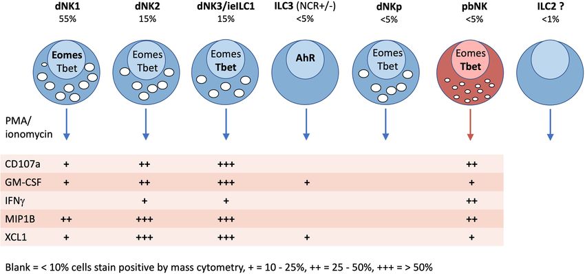
Huhn et al.                                                                                                                                          Uterine NK and ILC Subsets

                                        A

                                        B

                                        C

 FIGURE 1 | Possible roles of uterine NK cells and ILCs in human endometrium and placental development in humans and mice. (A) Important biological processes in
 endometrium during the human menstrual cycle are shown, which may be influenced by local immune cells. Following menstruation, the endometrium undergoes repair
 and proliferation under the influence of oestrogen from the ovary and blood supply is re-established involving both spiral arterioles and new capillaries. Following ovulation,
 progesterone action results in decidualization of the endometrial stroma and a rapid increase in the number of uNK cells, which comprise up to 70% of the leukocytes
 (shown in green) towards the end of the cycle. Dynamics of other ILC populations in human endometrium through the cycle are less well understood. Murine
 endometrium cycles by exposure to oestrogen then progesterone but the estrous cycle only lasts ~5 days and there is no menstruation. (B) The human maternal-fetal
 interface. Spiral arterioles (SpA) branch from the radial arteries and penetrate the decidua to supply the placenta. Right hand panel shows placental villi bathed in maternal
 blood which enters the intervillous space. Placental villi are covered by syncytiotrophoblast, beneath which is the villous cytotrophoblast layer (VT). Anchoring villi attach to
 decidua via columns of extravillous trophoblast (EVT) which differentiates from VT. Some EVT migrates down spiral arteries as endovascular trophoblast (ENDT) while
 other EVT cells migrate into the decidua as far as the myometrium where they transform into trophoblast giant cells (TGC). Interstitial EVT home to maternal spiral arteries
 and participate in their remodelling to provide a low pressure, high capacity blood supply to the placenta. During the first half of pregnancy, when spiral artery remodelling
 occurs, trophoblast invasion in humans is much deeper and more extensive than in mice. EVT invading into the decidua encounter maternal leukocytes (shown in green).
 In humans in the first trimester, these comprise: ~70% NK and ILCs, 20% macrophages and ~10% T-cells, which include CD4 and CD8 cells. Figure (B) adapted from
 Moffett and Colucci (1). (C) Schematic view of pregnant mouse uterus showing multiple implantation sites. Radial arteries branch from the arcuate artery to supply each
 developing fetus. The right hand panel shows a uterine cross section cut as indicated by the dotted line, with the arrangement of the placenta and decidua at gestation
 day (gd) ~12.5. The radial arteries penetrate through the myometrium, traversing a specialized structure that develops around mid-gestation, known as the mesometrial
 lymphoid aggregate of pregnancy (MLAP). The MLAP is not present in humans. Spiral arteries (SpA) branch from the radial artery into the decidua, rich in uterine NK cells
 but then merge at the interface between the placenta and the decidua to form a large blood canal that supplies the labyrinthine layer of the placenta where gaseous
 exchange takes place between maternal and fetal blood. This blood canal is lined with specialized fetal trophoblast cells (shown coloured purple). The boundary between
 the placenta and decidua is delineated by trophoblast giant cells (TGC), which show minimal invasion into the decidua - those few that do invade are largely perivascular.
 From gd12.5, glycogen rich trophoblast cells invade more extensively into the decidua (later than timepoint shown here), but spiral artery remodelling is largely complete
 by gd12.5. Panel C based on data from Adamson et al. (2).

Frontiers in Immunology | www.frontiersin.org                                           2                                              June 2021 | Volume 12 | Article 607669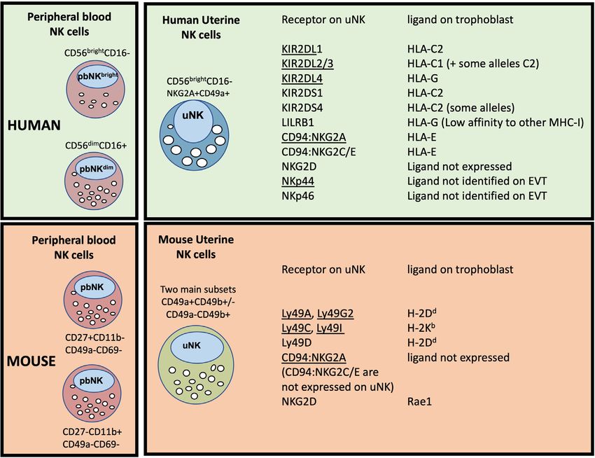
Huhn et al.                                                                                                               Uterine NK and ILC Subsets

and subsequent placental development (3). During the first                     diversity of the uNK niche within ILCs and how these new uNK
trimester of pregnancy, decidual glandular secretions nourish                 subsets may regulate normal placental development. We place
the developing embryo. Extravillous trophoblast cells (EVT),                  recent findings in the wider context of ILC biology and discuss
derived from the blastocyst, invade into the decidua and                      several outstanding questions that should guide future studies.
remodel maternal spiral arteries to become high conductance
vessels. These ensure a sufficient blood supply to the developing
foetus from ~10 weeks gestation until term (4). This arterial                 NK AND THE ILC FAMILY
transformation by trophoblast must be carefully regulated to                  OF LYMPHOCYTES
avoid both extremes of insufficient or excessive invasion.
Reduced invasion alters resource allocation between mother and                NK cells are members of the ILC family which also contains
foetus and is associated with common diseases of pregnancy                    ILC1, ILC2, ILC3 and lymphoid tissue inducer cells (LTi) (18).
including foetal growth restriction (FGR), pre-eclampsia, and                 Lineage studies show that, in both mice and humans, NK cells
miscarriage (5). Excessive invasion by trophoblast can also lead              and ILCs develop through a common lymphoid progenitor
to pregnancy complications, for example when the placenta                     (CLP) but NK and ILCs then diverge (Figure 2). The latter
attaches over caesarean scars or in ectopic pregnancies. Thus,                proceed through a common helper-like ILC precursor (ChILP)
proper development of the maternal-foetal interface is a tightly              dependent on the transcription factor PLZF (20). Subsequent
controlled balancing act. The uterine immune system is involved               development of the ILC1, ILC2 and ILC3 subtypes depends on
in regulating this process.                                                   differential expression of transcription factors T-bet, GATA-3
    Uterine natural killer cells (uNK) are the most abundant                  and RORgt respectively (21). In contrast, NK cell development
maternal immune cell in secretory phase endometrium and, if                   from CLPs proceeds via an NK precursor and depends upon
pregnancy ensues, constitute up to 70% of leukocytes in first                  Eomes in combination with T-bet (20, 22–25). Several precursors
trimester decidua. Numbers then decline substantially towards                 have been identified that can develop into tissue ILC subsets.
term (6). Non-NK ILCs are also present in the uterus, albeit in               Identification of specific intermediates in human and murine
lower numbers, throughout the reproductive cycle. These include               ILC development is still underway and the mechanisms
ILC1s, ILC3s and LTi-like cells, while the status of uILC2s is unclear.       controlling ILC development may differ from the proposed
A unique feature of uILCs is that during pregnancy they can                   scheme in certain tissues (26). Tissue ILCs also exhibit
encounter trophoblast from a genetically different individual- the            considerable plasticity within tissues, so the pathways leading
foetus. Therefore, uILC responses are regulated by interactions with          to NK and other ILCs appear to vary in different organs (19).
maternal stromal cells, leukocytes in the decidua and with ligands            Tissue residency is a hallmark of ILCs, which like NK cells, lack
expressed on EVT. Growing genetic and functional evidence                     antigen-specific receptors generated by somatic recombination.
suggests that interaction of uNK and other uILC subsets with                  Although parabiosis experiments and constitutive cell tagging
EVT regulates the depth of foetal trophoblast invasion (1, 7). In             approaches in mice show that, under physiological conditions,
addition, uILCs likely play important roles in homeostasis and                ILCs and some NK subsets in non-lymphoid organs are largely
tissue remodelling as well as in more canonical immune functions              tissue resident and maintained by self-renewal (27, 28), recent
such as controlling pathogens and malignancies. Indeed, uNK can               experiments suggest that ILC circulation is more extensive than
kill CMV-infected decidual stromal cells and are required for                 previously thought (29). ILC3s can migrate from the intestinal
effective responses to Chlamydia trachomatis (8, 9). Thus, uILCs              mucosa to local lymph nodes and low numbers of ILCs circulate
likely fulfil multiple functions depending on the context and their            in the blood (30). Thus, the extent to which individual ILC and
location (10). Moreover, these are likely to differ between species           NK subsets can exchange with the circulation in different organs
which have developed different mechanisms to sustain pregnancies.             including the uterus is still unclear.
The maternal-foetal interface in human and mouse, and the                         ILCs orchestrate context-specific immune responses and fulfil
potential roles uILCs play are summarised in Figures 1B, C. In                non-immunological roles including tissue remodelling and
both species, foetally-derived trophoblast cells invade the uterine           metabolic homeostasis (21, 31). Whilst the main ILC subtypes
mucosa during development of the placenta, although the extent of             have common features across different organs, they acquire
invasion is much greater in humans. This brings them into direct              distinctive characteristics in response to local cytokines and the
contact with decidual leukocytes, leading to the suggestion that uNK          tissue microenvironment. Growing evidence for plasticity
and uILCs regulate key processes during pregnancy.                            between some ILC subsets is also emerging, emphasising the
    The ability to perform a wide range of effector functions may be          need for detailed phenotypic and functional characterisation
facilitated by heterogeneity within the uILC compartment. The                 within each tissue (23, 32, 33). This variation has hampered
most studied uILC are uNK, originally defined as CD3- CD56+                    efforts to define the identity and function of uterine ILCs.
lymphocytes. There are several recent reviews of uterine ILCs
(11–14). The diversity of uNK was apparent from the first
phenotypic characterisations (15–17) and has become increasingly              UTERINE ILCs
evident as high dimensional single cell technologies have been
applied to the endometrium and decidua. Here, we focus on how                 Although all five main ILC subsets have been identified within
these new approaches have expanded our understanding of the                   the human uterus, the reports are somewhat conflicting and their

Frontiers in Immunology | www.frontiersin.org                             3                                   June 2021 | Volume 12 | Article 607669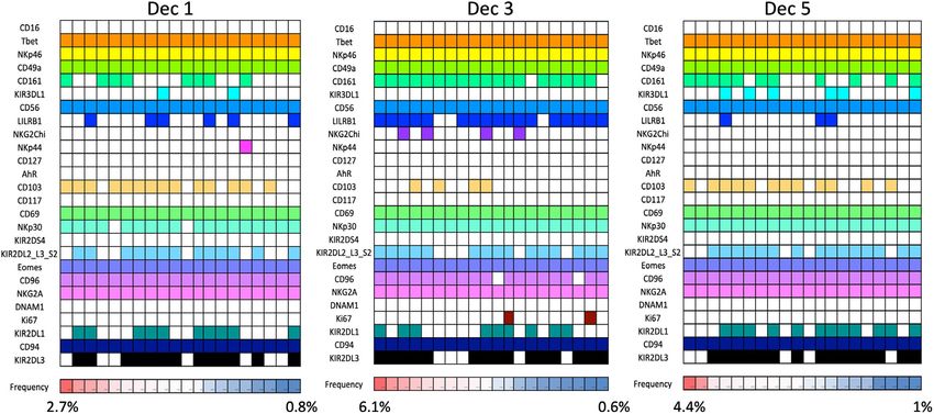
Huhn et al.                                                                                                                                 Uterine NK and ILC Subsets

 FIGURE 2 | Development and functions of NK cells and ILCs Simplified overview of key stages of ILC development. Scheme shown is largely based on murine data.
 Table shows typical cytokines that stimulate the main ILC subsets, together with their principal effector molecules and known immune functions. Phenotypic markers
 commonly used to identify human subsets are shown and assumes prior gating on lin-CD45+ cells. CLP common lymphoid progenitor, CILP, common innate
 lymphoid progenitor; CHILP, common helper ILP; ILCP, innate lymphoid cell precursor; NKP, NK progenitor; LTiP, lymphoid tissue inducer precursor; LTi, lymphoid
 tissue inducer; NFIL3, Nuclear factor, interleukin-3 regulated; ID2, inhibitor of DNA binding 2; GATA3, GATA binding protein 3; PLZF, promyelocytic leukemia zinc
 finger; T-bet, T-box transcription factor 21; Eomes, eomesodermin; AhR, aryl hydrocarbon receptor; ROR, Retinoic acid–related orphan receptor; TSLP, thymic
 stroma lymphopoietin; Areg, amphiregulin; LT, lymphotoxin; IFNg, interferon-gamma; IL, interleukin; NCR, natural cytotoxicity receptor; NK, natural killer. Figure and
 phenotypic markers adapted from Vivier et al. (18) and Guia and Mancinelli (19).

exact phenotypic profiles and functional roles remain undefined                          loss of key markers such as CD56 and NKp44; this is reduced by
(34–37). This is partly because studies of the functional                              use of mechanical dissociation of tissue (35).
properties of uterine ILCs and NK cells were based on
phenotypic markers derived from other tissues. This can be                             Uterine NK Cells
problematic because of the phenotypic variability in different                         NK cells were identified phenotypically as CD3−CD56+ granulated
locations. Enzymatic disaggregation of tissue can also result in                       lymphocytes in blood (38). Large granular lymphocytes had been

Frontiers in Immunology | www.frontiersin.org                                      4                                          June 2021 | Volume 12 | Article 607669
Huhn et al.                                                                                                              Uterine NK and ILC Subsets

described early in the 20th century in the uterine mucosa, but it was       decreased expression of KIR, LILRB1 and 9kDa granulysin.
only with the advent of immunohistochemistry that these were                There are also functional differences compared with their first
shown to belong to the NK lineage (15–17). Studies over decades             trimester counterparts; term dNK degranulate more readily in
have relied on gating on CD3−CD56+ to capture NK cells, but this            response to PMA/ionomycin stimulation or K562 but show
is now known to capture a mixture of both NK and non-NK ILCs in             decreased cytotoxicity to HCMV-infected stromal cells. Thus,
various tissues including the uterus (33, 37, 39, 40).                      uNK are a dynamic population which vary in both function and
    Endometrial NK Cells: NK cells in proliferative phase                   phenotype depending on the stage of the reproductive cycle and
endometrium (eNK) comprise ~20% of total leukocytes. After                  location within the tissue.
ovulation, eNK then proliferate vigorously during the secretory
phase which continues through early pregnancy (17, 41). This is             ILC1s
driven by IL-15 secreted by stromal cells in response to                    ILC1s, originally identified in the tonsils and gut mucosa, are
progesterone (42). eNK are CD56bright, express markers of tissue            Tbet+ cells that produce IFN-g in response to IL-12 stimulation
residency (CD49a, CD69 and CD9), and many canonical NK cell                 (61). Two human ILC1 subsets have been described: “classical”
markers (NKG2A, NKG2D and NKp46) but lack CD16 and                          ILC1s (CD56−CD94−CD127+CD117−NKp44−) and intra-
CD57. The family of killer cell immunoglobulin-like receptors               epithelial ILC1s (ieILC1s) (CD56+ CD103+) (62). The ILC1
(KIR) are also important in regulating NK activation. KIR                   cells identified in the decidua, resemble ieILC1s described in the
expression on eNK differs compared to matched pbNK as well                  gut, which express CD56, perforin and granzymes and are
as to dNK and is stable over multiple menstrual cycles (43, 44).            cytotoxic. ieILC1s express a unique integrin profile including
    Compared with decidual NK cells (dNK), there are very few               CD103 and are found enriched in epithelial regions (62). ILC1s
functional studies using eNK. They show cytotoxicity against                appear phenotypically heterogeneous and even display different
K562 targets and low levels of spontaneous cytokine secretion,              transcription factor requirements for their development in
which is increased upon activation with IL-15 (41, 45, 46). Their           different tissues so do not readily conform to the scheme
rapid increase after ovulation has led to suggestions that eNK              outlined in Figure 2 (63). In secretory phase endometrium no
may be important in implantation. Accurate counting is difficult             ‘classical CD127+ ILC1’ cells were detected (35) but both ILC1
because of their uneven distribution and very rapid changes in              and ieILC1 subsets are reported in decidua (36, 37). Indeed,
number through the cycle, but numerous studies have failed to               NKp44+CD103+ cells were identified as the major source of
establish any consistent alterations in numbers of eNK and                  IFN-g within the decidual Lin−CD56+ compartment (64).
implantation failure or recurrent miscarriage (47). Nor is it               Although CD56− ILC1s, ‘classical ILC1s’, are reported in term
clear that the wide variation in eNK numbers observed                       decidua, comprising ~3% of the Lin−CD127+ compartment,
between these patients is functionally significant since it is also          their existence is controversial (64). The gating strategy used to
seen in women with normal fertility (48).                                   identify ILC1s can capture a mixed population of other
    First Trimester Decidual NK Cells: In comparison to eNK,                leukocytes and hematopoietic stem cells in some tissues (33).
first trimester dNK have been far more extensively studied.                  Current evidence therefore supports the presence of uterine
Phenotypically they share many similarities with eNK                        ieILC1s but the presence of classical ILC1s needs confirming.
including expression of tissue residency markers, lack of CD16
and CD57, and increased proportion of cells that are KIR+ and               ILC2s
NKG2A+ (43, 49–52). Comparison of eNK from the secretory                    Mixed reports also exist for the presence of uterine ILC2s, typically
phase with first trimester dNK, identified over 150 transcripts               identified as Lin−CD127+CRTH2+ cells. A preliminary report
that differed >3 fold, highlighting significant changes that arise           showed ILC2s in small numbers in human non-pregnant
after the onset of pregnancy (53). As well as altered transcript            endometrium and decidua and suggested they can promote foetal
levels, there are changes in RNA splicing, resulting in expression          growth in mice (65). In human term decidua, ILC2s (Lin− CD56−
of inhibitory rather than activating isoforms of individual natural         CD127+ GATA-3+) were the most frequent CD56− CD127+ ILC,
cytotoxicity receptors (NCR). Ligation of NKp30 or NKp44                    with increased ILC2 in decidua basalis of women with preterm
therefore induces inhibitory responses in dNK but activation of             labour (64). Other studies did not detect CRTH2 expression in
pbNK (54). This may contribute to the low cytotoxicity displayed            endometrium or decidua (35, 36). These discrepancies may be
by dNK towards HLA class I null cell lines and to trophoblast               explained by the gate used to identify ILCs. As found for ILC1s in
(55). NK cells can mediate allo-recognition and dNK interact                the uterus, when gating on CD127+ cells, no ILC2s are detected in
with EVT as the latter invade into the decidua. Because some of             early decidua but CD127low/neg are fairly abundant (65). Whether
these receptors differ between mouse and human, the ligand/                 cells corresponding to ILC2s exist in the uterus at all stages of the
receptor interactions between dNK and foetal EVT are distinct in            reproductive cycle is therefore controversial.
the two species, requiring caution in extrapolating results
between the two (Figure 3).                                                 ILC3s
    Term Decidual NK Cells: As gestation proceeds, the                      ILC3s and Lymphoid Tissue-inducer-like cells (LTi-like) are
proportion of dNK decreases and by term lin−CD56+ cells                     defined by their expression of RORgt (66). LTi-like ILCs are
represent ~20% of CD45+ cells in decidua parietalis (60). They              phenotypically similar to LTi cells, which promote lymph node
retain expression of CD9, lack CD16 and CD57, and display                   formation in the developing foetus. ILC3s can be further

Frontiers in Immunology | www.frontiersin.org                           5                                    June 2021 | Volume 12 | Article 607669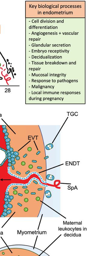
Huhn et al.                                                                                                                                Uterine NK and ILC Subsets

 FIGURE 3 | Major peripheral blood (pbNK) and uterine NK cell (uNK) subsets in human and mouse, with possible trophoblast MHC ligands for selected uNK
 receptors. The two main human pbNK subsets are Lin-CD56bright (~10% of total NK) and CD56dim (~90%); in mice the equivalent NK subsets are lin-CD27+
 CD11b+ and CD27-CD11b+ (56). In the human uterus, uNK cells are defined as lin-CD56+CD49a+ (or CD56+CD9+, since both CD49a and CD9 are markers of
 tissue residency in the uterus). Mouse uterine NK cells are typically gated as lin-NK1.1+NKp46+ and comprise two main subsets, distinguished on the basis of
 CD49a and CD49b expression (CD49b is often referred to as DX5). Recent studies have shown that uNK in both species exhibit further heterogeneity (described in
 more detail later). The right hand panel shows selected receptors expressed on uNK cells and whether the corresponding MHC ligands are expressed on human or
 murine trophoblast cells. Receptors that normally inhibit NK activity are underlined. Note that LILRB1 which is normally considered an inhibitory receptor, has been
 reported to function as an activating receptor in uNK cells by some authors (57, 58); conversely NKp44 may function as an inhibitory receptor in uNK (54), so is
 shown underlined. C2+HLA-C, indicates an HLA-C allele carrying a C2 epitope. Full details and original references describing expression of each ligand on
 trophoblast are available in Gaynor and Colucci (59).

subdivided based on NKp44 expression into NCR+ ILC3s and                               means it is problematic to identify ieILC1s, which in other
NCR− ILC3s; the latter are difficult to distinguish from LTi-like                       tissues are typically gated as CD56+CD94+/−CD103+ (33, 37,
ILCs and have typically been treated as one population. ILC3s are                      39, 40). For the same reason the presence of uterine ILC2s and
described in both human and murine uteri (34, 35, 37). In human                        classical ILC1s is not clearly established.
endometrium, the majority are RORgt+NKp44+ (~3% of
leukocytes) corresponding to NCR+ILC3s (35, 37). Within the                            Origin of Uterine ILCs
decidua, NCR+ILC3s produce IL-22 whilst NCR− ILC3s/LTis                                The dynamic changes in numbers of uNK and other uILCs in the
produce TNF upon activation (36). Both ILC3 and LTi subsets                            endometrium and decidua has led to interest in where they
also induce the upregulation of adhesion molecules ICAM-1 and                          originate. uNK cells have been the focus of most of these studies.
VCAM-1 on decidual stromal cells, suggesting they may be                               Since NK cells originate from bone marrow derived precursors,
involved in recruitment of other leukocytes or can perform LTi-                        work has centred on whether uNK cells derive from circulating
like functions (13, 36). Increased ILC3 proportions have also been                     CD34+ hematopoietic progenitor cells (HPCs) or NK lineage-
detected in decidua parietalis of women with preterm labour (64).                      committed precursors that migrate to the uterine mucosa. Others
    These studies suggest that ILCs are represented across all                         have examined whether mature blood NK cells can acquire uNK
stages of the human reproductive cycle and are dominated by                            characteristics in the uterine microenvironment. Following bone
uNK cells. However, the gating strategies needed to distinguish                        marrow transfer, donor-derived leukocytes, including NK cells,
these subsets from one another in the uterine mucosa are not                           are detected in endometrium and decidua in both humans and
yet clear. For example, new findings that human dNK are                                 mice, suggesting that in vivo these can arise from transferred
CD56+CD94+ but heterogeneous for CD103 expression                                      HPCs (67, 68). Human stage 3 NK-committed precursors can be

Frontiers in Immunology | www.frontiersin.org                                      6                                          June 2021 | Volume 12 | Article 607669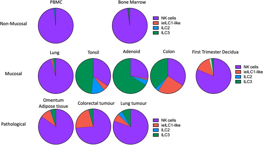
Huhn et al.                                                                                                               Uterine NK and ILC Subsets

detected in blood and can differentiate into more mature NK                    A complementary analysis of lin−CD45+ decidual cells by
cells in the presence of IL-15, which is expressed in the                  CyTOF using a panel of 41 antibodies identified 13 separate
endometrium and decidua (34). However CD34+ progenitors                    clusters of which 11 can be phenotypically assigned to NK or
are also detectable in both human endometrium and decidua and              ILC subsets (Figures 4A, B). This confirms the presence of
can rapidly differentiate into NK cells in vitro (69). This suggests       multiple dNK subsets at the protein level (39). Clusters 10-13
homing of NK precursors derived from bone marrow or                        (c10-13) correspond to dNK1 in the scRNAseq analysis but are
differentiation of stem cells resident in the tissue may both              further separated by differential KIR expression. Based on a similar
contribute to uNK populations. The latter is supported by the              receptor staining profile, including high expression of NKG2A and
finding that CD56+ NK cells develop in human endometrial                    low expression of KIR and LILRB1, c9 corresponds to dNK2, and
tissue xenografted into immunodeficient mice, in response to                c5 and c8, which differ based on NKG2A and NKp44 expression,
steroid hormones (70). Mature human NK cells circulating in                represent dNK3/ieILC1. Two small CD16+ pbNK-like clusters
blood also acquire characteristics of uNK cells, after treatment           (c1-2) are probably maternal blood contaminants as
with cytokines including TGF-b, suggesting that recruitment of             immunohistochemistry has revealed few CD56+CD16+ NK cells
pbNK can contribute to uNK populations (71). The idea that                 within the decidua itself (17). dNK1 are the most abundant dILC
both in situ differentiation from tissue resident precursors and           followed by dNK2 and dNK3/ieILC1, although proportions vary
recruitment from cells circulating in blood contribute to uNK              between donors and can also be affected by cryopreservation
populations is supported by elegant parabiosis studies in mice.            (Figure 4C). These ILC subsets and their phenotypes are
These show both mechanisms contribute to increases in uNK at               summarised in Figure 4D. Stimulation of ILCs by ‘missing self’
different times: tissue resident cells proliferate during early            (K562) or PMA/ionomycin revealed that these dNK subsets
pregnancy in response to decidualization, while a second wave              significantly differ in their responsiveness, with dNK2 and dNK3/
of recruitment of circulating NK cells augments uNK cell                   ieILC1 more responsive than dNK1. These are non-physiological
numbers in the second half of gestation (72, 73). Recent data              stimulations and how each subset responds in more relevant assays
from HLA mismatched uterus transplant recipients in humans                 such as co-cultures with trophoblast needs exploring.
suggests that uNK can indeed be replenished from the circulation               In the light of these new findings a re-evaluation of previous studies
(74). This approach could be applied to establish whether other            using pre-determined gating strategies to identify dNK subsets is
uILC subsets can also originate from the periphery and whether             possible. Three populations were found using NKp44 and CD103:
peripheral or tissue resident contributions vary during                    NKp44+CD103+, NKp44−CD103+ and NKp44−CD103− (37).
pregnancy, as seen in the mouse.                                           Although NKp44+CD103+ are enriched for dNK3/ieILC1, the
                                                                           remaining subsets do not directly correlate with those defined by
                                                                           CyTOF. pbNK cells can display memory-like properties with
HIGH RESOLUTION ANALYSIS OF                                                expansion of specific NK subsets in CMV infection (80).
DECIDUAL NK AND ILC POPULATIONS                                            Expansions of uNK subsets have been described in both mice (81)
                                                                           and humans (44, 57, 82). A population of NKG2Chigh LILRB1+,
It is now apparent that gating strategies used to define specific ILCs       termed pregnancy-trained decidual NK (PTdNK) is increased in
in one tissue may not readily translate to another because key             secondary and subsequent pregnancies. Phenotypically, these most
markers can differ. Recently single cell RNA sequencing                    closely resemble dNK1 and probably represent an expanded dNK1
(scRNAseq) and high dimensional mass cytometry (CyTOF) have                subpopulation. PTdNK have epigenetic and transcriptomic profiles
allowed ILCs and NK subsets to be reliably identified in different          that favour IFN-g and VEGF-A production although the functional
tissues despite this variation in phenotype and functional responses       studies were performed with dNK activated after IL-15 priming (57).
(33, 56). These techniques, as well as high parameter flow                  IFN-g and VEGF-A are not normally produced by freshly isolated
cytometry, have recently been applied to first trimester decidua to         dNK1 unless they are strongly activated after isolation, so their role in
provide a more accurate characterization of the phenotype and              vivo is unclear (83, 84). First pregnancies show a higher risk of low
functions of ILC subsets in the uterus (39, 40, 75–77).                    birth weight and pre-eclampsia, which may be due to better extra-
     Analysis by scRNAseq provided the first description of three           villous trophoblast invasion and arterial transformation in subsequent
distinct decidual NK populations, termed dNK1-3, as well as a              pregnancies (85–87). Further work is required to define the PTdNK
proliferating NK subset (dNKp) and ILC3s (40, 77). dNK1 are                subset more accurately to ascertain if they contribute to this effect.
characterised by expression of KIR, LILRB1, CD39 and increased
granzymes. dNK2 express high levels of ITGB2 and anti-
inflammatory ANXA1. Based on their expression signature,                    How Do Decidual ILC Populations
dNK3 resemble intra-epithelial ILC1s. No clusters corresponding            Compare With Other Tissues?
to ILC2s or classical ILC1s were identified. This analysis also             ILC subset composition varies between tissues and pathological
identified significant heterogeneity within other immune, stromal            states but in first trimester decidua it differs from other normal
and epithelial cells and allowed the first systematic analysis of           mucosal tissues (Figures 4C, 5) (33, 39). Some caution is needed
potential ligand/receptor interactions between uILCs and other             with this comparison as the gating strategies used in the two
decidual and placental cell types (40, 76, 78). scRNAseq analysis of       studies are not directly comparable. The lung contains relatively
luteal phase endometrium also identified several NK subsets                 fewer ieILC1 cells whilst colon, adenoid and tonsil all possess
suggesting that heterogeneity in uNK arises before pregnancy (79).         larger proportions of ILC3s than decidua. Intriguingly, in this

Frontiers in Immunology | www.frontiersin.org                          7                                      June 2021 | Volume 12 | Article 607669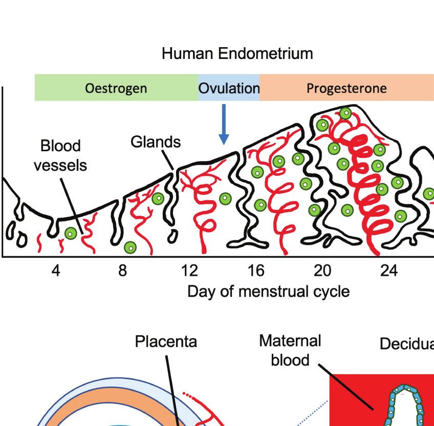
Huhn et al. Uterine NK and ILC Subsets
A B
C D
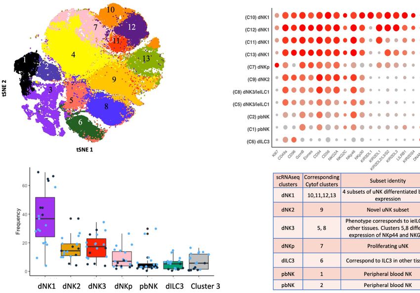
FIGURE 4 | ILC subsets identified by mass cytometry of first trimester decidual cells. (A) tSNE landscape of CD45+ CD3- CD19- CD14- HLA-DR- (Lin-)
cryopreserved decidual cells stained by mass cytometry. tSNE is coloured by clusters identified by DensVM clustering. 11 of these clusters can be assigned to NK or
ILC subsets. Cluster 3 is heterogeneous and cluster 4 appears to contain cells damaged by cryopreservation so these were excluded from subsequent analysis.
(B) Phenotypic characterisation of selected clusters. Size of the circle is representative of the proportion of the cluster positive for that marker. Circles are coloured
by intensity of staining for that marker. Intensities have been scaled by marker (ie within each column). Red corresponds to higher expression and grey to lower
expression. Percentage of cells in the cluster staining for each marker is indicated by size of the circles, scaled as shown on the right of the figure. (C) Box plots
show the proportions of designated clusters within the total CD45+ Lin- decidual compartment. Blue dots = cryopreserved samples, Black dots = fresh samples.
[Figure 4A adapted from Huhn et al. (39), where details of the antibody panel are described]. (D) Comparison of decidual NK and ILC clusters identified by single
cell RNA sequencing (scRNAseq) (40) and by mass cytometry (Cytof) (39). Identity of each cluster is based on their profiles of RNA or protein marker expression
respectively. Cytof clusters 10-13, correspond to scRNAseq cluster dNK1 and are distinguished by their KIR expression. Cytof clusters 5,8 are distinguished by NKp44/
NKG2A expression and correspond to scRNAseq cluster dNK3, representing ieILC. Cytof clusters 3 and 4 identified in panel A appear heterogeneous and don’t match
any scRNAseq clusters; identities are not established. ++ is > 75% positive staining of cells with antibody in Cytof; + is 25% to 75%; − is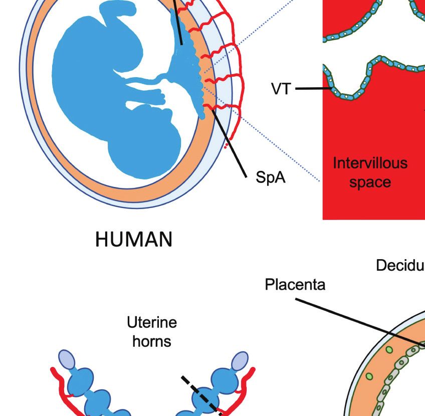
Huhn et al.                                                                                                                           Uterine NK and ILC Subsets

 FIGURE 5 | Frequencies of NK and ILC populations in decidua compared with other human tissues. Pie charts showing the frequencies of NK and ILC
 subsets in decidua (N = 7) compared with normal and pathological human tissues determined by mass cytometry. All charts, with the exception of first
 trimester decidua come from Simoni et al. (n = 4-9 separate donors for each tissue type) (33). For first trimester decidua, Live CD45+CD3-CD19-CD14-
 HLA-DR- cells were gated on as the parent population [data from Huhn et al. (39)]. Specified subsets were then identified on a tSNE landscape based on
 phenotypic profiles and their proportion within this population calculated. Unassigned cells (grey in decidua) could not be confidently labelled as a particular
 subset based on the markers included in the CyTOF panel. For all other tissues, samples were first depleted of T and B cells and then gated on Live
 CD45+ FcϵR1a– CD14– CD19– CD123–CD34-CD5- cells. Gating strategies to identify each subset were then as follows: NK cells (purple) = CD94+/-
 CD127+/-CD56+CD103-; ieILC1-like (red) = CD94+/-CD127+/-CD56+CD103+; ILC2 (blue) = CD94-CD127+CRTH2+; ILC3 =CD94-CD127+CRTH2-.
 [Figure adapted from Simoni et al. and Huhn et al. (33, 39)].

 FIGURE 6 | dNK receptor repertoire diversity Frequencies of the most common NK cell phenotypes were determined by mass spectrometry in freshly isolated
 CD45+Lin−CD56+ decidual NK cells for three selected individuals based on combinations of 26 individual phenotypic markers expressed on each cell. The twenty
 most frequent phenotypes detected are shown for each donor Dec1,3,5. Each column represents a phenotype shared by a number of cells; boxes are coloured if
 the marker is expressed in that phenotype. Frequencies of each phenotype as a percentage of total CD45+Lin−CD56+ are displayed as a heat map at the bottom of
 the figure. Based on analysis of data from Huhn et al. (39).

Frontiers in Immunology | www.frontiersin.org                                  9                                         June 2021 | Volume 12 | Article 607669
Huhn et al.                                                                                                                                    Uterine NK and ILC Subsets

TABLE 1 | Phenotypic diversity of CD45+Lin−CD56+ cells in human blood and decidua based on receptor profiles of individual cells analysed by mass cytometry.

Donor                  Tissue                  Input number                      Number of                   Frequency of                            Total frequency
                                                  of cells                       phenotypes                 most frequent                          captured by 20 most
                                                                                                           NK phenotype1(%)                     frequent phenotypes1(%)

Pb1                    Blood                       25683                            6173                            1.9                                     18.6
Pb2                    Blood                       9736                             4509                            1.9                                      13
Pb3                    Blood                       7892                             3563                            2.2                                     14.5
Pb4                    Blood                       10787                            3336                            4.6                                     25.9
Pb5                    Blood                       4164                             2882                            0.6                                     6.1
Pb6                    Blood                       14483                            4673                            2.2                                     19.6
Dec 1                  Decidua                     24031                            3792                            2.7                                     25.2
Dec 2                  Decidua                     24740                            4428                            3.8                                     32.5
Dec 3                  Decidua                     32197                            4745                            6.1                                     36.8
Dec 4                  Decidua                     24670                            3206                            5.6                                     36.2
Dec 5                  Decidua                     32507                            3658                            16                                      44.3
Dec 6                  Decidua                     32935                            3261                            4.4                                     31.4
1
Frequencies expressed as % of total CD45+Lin−CD56+ cells in blood or decidua. Blood and decidual samples not from matched donors. Data derived from Huhn et al. (39).

pbNK and dNK, the decidual NK cell repertoire is dominated by a                            ongoing uncertainty about which ILCs are present at different
more limited number of highly represented phenotypes, possibly                             stages of pregnancy. Implantation and early placental
selectively favoured during NK proliferation and maturation.                               development in the mouse do share some features with humans,
Some may represent developmental intermediates while others                                including decidualisation of stromal cells of the uterine mucosa,
include effectors that drive important physiological processes                             and invasion of foetal trophoblast cells into the decidua (Figure 1
during pregnancy. The immediate challenge is to determine how                              and Table 2). Although trophoblast invasion and spiral artery
the phenotypic diversity of uNK and ILC populations within and                             remodelling is necessary in both species to increase the blood
between women influences pregnancy outcomes.                                                supply to the developing placenta, this invasion is far more
                                                                                           extensive in humans. Human trophoblast invades both down
                                                                                           the blood vessels (endovascular) and through the decidua
                                                                                           (interstitial) extending as far as the inner myometrium
WHAT CAN WE LEARN ABOUT THE                                                                (Figure 1B). Trophoblast cells mediate the classical fibrinoid
FUNCTION OF UTERINE ILCs FROM                                                              necrosis of the arterial media that transforms these vessels (95,
MOUSE MODELS?                                                                              96). In mice, trophoblast invasion is minimal and limited to the
                                                                                           junctional zone of the placenta during the first 12 days of
Establishing the function of human NK cells and ILCs is                                    gestation. Instead, spiral artery modification is associated with
challenging because of the rapid changes in cell types and                                 NK cells which move into the arterial wall (59, 97, 98).

TABLE 2 | Key Features of human and murine pregnancy models.

Description                                                         Human                                                             Mouse

Duration                                 40 weeks                                                        ~19 days
Implantation sites                       Typically one                                                   Multiple implantation sites in each uterine horn
Decidualisation of endometrial stromal   Begins in non-pregnant endometrium. Progesterone                Triggered by embryo at site of attachment. Also progesterone
cells                                    dependent                                                       dependent
Uterine NK cell numbers                  Proliferate in endometrium before pregnancy Increase            Few in endometrium
                                         rapidly in first trimester then decline                          Rapid increase in trNK after implantation starting ~ gestation day
                                                                                                         (gd) 4.5
                                                                                                         Peak at mid-pregnancy ~gd 10 then decline
Trophoblast invasion                     Extensive: through decidua and even into myometrium by:         Limited: to junctional zone until gd12.
                                         Interstitial trophoblast, Endovascular trophoblast, Placental   Limited invasion of decidua thereafter by:
                                         bed giant cells                                                 Trophoblast giant cells and glycogen cells
Vascular remodelling                     Associated with endovascular trophoblast in lumen.              Associated with NK cell intravasation into smooth muscle of
                                                                                                         vessel
                                         Interstitial trophoblast in muscle wall.                        Mediated in part by IFNg from NK cells
                                         Some evidence for NK involvement before trophoblast             Endovascular Trophoblast limited to central canal in placenta
                                         invasion and away from placental bed
Localisation of NK cells                 Isolated cells and in aggregates In pregnancy, is limited to    NK cells in decidua and in the MLAp, embedded in the
                                         decidua, Not in myometrium                                      myometrium

(MLAp is the mesometrial lymphoid aggregate of pregnancy, not seen in humans).

Frontiers in Immunology | www.frontiersin.org                                         10                                         June 2021 | Volume 12 | Article 607669
Huhn et al. Uterine NK and ILC Subsets
Nonetheless, invading trophoblast cells do encounter maternal NK (Figure 7B). Prior to puberty, mouse endometrium contains
and ILCs in the murine decidua later in pregnancy after spiral predominantly ILC1s. These decline after puberty while cNK
artery remodelling is completed. and trNK increase (81). ILC2s and ILC3s are also present in low
There are also important differences in timing as well as numbers prior to pregnancy, the latter comprising both LTi-like
localisation of ILC populations. Decidualization is always and NCR+ ILC3s (35, 100). ILCs are distributed throughout the
characterized by extensive accumulation of NK cells in both tissue in a subset-specific manner. At gestational day 11.5 of
species, but in humans this process begins in the endometrium, pregnancy (gd 11.5), the decidua basalis and MLAp are
so large numbers of proliferating NK cells are present even before composed of mainly cNK or trNK respectively. ILC2 and ILC3s
pregnancy begins. In the mouse, NK numbers do not increase are absent from the decidua (35, 36). As outlined when discussing
until decidualization is triggered by implantation of the embryo. the origins of uNK and ILCs, different rates of subset proliferation,
In both species NK cells increase rapidly during the first influx from blood or conversion between ILC subsets within
trimester but decline by term (6, 11). In mice, at mid-gestation, the uterus may all contribute to these rapid changes in
ILCs are located in the mesometrial lymphoid aggregate of cellular composition.
pregnancy (MLAp) which develops in the myometrium (11). Relating human decidual ILC subsets to their murine
No such structure exists in humans and the function of the counterparts is difficult. dNK1 may be analogous to trNK that
MLAp is unknown. It may participate in the regulation of blood express high levels of Ly49, Eomes and Ki-67 (28, 81).
flow since the uterine artery that supplies each implantation site Murine cNK resemble circulating NK in humans but cNK are
coils through it (99). In addition, NK-derived IFNg is a key present in large numbers in murine decidua whilst CD16+ pbNK
cytokine in murine pregnancies but it is not a major cytokine are rarely seen in human decidual tissue (17, 35, 73). Murine
produced by the main dNK1 subset in humans. Despite these uILC1 expand in secondary pregnancies and express CXCR6, a
apparent differences in anatomy and cellular dynamics, human hallmark of antigen-specific NK memory cells (101). Thus, they
and murine pregnancy share key features. These include may be the functional counterparts to human PTdNK ‘memory’
trophoblast invasion into the decidua and the involvement of cells but differ in their low Ly49 expression (57, 81). How human
both foetal trophoblast and maternal uNK cells in spiral artery dNK2 and dNK3/ieILC1 relate to murine decidual ILCs is also
remodelling to ensure sufficient blood reaches the haemochorial unclear. Murine ieILC1 in the gut are Nfil3-dependent NKp46+
placenta. This suggests mice can provide a useful model to study NK1.1+ CD160+ (62). However, Nfil3-/- mice contain both
the role of NK cells and ILCs in placental development. uterine trNK and ILC1 but lack cNK cells (100). The
NK cells and ILC1s are the most abundant leukocytes in the difficulties in directly correlating murine and human dILC
pregnant mouse uterus (~30% of total leukocytes at mid- subsets may reflect the anatomical differences between human
gestation), significantly lower than in human first trimester and mouse placentation as well as the menstrual versus oestrus
decidua (35). There are three subsets: tissue resident NK (trNK), cycles in which ILCs and uNK develop preceding pregnancy.
CD49a+Eomes+, conventional NK (cNK), CD49a−Eomes+, and Nevertheless, further characterisation may reveal functional
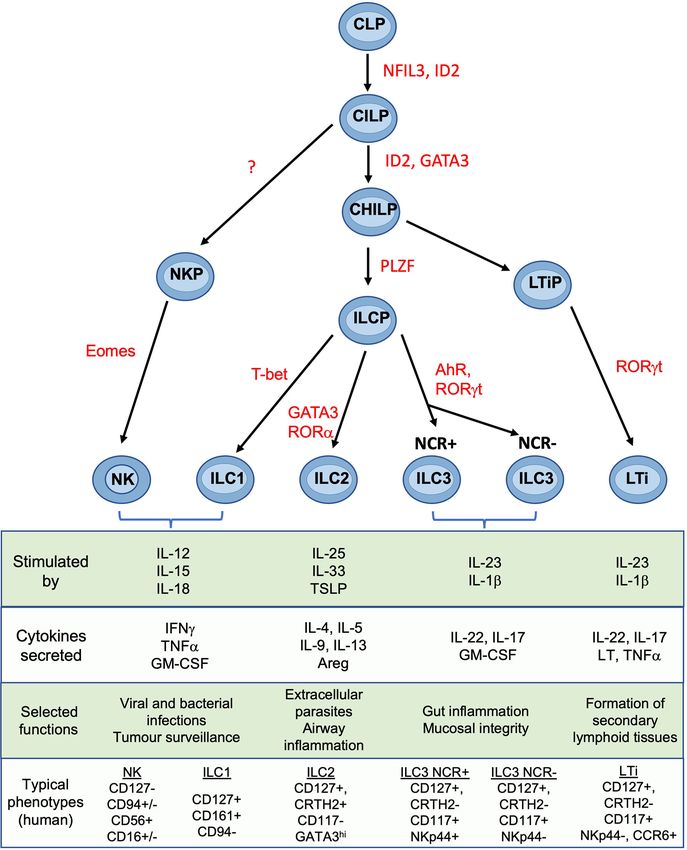
ILC1s, CD49a+ Eomes− (Figure 7A). These all change rapidly in homologies between phenotypically distinct uterine ILC
number and location throughout the reproductive cycle subsets between the species.
A B
FIGURE 7 | Dynamic changes of NK and ILC1s in the mouse uterus before and after pregnancy (A) Gating strategy to identify uterine NK and ILC1 subsets. Cells
isolated from whole uterus were analysed together. Representative 2D FACS plot is from gestational day 9.5 (gd 9.5) and gated on Live CD45+CD3−CD19−
CD11blow/- NK1.1+NKp46+ cells. ILC1s (red line) are CD49a+ Eomes−, tissue resident NK (trNK, blue) are CD49a+Eomes+, conventional NK (cNK, gold) are
CD49a− Eomes+. (B) Proportions of indicated uterine subsets at different stages of the reproductive cycle as a percentage of the total Live CD45+CD3−CD19−
CD11blow/- NK1.1+NKp46+ cells. w = weeks of age, gd = gestational day [Figure adapted from Filipovic et al. (81)].
Frontiers in Immunology | www.frontiersin.org 11 June 2021 | Volume 12 | Article 607669Huhn et al. Uterine NK and ILC Subsets
FUNCTIONS OF UTERINE ILC because both KIR and HLA-C genes are highly polymorphic so
POPULATIONS DURING PREGNANCY each pregnancy is characterized by different combinations of these
genes, resulting in variable activation or inhibition of dNK.
Evidence highlighting the importance of uNK cells in supporting Genetic studies of large pregnancy cohorts suggest that women
successful pregnancies has come from both humans and mice. with a KIR AA genotype are at increased risk of pregnancy
Loss of functional NK cells and/or ILCs in a variety of mouse disorders when the foetus has an HLA-C allele carrying a C2
models is associated with decidual abnormalities and reduced epitope (C2+HLA-C). This combination results in strong
vascular remodelling (102) and foetal growth restriction (98, 100, inhibitory signals to uNK via maternal inhibitory KIR2DL1
103, 104). For example, pregnancies from an NK- and T cell binding to foetal C2+HLA-C on EVT and is associated with
deficient mouse model, tgϵ26, exhibit smaller litters and lower increased risk of pre-eclampsia, foetal growth restriction and
birth weights. Normal birth phenotypes could be restored by miscarriage, although not all studies concur (110–113). In
bone marrow grafts from SCID mice that lack B and T cells contrast, combinations of maternal KIR and foetal HLA-C
but possess NK cells (105). These changes are in part due to which favour activation through KIR2DS1 or KIR2DS5 are
loss of IFNg secretion by NK cells, since they are rescued by associated with protection from pre-eclampsia and increased
systemic IFNg administration and similar effects are seen in mice birth weight (110, 112, 114–116).
lacking IFNg or its receptor (102, 103). Even deletion of single These are all disorders of pregnancy in which EVT migration
receptors on maternal NK cells can result in abnormal foetal and vascular remodelling is reduced (5). More recently the
development. Mice lacking Nkg2a, Ncr1 or AhR show defective presence of the strongly inhibitory allele KIR2DL1*003 in
NK cell maturation, abnormal spiral artery remodelling and mothers, combined with foetal C2+HLA-C ligand has been
lower foetal weights (106, 107). Depletion of NK cells in shown to further increase risk in a dose dependent manner
pregnant rats also causes abnormal arterial remodelling and (117). Thus, binding of a specific inhibitory receptor expressed
hypoxia, accompanied by differentiation of more invasive on uNK with the corresponding ligand on EVT can affect
trophoblast resulting in abnormal placental development (108). pregnancy. These effects are likely to be mediated by dNK1
The roles of other ILCs in pregnancy are much less well since they are the main KIR-expressing cells (Figure 4B). This
understood. Mice lacking the transcription factor Nfil3/E4bp4 idea is supported in a mouse model in which the addition of a
show greatly reduced numbers of cNK and ILC2s in the uterus single additional MHC molecule (H-2Dd) to the foetal genome
but retain trNK, ILC1 and ILC3s. This is associated with results in abnormal uterine artery remodelling and reduced foetal
placental abnormalities and reduced foetal growth indicating growth. This suggests that maternal recognition of a single
that the altered ILC repertoire affects the outcome of pregnancy paternally-derived H-2D d molecule influences pregnancy
(100). The foetal growth restriction in Nfil3-/- mice is reversed by outcome (98). Whether the mechanism involves direct
infusion of pleiotrophin, supporting the idea that NK or ILC recognition of paternal H2-Dd expressed on trophoblast by
subsets produce factors in addition to IFNg that promote inhibitory Ly49 receptors on maternal dNK cells, as seems to
placental development, but the detailed mechanisms occur for human dNK1 and C2+HLA-C on EVT, is not yet
responsible are unclear (104). certain. In humans, the role of other dILC subsets is less clear.
dNK2 and dNK3/ieILC1 express receptors for HLA-E and HLA-G
Regulation of Trophoblast Migration and other ligands on trophoblast, but these are not significantly
A unique feature of pregnancy is that dILCs interact with EVT polymorphic, so will not vary between pregnancies (39, 40). dNK2
from the foetus as well as with maternal cells in the decidua. and dNK3 respond vigorously to activation, but how they respond
Following implantation, in both humans and mice, foetally- to EVT in vivo and how the resulting cytokine responses
derived trophoblast migrate into the decidua where they contribute to pregnancy success, is not known. Patients with
encounter maternal immune cells. These foetal cells express an SCID due to mutations in IL2RG or JAK3, have very low levels
unusual repertoire of MHC class I antigens: human EVT is of ILC and NK cells in blood, gut and skin after hematopoietic
negative for HLA-A and -B but expresses polymorphic HLA-C stem cell transplantation (HSCT). Despite this, two women had
and oligomorphic HLA-E and HLA-G molecules (109). clinically normal pregnancies after HSCT and healthy babies of
Trophoblast are always MHC class II negative. Murine average weight (30). While this study did not investigate uterine
trophoblast from C57BL/6 mice express the MHC class I ILCs in these women, the results suggest NK cells and other ILCs
antigen H-2Kb but very low levels of H-2Db and Qa-1b. Many are not essential for reproduction under normal conditions, but
of the receptors on uNK that recognise these polymorphic MHC can influence pregnancy outcome by fine-tuning placental
molecules are also highly variable and include members of the development and hence foetal growth. This is likely achieved by
killer cell immunoglobulin-like receptors (KIRs) in humans or the fulfilling multiple functions including regulating trophoblast
Ly49 family in mice (Figure 3). The dNK1 subset in particular invasion, and remodelling of the vasculature and decidual tissue.
expresses relevant receptors for trophoblast HLA molecules, What are the critical responses triggered by KIR in dNK that
including NKG2A, LILRB1 and members of the KIR family might affect placental development and pregnancy outcome?
(Figures 3, 4B). Their activation status can therefore be dNK produce a wide array of chemokines and cytokines upon
influenced by both foetal and maternal ligands. Maternal KIR stimulation with PMA plus ionomycin, or by ligation of
recognition of HLA-C molecules on EVT is of particular interest activating receptors (37, 52, 118, 119). Ligation of activating
Frontiers in Immunology | www.frontiersin.org 12 June 2021 | Volume 12 | Article 607669Huhn et al. Uterine NK and ILC Subsets
A B
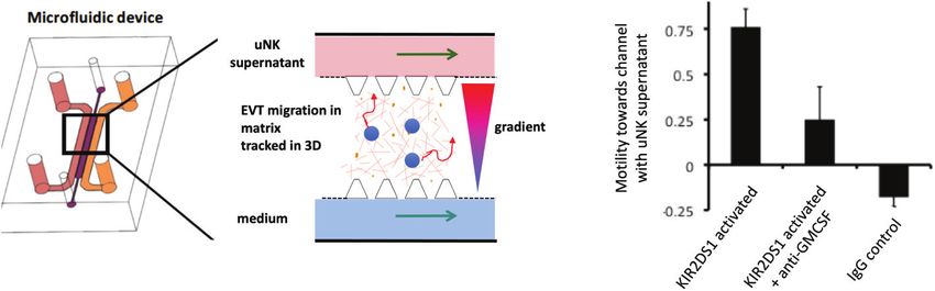
FIGURE 8 | Ligation of activating KIR on uNK affects human trophoblast migration in vitro. (A) Cartoon showing a cross section of a microfluidics device to study
effects of uNK supernatants on migration of extravillous trophoblast (EVT) . Primary human EVT isolated from first trimester placentas are stained with tracker dye
(blue) and embedded in Matrigel in the central channel. Cells are tracked in real time as they move between two side channels. These side channels have medium
constantly flowing through them and can be supplemented with other factors or supernatants conditioned by uNK cells to create a gradient. (B) Plot showing motility
of EVT towards or away from a microchannel containing uNK-conditioned medium. Freshly isolated uNK were purified from decidua using microbeads and KIR2DS1
was activated by EB6 antibody cross-linking for 12 hours. Stimulation conditions are shown on x-axis. On average, EVT display increased motility towards a channel
containing supernatant from uNK stimulated by KIR2DS1 cross-linking. This is reduced by the addition of neutralising anti-GM-CSF in the supernatant. Control is
ligation with isotype matched irrelevant IgG. Motility is calculated by subtracting the number of cells migrating upstream towards the uNK supernatant channel by the
number of cells migrating away and dividing this by the total number of cells, [Figure adapted from Abbas et al. (120)].
KIR to simulate dNK/EVT interactions in the decidua stimulates as their effect is partially abrogated by Ang-2 inhibition using in
secretion of GM-CSF and CXCL10, which have been shown to vitro models, little is known about the mechanisms by which
increase EVT migration (52, 115, 119). In a 3D microfluidics human uNK or EVT actually influence vascular remodelling in
model, primary trophoblast migrate towards a microchannel vivo (121, 128, 129). Evidence that NK cells can play a role in
containing uNK-conditioned medium following activation of regulating the vascular remodelling of the human endometrium
KIR2DS1 (Figure 8) (120). uNK have also been shown to prior to pregnancy is more compelling. Clusters of eNK around
secrete TGF-b which reduces EVT migration and this effect spiral arteries express angiogenic factors including VEGF-C and
changes with gestational age, so uNK responses can potentially PLGF which can regulate endothelial cell function (125).
enhance or decrease EVT migration (121). Whilst no migration Administration of a progesterone modulator (Asoprisnil) to
model can replicate the complex tissue environment of the women results in absence of eNK, altered arterial morphology
decidua, these genetic and functional studies suggest ligation of and no menstrual bleeding (42). Asoprisnil blocks IL-15 secretion
activating KIR on dNK can modulate EVT behaviour and thus by stromal cells in response to progesterone. Whether the
affect blood flow to the placenta. outcomes are due to the reduced uNK or other uILCs or a
consequence of blocking other actions of progesterone is
Vascular Remodelling in Decidua unclear. The role of uNK in modifying the arterial media needs
and Non-Pregnant Endometrium exploring further as the signals triggering menstruation and
The role of uNK-derived IFNg in vascular remodelling in murine decidual breakdown in miscarriage are still essentially unknown
decidua is well established (102). Impaired vascular remodelling (129). One suggestion is that eNK use the activating receptor
in the decidua of mice lacking NK cells or specific NK receptors, NKG2D to kill senescent stromal cells emerging during the
early in gestation shows that uNK cells may directly participate secretory phase (130). NK cells kill senescent cells in tumours
in this process prior to the arrival of trophoblast (122). This may and in mouse models of liver fibrosis, but whether this is
be a direct effect on the arteries as they infiltrate the media, unlike important for homeostasis in cycling human endometrium in
in humans where uNK are seen around the arteries but not in vivo needs verification (131, 132).
their wall. Murine uNK also secrete VEGF-C and mice lacking
the corresponding receptor VEGFR3 on endothelial cells show Regulation of Local Immune Responses
reduced vessel remodelling and foetal growth restriction. This in the Decidua
shows soluble factors secreted by uNK in mice can target It is clear that dNK can recognise both maternal and paternal
endothelium lining the arteries, as well as the surrounding HLA-C expressed by invading EVT, but allogeneic trophoblast
smooth muscle (123). There is indirect evidence that uNK may are also potential targets for maternal T cells. dNKs have been
also influence spiral arteries around which they are seen to shown to secrete cytokines and chemokines which have
cluster in human endometrium and decidua (95, 124). dNK immunoregulatory functions, leading to suggestions they
produce MMP-9 which breaks down extracellular matrix (ECM) contribute to foetal-specific T cell tolerance in the decidua via
of the vascular smooth muscle wall and angiogenic factors such a plethora of mechanisms (133, 134). For example, dNK
as VEGF-C and angiopoietins-1 and -2 (119, 125–127). activated in vitro to secrete IFNg, drives IDO production by
Although these could disrupt the vascular smooth muscle wall, decidual CD14+ cells and induces expansion of regulatory T cells
Frontiers in Immunology | www.frontiersin.org 13 June 2021 | Volume 12 | Article 607669You can also read