Burst mitofusin activation reverses neuromuscular dysfunction in murine - CMT2A - eLife
←
→
Page content transcription
If your browser does not render page correctly, please read the page content below
RESEARCH ARTICLE

                                     Burst mitofusin activation reverses
                                     neuromuscular dysfunction in murine
                                     CMT2A
                                     Antonietta Franco1†, Xiawei Dang1,2†, Emily K Walton1, Joshua N Ho3,4,
                                     Barbara Zablocka5, Cindy Ly6, Timothy M Miller6, Robert H Baloh7,
                                     Michael E Shy8, Andrew S Yoo3,4, Gerald W Dorn II1*
                                     1
                                      Department of Internal Medicine, Pharmacogenomics, Washington University
                                     School of Medicine, St Louis, United States; 2Department of Cardiology, The First
                                     Affiliated Hospital of Xi’an Jiao Tong University, Xi’an, China; 3Department of
                                     Developmental Biology, Washington University School of Medicine, St Louis, United
                                     States; 4Center for Regenerative Medicine, Washington University School of
                                     Medicine, St. Louis, United States; 5Mossakowski Medical Research Centre, Polish
                                     Academy of Sciences, Warsaw, Poland; 6Department of Neurology, Washington
                                     University School of Medicine, St Louis, United States; 7Department of Neurology,
                                     Cedars-Sinai Medical Center, Los Angeles, United States; 8Department of
                                     Neurology, Carver College of Medicine, University of Iowa, Iowa City, United States

                                     Abstract Charcot–Marie-Tooth disease type 2A (CMT2A) is an untreatable childhood peripheral
                                     neuropathy caused by mutations of the mitochondrial fusion protein, mitofusin (MFN) 2. Here,
                                     pharmacological activation of endogenous normal mitofusins overcame dominant inhibitory effects
                                     of CMT2A mutants in reprogrammed human patient motor neurons, reversing hallmark
                                     mitochondrial stasis and fragmentation independent of causal MFN2 mutation. In mice expressing
*For correspondence:
                                     human MFN2 T105M, intermittent mitofusin activation with a small molecule, MiM111, normalized
gdorn@wustl.edu
                                     CMT2A neuromuscular dysfunction, reversed pre-treatment axon and skeletal myocyte atrophy,
†
 These authors contributed           and enhanced axon regrowth by increasing mitochondrial transport within peripheral axons and
equally to this work                 promoting in vivo mitochondrial localization to neuromuscular junctional synapses. MiM111-treated
Competing interest: See              MFN2 T105M mouse neurons exhibited accelerated primary outgrowth and greater post-axotomy
page 22                              regrowth, linked to enhanced mitochondrial motility. MiM111 is the first pre-clinical candidate for
                                     CMT2A.
Funding: See page 22

Received: 15 July 2020
Accepted: 18 October 2020
Published: 19 October 2020
                                     Introduction
Reviewing editor: Joseph G
                                     Charcot–Marie-Tooth disease (CMT) describes a family of genetically diverse and clinically heteroge-
Gleeson, Howard Hughes
Medical Institute, The Rockefeller
                                     neous peripheral neuropathies (Fridman et al., 2015). Type 2A CMT (CMT2A) is caused by muta-
University, United States            tions of the mitochondrial fusion protein, mitofusin 2 (MFN2) (Züchner et al., 2004), and is
                                     distinguished from other CMT subtypes by onset of neuromuscular signs in early childhood and pro-
   Copyright Franco et al. This
                                     gressive loss of neuromuscular coordination and strength in arms throughout the first two decades
article is distributed under the
                                     of life, thought to be the consequence of dying-back of long peripheral nerves (Fridman et al.,
terms of the Creative Commons
Attribution License, which           2015; Feely et al., 2011; Bombelli et al., 2014; Yaron and Schuldiner, 2016; Berciano et al.,
permits unrestricted use and         2017). Because there are currently no disease-modifying treatments, CMT2A is managed with
redistribution provided that the     braces, wheelchairs, and social support.
original author and source are          Over 100 different dominant missense MFN2 mutations are implicated in CMT2A
credited.                            (Bere˛sewicz et al., 2018). MFN2 and related MFN1 are nuclear-encoded dynamin-family GTPases

Franco, Dang, et al. eLife 2020;9:e61119. DOI: https://doi.org/10.7554/eLife.61119                                                1 of 26
Research article                                                                                     Cell Biology Neuroscience

                                   eLife digest Charcot-Marie-Tooth disease type 2A is a rare genetic childhood disease where
                                   dying back of nerve cells leads to muscle loss in the arms and legs, causing permanent disability.
                                   There is no known treatment.
                                       In this form of CMT, mutations in a protein called mitofusin 2 damage structures inside cells
                                   known as mitochondria. Mitochondria generate most of the chemical energy to power a cell, but
                                   when mitofusin 2 is mutated, the mitochondria are less healthy and are unable to move within the
                                   cell, depriving the cells of energy. This particularly causes problems in the long nerve cells that
                                   stretch from the spinal cord to the arm and leg muscles.
                                       Now, Franco, Dang et al. wanted to see whether re-activating mitofusin 2 could correct the
                                   damage to the mitochondria and restore the nerve connections to the muscles. The researchers
                                   tested a new class of drug called a mitofusin activator on nerve cells grown in the laboratory after
                                   being taken from people suffering from CMT2A, and also from a mouse model of the disease.
                                   Mitofusin activators improved the structure, fitness and movement of mitochondria in both human
                                   and mice nerve cells. Franco, Dang et al. then tested the drug in the mice with a CMT2A mutation
                                   and found that it could also stimulate nerves to regrow and so reverse muscle loss and weakness.
                                       This is the first time scientists have succeeded to reverse the effects of CMT2A in nerve cells of
                                   mice and humans. However, these drugs will still need to go through extensive testing in clinical
                                   trials before being made widely available to patients. If approved, mitofusin activators may also be
                                   beneficial for patients suffering from other genetic conditions that damage mitochondria.

                                   located at the mitochondrial outer membrane-cytosol interface where they promote mitochondrial
                                   fusion essential to mitochondrial respiratory function and repair (Chan, 2012). Dominant inhibition
                                   by MFN2 mutants of mitochondrial fusion (Chen and Chan, 2006; Pareyson et al., 2015), mitoph-
                                   agy (Rizzo et al., 2016; Filadi et al., 2018), and/or neuronal mitochondrial transport
                                   (Pareyson et al., 2015; Baloh et al., 2007; Crunkhorn, 2018) are proposed to evoke neuronal
                                   degeneration in CMT2A.
                                      Because CMT2A is an autosomal dominant genetic condition, gene editing could potentially cor-
                                   rect causal MFN2 mutant alleles, but the large number of different causal CMT2A MFN2 mutations
                                   complicates an editing approach. Alternately, forced expression of normal mitofusins could oppose
                                   mutant MFN2 dysfunction, as demonstrated in a recent study in transgenic mice (Zhou et al.,
                                   2019). However, MFN gene therapy would be difficult to discontinue or reverse if postulated
                                   adverse effects of MFN overactivity are encountered (El Fissi et al., 2018). Here, we describe a ther-
                                   apeutic approach to CMT2A that is agnostic to MFN2 genotype and does not require genetic
                                   manipulation: intermittent or ‘burst’ activation of endogenous normal mitofusins. Pharmacological
                                   mitofusin activators improved mitochondrial morphology, fitness, and motility in human and mouse
                                   CMT2A neurons in vitro. Daily administration of a short acting mitofusin activator to mice with late
                                   stage CMT2A reversed neuromuscular dysfunction. Mechanistically, neuronal repair and regenera-
                                   tion were linked to enhanced mitochondrial transport to, and mitochondrial occupation within, axo-
                                   nal termini. Reversal of pre-existing CMT2A neuromuscular degeneration in vivo has not previously
                                   been achieved by any means, and provides a powerful rationale for advancing mitofusin activators
                                   to first in human trials.

                                   Results
                                   Genetically diverse CMT2A patient neurons exhibit similar
                                   mitochondrial phenotypes
                                   One of the central features of CMT2A is the large number of different MFN2 mutations that provoke
                                   the syndrome. Common MFN2 GTPase and coiled-coiled domain mutations induce more severe and
                                   earlier onset disease, whereas rare carboxy terminal domain mutations confer later onset and milder
                                   disease (Feely et al., 2011; Stuppia et al., 2015). We compared mitochondrial phenotypes in cells
                                   from four CMT2A patients, two having MFN2 mutations within the canonical dynamin/Fzo-like
                                   GTPase domain (MFN2 T105M in the G1 motif and MFN2 R274W between the G4 and G5 motifs),

Franco, Dang, et al. eLife 2020;9:e61119. DOI: https://doi.org/10.7554/eLife.61119                                                2 of 26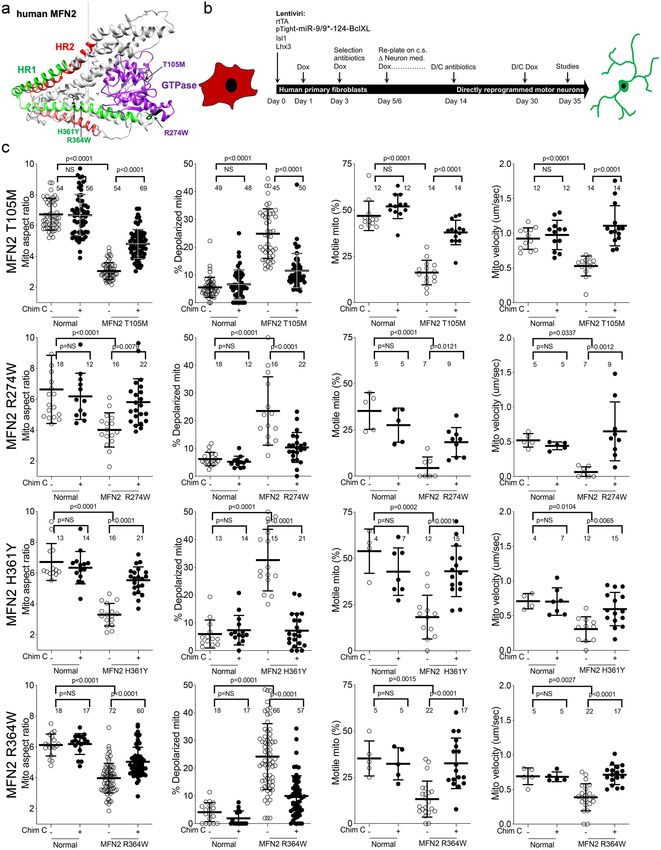
Research article                                                                                         Cell Biology Neuroscience

                                   and two with mutations in the MFN2 coiled-coiled helix bundle core (MFN2 H361Y and R364W).
                                   (Figure 1—figure supplement 1). Donor patient characteristics are in Table 1.
                                       To avoid loss of some CMT2A-associated mitochondrial phenotypes in iPSC-derived neurons
                                   (Rizzo et al., 2016; Saporta et al., 2015), we directly reprogrammed CMT2A patient fibroblasts
                                   into motor neurons via microRNA-mediated neuronal conversion (Figure 1b; Abernathy et al.,
                                   2017). Reprogramming efficiency was similar between CMT2A and control patient fibroblasts:>90%
                                   neurons (measured as b-III tubulin staining), and >85% motor neurons (measured as b-III tubulin,
                                   HB9/MNX1 co-staining) (Figure 1—figure supplement 2). Compared to neurons reprogrammed
                                   from individuals with no evident disease at the time of sampling and who had none of the tested
                                   MFN2 mutations by Sanger sequencing (‘normal’), all four CMT2A motor neuron lines exhibited frag-
                                   mented mitochondria (decreased mitochondrial aspect ratio; length/width) that is a consequence of
                                   impaired fusion in this context Franco et al., 2016; accompanying mitochondrial depolarization
                                   reflected characteristic functional impairment (Figure 1c; Crowley et al., 2016). Moreover, all four
                                   CMT2A motor neuron lines exhibited abnormal mitochondrial transport through axons, with dimin-
                                   ished proportion and velocity of motile mitochondria (Figure 1c). Mitochondrial fragmentation,
                                   respiratory dysfunction, and dysmotility observed in reprogrammed neurons are prototypical fea-
                                   tures of CMT2A (Baloh et al., 2007; Zhou et al., 2019; Verhoeven et al., 2006; Rocha et al.,
                                   2018).
                                       Dominant inhibition of normal MFN1 and MFN2 by CMT2A MFN2 mutants produces an imbal-
                                   ance between mitochondrial fission and fusion that underlies mitochondrial pathology in CMT2A
                                   (Zhou et al., 2019). This dynamic imbalance can be reversed in transfected mouse cells and in vivo
                                   mouse models by forced overexpression of normal MFN1 or MFN2 (Zhou et al., 2019; Detmer and
                                   Chan, 2007). We posited that pharmacological activation of normal endogenous human MFN1 and
                                   MFN2 would also reverse mitochondrial abnormalities in CMT2A patient motor neurons. Chimera C
                                   is one of a new class of direct mitofusin activators that promotes conformational activation of MFN1
                                   and MFN2, thereby stimulating endogenous mitofusins to improve mitochondrial dysmorphology
                                   and dysfunction (Rocha et al., 2018; Dang et al., 2020). Chimera C (100 nM, 48 hr) enhanced mito-
                                   chondrial fusion (i.e. it increased aspect ratio) and improved respiratory function (i.e. it reversed
                                   mitochondrial depolarization) in cells lacking either MFN1 or MFN2, but had no effects in cells lack-
                                   ing both mitofusin targets (Figure 1—figure supplement 3). Chimera C (100 nM, 48 hr) also
                                   improved mitochondrial aspect ratio, depolarization, and motility in all four CMT2A patient motor
                                   neuron lines (Figure 1c).

                                   Neuron-specific expression of MFN2 T105M in mice recapitulates key
                                   features of human CMT2A
                                   Children with CMT2A are typically healthy during early years, but develop signs of neuromuscular
                                   dysfunction during the mid first decade of life. Neurogenic distal limb muscular atrophy is progres-
                                   sive until the end of the second decade, at which time the disease stabilizes; longevity is normal, but

Table 1. Characteristics and sources of human primary fibroblasts used for motor neuron reprogramming studies.
Diseases            Mutation                     Age             Sex            Passage#       Source                           Fibroblast ID
CMT2A               MFN2 Thr105Met               41              F              P4-P10         Dr. Robert H. Baloh              -
CMT2A               MFN2 Arg274Trp               23              M              P4-P10         Dr. Barbara Zablocka             -
CMT2A               MFN2 His361Tyr               41              M              P4-P10         Dr. Robert H. Baloh              -
CMT2A               MFN2 His364Trp               28              F              P6-P10         Dr. Michael E. Shy               -
CMT1A               PMP22 DUP                    28              F              P4-P10         Coriell Institute                GM05167
CTRL 1              -                            68              F              P3-P7          NINDS                            ND34769
CTRL 2              -                            71              F              P3-P7          NINDS                            ND36320
CTRL 3              -                            55              F              P3-P7          NINDS                            ND29510
CTRL 4              -                            66              M              P8-P10         NINDS                            ND29178
CTRL 5              -                            72              M              P3-P7          NINDS                            ND34770
CTRL 6              -                            55              M              P4-P10         NINDS                            ND38530

Franco, Dang, et al. eLife 2020;9:e61119. DOI: https://doi.org/10.7554/eLife.61119                                                    3 of 26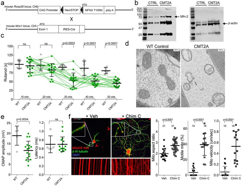
Research article                                                                                          Cell Biology Neuroscience

Figure 1. Mitochondrial abnormalities in reprogrammed CMT2A patient motor neurons and their improvement after mitofusin activation. (a) Model
structure of MFN2 showing location of CMT2A patient mutations. (b) Schematic depiction of fibroblast reprogramming procedure to produce motor
neurons. (c) Mitochondrial testing in reprogrammed motor neurons from four CMT2A patients with different MFN2 mutations and representative of
Figure 1 continued on next page

Franco, Dang, et al. eLife 2020;9:e61119. DOI: https://doi.org/10.7554/eLife.61119                                                       4 of 26
Research article                                                                                                   Cell Biology Neuroscience

Figure 1 continued
three normal control subjects. Open circles are baseline; closed circles are 48 hr after addition of mitofusin activator Chimera C (100 nM). Each circle is
one neuron from two or three independent reprogrammings. P values from ANOVA.
The online version of this article includes the following figure supplement(s) for figure 1:
Figure supplement 1. Genotyping of CMT2A patient cells.
Figure supplement 2. Direct reprogramming of human skin fibroblasts to neurons.
Figure supplement 3. Chemical characteristics and functional profiling of mitofusin activators used in this study.

                                      disability is permanent (Fridman et al., 2015; Feely et al., 2011). No mouse models of CMT2A reca-
                                      pitulate all of these key clinical features in the absence of confounding developmental phenotypes
                                      (Zhou et al., 2019; Detmer et al., 2008; Cartoni et al., 2010; Bannerman et al., 2016; Dorn, 2020).
                                      Therefore, a prerequisite for proof-of-concept testing of mitofusin activation in vivo was to generate
                                      a mouse CMT2A model having greater similarity to the human condition.
                                         By combining Rosa26  (Bannerman et al., 2016) and Mnx1-Cre (HB9)
                                      (Yang et al., 2001) alleles (Figure 2a) we drove human MFN2 T105M expression in mouse neurons
                                      (Figure 2b; CMT2A mouse). Neuromuscular functional integrity over time was assessed as the dura-
                                      tion mice could walk on an elevated accelerating rotating cylinder without falling off (RotaRod
                                      latency). RotaRod latency of CMT2A mice was normal at 10 weeks of age, progressively declined
                                      thereafter, and stabilized beyond 30 weeks (Figure 2c). As in clinical CMT2A, axonal mitochondria
                                      of MFN2 T105M mice were fragmented with disorganized cristae (Sole et al., 2009; Figure 2d).
                                         Neuroelectrophysiological testing of CMT2A patients characteristically reveals reduced com-
                                      pound muscle action potentials (CMAP) with normal nerve conduction velocities (Berciano et al.,
                                      2017; Harding and Thomas, 1980). Recapitulating this clinical finding, sciatic nerve-tibialis muscle
                                      CMAP amplitudes of 50-week-old MFN2 T105M mice were diminished with no change in signal
                                      latency, which reflects conduction velocity (Figure 2e). Tibialis myofiber atrophy and loss of large
                                      axons without demyelination in the MFN2 T105M mouse (vide infra) also mimicked clinical CMT2A
                                      (Verhoeven et al., 2006; Muglia et al., 2001; Neves and Kok, 2011).
                                         To further evaluate the relevance of the MFN2 T105M mouse to human CMT2A, dorsal root gan-
                                      glion (DRG) sensory neurons were isolated and placed in culture, the MFN2 T105M transgene
                                      induced with Adeno-Cre, and neurons assayed for the mitochondrial pathologies delineated in
                                      reprogrammed CMT2A patient motor neurons (vide supra). CMT2A-associated abnormalities in
                                      axon mitochondrial aspect ratio and transport (Figure 2f) and polarization status (Figure 2—figure
                                      supplement 1) were each mimicked in mouse CMT2A DRGs. As in reprogrammed human CMT2A
                                      motor neurons, mitofusin activation improved these abnormalities (Figure 2f and Figure 2—figure
                                      supplement 1, compare to Figure 1c).

                                      Burst mitofusin activation reverses neuromuscular dysfunction in
                                      CMT2A mice
                                      Collectively, the above results show that activating mitofusins can improve multiple mitochondrial
                                      abnormalities manifested by cultured human and mouse CMT2A neurons. To determine whether
                                      benefits of mitofusin activation in cultured neurons would translate to therapeutic effects on neuro-
                                      muscular dysfunction in CMT2A we contemplated an in vivo trial in our CMT2A mouse. However,
                                      Chimera C is rapidly degraded by the liver and undergoes first-pass metabolism, making it impracti-
                                      cal for in vivo studies (Dang et al., 2020). We therefore evaluated in vivo efficacy of mitofusin activa-
                                      tion in CMT2A using MiM111, a structurally distinct compound having a mitofusin activation profile
                                      similar to Chimera C (Figure 1—figure supplement 3), but which is metabolically stable with good
                                      nervous system bioavailability (Dang et al., 2020). We hypothesized that intermittent or ‘burst’ mito-
                                      fusin activation (a dosing schedule that reversed mitochondrial dysfunction for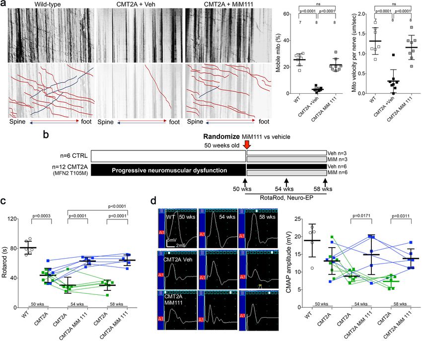
Research article                                                                                                  Cell Biology Neuroscience

Figure 2. Characteristics of a neuron-specific MFN2 T105M mouse model of CMT2A. (a) Schematic depiction Mfn2 expression strategy. (b)
Immunoblot analysis of MFN2 expression in mouse sciatic nerves. (c) Serial RotaRod latency studies; CMT2A is green squares (n = 16), wild-type (WT)
control is open circles (n = 6). (d) Electron micrographs of axonal mitochondrial from sciatic nerves (50 weeks). (e) Comparative neuro-electrophysiology
study results of 50-week-old mice in panel c. (f) Response of CMT2A dorsal root ganglion neurons to mitofusin activation with Chimera C (100 nM, 48
hr). Top images are confocal micrographs of DRGs stained for mitochondria (red) and axons (green). Insets are higher power magnification to see
mitochondrial morphology. Bottom images are kymographs showing mitochondrial (red) motility. Vertical columns are stationary mitochondria; lines
transiting left to right or right to left are moving. P values are from t-test from 3 or four independent experiments.
The online version of this article includes the following figure supplement(s) for figure 2:
Figure supplement 1. Flow cytometric profiling of mitochondrial polarization status in mouse dorsal root ganglion (DRG) neurons.

                                     dysmotility in CMT2A mice for ~12 hr out of every 24 hr. Indeed, Figure 3—figure supplement 1
                                     show that mitochondrial motility in sciatic nerve axons of MFN2 T105M mice was normalized 4 hr
                                     after a single intramuscular injection of MiM111 (30 mg/kg), declined by approximately half after 12
                                     hr, and returned to CMT2A baseline after 24 hr.
                                         If CMT2A neuron die-back is reversible then burst mitofusin activation should improve neuromus-
                                     cular degeneration in MFN2 T105M mice who had progressed to the severe and stable CMT2A phe-
                                     notype. To test this notion, 50-week-old MFN2 T105M mice and littermate controls were
                                     randomized to receive daily MiM111 or its vehicle. Researchers blind to genotype and treatment
                                     group performed Rotarod and neuro-electrophysiological testing after 4 and 8 weeks (Figure 3b).

Franco, Dang, et al. eLife 2020;9:e61119. DOI: https://doi.org/10.7554/eLife.61119                                                                6 of 26
Research article Cell Biology Neuroscience
Figure 3. Mitofusin activation reverses neuromuscular dysfunction in MFN2 T105M mice. (a) Ex vivo mitochondrial motility in CMT2A mouse sciatic
nerve axons 4 hr after intramuscular administration of mitofusin activator MiM111 or vehicle. Top panel is kymographs. Bottom panel emphasizes motile
mitochondria with red and blue lines transiting antegrade or retrograde, respectively. (Note, mitochondrial transport in ex vivo sciatic nerves favors the
antegrade [spine to foot] direction because mitochondria are recruited to the site of nerve injury at the distal amputation site [Zhou et al., 2016]). (b)
Experimental design to evaluate efficacy of MiM111 in late murine CMT2A. (c) RotaRod latency in vehicle- (green) and MiM111-treated (blue) MFN2
T105M mice. (d) Neuroelectrophysiology studies: (left) representative CMAP tracings; (right) quantitative data. Each symbol in c and d is one mouse. P
values from ANOVA. WT control values are open circles in panels c and d; complete WT control data are in Figure 3—figure supplement 2.
The online version of this article includes the following figure supplement(s) for figure 3:
Figure supplement 1. In vivo pharmacokinetics and target engagement of MiM111 administered intramuscularly.
Figure supplement 2. Effects of MiM111 on neuromuscular function in control mice.
The characteristic decreases in RotaRod latency and CMAP amplitude in MFN2 T105M mice (see
Figure 2c and e) were reversed 4 weeks after MiM111 treatment and remained near normal after 8
weeks (Figure 3c and d); MiM111 had no effect on control mice (Figure 3—figure supplement 2).
Compared to sciatic nerves of vehicle-treated MFN2 T105M CMT2A mice, MiM111 treatment
reduced axon damage (Figure 4a), increased axon diameter (Figure 4b), and increased staining for
superior cervical ganglion 10 (SCG10; a marker of neuron regeneration) (Shin et al., 2014;
Franco, Dang, et al. eLife 2020;9:e61119. DOI: https://doi.org/10.7554/eLife.61119 7 of 26Research article Cell Biology Neuroscience Figure 4. Mitofusin activation reverses histopathological findings in MFN2 T105M mice. (a) Toluidine blue stained sections of mouse mid tibial nerves. Arrows show blue-stained damaged axons in CMT2A mice. Quantitative group data for damaged axons and SCG10-regenerating axons (see Figure 4—figure supplement 1) are on the right. (b) Electron micrographs of mid-tibial nerve axons from CMT2A mouse study groups after 8 weeks of therapy. Note heterogeneity in axon size (top images; left graph) and mitochondrial abnormalities (bottom images, right graph). (c) Wheat germ agglutinin (WGA) labeled sections of tibialis anterior muscle and quantitative myocyte cross sectional area. (d) Confocal micrographs of neuromuscular junctions to show mitochondrial occupancy yellow organelles within red synapses (also see Figure 4—figure supplement 1). Each symbol represents results from one mouse. Data are means ± SD; p values are 1- or 2-way ANOVA. Figure 4 continued on next page Franco, Dang, et al. eLife 2020;9:e61119. DOI: https://doi.org/10.7554/eLife.61119 8 of 26
Research article Cell Biology Neuroscience
Figure 4 continued
The online version of this article includes the following figure supplement(s) for figure 4:
Figure supplement 1. Effects of MiM111 on neuromuscular integrity in CMT2A MFN2 T105M mice.
Figure 4—figure supplement 1). These findings suggest that mitofusin activation reversed CMT2A-
induced neuronal degeneration.
Skeletal myocytes of CMT2A mouse tibialis muscle innervated by the sciatic nerve were abnor-
mally small (Figure 4c), reflecting neurogenic muscle atrophy (because the MFN2 T105M transgene
is directed by neuron-specific HB9-Cre). In agreement with muscle atrophy being a secondary effect,
skeletal myocyte mitochondria of CMT2A mice appeared structurally normal (Figure 4—figure sup-
plement 1) compare to sciatic nerve axon mitochondria in Figures 2d and 4b. Therefore, normaliza-
tion of tibialis myocyte diameter after mitofusin activation (Figure 4c) indicates restoration of
neuromuscular integrity.
Collectively, the above findings provide indirect support for the idea that CMT2A mice suffer
from distal neuron dieback that can be reversed by activating mitofusins. Reasoning that decreased
neuromuscular junction density in CMT2A mice would constitute direct evidence for dieback, we
quantified neuromuscular junctional synapses containing receptors for the neurotransmitter acetyl-
choline (AchR) in tibialis muscles of CMT2A mice. Compared to normal mice, CMT2A mice
had ~50% fewer synaptic junctions/myocyte, which was reversed after mitofusin activator treatment
(Figure 4—figure supplement 1). Strikingly, mitochondrial occupancy of vehicle-treated CMT2A
neuromuscular synaptic junctions was also reduced by ~half compared to normal mice, and was nor-
malized by MiM111 treatment (Figure 4d). Because mitochondrial transport can play a central role
in neuron repair and regeneration (Sheng, 2017), the observation that MiM111 promoted mitochon-
drial localization within terminal neuromuscular synaptic junctions provided a plausible mechanistic
link between mitofusin activation, mitochondrial motility, neuronal regrowth, and reversal of neuro-
muscular dysfunction in this preclinical CMT2A model.
Enhanced mitochondrial function in mitofusin-activated CMT2A DRGs
leads to accelerated axon growth
Reversal of CMT2A-induced distal neuron die back implies neuronal regrowth. Indeed, sensory DRG
neurons isolated from CMT2A mice and cultured in the presence of MiM111 (100 nM, 48 hr) exhib-
ited not only enhanced mitochondrial fusion (increased aspect ratio) and transport (greater mito-
chondrial motility and velocity), but axon outgrowth (length and branching) (Figure 5). Similar
effects were seen in CMT2A DRGs treated with Chimera C (100 nM, 48 hr) (Figure 5—figure supple-
ment 1). Both MiM111 and Chimera C provoked mitochondrial redistribution to axonal termini of
cultured CMT2A DRGs (Figure 5—figure supplement 1) recapitulating mitochondrial occupation of
neuromuscular synapses after MiM111 treatment of CMT2A mice in vivo (see Figure 4d).
Comparing the mitochondrial motility, aspect ratio, and neuron growth responses at different
times after mitofusin activation revealed significantly increased mitochondrial trafficking within 2 hr,
whereas enhanced axon outgrowth was significant after 24 and 28 hr, and mitochondrial aspect ratio
(i.e. fusion) was significant only after 48 hr (Figure 5—figure supplement 2). Given the established
role for mitochondrial transport in neuronal repair (Sheng, 2017), this temporal sequence lends cre-
dence to the idea that accelerated neuron growth is a consequence of enhanced mitochondrial func-
tion and redistribution.
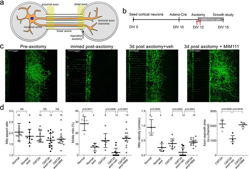
Mitofusin activation accelerates in vitro CMT2A axon regeneration
after axotomy
DRG outgrowth measures in vitro regrowth of neuronal extensions that are amputated during the
cell isolation trituration procedure. We considered that a more appropriate model of regrowth after
dieback in CMT2A would test intact neurons lacking only the distal axons. Because CMT2A mouse
neurons grow poorly in tissue culture in the absence of mitofusin activators (vide supra), this was not
feasible using DRGs. Therefore, we seeded cortical neurons collected from MFN2 T105M allele mice
in chambers separated from empty chambers by linear microchannels. In the absence of Cre-recom-
binase these ‘normal’ neurons grew axons through the microchannels that branched into the empty
Franco, Dang, et al. eLife 2020;9:e61119. DOI: https://doi.org/10.7554/eLife.61119 9 of 26Research article Cell Biology Neuroscience Figure 5. Mitofusin activation reverses mitochondrial pathology and stimulates growth of CMT2A dorsal root ganglion neurons in vitro. (a) Confocal micrographs of CMT2A mouse DRGs cultured for 48 hr with MiM111 or its vehicle. Note greater neuronal process length and branching in MiM111- treated neuron. Exploded insets (right) show neuronal process termini. Mitochondria express mitoDS Red; neuronal processes stained for b-III tubulin are green. (b) Kymographs of mitochondrial motility in neuronal processes of live DRGs from studies shown in (a). Top panel is raw data. Bottom panels emphasize motile mitochondria with red and blue lines transiting left to right or right to left, respectively. (c-f) Quantitative group data demonstrating effect of MiM111 on CMT2A DRG mitochondrial aspect ratio (c), motility (d, e), neuronal process length and branching (e), and proportion of neuronal process termini containing mitochondria (f). The online version of this article includes the following figure supplement(s) for figure 5: Figure supplement 1. Mitofusin activation with Chimera C reverses mitochondrial pathology and stimulates growth of CMT2A dorsal root ganglion neurons in vitro. Figure supplement 2. Time course studies of DRG mitochondria responses to mitofusin activation after aspiration axotomy. Franco, Dang, et al. eLife 2020;9:e61119. DOI: https://doi.org/10.7554/eLife.61119 10 of 26
Research article                                                                                               Cell Biology Neuroscience

                                    chambers (Figure 6a). Adenoviral Cre was then used to activate the CMT2A MFN2 T105M trans-
                                    gene, followed after 48 hr by aspiration amputation of the branched axonal termini (Figure 6b and
                                    c). Mitochondrial motility and aspect ratio were measured 1 hr before and after axotomy; axon
                                    regrowth was measured 3 days after axotomy. The aspect ratio of mitochondria in the distal linear
                                    axons of normal and CMT2A neurons was unaffected either by axotomy or by MiM111 (Figure 6d,
                                    left panel). By contrast, and consistent with a previous study in normal neurons (Zhou et al., 2016),
                                    mitochondrial motility was reduced by axotomy (Figure 6d middle panels). Mitofusin activation with
                                    MiM111 after axotomy restored mitochondrial motility and neuronal outgrowth to pre-axotomy lev-
                                    els. Thus, the link between experimentally activating mitofusins, the subsequent increase in mito-
                                    chondrial transport, and enhanced neuronal growth/repair was consistent for mouse CMT2A sciatic
                                    nerve axons in vivo, cultured CMT2A DRG neuron outgrowth, and cultured CMT2A cortical nerve
                                    regrowth after distal axotomy.

                                    Discussion
                                    These preclinical studies show that activating endogenous normal mitofusins can improve stable
                                    neuromuscular dysfunction caused by a CMT2A MFN2 mutant. Pharmacological mitofusin activation
                                    enhanced CMT2A neuron growth in vivo and in vitro by promoting mitochondrial fitness and

Figure 6. Mitofusin activation stimulates post-axotomy regrowth of CMT2A cortical neurons in vitro. (a) Schematic depiction of microfluidic platform.
Yellow areas show proximal axon where mitochondrial motility was measured and distal axon where mitochondrial aspect ratio was measured. (b)
Experimental design. DIV is days in vitro. Red arrows are times of pre- and immediate post-axotomy mitochondrial studies. (c) Representative images of
CMT2A neuron terminal branches at different times relative to aspiration axotomy. (d) Quantitative group data demonstrating effect of MiM111 on
CMT2A cortical neuron mitochondrial aspect ratio (left panel), motility (middle panels), and axon length (right panel).

Franco, Dang, et al. eLife 2020;9:e61119. DOI: https://doi.org/10.7554/eLife.61119                                                            11 of 26
Research article Cell Biology Neuroscience
transport, thereby reversing CMT2A-associated mitochondrial fragmentation, depolarization and
stasis. We believe the key benefit that accrued from directly activating mitofusin-mediated mito-
chondrial fusion and motility was improved delivery of healthy mitochondria to neuromuscular junc-
tions and axon growth buds. To our knowledge, this is the first report of any experimental
intervention that fully reverses in vivo CMT2A phenotypes, demonstrating the feasibility of a clinically
translatable disease-modifying therapeutic modality for this incurable condition.
Our studies integrated findings from multiple complementary experimental platforms. Motor neu-
rons directly reprogrammed from CMT2A patient fibroblasts have not previously been described,
and provided a platform in which effects of therapeutic interventions could be assessed on different
patients’ mutations and individual genetic backgrounds using a disease-relevant cell type. Com-
pared to iPS cell-derived CMT2A neurons (Rizzo et al., 2016; Saporta et al., 2015), direct reprog-
ramming more faithfully reproduced prototypical CMT2A mitochondrial phenotypes. Compared to
the parental patient fibroblasts (Dang et al., in preparation), neurons permitted assessment of the
CMT2A-associated mitochondrial motility disorders. While CMT2A has both sensory and motor neu-
ropathy components, we reprogrammed specifically for motor neurons because motor components
of this disease are the major source of patient disability.
All mouse disease models have advantages and limitations. Our CMT2A mouse model expresses
human MFN2 T105M using a ‘motor neuron selective’ promoter (vide infra), and therefore does not
exhibit sensory nerve involvement that is sometime manifested in clinical CMT2A. However, com-
pared to other CMT2A mice, the current model more faithfully recapitulates CMT2A neuromuscular
dysfunction that is the dominant cause of morbidity in the human condition (Fridman et al., 2015;
Feely et al., 2011; Bombelli et al., 2014; Yaron and Schuldiner, 2016; Berciano et al., 2017). We
previously used young adult mice carrying this combination of MFN2T105M and HB9-Cre alleles to
evaluate effects of a topically applied prototype mitofusin activator on mitochondrial motility in sci-
atic nerve axons ex vivo (Rocha et al., 2018). It was not known if, with age, these mice would
develop neuromuscular signs similar to clinical CMT2A. As shown here, motor function in these mice
is normal at age 10 weeks, but declines until age 30 weeks whereupon it stabilized for at least
another 20 weeks. This pattern is similar to the clinical course of CMT2A, in which apparently normal
children typically manifest neuromuscular signs in the mid first decade of life, exhibit progressive
loss of motor function in distal extremities over the next 10–15 years, and then stabilize. Moreover,
the functional (neuroelectrophysiological), histological, and ultrastructural features of axonal tissue in
the mice were similar to the human condition. Together with the positive response to mitofusin acti-
vation in patient neurons, the improvement in neuromuscular function and cell/organelle pathology
in MiM111-treated CMT2A mice supports the approach of mitofusin activation for the clinical
disease.
Perhaps the most remarkable finding here is that mitofusin activation reversed the signs of
CMT2A in mice with severe, stable disease. Every measured endpoint was improved, including gross
neuromuscular function (RotaRod), electrophysiological metrics of neuromuscular integrity (CMAP),
read-outs for axon degeneration, and multiple histological and ultrastructural assays of mitochon-
drial pathology in neurons. In vivo and in vitro results pointed to enhanced CMT2A neuron repair
and regrowth as a central reason for phenotype reversal. Because it is not possible to functionally
dissociate mitofusin-mediated increases in mitochondrial fusion and motility, it is unclear if one or
the other of these responses preferentially underlies the neuroregenerative effects of mitofusin acti-
vation. However, it seems reasonable to postulate that mitochondrial delivery to distal neurons has
greatest importance in the long peripheral nerves innervating hands, forearms, feet, and forelegs,
i.e. those areas most impacted in CMT2A (Fridman et al., 2015; Feely et al., 2011; Bombelli et al.,
2014; Yaron and Schuldiner, 2016; Berciano et al., 2017). In agreement with this notion, we
observed a positive correlation between mitochondrial delivery to or occupancy of axonal termini
and CMT2A neuron growth in vivo and in vitro.
As introduced above, damaging MFN2 mutations are a straightforward cause of CMT2A, but
MFN2 multifunctionality complicates delineating the underlying cellular pathology (Filadi et al.,
2018; Dorn, 2020). For this reason, the specific functional benefits accruing from mitofusin activa-
tion in CMT2A cannot unambiguously be defined. Mitochondrial fusion and motility are impaired in
CMT2A (Chen and Chan, 2006) and allosteric mitofusin activation corrects both of these parameters
(Franco et al., 2016; Rocha et al., 2018; Dang et al., 2020). Mitochondrial respiratory dysfunction,
measured here as loss of inner membrane polarization, is a consequence of diminished fusion-
Franco, Dang, et al. eLife 2020;9:e61119. DOI: https://doi.org/10.7554/eLife.61119 12 of 26Research article Cell Biology Neuroscience
mediated homeostatic repair (Chen and Chan, 2006), and its improvement can therefore also be
explained by enhanced fusogenicity. MFN2 has a role in mitophagic mitochondrial quality and quan-
tity control (Chen and Dorn, 2013; Gong et al., 2015), and allosteric mitofusin activation sup-
pressed the increase in autophagy/mitophagy induced by CMT2A mutant MFN2 T105M in cultured
cells (Rocha et al., 2018). Finally, MFN2 can mediate physical interactions and calcium signaling
between mitochondria and endoplasmic reticulum that may also have a role in CMT2A
(Larrea et al., 2019), but effects of mitofusin activation on mitochondria-reticular interactions have
not been described.
Here, we studied two structurally distinct but functionally similar allosteric mitofusin activators, a
new class of drug that is the first to directly enhance mitochondrial fusion and transport. Although
the prototype compounds were found not to be ‘druggable’, a new generation of mitofusin activa-
tors have addressed pharmaceutical limitations of the initial chemical series (Dang et al., 2020). As
described previously (Rocha et al., 2018; Dang et al., 2020), mitofusin activators have minimal
effects in normal cells, likely because increasing the probability that mitofusins are in their open/
‘active’ conformation is a subtle intervention that can be readily compensated for in the absence of
a pre-existing imbalance between mitochondrial fusion and fission. The current in vivo studies used a
short-acting compound administered once daily to evaluate the effects of intermittent, or burst,
mitofusin activation on CMT2A neuromuscular dysfunction. We considered that continuous long-
term activation of mitochondrial fusion and transport might possibly be deleterious (El Fissi et al.,
2018) (although it is worth noting that adverse effects of MFN1 and MFN2 overexpression in trans-
genic mice have not been reported). Moreover, we reasoned that the problem underlying CMT2A is
the cumulative effects of long-term mitochondrial stasis and dysfunction on mitochondrial fitness
and neuromuscular integrity. This scenario can explain why CMT2A progresses over many years in
people and many weeks in mice. Our aim with burst activation was to turn back the disease clock
through daily re-setting of mitochondrial function. By intermittently mobilizing healthy mitochondria
to distal areas of physiological need, and simultaneously removing senescent or impaired mitochon-
dria, neuron repair, renewal, and neuromuscular signaling were improved.
A mouse is not a man and human neuroregenerative capacity declines with age (Mattson and
Magnus, 2006). For this reason we do not expect that mitofusin activation can fully reverse CMT2A
phenotypes in older human patients with long-term stable disease. Nevertheless, the current results
suggest that pharmacological mitofusin activation could offer the first disease-altering therapy for
younger CMT2A patients. An intriguing possibility is that mitofusin activation may also have a thera-
peutic role in some of the many other neurodegenerative conditions not directly caused by mitofusin
defects wherein mitochondrial fusion or transport are defective (Dang et al., in preparation;
Knott et al., 2008; Burté et al., 2015).
Materials and methods
Key resources table
Reagent type
(species) or Source or Additional
resource Designation reference Identifiers information
Gene Mfn-2 NCBI Gene Gene ID: 170731 MFN2
Mus ENSMUSG00
musculus 000029020
Gene MFN-2 NCBI Gene Gene ID: 9927 MFN2
(Human) ENSG0000
0116688
Genetic reagent Rosa-STOP-mMFN (C57BL/6 Gt(ROSA) The Jackson C57Bl/6
(M. musculus) Thr105Met 26 Sortm1 (CAG Laboratory:
(T105M) mice MFN2*T105M) 025322
Dple/)
Genetic reagent HB9-Cre mice (B6.129S1-Mnx1tm4 The Jackson C57Bl/6
M. musculus (cre)Tmj/J) Laboratory :
006600
Continued on next page
Franco, Dang, et al. eLife 2020;9:e61119. DOI: https://doi.org/10.7554/eLife.61119 13 of 26Research article Cell Biology Neuroscience
Continued
Reagent type
(species) or Source or Additional
resource Designation reference Identifiers information
Genetic reagent C57BL/6J mice C57Bl/6 The Jackson C57Bl/6
M. musculus Laboratory :
000664
Mfn2 null Mfn2 null MEFs ATCC CRL-2994 Murine
M. musculus embryonic
fibroblasts
Mfn1/Mfn2 null Mfn1 and Mfn2 ATCC CRL-2993 Murine
M. musculus double knock embryonic
out MEFs fibroblasts
Mfn1 null Mfn1 null MEFs ATCC CRL-2992 Murine
M. musculus embryonic
fibroblasts
Cell line Dermal Dr. Robert Female
(H. sapiens) fibroblast H. Baloh
(MFN2 T105M) (Cedars Sinai)
Cell line Dermal Dr. Robert Male
(H. sapiens) fibroblast H. Baloh
(MFN2 H361Y) (Cedars Sinai)
Cell line Dermal fibroblast Dr. Barbara PMID:28076385 Male
(H. sapiens) (MFN2 R274W) Zablocka
(Mossakowski
Med Res Ctr)
Cell line Dermal fibroblast Dr. Michael Female
(H. sapiens) (MFN2 R364W) E. Shy (University
of Iowa)
Cell line Dermal NINDS ND34769 Female
(H. sapiens) fibroblast
(Normal)
Cell line Dermal NINDS ND36320 Female
(H. sapiens) fibroblast
(Normal)
Cell line Dermal NINDS ND29510 Female
(H. sapiens) fibroblast
(Normal)
Transfected Adenovirus Vector Biolabs Cat#: 1080
construct (Human b-galactosidase
Adenovirus
Type5 (dE1/E3))
Transfected Adenovirus Signagen Cat#: 12259
construct Mito-Ds-Red2
(Human Adenovirus
Type5 (dE1/E3))
Transfected Adenovirus Vector Biolabs Cat#: 1794
construct Cre-recombinase
(Human Adenovirus
Type5 (dE1/E3))
Recombinant rtTA-N144 Addgene Cat#: 66810 Lentiviral construct
DNA reagent (plasmid) to transfect and
express the plasmid
Recombinant pTight-9- Addgene Cat#: 60857 Lentiviral construct
DNA reagent 124-BclxL to transfect and
(plasmid) express the plasmid
Recombinant LHX3-N174 PMID:28886366 Lentiviral construct
DNA reagent and ISL1- to transfect and
N174 (plasmid) express the plasmid
Continued on next page
Franco, Dang, et al. eLife 2020;9:e61119. DOI: https://doi.org/10.7554/eLife.61119 14 of 26Research article Cell Biology Neuroscience
Continued
Reagent type
(species) or Source or Additional
resource Designation reference Identifiers information
Antibody Anti-Mfn-2 AbCAM Cat#: ab56889 (1:1000)
(Mouse
monoclonal)
Antibody Anti-COX-IV AbCAM Cat#: ab16056 (1:1000)
(Rabbit
polyclonal)
Antibody Anti-Stathmin-2 Novus Cat#: NBP1-49461 (1:1000)
(Rabbit polyclonal) Biologicals
Antibody Anti-GAPDH AbCAM Cat#: ab8245 (1:3000)
(Mouse
monoclonal)
Antibody Anti-FSP-1 Novus Cat#: NBP1-49461 (1:400)
(Rabbit Biologicals
polyclonal)
Antibody Anti-MNX1 DSHB Cat#: 81.5C10 (2 mg/ml)
(Mouse
monoclonal)
Antibody Anti-b-tubulin III Biolegend Cat#: 801201 (1:200)
(Mouse
monoclonal)
Antibody Alexa-Fluor 488 ThermoFisher Cat#: A11029 (1:400)
(Goat
anti-mouse)
Antibody Alexa- Fluor 488 ThermoFishe Cat#: A11008 (1:400)
(Goat anti-
rabbit)
Antibody (Goat anti- ThermoFisher Cat#: 31460 (1:3000)
rabbit IgG)
Antibody Alexa- Fluor 594 ThermoFisher Cat#: A32740 (1:400)
(Goat anti
rabbit)
Antibody (Peroxidase- Cell Signaling Cat#: 7076S (1:3000)
conjugated
anti-mouse IgG)
Antibody (a-Bungarotoxin ThermoFisher Cat#: B12423 (0.5 mg/ml)
Alexa flour 594)
Sequence- HB9CRE Fw The Jackson 006600 CTAGGCCACAGA
based Laboratory ATTGAAAGATCT
reagent
Sequence- HB9CRE Rv The Jackson 006600 GTAGGTGGAAA
based Laboratory TTCTAGCATCATCC
reagent
Sequence- HB9CRE TG Fw The Jackson 006600 GCGGTCTGGCA
based Laboratory GTAAAAACTATC
reagent
Sequence- HB9CRE TG Rv The Jackson 006600 GTGAAACAGCAT
based Laboratory TGCTGTCACTT
reagent
Sequence- Mfn2 T105M The Jackson 025322 GACCCCGTT
based M Fw Laboratory ACCACAGAAGA
reagent
Sequence- Mfn2 T105M The Jackson 025322 AACTTTGTCC
based M Rv Laboratory CAGAGCATGG
reagent
Continued on next page
Franco, Dang, et al. eLife 2020;9:e61119. DOI: https://doi.org/10.7554/eLife.61119 15 of 26Research article Cell Biology Neuroscience
Continued
Reagent type
(species) or Source or Additional
resource Designation reference Identifiers information
Sequence- Mfn2 T105M The Jackson 025322 AAGGGAGCTGC
based Wt Fw Laboratory AGTGGAGTA
reagent
Sequence- Mfn2 T105M The Jackson 025322 CCGAAAATCT
based Wt Rv Laboratory GTGGGAAGTC
reagent
Sequence- MFN2 T105M Fw This paper PCR primers for TTGCACTGAA
based cell line mutation TAGGGCTTTG
reagent validation
Sequence- MFN2 T105M Rv This paper PCR primers for CATTCACCTC
based cell line mutation CACAGGGTG
reagent validation
Sequence- MFN2 R274W Fw This paper PCR primers for CGTGGTAGGTG
based cell line mutation TCTACAAGAAGC
reagent validation
Sequence- MFN2 R274W Rv This paper PCR primers for CTGGTGAGG
based cell line mutation GCTGATGAAAT
reagent validation
Sequence- MFN2 H361Y This paper PCR primers for CCTGGCAGTGA
based reagent and R364W Fw cell line mutation AAACCAGAG
validation
Sequence- MFN2 H361Y This paper PCR primers for AAGGCGTGT
based and R364W Rv cell line mutation CCTAACTGCC
reagent validation
Chemical Trans-MiM111 Mitochondria Cpd 13b in
compound, in Motion, Inc PMID:32506913
drug
Chemical Chimera C Paraza Pharma Cpd 2 in
compound, PMID:32506913
drug
Chemical Papain Sigma Cat#: P4762
compound,
drug
Chemical Laminin Sigma Cat#: L2020
compound,
drug
Chemical Poly-d-Lysine Sigma Cat#: P7886
compound,
drug
Chemical Poly-ornithine Sigma-Aldrich Cat#: P4957
compound,
drug
Chemical Fibronectin Sigma-Aldrich Cat#: F4759
compound,
drug
Chemical Polybrene Sigma-Aldrich Cat#: H9268
compound,
drug
Chemical Doxycycline Sigma-Aldrich Cat#: D9891
compound,
drug
Chemical G418/Geneticin Invitrogen Cat#: 10131-035
compound,
drug
Continued on next page
Franco, Dang, et al. eLife 2020;9:e61119. DOI: https://doi.org/10.7554/eLife.61119 16 of 26Research article Cell Biology Neuroscience
Continued
Reagent type
(species) or Source or Additional
resource Designation reference Identifiers information
Chemical Retinoic Acid Sigma Cat#: R2625
compound,
drug
Chemical BDNF, NT-3, Peprotech Cat#: 450-02,
compound, CNTF, GDNF Cat#: 450-03,
drug Cat#: 450-13,
Cat#: 450-10
Chemical Dibutyryl cAMP Sigma Cat#: D0627
compound,
drug
Chemical Valproic acid Sigma Cat#: 676380
compound,
drug
Chemical Puromycin Invitrogen Cat#: A11138-03
compound,
drug
Chemical Collagenase Worthington Cat#: 41J12861
compound, Biochemical
drug
Chemical (2-Hydroxypropyl)- Sigma Cat#: 332607
compound, b-cyclodextrin
drug
Chemical Carbonyl Sigma Cat#: C2759
compound, cyanide-p-trifluoro
drug methoxyphenyl
hydrazone
Chemical B27 supplement Gibco Cat#: 17504-044
compound,
drug
Chemical Insulin-transferrin- Sigma Cat#: 1884
compound, sodium selenite
drug
Chemical Glucose Sigma Cat#: G5767
compound,
drug
Chemical L-glutamine Gibco Cat#: 25030-149
compound,
drug
Chemical Goat serum Jackson Cat#: 005-000121
compound, Immunoresearch
drug
Chemical Glutaraldehyde Electron Cat#: 16216
compound, Microscopy
drug Science
Chemical MitoTracker Thermo Fisher Cat#: M7514
compound, Green
drug
Chemical Calcein AM Thermo Fisher Cat#: C3100MP
compound,
drug
Chemical Hoechst Thermo Fisher Cat#: H3570
compound,
drug
Chemical MitoTracker Thermo Fisher Cat#: M7510
compound, Orange
drug
Continued on next page
Franco, Dang, et al. eLife 2020;9:e61119. DOI: https://doi.org/10.7554/eLife.61119 17 of 26Research article Cell Biology Neuroscience
Continued
Reagent type
(species) or Source or
resource Designation reference Identifiers
Chemical Tetramethylrhodamine Thermo Fisher Cat#: T-669
compound, ethyl ester
drug
Software, ImageJ C. A. Schneider https://imagej.
algorithm net/Sholl_Analysis
Software, Viasys Healthcare Middleton Cat#: OL060954
algorithm Nicolet Biomedical
instrument with
Viking Quest version Additional
11.2 software information
Software, Gallios instrument Beckman N/A
algorithm with FlowJo Coulter
10 software
Other RotaRod Ugo Basile Cat#: 47650
Other XonaChips Xona Cat#: XC450
Microfluidics
Mouse lines
Rosa-STOP-mMFN Thr105Met (T105M) mice (C57BL/6 Gt(ROSA)26 Sortm1 (CAG-MFN2*T105M)
Dple/J) from The Jackson Laboratory (Bar Harbor, Maine, USA; Stock No: 025322, RRID:MGI:_JAX:
025322) were crossed to HB9-Cre mice (B6.129S1-Mnx1tm4(cre)Tmj/J) from The Jackson Laboratory
(Stock No: 006600, (RRID:MGI:_JAX:006600)) to generate neuron-targeted MFN2 T105M mice. HB9
is a motoneuron-specific promoter (Yang et al., 2001), but JAX data indicates that this HB9-Cre line
also drives expression in some sensory DRG neurons (https://images.jax.org/webclient/img_detail/
20564/). All experimental procedures were approved by Washington University in St. Louis School of
Medicine Animal Studies Committee; IACUC protocol number 19–0910, Exp:12/16/2022.
Cell lines
Normal mouse embryonic fibroblasts (MEFs) were prepared by enzymatic dissociation from embry-
onic day E.13.5–14.5 C57BL/6J mice (The Jackson Laboratory Cat:# 000664, RRID:IMSR_JAX:
000664). Mfn1 null and Mfn2 null Mfn1/Mfn2 double null MEFs fibroblasts were purchased from
American Type Culture Collection (ATCC Manassas, Virginia, USA) (CRL-2992, RRID:CVCL_L691 and
CRL-2994, RRID:CVCL_L692 and CRL-2993, RRID:CVCL_L693 respectively). Human fibroblast: Der-
mal fibroblast (MFN2 T105M) and Dermal fibroblast (MFN2 H361Y) from Dr. Robert H. Baloh
(Cedars Sinai), Dermal fibroblast (MFN2 R274W) from Dr. Barbara Zablocka (Mossakowski Med Res
Ctr), Dermal fibroblast (MFN2 R364W) from Dr. Michael E. Shy (University of Iowa). Dermal fibroblast
(Normal) from NINDS respectively: ND34769, (RRID:CVCL_EZ04, ND36320, RRID:CVCL_EZ16 and
ND29510, RRID:CVCL_Y813).
Viral vectors
Adenovirus expressing human FLAG-hMFN2 -T105M was prepared at Vector Biolabs (Malvern, PA,
USA). Adenoviri expressing b-galactosidase (Ad-CMV-b-Gal; #1080), and Ad-Cre (#1794) were pur-
chased from Vector Biolabs. Adenovirus for Mito-Ds-Red2 came from Signagen (Cat:#SL1007744).
Lentivirus packaging vectors: psPAX2 (Addgene, Cat#: 12260) pMD2.G (Addgene, Cat#: 12259),
Lentiviral vectors with recombinant DNA: rtTA-N144 (Addgene, Cat#: 66810) pTight-9–124-BclxL
(Addgene, Cat#: 60857), human LHX3-N174 and human ISL1-N174 were packaged and used as
described (Abernathy et al., 2017).
Antibodies
Mouse monoclonal anti-mitofusin 2 (Cat # ab56889 - 1:1000, RRID:AB_2142629), anti-COX-IV (Cat
#ab16056 - 1:1000, RRID:AB_443304) and anti-GAPDH (Cat #ab8245 - 1:1000, RRID:AB_2107448)
Franco, Dang, et al. eLife 2020;9:e61119. DOI: https://doi.org/10.7554/eLife.61119 18 of 26Research article Cell Biology Neuroscience
were from AbCAM (Cambridge, MA, USA). Rabbit polyclonal anti-Stathmin-2/Superior Cervical Gan-
glion 10 (SCGN10; Cat # NBP1-4946, RRID:AB_10011569) was from Novus Biologicals (Littleton,
CO, USA).Rabbit polyclonal FSP-1 was from Sigma Millipore (Cat # 07–2274, RRID:AB_10807552).
Anti-mouse monoclonal -MNX1was from DSHB (1:10, Cat# 81.5C10, RRID:AB_2145209). Mouse
monoclonal anti-b -tubulin III (Cat # 801201- 1:500, RRID:AB_2313773) was from Biolegend (San
Diego, CA, USA). Peroxidase-conjugated anti-mouse IgG (Cat #7076S - 1:1000, RRID:AB_330924)
was from Cell Signaling (Danvers, MA, USA). Goat anti-rabbit IgG (Spicier reactivity Goat, Host/Iso-
type Rabbit/IgG; Cat #31460, RRID:AB_228341) and Alexa-Fluor 488 anti-mouse ThermoFisher (Wal-
tham, MA, USA Cat #A11008, RRID:AB_143165 ). a-Bugarotoxin Alexa flour 594 was from
ThermoFisher, Waltham, MA, USA Cat:# B12423.
PCR genotyping of MFN2 mutations in CMT2A patient fibroblasts
DNA was extracted from 5 106 primary human fibroblasts using the DNeasy blood and tissue kit
(Qiagen, Cat#: 69506) according to manufacturer’s protocol. PCR used Taq Plus Master Mix 2X
(Cat#: BETAQR-L, Bulls eye). 50 ng of genomic DNA template, and the following primers:
(MFN2 T105M) - 5’- TTGCACTGAATAGGGCTTTG- 3’
5’- CATTCACCTCCACAGGGTG- 3’
(MFN2 R274W) - 5’- CGTGGTAGGTGTCTACAAGAAGC- 3’
5’- CTGGTGAGGGCTGATGAAAT- 3’
(MFN2 H361Y, R364W) - 5’-CCTGGCAGTGAAAACCAGAG- 3’
5’- AAGGCGTGTCCTAACTGCC- 3’.
PCR products were purified using PureLink Quick Gel Extraction Kit (Cat#: K21000-12, Invitro-
gen). Sanger sequencing of PCR products was performed at GENEWIZ.
Cultured cells
Direct reprogramming of human motor neurons from patient fibroblasts used the procedure as
described (Abernathy et al., 2017). Reprogramming cocktail consisted of 1 ml concentrated lentivi-
rus containing the reverse tetracycline-controlled transactivator (rtTA; Addgene, Cat# 66810), 1 ml
virus containing pT-BclXL-9/9* 124, 125 ml virus containing motor neuron transcription factor ISL1,
and 125 ml virus containing motor neuron transcription factor LHX3 with polybrene (8 mg/ml; Sigma-
Aldrich, Cat# H9268). Human skin fibroblasts of low passage number (P4-P7) were spinfected at 37˚
C for 30 min at 1,000 G. Doxycycline (Dox, 1 mg/ml; Sigma Aldrich, Cat# D9891) and antibiotics
for respective vectors (Puromycin, 3 mg/ml; Invitrogen, Cat# A11138-03; Geneticin, 400 mg/ml; Invi-
trogen, Cat# 10131–035) were added to culture medium for 4 days after viral transduction. On day 5
cells were re-plated on poly-ornithine/laminin/fibronectin (Sigma, Cat# P4957, # L2020, # F4759)
coated 18 mm glass coverslips and on the following day changed to neuronal medium supple-
mented with Dox (1 mg/ml added every other day), valproic acid (1 mM; Sigma, Cat# 676380), dibu-
tyryl cAMP (200 mM; Sigma, Cat# D0627), BDNF, NT-3, CNTF, GDNF (all 10 ng/ml, Peprotech, Cat#
450–02, #450–03, #450–13, #450–10), retinoic Acid (1 mM; Sigma, Cat# R2625) and antibiotics. Neu-
ronal medium was refreshed by replacing half every 4 days. Antibiotics were discontinued on day 14;
Dox was discontinued on day 30. Cells underwent studies beginning on day 35. Motor neurons were
identified after formalin fixation by labeling with mouse anti-MNX1 (1:10; DSHB, Cat# 81.5C10) and
mouse anti-TUBB3B (1:2000; Biolegend, Cat#PRB-435P-100). Fibroblasts were identified by labeling
with rabbit anti FSP-1 (1:200; Sigma, Cat: # 07–2274).
Adult mouse dorsal root ganglion (DRG) neurons were prepared from ~8-week-old HB9Cre-
MFN2 Thr105Met flox-stop transgenic mice as previously described (Rocha et al., 2018). To com-
prehensively induce MFN2 T105M transgene expression, the DRGs were infected with Adeno-Cre
(M.O.I. of 50) 48 hr prior to study. DRG neurons were distinguished from non-neuronal cells by stain-
ing with anti-b-III tubulin.
Mouse cortical neurons were isolated from individual embryonic day E.18.5 MFN2 Thr105Met
flox-stop transgenic mice by papain digestion and mechanical dispersion using a published proce-
dure (Sobieski et al., 2015). Briefly, mouse brain cortices were isolated under a dissecting micro-
scope and sliced into 0.5–1 mm thick sections in Leibovitz’s L-15 Medium (Gibco Cat:#11415–064)
containing BSA (0.23 mg/ml, Sigma Cat:#A7030). Papain (1 mg/ml, Sigma Cat #P4762) was added
and the tissue digested for 20 min at 37˚C. The papain solution was replaced and micropipettes
Franco, Dang, et al. eLife 2020;9:e61119. DOI: https://doi.org/10.7554/eLife.61119 19 of 26Research article Cell Biology Neuroscience
used to triturate the solution until no more tissue was visible. Cortical neurons were plated in micro-
fluidic neuron XonaChip chambers as described below.
Imaging
Static confocal imaging of cultured neurons used triple-stained with MitoTracker Green (200 nM;
Invitrogen, Thermo Fisher Scientific Cat:# M7514) to visualize mitochondria, tetramethylrhodamine
ethyl ester (TMRE, 200 nM, Invitrogen Thermo Fisher Scientific Cat:# T-669) that labels mitochondria
with normal polarization of the mitochondrial inner membrane, and Hoechst (10 mg/ml; Invitrogen,
Thermo Fisher Scientific Cat:# H3570) that stains nuclei blue as described (Franco et al., 2016).
Static live cell images were acquired on a Nikon Ti Confocal microscope using either a 60 1.3 NA
oil-immersion objective or 10 0.3 NA dry objective, in Krebs-Henseleit buffer (138 NaCl, 3.7 nM
KCL, 1.2 n M KH2PO4, 15 nM glucose, 20 nM HEPES pH: 7.2–7.5, and 1 mM CaCl2): laser excitation
was 488 nm with emission at 510 nm for MitoTracker Green and Ad-Mito GFP, 549 nm with emission
at 590 nm for TMRE, and 306 nm with emission 405 nm for Hoecsht and DAPI.
Axon branching analysis of CMT2A mouse DRGs was performed at various times after isolation
and plating, as indicated. In some studies neurons were infected with Ad-mito-RFP to label mito-
chondria red. Cells were fixed and labeled with anti-b-tubulin III (1:200 in 10% goat serum in PBS) to
identify neurons. Images were acquired using the 10x objective and excitation at 488 nm/emission
510 nm for Alexa-Flour 488 and 579 excitation/599 emission for mito-RFP. Sholl analysis of axonal
branching used ImageJ (Schneider et al., 2012) and an open source plugin (https://imagej.net/
Sholl_Analysis). Briefly, a starting radius was set to encompass the soma of b-tubulin III-positive DRG
neurons and concentric circles established at 10 micron increments, to 40 microns. Numbers of axon
and radii intersections were totaled for all circles to derive intersection number, which is a measure
of axonal branching. Special attention was given to ensure that there was uniform staining along all
parts of the DRG soma and axons so that the plugin was able to accurately assess the number of
intersections accurately.
Video confocal studies of mitochondrial motility studies in neurons and sciatic nerves used time-
lapse imaging (1 frame every 5 s) for 121 frames (10 min, sciatic nerve) or 180 frames (15 min, cul-
tured neurons) at 37˚C on a Nikon A1Rsi Confocal Microscope using a 40x oil objective as described
(Rocha et al., 2018). Cultured neuron mitochondria were labeled with Adeno-mitoDsRed2 or Mito-
Tracker Orange (200 nM, Invitrogen Thermo Fisher Scientific Cat:# M7510) excited at 561 nm, emis-
sion 585 nm. Sciatic nerve axon mitochondria were labeled with TMRE. Kymographs and
quantitative data were generated using an Image-J plug-in.
In vitro microfluidic studies of axon growth used primary cortical neurons isolated from embryonic
day 18.5 MFN2 T105M flox-stop mice. 50,000–90,000 suspended cells in 20 ml of Earle’s Minimal
Essential Medium (MEM; #11090–081; Gibco) supplemented with 5% FBS (Gibco #16140–063), 5%
horse serum (HS) (Gibco #26050–070), 400 mM L-glutamine (Gibco #25030–149), 50 units/ml each
penicillin/streptomycin (Gibco #15070–063) and 0.3% glucose (Sigma G 5767) (5–5 media) was
added to the left chambers of XonaChips with 450 mm microgroove barriers (#XC450; Xona Micro-
fluidics, Temecula, CA, USA) coated with 0.5 mg/ml poly(D)lysine (Sigma #P7280). Ten minutes
thereafter, 150 ml of 5–5 medium supplemented with 0.5 ml insulin-transferrin-sodium selenite (Sigma
I 1884) was added to each well and neurons cultured under standard conditions. After 24 hr the
medium was changed to neurobasal medium (#21103–049; Gibco, Carlsbad, CA, USA), 1x B27 sup-
plement (#17504–044, Gibco, Carlsbad, CA, USA), 50 units/ml each penicillin/streptomycin (#15070–
063; Gibco, Carlsbad, CA, USA) and 400 mM L-glutamine (#25030–149; Gibco, Carlsbad, CA, USA)
with feeding every 2 to 3 days until axotomy (DIV 12), and infected with adeno-Cre for 48 hr to
induce MFN2 Thr105Met expression. Vacuum aspiration axotomy and post-axotomy regrowth analy-
ses were performed as described (Zhou et al., 2016). Aspiration axotomy was followed by applica-
tion of fresh neuron feeding medium containing MIM111 (100 nM) or its vehicle (Me2SO, 1:1,000).
Cells were fixed in situ; axonal outgrowth and post-axotomy regrowth were analyzed by confocal
analysis of b-III tubulin positive cells. The area of bIII-tubulin signals above the same threshold within
a 1024 1024 image that covers all axon segments extending from microgrooves was measured
using ImageJ (NIH) and reported as pixels density of axon segments extending from an average of
all microgrooves.
Immunoblot analysis was performed on mouse sciatic nerve proteins size-separated on 10% SDS-
PAGE gels (Biorad Cat# 456–1036) and transferred to 0.45 mM Polyvinylidene fluoride (PVDF)
Franco, Dang, et al. eLife 2020;9:e61119. DOI: https://doi.org/10.7554/eLife.61119 20 of 26You can also read