A fungal member of the Arabidopsis thaliana phyllosphere antagonizes Albugo laibachii via a GH25 lysozyme
←
→
Page content transcription
If your browser does not render page correctly, please read the page content below
RESEARCH ARTICLE

                                    A fungal member of the Arabidopsis
                                    thaliana phyllosphere antagonizes Albugo
                                    laibachii via a GH25 lysozyme
                                    Katharina Eitzen1,2, Priyamedha Sengupta1, Samuel Kroll2, Eric Kemen2,3*,
                                    Gunther Doehlemann1*
                                    1
                                     Institute for Plant Sciences and Cluster of Excellence on Plant Sciences (CEPLAS),
                                    University of Cologne, Center for Molecular Biosciences, Cologne, Germany; 2Max
                                    Planck Institute for Plant Breeding Research, Cologne, Germany; 3Department of
                                    Microbial Interactions, IMIT/ZMBP, University of Tübingen, Tübingen, Germany

                                    Abstract Plants are not only challenged by pathogenic organisms but also colonized by
                                    commensal microbes. The network of interactions these microbes establish with their host and
                                    among each other is suggested to contribute to the immune responses of plants against
                                    pathogens. In wild Arabidopsis thaliana populations, the oomycete pathogen Albugo laibachii plays
                                    an influential role in structuring the leaf phyllosphere. We show that the epiphytic yeast
                                    Moesziomyces bullatus ex Albugo on Arabidopsis, a close relative of pathogenic smut fungi, is an
                                    antagonistic member of the A. thaliana phyllosphere, which reduces infection of A. thaliana by A.
                                    laibachii. Combination of transcriptomics, reverse genetics, and protein characterization identified a
                                    GH25 hydrolase with lysozyme activity as a major effector of this microbial antagonism. Our
                                    findings broaden the understanding of microbial interactions within the phyllosphere, provide
                                    insights into the evolution of epiphytic basidiomycete yeasts, and pave the way for novel biocontrol
                                    strategies.

*For correspondence:
eric.kemen@uni-tuebingen.de
(EK);
g.doehlemann@uni-koeln.de (GD)      Introduction
                                    Plants are colonized by a wide range of microorganisms. While some microbes enter the plant and
Competing interests: The
                                    establish endophytic interactions with a broad range of outcomes from beneficial to pathogenic,
authors declare that no
                                    plant surfaces harbor a large variety of microbial organisms. Recent research has focused largely on
competing interests exist.
                                    the importance of the rhizosphere microbiota in nutrient acquisition, protection from pathogens,
Funding: See page 19                and boosting overall plant growth and development (Ritpitakphong et al., 2016; Walker et al.,
Received: 30 November 2020          2003; Bulgarelli et al., 2013). However, the above ground parts of the plant including the phyllo-
Accepted: 10 January 2021           sphere are colonized by diverse groups of microbes that also assist in plant protection and immunity
Published: 11 January 2021          (Busby et al., 2016; Mikiciński et al., 2016). The environment has a major impact on the microbial
                                    communities of the leaf surface, ultimately influencing their interactions with the host (Stone et al.,
Reviewing editor: Caroline
Gutjahr, Technical University of
                                    2018).
Munich, Germany                        Scale-free network analysis was performed with the leaf microbial population of Arabidopsis thali-
                                    ana (Agler et al., 2016). The majority of the interactions between kingdoms, e.g. fungi and bacteria,
   Copyright Eitzen et al. This
                                    were found to be negative, consistent with the fact that rather the antagonistic interactions stabilize
article is distributed under the
                                    a microbial community (Coyte et al., 2015). Phyllosphere network analysis of A. thaliana identified a
terms of the Creative Commons
Attribution License, which          small number of microbes as ‘hub’ organisms, i.e. influential microbes that have severe effects on
permits unrestricted use and        the community structure. The major hub microbe in the A. thaliana phyllosphere is the oomycete
redistribution provided that the    Albugo laibachii, which is a pathogenic symbiont biotrophic of Arabidopsis (Agler et al., 2016). This
original author and source are      pathogen has been shown to significantly reduce the bacterial diversity of epiphytic and endophytic
credited.                           leaf habitats. Since bacteria generally comprise a large proportion of the phyllosphere microbiome

Eitzen et al. eLife 2021;10:e65306. DOI: https://doi.org/10.7554/eLife.65306                                                        1 of 23
Research article                                                                                                   Plant Biology

                                    eLife digest Much like the ‘good bacteria’ that live in our guts, many microscopic organisms
                                    can co-exist with and even benefit the plants they live on. For instance, the yeast Moesziomyces
                                    bullatus ex Albugo (MbA for short) can shield the leaves of its plant host against white rust, a
                                    disease caused by the organism Albugo laibachii. Studies have started to unveil how the various
                                    microbes at the surface of leaves interact and regulate their own community, yet the genetic
                                    mechanisms at play are less well-known.
                                       To investigate these processes, Eitzen et al. examined the genes that were switched on when
                                    MbA cells were in contact with A. laibachii on a leaf. This experiment revealed a few gene
                                    candidates that were then deleted, one by one, in MbA cells. As a result, a gene emerged as being
                                    key to protect the plant from white rust. It produces an enzyme known as the GH25 hydrolase,
                                    which, when purified, could reduce A. laibachii infections on plant leaves.
                                       Bacteria, fungi and other related microorganisms cause many diseases which, like white rust, can
                                    severely affect crops. Chemical methods exist to prevent these infections but they can have many
                                    biological and ecological side effects. A solution inspired by natural interactions may be safer and
                                    more effective at managing plant diseases that affect valuable crops. Harnessing the interactions
                                    between microbes living on plants, and the GH25 enzyme, may offer better disease control.

                                    (Vorholt, 2012), phylogenetic profiling of A. thaliana was also directed toward identifying a small
                                    group of bacteria that frequently colonize A. thaliana leaves. The analysis helped to develop a syn-
                                    thetic community of bacteria for experiments in gnotobiotic plants.
                                       Besides bacteria and oomycetes, the microbiota of the A. thaliana leaf also comprises a broad
                                    range of fungi. Among those fungi, basidiomycete yeasts are frequently found and the most fre-
                                    quent ones are the epiphytic basidiomycete genus Dioszegia (Agler et al., 2016), as well as an ana-
                                    morphic yeast associated with A. laibachii infection belonging to the Ustilaginales. This order
                                    includes many pathogens of important crop plants, for example corn smut and loose smut of oats,
                                    barley, and wheat are caused by Ustilago maydis, U. avenae, U. nuda, and U. tritici, respectively.
                                    Generally, the pathogenic development of smut fungi is linked with sexual recombination and plant
                                    infection is only initiated upon mating, when two haploid sporidia form a dikaryotic filament
                                    (Brefort et al., 2009). Ustilaginales Pseudozyma sp. yeasts, however, are found mostly in their ana-
                                    morphic stage in nature. They tend to epiphytically colonize a wide range of habitats, where an infre-
                                    quent sexual recombination might occur when they meet on a susceptible host (Kruse et al., 2017).
                                    Phylogenetic reconstruction (Kruse et al., 2017; Wang et al., 2015) showed that the smut pathogen
                                    of millet, Moesziomyces bullatus and four species of Pseudozyma, namely P. antarctica, P. aphidis,
                                    P. parantarctica, and P. rugulosa form a monophyletic group. The latter do represent anamorphic
                                    and culturable stages of M. bullatus and, hence, can be grouped to this genus. Moesziomyces strains
                                    have been reported in a number of cases to act as microbial antagonists. A strain formerly classified
                                    as Pseudozyma aphidis (now M. bullatus) inhibited Xanthomonas campestris pv. vesicatoria, X. cam-
                                    pestris pv. campestris, Pseudomonas syringae pv. tomato, Clavibacter michiganensis, Erwinia amylo-
                                    vora, and Agrobacterium tumefaciens in vitro and also led to the activation of induced defense
                                    responses in tomato against the pathogen (Barda et al., 2015). It was reported that P. aphidis can
                                    parasitize the hyphae and spores of Podosphaera xanthii (Gafni et al., 2015). Pseudozyma churashi-
                                    maensis was reported to induce systemic defense in pepper plants against X. axonopodis, Cucumber
                                    mosaic virus, Pepper mottle virus, Pepper mild mottle virus, and broad bean wilt virus (Lee et al.,
                                    2017).
                                       In the present study, we explored the antagonistic potential of an anamorphic Ustilaginales yeast
                                    within the leaf microbial community of A. thaliana. We show that Moesziomyces bullatus ex Albugo
                                    on Arabidopsis (which will be referred to as MbA from further on) prevents infection by the oomy-
                                    cete pathogen A. laibachii and identified fungal candidate genes that were upregulated in the pres-
                                    ence of A. laibachii, when both microbes were co-inoculated to the host plant. A knockout mutant
                                    of one of the candidates, which belongs to the glycoside hydrolase – family 25 (GH25) – was found
                                    to lose its antagonistic activity toward A. laibachii, providing mechanistic insights into fungal-

Eitzen et al. eLife 2021;10:e65306. DOI: https://doi.org/10.7554/eLife.65306                                                        2 of 23
Research article                                                                                                    Plant Biology

                                    oomycete antagonism within the phyllosphere microbiota. Functional characterization of GH25 will
                                    be an important step toward establishing MbA as a suitable biocontrol agent.

                                    Results
                                    In a previous study we isolated a basidiomycetous yeast from Arabidopsis thaliana leaves infected
                                    with the causal agent of white rust, Albugo laibachii (Agler et al., 2016). This yeast was tightly asso-
                                    ciated with A. laibachii spore propagation. Even after years of subculturing in the lab and re-inocula-
                                    tion of plants with frozen stocks of A. laibachii isolate Nc14, this yeast remained highly abundant in
                                    spore isolates. Phylogenetic analyses based on fungal ITS-sequencing identified the yeast as Pseudo-
                                    zyma sp. Those yeasts can be found across the family of Ustilaginaceae, being closely related to
                                    pathogens of monocots like maize, barley, sugarcane, or sorghum (Zuo et al., 2019). Based on phy-
                                    logenetic similarity to the pathogenic smut Moesziomyces bullatus which infects millet, several ana-
                                    morphic Pseudozyma isolates were suggested to be renamed and grouped to M. bullatus
                                    (Wang et al., 2015). Since the Pseudozyma sp. that was isolated from A. laibachii spores groups
                                    into the same cluster, we classified this newly identified strain as MbA (Moesziomyces bullatus ex
                                    Albugo on Arabidopsis).
                                        Based on the identification of MbA as having a significant effect on bacterial diversity in the Ara-
                                    bidopsis phyllosphere, we tested its interaction with 30 bacterial strains from 17 different species of
                                    a synthetic bacterial community (SynCom, Supplementary file 1) of Arabidopsis leaves in one-to-
                                    one plate assays. This experiment identified seven strains being inhibited by Moesziomyces, as indi-
                                    cated by halo formation after 7 days of co-cultivation (Figure 1—figure supplement 1). Interest-
                                    ingly, this inhibition was not seen when the pathogenic smut fungus U. maydis was co-cultivated
                                    with the bacteria, indicating a specific inhibition of the bacteria by MbA (Figure 1—figure supple-
                                    ment 1).
                                        The primary hub microbe in the Arabidopsis phyllosphere was found to be the pathogenic oomy-
                                    cete A. laibachii, which was isolated in direct association with Moesziomyces (Agler et al., 2016). To
                                    test if both species interfere with each other, we deployed a gnotobiotic plate system and quantified
                                    A. laibachii infection symptoms on Arabidopsis. In control experiments, spray inoculation of only A.
                                    laibachii spores on Arabidopsis leaves led to about 33% infected leaves at 14 days post infection
                                    (dpi) (Figure 1). When the bacterial SynCom was pre-inoculated on leaves 2 days before A. laibachii
                                    spores a significant reduction of A. laibachii infection by about 50% was observed (Figure 1). How-
                                    ever, if Moesziomyces was pre-inoculated with the bacterial SynCom, A. laibachii spore production
                                    was almost completely abolished. Similarly, the pre-inoculation of only MbA resulted in an almost
                                    complete loss of A. laibachii infection, independently of the presence of a bacterial community (Fig-
                                    ure 1). The antagonistic effect of MbA toward A. laibachii was further confirmed using Trypan blue
                                    staining of A. laibachii infected A. thaliana leaves. A. laibachii forms long, branching filaments on
                                    Arabidopsis leaves at 15 dpi. Contrarily, in the presence of MbA, we observed mostly zoospores
                                    forming either no or very short hyphae, while further colonization of the leaf with long, branching
                                    was not observed (Figure 1—figure supplement 2). Together, our findings demonstrate that MbA
                                    holds a strong antagonistic activity toward A. laibachii, resulting in efficient biocontrol of pathogen
                                    infection. Thus, MbA is an important member of the A. thaliana phyllosphere microbial community,
                                    with a strong impact on its quantitative composition. However, despite several reports of the basid-
                                    iomycete yeasts acting as antagonists, genome information of this group is rather limited. To enable
                                    a molecular understanding of how MbA acts on other members of the phyllosphere community,
                                    MbA genome information is required. We therefore sequenced the genome of MbA and established
                                    molecular tools including a protocol for stable genomic transformation to allow functional genetic
                                    approaches.

                                    The genome of MbA
                                    Genome sequence of MbA was analyzed by Single Molecule Real-Time sequencing (Pacific Bioscien-
                                    ces, Menlo Park, CA), which lead to 69,674 mapped reads with an accuracy of 87.3% and 8596 bp
                                    sub-read length. Sequence assembly using the HGAP-pipeline (Pacific Biosciences) resulted in 31
                                    contigs with an N50 Contig Length of 705 kb. The total length of all contigs results in a predicted
                                    genome size of 18.3 Mb (Table 1). Gene prediction for the MbA genome with Augustus
                                    (Stanke et al., 2004) identified 6653 protein coding genes, of which 559 carry a secretion signal.

Eitzen et al. eLife 2021;10:e65306. DOI: https://doi.org/10.7554/eLife.65306                                                         3 of 23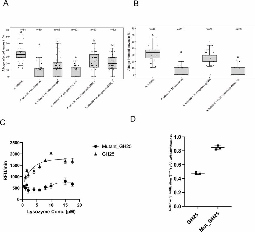
Research article                                                                                                            Plant Biology

Figure 1. Infection assay of A. laibachii on A. thaliana. Addition of a bacterial SynCom reduces the infection symptoms of A. laibachii at
14 days post infection. Those symptoms can be almost abolished by spraying MbA to the plant, independently of the presence of the bacterial
community. Infections were performed in six individual replicates with 12 technical replicates. N indicates the number of infected plants that were
scored for symptoms. An analysis of variance (ANOVA) model was used for pairwise comparison of the conditions, with Tukey’s HSD test to determine
differences among them. Different letters indicate significant differences (p-values
Research article                                                                                                 Plant Biology

Table 1. Comparison of genomes and genomic features of known pathogenic and anamorphic Ustilaginomycetes.
                                  MbA     U. bromivora S. scitamineum S. reilianum U. maydis U. hordei M. pennsylvanicum A. flocculosa
Assembly statistics
Total contig length (Mb)          18.3                    19.5             18.2     19.7       20.7       19.2               23.2
Total scaffold length (Mb)                20.5            19.6             18.4     19.8       21.15      19.2               23.3
Average base coverage             50     154            30              20      10        25        339               28
N50 contig (kb)                   705.1                   37.6             50.3     127.4      48.7       43.4               38.6
N50 scaffold length (kb)                  877             759.2            738.5    817.8      307.7      121.7              919.9
Chromosomes                       21      23                               23       23         23
GC-content (%)                    60.9    52.4            54.4             59.7     54         52         50.9               65.1
Coding (%)                        62.8    54.4            57.8             62.6     56.3       54.3       54                 66.3
Coding sequence
Percentage CDS (%)                69.5    59.8            62               65.9     61.1       57.5       56.6               54.3
Average gene size (bp)            1935    1699            1819             1858     1836       1708       1734               2097
Average gene density (gene/kb)    0.36    0.35            0.34             0.37     0.34       0.34       0.33               0.30
Protein-coding genes              6653    7233            6693             6648     6786       7113       6279               6877
Exons                             11,645 11,154           10,214           9776     9783       10,907     9278               19,318
Average exon size (bp)            1091    1101            1191             1221     1230       1107       527                658
Exons/gene                        1.75    1.5             1.5              1.47     1.44       1.53       1.48               2.8
tRNA genes                        150     133             116              96       111        110        126                176
Noncoding sequence
Introns                           9333    3921            3521             3103     2997       3161       2999               12,427
Introns/gene                      1.40    0.54            0.53             0.47     0.44       0.44       0.48               1.81
Average intron length (base)      163     163             130.1            144      142        141        191.4              141
Average intergenic distance (bp) 769      1054            1114             929      1127       1186       1328               1273
Secretome
Protein with signal peptide       559                     622              632      625        538        419                622
Secreted without TMD              380                                               467                                      737
– with known domain               260                                               264                                      554

[Brefort et al., 2014; Nadal and Gold, 2010].

                                       Also the third major recombination event, affecting MbA contig 8, changes the genomic context
                                    genes encoding essential virulence factors in U. maydis (stp1 and pit1/2), as well as the A mating
                                    type locus, which is important for pheromone perception and recognition of mating partners
                                    (Bölker et al., 1992). Based on the strong antibiotic activities of MbA, we mined the genome of
                                    MbA for the presence of secondary metabolite gene clusters. Using AntiSMASH, we were able to
                                    predict 13 of such clusters, of which three can be assigned to terpene synthesis, three contain nonri-
                                    bosomal peptide synthetases, and one cluster has a polyketide synthase as backbone genes (Fig-
                                    ure 3—figure supplement 1). Interestingly, the secondary metabolite cluster that is involved in the
                                    production of the antimicrobial metabolite ustilagic acid in other Ustilaginomycetes is absent in
                                    MbA (Figure 3—figure supplement 1B). On the contrary, we could identify three MbA-specific
                                    metabolite clusters that could potentially be involved in the antibacterial activity of MbA (Figure 3—
                                    figure supplement 1C).
                                       A previous genome comparison of the related Ustilaginales yeast A. flocculosa with U. maydis
                                    concluded that this anamorphic strain had lost most of its effector genes, reflecting the absence of a
                                    pathogenic stage in this organism (Lefebvre et al., 2013). In contrast, MbA contains 1:1 homologs
                                    of several known effectors with a known virulence function in U. maydis (Table 2). We previously
                                    found that Moesziomyces sp. possess functional homologues of the pep1 gene, a core virulence
                                    effector of U. maydis (Sharma et al., 2019), suggesting that such anamorphic yeasts have the

Eitzen et al. eLife 2021;10:e65306. DOI: https://doi.org/10.7554/eLife.65306                                                         5 of 23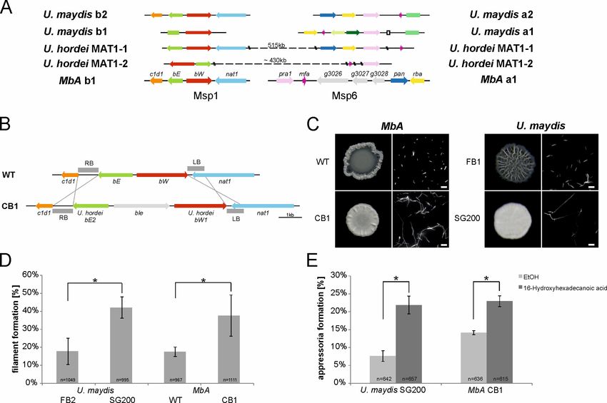
Research article                                                                                                         Plant Biology

Figure 2. Circos comparison of MbA and U. maydis chromosome structure (A). We highlighted potential secondary metabolite clusters, secreted
proteins, and gene predictions on both strands (±). (B) Homology-based comparisons identified three chromosomal recombination events, which
affects the MbA contigs 2, 6, and 8.
The online version of this article includes the following figure supplement(s) for figure 2:
Figure supplement 1. Genome comparison of MbA and Moesziomyces antarctica T-34.

                                    potential to form infectious filamentous structures by means of sexual reproduction (Kruse et al.,
                                    2017). To assess the potential virulence activity of MbA effector homologs, we expressed the homo-
                                    log of the U. maydis core effector Pep1 in an U. maydis pep1 deletion strain (SG200D01987). This
                                    resulted in complete restoration of U. maydis virulence, demonstrating that MbApep1 encodes a
                                    functional effector protein (Figure 3—figure supplement 2).
                                       A hallmark of the U. maydis genome structure is the presence of large clusters with effector
                                    genes, the expression of which is only induced during plant infection (Kämper et al., 2006). To
                                    assess the presence of potential virulence clusters in MbA, we compared all U. maydis effector gene
                                    clusters to the MbA genome, based on homology. This revealed that the 12 major effector clusters
                                    of U. maydis are present in MbA. However, while many of the clustered effector genes are dupli-
                                    cated in pathogenic smut fungi, MbA carries only a single copy of each effector gene. This results in
                                    the presence of ‘short’ versions of the U. maydis gene effector clusters (Figure 3—figure supple-
                                    ment 3). This gets particularly obvious for the biggest and most intensively studied virulence cluster
                                    of smut fungi, the effector cluster 19A (Schirawski et al., 2010; Brefort et al., 2014; Dutheil et al.,
                                    2016). In MbA, only 5 out of the 24 effector genes present in U. maydis are conserved in this cluster
                                    (Figure 3). Interestingly, some anamorphic yeasts like Kalmanozyma brasiliensis and A. flocculosa
                                    completely lost virulence clusters, while another non-pathogenic member of the Ustilaginales, Pseu-
                                    dozyma hubeiensis, shows an almost complete set of effectors when compared to U. maydis
                                    (Figure 3).

Eitzen et al. eLife 2021;10:e65306. DOI: https://doi.org/10.7554/eLife.65306                                                              6 of 23
Research article Plant Biology
Table 2. MbA proteins homologous to U. maydis effector genes with known virulence function.
Query E- Identity
Name Homologue cover value (%) U. maydis knockout phenotype Reference
g1653 UMAG_01987 82% 3-e56 60.96 Complete loss of tumor formation – blocked in early stages of Doehlemann et al.,
(Pep1) infection 2009
g1828 UMAG_01829 99% 0.0 71.57 Organ-specific effector – reduced virulence in seedling leaves Schilling et al., 2014
(Afu1)
g2626 UMAG_12197 98% 1e-48 60.16 Complete loss of tumor formation – blocked in early stages of Seitner et al., 2018
(Cce1) infection
g2765 UMAG_11938 100% 1e-73 93.44 Reduced in virulence Krombach et al., 2018
(Scp2)
g2910 UMAG_02475 32% 3e-42 60.71 Complete loss of tumor formation – blocked in early stages of Schipper, 2009
(Stp1) infection
g3652 UMAG_02239 43% 9e-11 54.90 Organ-specific effector – reduced virulence in seedling leaves Redkar et al., 2015
(See1)
g3113 UMAG_01375 (Pit2) * * * Complete loss of tumor formation – blocked in early stages of Doehlemann et al.,
infection 2011
g3279 UMAG_03274 10% 5e-20 70.11 Strong attenuation of virulence – reduced tumor size and number Ma et al., 2018
(Rsp3)
g5296 UMAG_05731 98% 3e-70 43.84 Reduced virulence Djamei et al., 2011
(Cmu1)
g6183 UMAG_06098 100% 0.0 81.85 Reduced virulence Ökmen et al., 2018
(Fly1)
g5835 UMAG_05302 87% 8e-24 37.81 Minor impact on tumor formation – reduced anthocyanin Brefort et al., 2014
(Tin2) biosynthesis
Genetic characterization of MbA
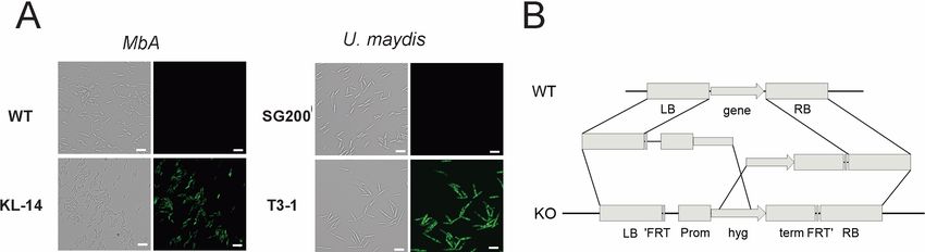
To perform reverse genetics in MbA, we established a genetic transformation system based on pro-
toplast preparation and polyethylene glycol (PEG)-mediated DNA transfer. In preliminary transfor-
mation assays, we expressed a cytosolic GFP reporter-gene under control of the constitutive o2tef-
Promoter (Figure 4A). For the generation of knockout strains, a split marker approach was used to
avoid ectopic integrations (Figure 4B). To allow generation of multiple knockouts, we used a selec-
tion marker-recycling system (pFLPexpC) that allows selection marker excision at each transforma-
tion round (Khrunyk et al., 2010).
We decided to apply the transformation system to study the MbA mating type loci in more detail.
Although phylogenetically closely related to U. hordei, which has a bi-polar mating system, MbA
owns a tetrapolar mating system whereby both mating type loci are physically not linked. This situa-
tion is similar to the mating type structure in the pathogenic smut U. maydis (Figure 4A). The a-
locus, which encodes a pheromone receptor system that is required for sensing and fusion of com-
patible cells, is located on contig 6. The b-locus can be found on contig 1. This multiallelic mating
locus contains two genes (b-East and b-West), which code for a pair of homeodomain transcription
factors. Upon mating of compatible cells, pathogenic and sexual development are triggered by a
heterodimeric bE/bW complex (Brefort et al., 2009). Since the MbA genome is completely
equipped with mating type genes, we first deployed a screen for potential mating partners. To this
end, we screened wild M. bullatus isolates to find a suitable mating partner, but we could not
observe any mating event (Figure 5—figure supplement 1). To test if MbA is able to undergo path-
ogenic differentiation in the absence of mating, we generated a self-compatible strain (CB1)
that carries compatible b-mating alleles: to construct the CB1 strain, we used compatible alleles of
the b-East and b-West genes of the barley smut U. hordei, a pathogen that is the phylogenetically
most closely related to MbA and amenable to reverse genetics. The native MbA locus was replaced
by the compatible U. hordei b-East and b-West gene alleles via homologous recombination
(Figure 5B).
Incubation of the MbA CB1 on charcoal plates led to the formation of aerial hyphae with the char-
acteristic fluffy phenotype of filamentous strains like the self-compatible, solopathogenic U. maydis
SG200 strain (Figure 5C). A second established method to induce filament formation in smuts is on
Eitzen et al. eLife 2021;10:e65306. DOI: https://doi.org/10.7554/eLife.65306 7 of 23Research article Plant Biology
Figure 3. Structure of the largest virulence cluster (Cluster 19A) in pathogenic smut fungi and anamorphic smut yeasts (marked with*). Colors indicate
genes with homology to each other: related gene families are indicated in orange, yellow, blue, green, and brown, whereas unique effector genes are
shown in gray. Genes encoding proteins without a secretion signal are shown in white (Brefort et al., 2014).
The online version of this article includes the following figure supplement(s) for figure 3:
Figure supplement 1. Secondary metabolite gene in the genome of MbA.
Figure supplement 2. Protein alignment of the core effector Pep1 (Hemetsberger et al., 2015) from different Ustilaginomycetes.
Figure supplement 3. Comparison of known virulence clusters (Kämper et al., 2006) between U. maydis and MbA.
hydrophobic parafilm (Mendoza-Mendoza et al., 2009). Quantification after 18 hr incubation of
MbA CB1 on parafilm resulted in the formation of filaments comparable to those of the U. maydis
SG200 strain (Figure 5D). While about 17% of MbA wild-type cells showed filaments, the CB1 strain
with compatible b-genes showed 38% filamentous growth.
Figure 4. Genetic transformation of MbA. (A) Stable transformants that express cytosolic GFP could be obtained by generating protoplasts with
Glucanex and ectopically integrating linear DNA fragments into the genome via polyethylene glycol (PEG)-mediated transformation. (B) Overview of the
split-marker approach that was used to generate deletion mutants via homologous recombination.
Eitzen et al. eLife 2021;10:e65306. DOI: https://doi.org/10.7554/eLife.65306 8 of 23Research article Plant Biology
Figure 5. The self-compatible MbA strain CB1. (A) MbA mating type genes, unlike the ones of U. hordei, can be found on two different chromosomes
similar to the tetrapolar mating type system of U. maydis. (B) To generate a self-compatible strain (CB1), the b-mating genes of U. hordei were
integrated at the native MbA b-locus. (C) Unlike the MbA wild-type strain (top left), strain CB1 (bottom left) shows a fluffy phenotype on charcoal plates
and filamentous growth. U. maydis haploid F1 strain (top right) and self-compatible SG200 strain (bottom right) were used as negative and positive
control, respectively. (D and E) Induction of filamentation and appressoria formation in strain CB1 was studied in three independent experiments. For
this around 1000 cells for filament formation and around 600 cells for appressoria formation were analyzed and error bars indicate standard error. After
incubation on a hydrophobic surface, both, filament and appressoria formation in strain CB1, were significantly different (*chi-squared test for
Independence – a = 0.0001) when compared to MbA wild type and similar to the level of the self-compatible U. maydis strain SG00. U. maydis haploid
F2 strain was used as negative control (Kämper et al., 2006). Scale bar: 20 mm. 16-HDD: 16-hydroxyhexadecanoic acid.
The online version of this article includes the following source data and figure supplement(s) for figure 5:
Source data 1. Appressoria quantification of MbA.
Figure supplement 1. Mating assays of MbA and different M. bullatus isolates.
Formation of appressoria is a hallmark of pathogenic development in smut fungi (Mendoza-
Mendoza et al., 2009). While the switch from yeast-like growth to filamentous development is the
first step in the pathogenic development of smut fungi, host penetration is accompanied by the for-
mation of a terminal swelling of infectious hyphae, termed ‘appressoria’. Induction of
appressoria formation in vitro can be induced by adding 100 mM of the cutin monomer 16-hydroxy-
hexadecanoic acid (HDD) to the fungal cells prior to cell spraying onto a hydrophobic surface (Men-
doza-Mendoza et al., 2009). In the absence of HDD, only about 8% of the U. maydis SG200 cells
and 14% of the MbA cells formed appressoria on parafilm 24 hr after spraying (Figure 5E). Addition
of 100 mM HDD resulted in a significant induction of appressoria in both U. maydis and MbA, dem-
onstrating that MbA does hold the genetic repertoire to form infection structures in vitro. Together,
the analysis of the recombinant CB1 strain indicates that MbA can sense pathogenesis-related
Eitzen et al. eLife 2021;10:e65306. DOI: https://doi.org/10.7554/eLife.65306 9 of 23Research article Plant Biology Figure 6. Transcriptome analysis of MbA. (A) Experimental setup used for the transcriptomic (RNA-Sequencing) analysis in MbA. (B) Venn diagrams showing differential regulated MbA genes after spraying of haploid cells onto the A. thaliana leaf surface. A total number of 801 genes were upregulated in response to A. laibachii in the presence and absence of bacterial SynCom. Of the 801 genes, 216 were upregulated in both conditions (Supplementary file 2). (C) Hierarchical clustering of the 18 A. laibachii-induced MbA genes that are predicted to encode secreted proteins. Of these genes, nine were selected as candidate microbe–microbe effector genes, based on their transcriptional upregulation and prediction to encode for extracellularly localized proteins. The online version of this article includes the following source data and figure supplement(s) for figure 6: Source data 1. Gene expression data of candidate effector genes. Figure supplement 1. Gene expression analysis of MbA in response to SynCom bacteria and A. laibachii. Figure 6 continued on next page Eitzen et al. eLife 2021;10:e65306. DOI: https://doi.org/10.7554/eLife.65306 10 of 23
Research article                                                                                                      Plant Biology

Figure 6 continued
Figure supplement 2. Gene ontology (GO) analysis of differentially regulated MbA genes.

                                    surface cues and produce penetration structures to a similar level as that seen for the pathogenic
                                    model organism U. maydis.

                                    Identification of microbe–microbe effector genes by RNA-Seq
                                    To study the transcriptomic response of MbA to different biotic interactions, RNA sequencing was
                                    performed. The MbA transcriptome was profiled in five different conditions (Figure 6A; cells in axe-
                                    nic culture versus cells on-planta, on-planta + SynCom, on-planta + A. laibachii, on-planta + SynCom
                                    + A. laibachii). Inoculations of A. thaliana leaves were performed as described above for A. laibachii
                                    infection assays (Figure 6A). For MbA RNA preparation, the epiphytic microbes were peeled from
                                    the plant tissue by using liquid latex (see Materials and methods section for details).
                                        The libraries of the 15 samples (five conditions in three biological replicates each) were generated
                                    by using a poly-A enrichment and sequenced on an Illumina HiSeq4000 platform. The paired end
                                    reads were mapped to the MbA genome by using Tophat2 (Kim et al., 2013). The analysis revealed
                                    that MbA cells on A. thaliana leaves (on-planta) downregulated 1300 and upregulated 1580 genes
                                    compared to cells in axenic culture (Figure 6B, Supplementary file 2). Differentially expressed
                                    genes were determined with the ‘limma’-package in R on ‘voom’ (Figure 6—figure supplement 1)
                                    using a False discovery rate threshold of 0.05 and log2FC > 0. A gene ontology (GO) terms analysis
                                    revealed that, among the downregulated genes, 50% were associated with primary metabolism (Fig-
                                    ure 6—figure supplement 2). In the two conditions in which A. laibachii was present, we observed
                                    upregulation of 801 genes. Among these genes, 411 genes were specific to co-incubation of MbA
                                    with A. laibachii and SynCom while 174 were specific to incubation with A. laibachii only. A set of
                                    216 genes was shared in both conditions (Figure 6B).
                                        In the presence of A. laibachii, mainly metabolism- and translation-dependent genes were upre-
                                    gulated, which might indicate that MbA can access a new nutrient source in the presence of A. laiba-
                                    chii (Figure 6—figure supplement 2). Among all A. laibachii-induced MbA genes, 18 genes encode
                                    proteins carrying a secretion signal peptide and having no predicted transmembrane domain
                                    (Figure 6C). After excluding proteins being predicted to be located in intracellular organelles, nine
                                    candidate genes remained as potential microbe–microbe-dependent effectors, i.e. MbA genes
                                    that are induced by A. laibachii, show no or low expression in axenic culture, and encode for putative
                                    secreted proteins (Figure 6C). Interestingly, four of these genes encode putative glycoside hydro-
                                    lases. Furthermore, two genes encode putative peptidases, one gene likely encodes an alkaline
                                    phosphatase and two encode uncharacterized proteins (Figure 6C).
                                        To directly test the eventual antagonistic function of those genes toward A. laibachii, we selected
                                    the two predicted glycoside hydrolases-encoding genes g5 and g2490 (GH43 and GH25) and the
                                    gene encoding the uncharacterized protein g5755 for gene deletion in MbA. The respective mutant
                                    strains were tested in stress assays to assess, whether the gene deletions resulted in general growth
                                    defects. Wild-type and mutant MbA strains were exposed to different stress conditions including
                                    osmotic stress (sorbitol, NaCl), cell wall stress (calcofluor, Congo-red), and oxidative stress (H2O2).
                                    Overall, in none of the tested conditions we observed a growth defect of the deletion mutants in
                                    comparison to wild-type MbA (Figure 7—figure supplement 1). To test an eventual impact of the
                                    deleted genes in the antagonism of the two microbes, the MbA deletion strains were each pre-inoc-
                                    ulated on A. thaliana leaves prior to A. laibachii infection. Deletion of g5 resulted in a significant but
                                    yet marginal increase of A. laibachii disease symptoms, while deletion of g5755 had no effect on A.
                                    laibachii. We therefore considered these two genes being not important for the antagonism of MbA
                                    toward A. laibachii. Strikingly, the MbA Dg2490 strain almost completely lost its biocontrol activity
                                    toward A. laibachii. This phenotype was reproduced by two independent g2490 deletion strains
                                    (Figure 7A). To check if this dramatic loss of microbial antagonism is specific to the deletion of
                                    g2490, in-locus genetic complementation of strain Dg2490_1 was performed via homologous recom-
                                    bination. The resulting strain MbA Dg2490/compl regained the ability to suppress A. laibachii

Eitzen et al. eLife 2021;10:e65306. DOI: https://doi.org/10.7554/eLife.65306                                                          11 of 23
Research article Plant Biology
infection, confirming that the observed phenotype specifically resulted from the deletion of the
g2490 gene (Figure 7B). Together, these results demonstrate that the biocontrol of the pathogenic
oomycete A. laibachii by the basidiomycete yeast MbA is determined by the secretion of a previ-
ously uncharacterized GH25 enzyme, which is transcriptionally activated specifically when both
microbes are co-colonizing the A. thaliana leaf surface.
Functional characterization of the secreted MbA hydrolase
To characterize the protein function of the GH25 encoded by MbA g2490, we were using Pichia pas-
toris for heterologous expression. The recombinant protein was tagged with polyhistidine tag for Ni-
NTA affinity purification. The purified protein was detected at an expected size of 27 kDa (Figure 7—
figure supplement 2). In addition, via site directed mutagenesis a mutated version of the protein
was generated, carrying a single amino exchange at the predicted active site (GH25_D124E). Both
active and mutated versions of the GH25 hydrolase were subjected to a quantitative lysozyme activ-
ity assay using the fluorogenic substrate Micrococcus lysodeikticus with commercial Hen egg-white
lysozyme (HEWL) as a control. We noticed a concentration-dependent increase in relative fluores-
cence unit (RFU)/min for the active GH25 in molar concentrations from 2 mM to 10 mM. Whereas, for
similar concentrations, mutated GH25 (GH25mut) showed no significant increase in RFU/min com-
pared to the active version. Commercial HEWL showed a steady increase in RFU/min from 1 mM to
5.5 mM concentrations (Figure 7C; Figure 7—figure supplement 2). Thus, the recombinant protein
represents a functional GH25 hydrolase with a lysozyme activity.
To test for a direct function of the GH25 lysozyme, we treated A. laibachii-infected Arabidop-
sis plants with the recombinant protein. To quantify the impact of GH25 treatment on A. laiba-
chii infection, we performed quantitative PCR to determine the relative A. laibachii biomass on
Arabidopsis in response to GH25. Strikingly, we observed a significant reduction of A. laibachii
colonization in leaves treated with the active GH25 lysozyme, while the mutated enzyme
GH25_D124E did not significantly influence infection (p-value ofResearch article Plant Biology Figure 7. A reverse-genetic approach to identify the MbA gene that is responsible for the suppression of A. laibachii infection. (A) Three candidate microbe–microbe effector genes (g5, g5755, and g2490) were deleted in MbA and deletion strains were individually inoculated on A. thaliana together with A. laibachii. Inoculation of two independent g2490 null strains (Dg2490_1; Dg2490_2) resulted in significant and almost complete loss of the biocontrol activity of MbA. While deletion of g5 resulted in a marginal reduction of disease symptoms at 14 days post infection, deletion of g5755 had no effect on A. laibachii. (B) Genetic complementation of the g2490 deletion restores the biocontrol activity to wild-type levels. Infections in (A) were performed in six, in (B) in three individual replicates. In each replicate 12 plants were infected. N indicates the number of infected plants that were scored for symptoms. Different letters indicate significant differences (p-values
Research article                                                                                                             Plant Biology

Figure 7 continued
sets (only Albugo) with A. laibachii treated with GH25 and A. laibachii treated with Mutant_GH25 by ddCT method. Unpaired t-test between GH25 and
Mutant_GH25 sets gave a p-value of
Research article                                                                                                      Plant Biology

                                    The presence of A. laibachii resulted in the induction of primary metabolism and biosynthesis path-
                                    ways, which might reflect enhanced growth of MbA in the presence of A. laibachii.
                                        A set of MbA genes encoding secreted hydrolases was induced by A. laibachii and one of these
                                    genes which encodes a putative GH25 hydrolase with similarity to Chalaropsis type lysozymes
                                    appeared to be essential for the biocontrol of A. laibachii. Initially discovered in the fungi Chalarop-
                                    sis sp., this group of proteins is largely present in bacteria as well as phages, for example the germi-
                                    nation-specific muramidase from Clostridium perfringens S40 (Chen et al., 1997). The bacterial
                                    muramidase, cellosyl from Streptomyces coelicolor (Rau et al., 2001), also belongs to the
                                    Chalaropsis type of lysozyme. These proteins are proposed to cleave the b-1,4-glycosidic bond
                                    between N-acetylmuramic acid (NAM) and N-acetylglucosamine (NAG) in the bacterial peptidogly-
                                    can. Specifically, the b-1,4 N,6-O-diacetylmuramidase activity allows the Chalaropsis type lysozyme
                                    to degrade the cell wall of Staphylococcus aureus, in contrast to the commercially available HEWL
                                    (Rau et al., 2001). Despite differences in structure and molecular weight from HEWL, the GH25 of
                                    MbA has lysozyme activity against the gram positive bacterium Micrococcus lysodeikticus in a fluoro-
                                    genic assay. This highlights the overall biochemical functionality of the recombinant glycoside hydro-
                                    lase. The glycoside Hydrolase 25 family is predicted to have an active site motif DXE that is highly
                                    conserved across the fungal kingdom (Figure 7—figure supplement 4). The structure of glycoside
                                    hydrolase family 25 from Aspergillus fumigates was characterized and the presence of N-terminal
                                    signal peptide was considered to indicate an extracellular secretion of the protein with possible anti-
                                    microbial properties (Korczynska et al., 2010). The role of the secreted hydrolase in the fungal king-
                                    dom is not completely explored yet. The presence of such hydrolases has in many cases been
                                    hypothesized to be associated with hyperparasitism of fungi parasitizing fungi (Hyde et al., 2019) or
                                    oomycetes parasitizing oomycetes (Horner et al., 2012). Our results might therefore indicate a cross
                                    kingdom hyperparasitism event between a fungus and an oomycete. Previous work on microbial
                                    communities has indicated that negative interactions stabilize microbial communities. Hyperparasit-
                                    ism is such a negative interaction with a strong eco-evolutionary effect on pathogen–host interac-
                                    tions and therefore on community stability (Parratt and Laine, 2016). MbA might therefore regulate
                                    A. laibachii infection and reduce disease severity. The qPCR evaluation of oomycete biomass
                                    strongly points toward the idea that A. laibachii is a direct target of antagonism for MBA. Since we
                                    observed reduced formation of A. laibachii in the presence of MbA, we also tested if the GH25 lyso-
                                    zyme would suppress zoospore germination. However, we could not detect a significant reduction
                                    of A. laibachii zoosporangia germination upon treatment with active GH25 lysozyme (Figure 7—fig-
                                    ure supplement 5), suggesting that the GH25 lysozyme interferes with A. laibachii at a later stage of
                                    infection. As A. laibachii has been shown to reduce microbial diversity (Agler et al., 2016), MbA
                                    might increase diversity through hyperparasitism of A. laibachii. At the same time this increased
                                    diversity might have caused the need for more secondary metabolites to evolve in the MbA genome
                                    to defend against niche competitors. Through its close association with A. laibachii, MbA could be a
                                    key regulator of the A. thaliana microbial diversity and therefore relevant for plant health beyond
                                    the regulation of A. laibachii infection.
                                        In conclusion, the secreted hydrolase we identified as a main factor of A. laibachii inhibition has
                                    great potential to act as antimicrobial agent. The isolated compound is not only valuable per se in
                                    an ecological context. It can further lay the grounds for exploring other microbial bioactive com-
                                    pounds that mediate inter-species and inter-kingdom crosstalk. A main goal of our future studies will
                                    be to understand on the mechanistic level, how the GH-25 suppresses A. laibachii, and at which
                                    developmental step the oomycete infection is blocked. It will be particularly interesting to elucidate
                                    if and how the GH25 enzyme activity directly interferes with the A. laibachii cell wall. While the
                                    canonical lysozyme substrate is found in bacterial cell walls, a detailed biochemical characterization
                                    of substrate specificity will be required to pinpoint potential target sites in the oomycete cell wall.
                                    Also, to the best of our knowledge there is no detailed information on the A. laibachii cell wall struc-
                                    ture and composition. One could speculate if GH25 activity directly affects cell wall integrity of A. lai-
                                    bachii, or if a modification of the cell wall structure interferes with pathogenic development, e.g. by
                                    interfering with cellular differentiation, blocking signal perception, or by triggering a host defense
                                    response.
                                        Since the GH-25 enzyme is well conserved among Ustilaginales including pathogenic species, it
                                    will also be tempting to elucidate whether the species-specific antagonism identified here is broadly
                                    conserved among Ustilaginales fungi and oomycetes. We further will investigate potential responses

Eitzen et al. eLife 2021;10:e65306. DOI: https://doi.org/10.7554/eLife.65306                                                          15 of 23
Research article Plant Biology
by the host plant and how this impacts A. laibachii growth upon MbA colonization. Functional inves-
tigation of these interactions can provide meaningful insights as to why certain yeasts prefer to colo-
nize specific environments. At the same time, it will be worth exploring how the basidiomycete
yeasts influence the bacterial major colonizers of the phyllosphere.
Materials and methods
Key resources table
Reagent type
(species) or Source or
resource Designation reference Identifiers Additional information
Gene g2490 This paper
(MbA_g2490)
Strain, strain DH5a Other Doehlemann lab
background
(Escherichia coli)
Genetic reagent KM71H-OCH Other Doehlemann lab
(Pichia pastoris)
Antibody Monoclonal 6x- Sigma (St. Louis; 1/10,000
His tag antibody Mississippi; USA)
Antibody Mouse IgG Thermo Fischer 1/3000
(Monoclonal) Scientific
(Waltham;
Massachusetts;
USA)
Recombinant pGAPza Invitrogen,
DNA reagent (plasmid) Carlsbad, CA,
USA
Sequence-based A. thaliana EF1- Ruhe et al., PCR primers AAGGAGGCTGCTGAGATGAA
reagent a:forward 2016
Sequence-based A. thaliana EF1- Ruhe et al., PCR primers TGGTGGTCTCGAACTTCCAG
reagent a:reverse 2016
Sequence-based Oomycete Ruhe et al., PCR primers ACTTTCAGCAGTGGATGTCTA
reagent internal 2016
transcribed
spacer (ITS) 5.8
s: forward
Sequence-based Oomycete Ruhe et al., PCR primers GATGACTCACTGAATTCTGCA
reagent internal 2016
transcribed
spacer (ITS) 5.8
s: reverse
Commercial EnzChek Invitrogen E22013 Lysozyme activity assay
assay or kit Lysozyme Assay
Kit
Chemical Trypan blue Sigma Aldrich CAS-Number: 72-57-1
compound, drug stain (No. 302643)
Strains and growth conditions
MbA wild-type strain was isolated from A. laibachii infected A. thaliana leaves [7]. Wild-type MbA (at
22˚) and U. maydis (at 28˚) strains were grown in liquid YEPS light medium and maintained on potato
dextrose agar plates. King’s B medium was used for culturing Syn Com bacterial members at 22˚. All
the strains were grown in a rotary shaker at 200 rpm. All the recipes for medium and solutions can
be found in Supplementary file 3. Stress assays for fungi: wild-type and mutant strains of MbA
grown to an optical density (600 nm) of 0.6–0.8 were centrifuged at 3500 rpm for 10 min and sus-
pended in sterile water to reach an OD of 1.0. Next, a dilution series from 100 to 10–4 was prepared
in sterile H2O. In the end, 5 ml of each dilution was spotted on CM plates supplemented with the
indicated stress agents. The plates were incubated for 2 days at 22˚C. Confrontation assays: at first,
Eitzen et al. eLife 2021;10:e65306. DOI: https://doi.org/10.7554/eLife.65306 16 of 23Research article Plant Biology
MbA and SynCom bacterial strains were grown to an O.D of 0.8–1. MbA cultures (10 ml) were
dropped in four quadrants of a potato dextrose agar plate, previously spread with a bacterial cul-
ture. Plates were incubated for 2–4 days at 22˚C.
Transformation of MbA and plasmid construction for generation of
knockout mutants
Fungal strains were grown in YEPSL at 22˚C in a rotary shaker at 200 rpm until an O.D. of 0.6 was
reached and centrifuged for 15 min at 3500 rpm. The cells were washed in 20 ml of SCS
(Supplementary file 3) and further centrifuged for 10 min at 3000 rpm, before being treated with 3
ml SCS solution with 20 mg/ml of Glucanex (Lysing Enzyme from Trichoderma harzianum, # L1412,
Sigma). After 20 min of incubation at room temperature, as cell wall lysis occurred, cold SCS was
added to the mixture and protoplasts spun down for 10 min at 2400 rpm. They were then washed
twice with SCS and resuspended with 10 ml STC (Supplementary file 3) to be centrifuged at 2000
rpm for 10 min. Finally, the pellet was dissolved in 500 ml STC and stored in aliquots of 50 ml at 80˚
C. Five micrograms of plasmid DNA along with 15 mg heparin was added to 50 ml protoplasts. After
incubation on ice for 10 min, STC/40% PEG (500 ml) was added to it and mixed gently by pipetting
up and down; this step was followed by another 15 min on ice. The transformation mix was added
to 10 ml of molten regeneration (reg) agar and poured over a layer of already solidified reg agar
containing appropriate antibiotic solution. For the bottom layer, we used 400 mg/ml hygromycin/8
mg/ml carboxin/300 mg/ml nourseothricin (NAT).
Plasmids were cloned using Escherichia coli DH5a cells (Invitrogen, Karlsruhe, Germany). Con-
struction of deletion mutants was performed by homologous recombination; the 5’ and 3’ flanking
regions of the target genes were amplified and ligated to an antibiotic resistance cassette (Käm-
per, 2004). The ligated fragment was subsequently transformed into MbA. Homologous integration
of the target gene was verified via PCR on the antibiotic resistant colonies. Oligonucleotide pairs for
knockout generation and verification can be found in Supplementary file 4. PCR amplification was
done using Phusion DNA polymerase (Thermo Scientific, Bonn, Germany), following the manufac-
turer’s instructions, with 100 ng of genomic DNA or cDNA as template. Nucleic acids were purified
from 1% TAE agarose gels using Macherey-Nagel NucleoSpin Gel and PCR Clean-up Kit.
Mating assay and generation of the self-compatible MbA strain CB1
Haploid strains of MbA were grown in liquid cultures, mixed, and drops arranged on PD plates with
charcoal to induce filament formation. Plate with the haploid U. maydis strains FB1 and FB2 and the
solopathogenic strain SG200 served as internal control.
The complete b-locus of the solopathogenic U. hordei strain DS200 was amplified (Figure 1—fig-
ure supplement 2) and inserted into the MbA b-locus by homologous recombination. The strain
obtained, known as compatible b1 (CB1), was tested positive by amplification of the right border
and left border areas with primers specific for the genomic locus and for the plasmid region. Addi-
tionally, two primers specific for the MbA bE and bW genes were chosen to amplify parts of the
native locus. To induce filament and appressoria formation in vitro we used a Moesziomyces YEPSL
culture at OD600 0.6–0.8. The cells were diluted to an OD600 of 0.2 in 2% YEPSL (for appressoria for-
mation 100 mM 16-hydroxyhexadecanoic acid [Sigma-Aldrich] or 1% ethanol was added) and sprayed
the yeast like cells on parafilm which mimics the hydrophobic plant surface. After 18 hr of incubation
at 100% humidity the number of cells grown as filaments (or generating appressoria) was deter-
mined relative to the total number of total cells by using a light microscope.
Arabidopsis thaliana leaf infections and quantification of albugo
biomass quantification by qPCR
Sterilized Arabidopsis thaliana seeds were subjected to cold treatment for 7 days and sown on 1/2
strength Murashige Skoog (MS) medium (Supplementary file 3). The MS plates are directly trans-
ferred to growth chambers having 22˚C on a short-day period (8 hr light) with (33–40%) humidity
and grown for 4 weeks before inoculation. Overnight liquid cultures of MbA and SynCom bacterial
strains were grown to an OD600 of 0.6. The cultures were spun down at 3500 rpm for 10 min and the
pellets dissolved in MgCl2. Five hundred microliters of each culture was evenly sprayed on 3-week
old A. thaliana seedlings using airbrush guns. Two days later, a spore solution of A. laibachii was
Eitzen et al. eLife 2021;10:e65306. DOI: https://doi.org/10.7554/eLife.65306 17 of 23Research article Plant Biology
then sprayed on the seedlings following the protocol of Ruhe et al., 2016. Two weeks later, the dis-
ease symptoms on the leaves were scored as a percentage between infected and non-infected
leaves.
Four weeks old A. thaliana seedlings on MS plates were sprayed with A. laibachii as a control and
GH25+ A. laibachii and Mut_GH25+A. laibachii as treatments. After 10 dpi, the seedlings were har-
vested, frozen in liquid nitrogen, and kept at 80˚C. For DNA extraction, the frozen plant material
was ground into a fine powder with mortar and pestle and treated with extraction buffer (50 mM
Tris pH 8.0, 200 mM NaCl, 0.2 mM ethylenediaminetetraacetic acid [EDTA], 0.5% SDS, 0.1 mg/ml
proteinase K [Sigma–Aldrich]). This was followed by centrifugation after the addition of one volume
phenol/chloroform/isoamylalkohol, 25:24:1 (Roth). The top aqueous layer was removed and added
to one volume of isopropanol to precipitate the nucleic acids. DNA pellet obtained after centrifuga-
tion was washed with 70% EtOH and finally dissolved in 50 ml nuclease-free water. For qPCR meas-
urements, 10 ml of GoTaq qPCR 2 Master Mix (Promega, Waltham, Madison, USA), 5 ml of DNA
(~50 ng), and 1 ml of forward and reverse primer (10 mM) up to a total volume 20 ml were used. Sam-
ples were measured in triplicates in a CFX Connect real-time PCR detection system (Bio-Rad) follow-
ing the protocol of Ruhe et al., 2016. Amount of A. laibachii DNA was quantified using the
following oligonucleotide sequences: A. thaliana EF1-a: 50 -AAGGAGGCTGCTGAGATGAA-30 , 50 -
TGGTGGTCTCGAACTTCCAG-30 ; Oomycete internal transcribed spacer (ITS) 5.8 s: 50 -ACTTTCAG-
CAGTGGATGTCTA-30 , 50 -GATGACTCACTGAATTCTGCA-30 . Cq values obtained in case of the
oomycete DNA amplification was normalized to A. thaliana DNA amplicon and then the difference
between control (only Albugo) and treatment (Albugo+ GH25/Mut_GH25) was calculated by ddCq.
The relative biomass of Albugo was analyzed by the formula (2 ddCq). Each data point in the graph
represents three independent biological replicates.
Nucleic acid methods
RNA-Extraction of Latex-peeled samples: Four weeks old A. thaliana plants were fixed between two
fingers and liquid latex was applied to the leaf surface by using a small brush. The latex was dried
using the cold air option of a hair dryer, carefully peeled off with a thin tweezer, and immediately
frozen in liquid nitrogen. Afterwards, the frozen latex pieces were grinded with liquid nitrogen and
the RNA was isolated by using Trizol Reagent (Invitrogen, Karlsruhe, Germany) according to the
manufacturer’s instructions. Turbo DNA-Free Kit (Ambion, Life Technologies, Carlsbad, California,
USA) was used to remove any DNA contamination in the extracted RNA. Synthesis of cDNA was per-
formed using First Strand cDNA Synthesis Kit (Thermo Fischer scientific, Waltham, Massachusetts,
USA) according to recommended instruction starting with a concentration of 10 mg RNA. QIAprep
Mini Plasmid Prep Kit (QIAGEN, Venlo, The Netherlands) was used for isolation of plasmid DNA
from bacteria after the principle of alkaline lysis. Genomic DNA was isolated using phenol–chloro-
form extraction protocol (Kämper et al., 2006).
RT-qPCR oligonucleotide pairs were designed with Primer3 Plus. The oligonucleotide pairs were
at first tested for efficiency using a dilution series of genomic DNA. The reaction was performed in a
Bio-Rad iCycler system using the following conditions: 2 min at 95˚C, followed by 45 cycles of 30 s at
95˚C, 30 s at 61˚C, and generation of melting curve between 65˚C and 95˚C.
Bioinformatics and computational data analysis
Sequence assembly of MbA strains was performed using the HGAP pipeline (Pacific Biosciences).
MbA genome was annotated with the Augustus software tool. Secretome was investigated using
SignalP4.0. Analysis of functional domains in the secreted proteins was done by Inter-Pro Scan. Anti-
Smash was used to predict potential secondary metabolite clusters. RNA sequencing was done at
the Cologne Center for Genomics (CCG) by using a poly-A enrichment on an Illumina HiSeq4000
platform. The achieved paired end reads were mapped to the MbA and A. thaliana TAIR10 genome
by using Tophat2 (Kim et al., 2013). RNA-Seq reads of MbA axenic cultures were used to generate
exon and intron hints and to start a second annotation with Augustus. Heat maps were performed
using the heatmap.2 function of the package gplots (version 3.0.1) in R-studio (R version 3.5.1). An
analysis of variance (ANOVA) model was used for pairwise comparison of the conditions, with
Tukey’s HSD test to determine the significant differences among them (p-valuesResearch article Plant Biology
Heterologous protein production and GH25 activity assay
The Pichia pastoris KM71H-OCH gene expression system was used to produce MBA_GH25 domain
tagged with an N-terminal Polyhistidine tag (6xHis) and a C-terminal peptide containing the c-myc
epitope and a 6xHis tag. The His-MspGH25 cloned into pGAPZaA vector (Invitrogen, Carlsbad, CA,
USA) under the control of a constitutive promotor with an a-factor signal peptide for secretion.
Expression and purification of recombinant proteins were performed according to manufacturer’s
instructions (Invitrogen Corporation, Catalog no. K1710-01): YPD medium supplemented with 100
mg ml 1 zeocin was used for initial growth of P. pastoris strains at 28˚C and 200 rpm (for liquid cul-
tures). Production of the recombinant protein was performed in 1 L buffered (100 mM potassium
phosphate buffer, pH 6.0) YPD medium with 2% sucrose at 28˚C for 24 hr with 200 rpm shaking.
Next the protein was subjected to affinity purification with a Ni-NTA-matrix according to manufac-
turer’s instructions (Ni-Sepharose 6 Fast-Flow, GE-Healthcare; Freiburg, Germany). After purification,
the His-MspGH25 protein was dialyzed in an exchange buffer (0.1 M NaPi, 0.1 M Nacl, pH = 7.5).
The purified protein was kept in 100 ml aliquots at 4˚C.
Site-directed mutagenesis was performed on pGAPZa-His- MspGH25 vector according to the
instructions of the QuikChange Multi Site-Directed Mutagenesis Kit (Agilent Technologies, Santa
Clara, United States) with primers targeting nucleotides of the active site of GH25.
Purified glycoside hydrolase of MBA from P. pastoris was quantified according to a sensitive fluo-
rescence-based method using Molecular Probes EnzChek Lysozym-Assay-Kit (ThermoFisher Scien-
tific, Katalognummer: E22013). DQ lysozyme substrate (Micrococcus lysodeikticus) stock suspension
(1.0 mg/ml) and 1000 units/ml HEWL stock solution were prepared according to the
manufacturer’s instruction. Molar concentration of the HEWL stock solution was calculated using the
following website (https://www.bioline.com/media/calculator/01_04.html) and was found to be 11
mM. Protein concentration of MspGH25 both active and mutated version was measured in the Nano-
drop 2000c spectrophotometer (Thermo Fischer scientific, Waltham, Massachusetts, USA) according
to the manufacturer’s instructions using 100 ml of sample after using 100 ml of the appropriate buffer
as a blank control in glass cuvette. The molar concentrations of recombinant proteins were also cal-
culated as above.
At the start of the reaction 50 ml of the DQ lysozyme substrate working suspension was added to
each microplate well containing reaction buffer with either HEWL (in molar concentrations ranging
from 0.1 to 5.5 mM) or MspGH25 (in molar concentration from 0.5 to 17.5 mM). Fluorescence inten-
sity of each reaction was measured every 5 min to follow the kinetic of the reaction at 37˚C for 60
min, using fluorescence microplate reader with fluorescein filter Tecan Infinite 200 Pro plate reader
(Tecan Group Ltd., Männendorf, Switzerland). Digestion products from the DQ lysozyme substrate
have an absorption maximum at ~494 nm and a fluorescence emission maximum at ~518 nm.
Acknowledgements
This work was funded through by the Deutsche Forschungsgemeinschaft (DFG, German Research
Foundation) under Germany’s Excellence Strategy EXC-2048/1, Project ID 390686111, and the DFG
priority program SPP2125 ‘DECRyPT’. We are grateful to Marco Thines for generously providing M.
bullatus wild-type strains. We thank Libera Lo Presti for critically reading the manuscript and helpful
comments and suggestion.
Additional information
Funding
Funder Grant reference number Author
Deutsche Forschungsge- SPP 2125 DECRyPT Katharina Eitzen
meinschaft Priyamedha Sengupta
Deutsche Forschungsge- EXC-2048/1 Katharina Eitzen
meinschaft
Deutsche Forschungsge- Project ID 390686111 Katharina Eitzen
meinschaft
Eitzen et al. eLife 2021;10:e65306. DOI: https://doi.org/10.7554/eLife.65306 19 of 23You can also read