In vivo proteomic mapping through GFP-directed proximity-dependent biotin labelling in zebrafish - eLife
←
→
Page content transcription
If your browser does not render page correctly, please read the page content below
TOOLS AND RESOURCES

                                   In vivo proteomic mapping through GFP-
                                   directed proximity-dependent biotin
                                   labelling in zebrafish
                                   Zherui Xiong1, Harriet P Lo1, Kerrie-Ann McMahon1, Nick Martel1, Alun Jones1,
                                   Michelle M Hill2, Robert G Parton1,3*, Thomas E Hall1*
                                   1
                                    Institute for Molecular Bioscience, The University of Queensland, Brisbane,
                                   Australia; 2QIMR Berghofer Medical Research Institute, Herston, Australia; 3Centre
                                   for Microscopy and Microanalysis, The University of Queensland, Brisbane, Australia

                                   Abstract Protein interaction networks are crucial for complex cellular processes. However, the
                                   elucidation of protein interactions occurring within highly specialised cells and tissues is
                                   challenging. Here, we describe the development, and application, of a new method for proximity-
                                   dependent biotin labelling in whole zebrafish. Using a conditionally stabilised GFP-binding
                                   nanobody to target a biotin ligase to GFP-labelled proteins of interest, we show tissue-specific
                                   proteomic profiling using existing GFP-tagged transgenic zebrafish lines. We demonstrate the
                                   applicability of this approach, termed BLITZ (Biotin Labelling In Tagged Zebrafish), in diverse cell
                                   types such as neurons and vascular endothelial cells. We applied this methodology to identify
                                   interactors of caveolar coat protein, cavins, in skeletal muscle. Using this system, we defined
                                   specific interaction networks within in vivo muscle cells for the closely related but functionally
                                   distinct Cavin4 and Cavin1 proteins.

*For correspondence:
r.parton@imb.uq.edu.au (RGP);
                                   Introduction
thomas.hall@imb.uq.edu.au (TEH)    The understanding of the biological functions of a protein requires detailed knowledge of the mole-
                                   cules with which it interacts. However, robust elucidation of interacting proteins, including not only
Competing interests: The           strong direct protein-protein interactions, but also weak, transient or indirect interactions is challeng-
authors declare that no
                                   ing. Proximity-dependent biotin labelling (BioID) using genetically engineered biotin ligases has
competing interests exist.
                                   emerged as a novel approach for studying protein-protein interactions and the subcellular proteome
Funding: See page 20               in living cells (Roux et al., 2012; Kim et al., 2016; Branon et al., 2018; Ramanathan et al., 2018).
Received: 05 November 2020         When fused to a protein of interest (POI) and expressed in cells, the promiscuous biotin ligases cova-
Accepted: 15 February 2021         lently attach biotin to all proteins within a 10 nm radius, which can be subsequently isolated by
Published: 16 February 2021        streptavidin purification and identified by mass spectrometry. Compared with traditional affinity
                                   purification with protein-specific antibodies or affinity purification tags, the BioID method has the
Reviewing editor: Lilianna
                                   advantage of being able to capture weak and transient interactions. In addition, unlike conventional
Solnica-Krezel, Washington
University School of Medicine,
                                   methods such as affinity purification, where stringent extraction conditions may disrupt protein-pro-
United States                      tein interactions, the BioID method does not require proteins to be isolated in their native state.
                                   Therefore, harsh protein extraction and stringent wash conditions can be applied, which can improve
   Copyright Xiong et al. This
                                   solubilisation of membrane proteins and reduce false positives (Varnaitė and MacNeill, 2016;
article is distributed under the
                                   Gingras et al., 2019).
terms of the Creative Commons
Attribution License, which             The BioID method has been widely applied in cell biology to study protein-protein interactions in
permits unrestricted use and       cultured cells, providing valuable information for building protein interaction networks. However,
redistribution provided that the   the reductionist in vitro applications described to date, while powerful in their own right, lack the
original author and source are     complexity and context to address phenomena that can only be modelled in vivo, for example the
credited.                          differentiation of specialised cell types such as those found in muscle, the nervous system, and

Xiong et al. eLife 2021;10:e64631. DOI: https://doi.org/10.7554/eLife.64631                                                           1 of 24
Tools and resources                                                                                                   Cell Biology

                                   vasculature. The most recent generation of biotin ligases has been applied in vivo in invertebrate
                                   models; flies (Drosophila melanogaster) and worms (Caenorhabditis elegans) as well as plants (Arabi-
                                   dopsis and Nicotiana benthamiana) (Branon et al., 2018; Mair et al., 2019; Zhang et al., 2019).
                                   Until now, however, the applicability of BioID has been limited by the necessity to genetically tag
                                   each POI directly with a biotin ligase and generate transgenic organisms. Here, we describe a more
                                   versatile approach to the in vivo application of BioID in a vertebrate model organism, the zebrafish.
                                   Instead of directly fusing the biotin ligase to a POI, we developed a modular system for GFP-
                                   directed proteomic mapping by combining BioID with a GFP-binding nanobody (Hamers-
                                   Casterman et al., 1993; Rothbauer et al., 2008; Tang et al., 2015; Ariotti et al., 2015a). This sys-
                                   tem couples the power of the BioID system with the ability to use existing GFP-tagged transgenic
                                   zebrafish lines for proteomic mapping between different tissues and/or different proteins. We dem-
                                   onstrate the application of this system in screening for proteins associated with the caveolar cast
                                   proteins, Cavin1 and Cavin4, in differentiated skeletal muscle which has, to date, been difficult to
                                   achieve in culture. These analyses reveal proteins and pathways that are both overlapping and spe-
                                   cific to Cavin1 and Cavin4.

                                   Results
                                   Proximity biotinylation in live zebrafish embryos
                                   We first tested the ability of a number of biotin ligases to catalyse protein biotinylation in live zebra-
                                   fish embryos. Initial attempts using BirA* or BioID2 biotin ligases in vivo in zebrafish were unsuccess-
                                   ful and resulted in no detectable biotinylation in zebrafish embryos as assessed by streptavidin
                                   western blotting (Figure 1—figure supplement 1). In recent years, the new bioID biotin ligases,
                                   BASU, TurboID, and miniTurbo, have been developed and showed greatly improved catalytic effi-
                                   ciency and enhanced proximity labelling in cultured cells (Branon et al., 2018; Ramanathan et al.,
                                   2018). We therefore tested their ability to catalyse protein biotinylation in live zebrafish embryos.
                                   Untagged, cytoplasmically localised biotin ligases were transiently expressed in zebrafish embryos
                                   by RNA injection, and the level of protein biotinylation was assessed using streptavidin immunoblot
                                   analysis (experimental regimen illustrated in Figure 1A; Ramanathan et al., 2018; Branon et al.,
                                   2018). The biotin ligases were fused to an EGFP tag for selection of transgene expressing embryos,
                                   and a Myc tag for detection by western blot. At 24 hr post injection, the GFP-positive embryos were
                                   dechorionated before incubation in biotin-supplemented media for a further 18 hr (Figure 1A). Total
                                   protein extracts from fish embryos were then subjected to SDS-PAGE and streptavidin immunoblot-
                                   ting (Figure 1B). TurboID injected embryos exhibited the strongest biotinylation of endogenous pro-
                                   teins among the three new biotin ligases with 500 mM biotin incubation. Of note, BASU and
                                   miniTurbo were expressed at a lower level than TurboID despite equal amount of RNA injection.
                                   While we did not explore the underlying reasons for this, greater instability of miniTurbo has been
                                   reported previously and other biotin ligases have shown poor expression in cell cultures (May et al.,
                                   2020; Branon et al., 2018). Therefore, we chose TurboID for all subsequent experiments. Note the
                                   two prominent bands consistently detected around 70 and 135 kDa in all samples likely represent
                                   endogenously biotinylated proteins (Housley et al., 2014; Ahmed et al., 2014).
                                       To visualise TurboID-catalysed biotinylation in situ, TurboID-expressing embryos were stained
                                   with NeutrAvidin-DyLight 550 after biotin incubation (Figure 1C). The mosaic expression of Tur-
                                   boID-GFP in the muscle fibres, as well as expression in the yolk, corresponded with strong NeutrAvi-
                                   din staining. The mRNA injections frequently gave rise to differing levels of expression between
                                   individual muscle cells within the same animal. Therefore, muscle fibres with little or no TurboID-GFP
                                   expression served as an internal negative control.
                                       Biotin concentration and incubation time are two crucial factors that affect biotin ligase efficiency
                                   in cultured cells (Roux et al., 2012; Kim et al., 2016; Branon et al., 2018; Ramanathan et al.,
                                   2018). To achieve the most effective experimental conditions for TurboID application in zebrafish,
                                   we sought to optimise these parameters. From our initial experiments with BirA*, we knew that
                                   zebrafish embryos are able to tolerate a biotin concentration as high as 800 mM with no obvious
                                   morphological abnormalities (Figure 1—figure supplement 2). To determine the optimal biotin con-
                                   centration for TurboID in zebrafish, TurboID-expressing embryos were incubated in embryo medium
                                   containing biotin concentration ranging from 0 to 750 mM for 18 hr, followed by lysis and

Xiong et al. eLife 2021;10:e64631. DOI: https://doi.org/10.7554/eLife.64631                                                           2 of 24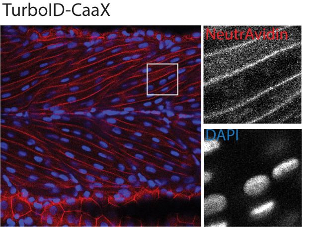
Tools and resources Cell Biology
A
Myc Biotin Ligase eGFP pA
24 hpf
BASU
TurboID
Incubate embryos in medium
MiniTurbo
Transient expression Chorion removal supplemented with biotin
3 versions of biotin ligase by mRNA injection by pronase
Embryo solubilisation Embryo permeabilisation
and Western Blot and NeutrAvidin staining
B C
SU o
o
BA rb
rb
m ID
m ID
iTu
iTu
o
o
SU
rb
rb
in
in
T
T
BA
NeutrAvidin-DyLight 550 Merge
Tu
Tu
W
W
eGFP
Biotin (μM): 0 500 50 50 50 500 500 500
kDa
180 endogenous
TurboID-eGFP Trunk
135
100 biotinylated
Streptavidin-HRP
75 proteins
63
48 yolk
35 100 µm 100 µm
25
Trunk
17
Control
IB: Actin
yolk
IB: myc
100 µm 100 µm
D E
WT TurboID-eGFP WT TurboID-eGFP WT
Biotin (μM): 0 750 0 50 100 250 500 750 Incubation (h): 0 0 1 2 4 6 18 18
kDa kDa
180 180
135 135
100 100
Streptavidin-HRP
75
75
Streptavidin-HRP
63
63
48
48
35
35
25
25
17
48 48
IB: Actin IB: Actin
63
IB: Myc 63 IB: Myc
Figure 1. Testing and optimising biotin ligases: BASU, TurboID, and miniTurbo. (A) A schematic overview of the workflow. The BASU/TurboID/
MiniTurbo was transiently expressed in zebrafish embryos by RNA injection. Chorion-removed fish embryos with green fluorescence were selected for
incubation in biotin supplemented embryo media for 18 hr. After biotin incubation, embryos were analysed by western blotting and
immunofluorescence. (B) The streptavidin-HRP blot showing biotinylated proteins in two dpf zebrafish embryos expressing eGFP-tagged BASU,
Figure 1 continued on next page
Xiong et al. eLife 2021;10:e64631. DOI: https://doi.org/10.7554/eLife.64631 3 of 24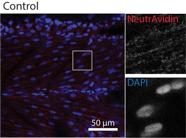
Tools and resources                                                                                                               Cell Biology

Figure 1 continued
TurboID, and miniTurbo. Fish embryos were incubated in biotin concentrations of 50 or 500 mM biotin for 18 hr before embryo solubilisation and
Western blot analysis. Actin immunoblot (IB:Actin) serves as a loading control; the anti-Myc immunoblot (IB:Myc) reflects the protein level of each biotin
ligases; each sample is a pool of 30 embryos. (C) Representative images of NeutrAvidin staining of biotinylated proteins in 2 dpf zebrafish embryo
transiently expressing TurboID-eGFP. Fish muscle and yolk were outlined with dashed lines. White arrows indicate muscle fibres expressing TurboID-
eGFP. n = 6. Scale bar denotes 100 mm (D and E) Dependency of TurboID activity on biotin concentration and incubation time. Zebrafish embryos
transiently expressing TurboID-eGFP were incubated with embryo media containing 0 to 750 mM biotin for 18 hr (D) or incubated with 500 mM biotin for
0 to 18 hr (E) before protein extraction and immunoblot analysis with streptavidin-HRP, anti-Actin and anti-Myc antibodies; each sample is a pool of 30
embryos. For immunoblots showing the biotinylation of BioID and BioID2 in zebrafish embryos see Figure 1—figure supplement 1. For biotin
tolerance of zebrafish embryos see Figure 1—figure supplement 2. For original western blot images see Figure 1—source data 1.
The online version of this article includes the following source data and figure supplement(s) for figure 1:
Source data 1. Raw images of blots.
Figure supplement 1. Testing BioID and BioID2 in transgenic zebrafish.
Figure supplement 2. Determining biotin toxicity and tolerance in zebrafish embryos.

                                     streptavidin immunoblotting. Weak labelling could be seen with 50 mM biotin, increasing through
                                     250 mM, with the strongest labelling at concentration of 500 and 750 mM (Figure 1D). Unlike its
                                     application in cultured cells and yeast (Branon et al., 2018), TurboID did not produce detectable
                                     exogenous biotinylation without the addition of biotin (Figure 1D). This provides the opportunity for
                                     temporal resolution by addition of exogenous biotin at specific developmental stages. Unexpect-
                                     edly, the anti-Myc immunoblot showed that a higher biotin concentration resulted in more TurboID
                                     in the total protein extracts (Figure 1D). Concomitantly, the addition of exogenous biotin did not
                                     change the level of endogenous biotinylated proteins in the WT embryos (Figure 1D).
                                        In mammalian cell culture, a 10-min biotin incubation with TurboID is sufficient to visualise biotiny-
                                     lated proteins by immunoblotting and to perform analysis of different organellar proteomes
                                     (Branon et al., 2018). However, we did not observe rapid biotinylation in zebrafish within the first 2
                                     hr of biotin incubation (Figure 1E). TurboID-induced biotin labelling was only weakly detected after
                                     4–6 hr incubation and adequate biotinylation was only detected after overnight incubation (18 hr).

                                     In vivo proximity biotinylation targeted to a specific subcellular region
                                     or a protein of interest
                                     Next, we tested the spatial resolution of TurboID-catalysed biotinylation in zebrafish when TurboID
                                     was targeted to a specific subcellular region and to a POI. We tagged TurboID with a nuclear local-
                                     isation signal (NLS), a plasma membrane localisation motif (CaaX), the transmembrane protein
                                     CD44b and the muscle T-tubule enriched membrane protein Cavin4b (Figure 2A). After biotin treat-
                                     ment, the TurboID fusion proteins produced a biotinylation pattern corresponding to the appropri-
                                     ate subcellular location of targeting sequences/proteins in zebrafish embryos (Figure 2B). The
                                     spatial resolution of the biotin labelling was remarkable as even the T-tubule structure, which is diffi-
                                     cult to resolve in fixed embryos, was clearly visible by NeutrAvidin staining in the embryos express-
                                     ing Cavin4b-TurboID. Furthermore, the biotinylated protein derived from each TurboID construct
                                     gave rise to a unique barcode of protein bands on the streptavidin blot, indicative of proteins spe-
                                     cific to each corresponding subcellular compartment (Figure 2C). These results demonstrated that
                                     TurboID was able to specifically label a selective subpopulation of endogenous proteins when tar-
                                     geted to a specific subcellular region or protein in zebrafish embryos. Moreover, the TurboID-bioti-
                                     nylated proteins were recoverable from crude fish lysates by affinity purification with streptavidin-
                                     conjugated beads (Figure 2D), ready for downstream applications such as identification by mass
                                     spectrometry.
                                         Overall, TurboID showed robust biotin labelling with high spatial resolution in zebrafish embryos.
                                     These properties rendered it suitable for pursuing in vivo proteomic analyses.

                                     Conditionally stabilised GFP-binding protein (dGBP) is able to target
                                     GFP-tagged proteins in zebrafish
                                     Although we were able to achieve proximity-dependent biotin labelling in zebrafish embryos tran-
                                     siently expressing TurboID by mRNA injection, this method requires the direct injection of a large
                                     number of newly fertilised embryos in order to obtain sufficient protein for subsequent mass

Xiong et al. eLife 2021;10:e64631. DOI: https://doi.org/10.7554/eLife.64631                                                                        4 of 24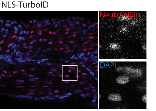
Tools and resources                                                                                                              Cell Biology

    A

    B

        C                                                                        D

Figure 2. Spatial resolution of TurboID-catalysed biotinylation in zebrafish embryos. (A) Schematic representation of eGFP-, NLS-, CaaX-, CD44b-, and
Cavin4b-tagged TurboID constructs for mRNA injection in zebrafish embryos. TurboID-eGFP was used as a positive control. (B) Representative images
showing the distribution of biotinylated proteins in two dpf zebrafish embryos transiently expressing different TurboID constructs. Negative control fish
were injected with eGFP only. Fish embryos were fixed and permeabilised before NeutrAvidin-DyLight staining for biotin and DAPI staining to indicate
Figure 2 continued on next page

Xiong et al. eLife 2021;10:e64631. DOI: https://doi.org/10.7554/eLife.64631                                                                       5 of 24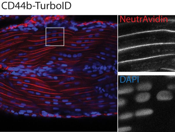
Tools and resources                                                                                                             Cell Biology

Figure 2 continued
nuclei. Regions within the white box were magnified and shown in the gray scale for NeutrAvidin and DAPI staining in the right panel; n = 3. Scale bar
represents 50 mm. (C) Streptavidin-HRP blots showing the ‘protein barcode’ produced by biotinylated proteins in fish embryo transiently expressing
different TurboID constructs. Actin immunoblot served as a loading control. Each sample is a pool of 30 embryos. (D) SYPRO Ruby protein stain
showing proteins isolated by streptavidin-pulldown. Approximately three hundred embryos transiently expressing each TurboID constructs were
subjected to streptavidin-pulldown after biotin incubation and embryo lysis. For original western blot/gel images see Figure 2—source data 1.
The online version of this article includes the following source data for figure 2:
Source data 1. Raw images of blots.

                                      spectrometry sequencing. It is a labour-intensive exercise when potentially analysing multiple POIs,
                                      and new genetic constructs must be generated for each POI. In addition, the protein expressed
                                      from mRNA injected at the one-cell stage becomes progressively depleted and is present only in
                                      trace amounts beyond 3 days post fertilisation. As such, this methodology is limited to early stage
                                      embryos. To circumvent these issues, we envisaged a modular system that would utilise the many
                                      existing stable zebrafish lines which express GFP-tagged proteins. Previously, we demonstrated that
                                      a GFP-binding peptide (GBP; a 14 kDa nanobody) is able to target a peroxidase (APEX2) to GFP-
                                      tagged POIs in both cell culture and zebrafish systems (Ariotti et al., 2015b), and can be used for
                                      ultrastructural localisation. Based on these findings, we reasoned that genetically fusing TurboID
                                      with GBP would target the TurboID-GBP fusion protein to GFP-labelled POIs and/or subcellular
                                      compartments in zebrafish, enabling GFP-directed proximity biotinylation in vivo. Furthermore, gen-
                                      eration of a stable zebrafish line expressing TurboID-GBP would allow delivery of the transgene by a
                                      simple genetic cross, circumventing the need for microinjection and enabling continued expression
                                      beyond the embryonic stages.
                                          As proof-of-principle, we fused a red fluorescent protein (mRuby2) with the GBP nanobody and
                                      transiently expressed it in a transgenic fish line already expressing Cavin1a-Clover. Clover is a GFP
                                      derivative recognised by GBP (Shaner et al., 2013), and Cavin1a is an ortholog of caveolae-associ-
                                      ated protein one in zebrafish (Lo et al., 2015). When expressed at low levels, mRuby2-GBP showed
                                      clear colocalisation with Cavin1a-Clover at the plasma membrane in the mRuby2-positive muscle
                                      cells (Figure 3A). However, when mRuby-GBP was expressed at higher levels, red fluorescence was
                                      observed in the cytoplasm in addition to the plasma membrane, likely due to the saturation of bind-
                                      ing between GBP and GFP. This observation raised concerns about the potential of non-specific
                                      labelling from unbound TurboID-GBP under these conditions. As a solution, we substituted the GBP
                                      with a conditionally stabilised GFP-nanobody (destabilised GBP or ‘dGBP’) that is rapidly degraded
                                      unless the GFP-binding site is occupied (Tang et al., 2016; Ariotti et al., 2018a). Using this
                                      approach, we observed tight association of mRuby2-dGBP and Cavin1a-Clover in all muscle cells
                                      regardless of expression level (Figure 3B). We reasoned that a system utilising the conditionally sta-
                                      bilised nanobody would be less likely to result in non-specific biotin labelling within target cells in
                                      vivo. Furthermore, use of the conditionally stabilised GBP gives potential for modularity, since tissue
                                      or cell type specific biotinylation will only occur in cells expressing both GFP-POI and TurboID-dGBP
                                      fusion proteins.

                                      Development of BLITZ; biotin labelling in tagged zebrafish
                                      We next generated a number of fish lines expressing TurboID-dGBP under the ubiquitous beta actin
                                      2 (actb2) promoter (Casadei et al., 2011). To facilitate selection of appropriate transgenic integra-
                                      tions, we added a cytoplasmic red fluorescent protein, mKate2, as a visible reporter upstream of
                                      TurboID-dGBP linked by a P2A sequence (Donnelly et al., 2001; Kim et al., 2011). The P2A
                                      sequence is a short ribosome-skipping sequence which separates the upstream mKate2 from down-
                                      stream TurboID-dGBP, reducing the potential interference from the fluorescent protein. The expres-
                                      sion of transgene is stable in our zebrafish lines and demonstrates Mendelian inheritance over four
                                      generations, indicating a stable single transgenic integration.
                                         We first tested whether these zebrafish lines were able to catalyse specific biotinylation in tissues
                                      expressing GFP. The TurboID-dGBP fish were outcrossed with transgenic lines expressing cyto-
                                      plasmic GFP in the vasculature (kdrl:EGFP) (Beis et al., 2005) and the motor neurons (MotoN:EGFP)
                                      (Punnamoottil et al., 2015; Figure 4A). Biotinylated proteins were examined in three dpf embryos
                                      after overnight biotin incubation. In embryos co-expressing ubiquitous TurboID-dGBP and tissue-

Xiong et al. eLife 2021;10:e64631. DOI: https://doi.org/10.7554/eLife.64631                                                                     6 of 24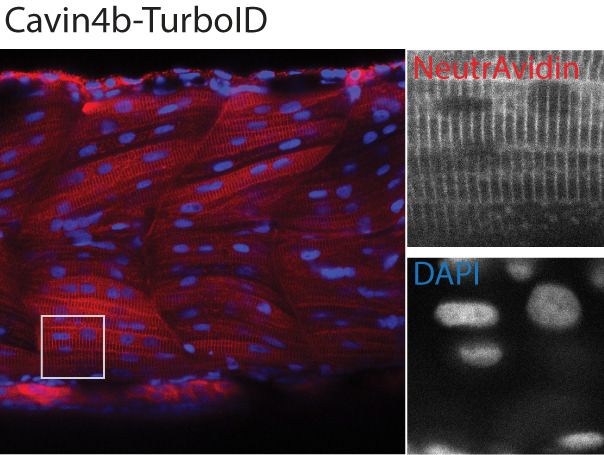
Tools and resources Cell Biology
A
mRuby2-GBP Merge
4000 Clover
3 dpf ~0.05 ng
mRuby2
Arbitrary Unit
3000
2000
1000
0
0 2 4 6 8
Distance (μm)
3 dpf ~0.5 ng 4000 Clover
mRuby2
Arbitrary Unit
3000
2000
1000
0
0 2 4 6 8
Distance (μm)
3 dpf ~1 ng 4000 Clover
mRuby2
Arbitrary Unit
3000
2000
1000
0
0 2 4 6 8
Distance (μm)
B
Cavin1a-Clover mRuby2-dGBP Merge
3 dpf ~0.05 ng Clover
30000
mRuby2
Arbitrary Unit
20000
10000
0
0 2 4 6 8
Distance (μm)
Clover
3 dpf ~0.5 ng 30000
mRuby2
Arbitrary Unit
20000
10000
0
0 2 4 6 8
Distance (μm)
3 dpf ~1 ng 30000 Clover
mRuby2
Arbitrary Unit
20000
10000
0
0 2 4 6 8
Distance (μm)
Figure 3. In vivo binding of GFP-nanobody, GBP and dGBP, in stable transgenic zebrafish embryos. (A and B) Representative images showing the
colocalisation between Cavin1a-Clover and mRuby2-GBP/dGBP in live zebrafish embryos. Cavin1a-Clover zebrafish embryos transiently expressing
mRuby2-tagged GBP (A) or dGBP (B). Injected embryos were imaged at three dpf. The approximate amount of injected RNA was indicated in the
Figure 3 continued on next page
Xiong et al. eLife 2021;10:e64631. DOI: https://doi.org/10.7554/eLife.64631 7 of 24
Tools and resources                                                                                                         Cell Biology

Figure 3 continued
mRuby2 images. Line scan (indicated by the blue line) shows the fluorescent intensity of Clover and mRuby2 across the sarcolemma of mRuby2-positive
muscle cells. Scale bar denotes 10 mm in both (A) and (B).

                                    specific GFP, TurboID-catalysed biotinylation was detected in the intersegmental vessels and the spi-
                                    nal cord motor neurons in the kdrl:EGFP and MotoN:EGFP lines, respectively (Figure 4A). These
                                    results demonstrate that our TurboID-dGBP system can produce biotinylation with tissue specificity.
                                        To test the biotinylation on a subcellular level, the TurboID-dGBP fish were outcrossed with Cav-
                                    in1a-Clover and Cavin4a-Clover transgenic fish lines expressing Cavin1a-Clover and Cavin4a-Clover
                                    under the control of the muscle specific actin promoter, actc1b. Cavin1a and Cavin4a are ortho-
                                    logues of human CAVIN1 and CAVIN4, which are caveola-associated proteins involved in caveolar
                                    formation. With the same procedures, we observed clear colocalisation between biotinylated pro-
                                    teins and Clover-tagged cavins in muscle fibres, at the sarcolemma and T-tubules, suggesting our
                                    TurboID-dGBP system can produce proximity-dependent biotinylation with subcellular resolution.
                                    Without biotin treatment or without the expression of GFP, there was no detectable biotinylation
                                    effected by TurboID. Notably, the specificity of GFP-directed biotinylation was not compromised in
                                    fish lines expressing higher levels of TurboID-dGBP (Figure 4—figure supplement 1).
                                        We next visualised the proteins biotinylated by TurboID-dGBP on streptavidin blots (Figure 4B).
                                    The two prominent bands representing endogenously biotinylated proteins were again observed in
                                    embryos carrying both TurboID-dGBP and Cavin1a-clover; omitting the biotin supplement resulted
                                    in no exogenous biotinylation. Intriguingly, in the absence of Cavin1a-Clover, a weak biotinylation
                                    was still observed in the embryos carrying only the TurboID-dGBP transgene, despite the level of
                                    TurboID-dGBP being undetectable on anti-Myc immunoblot. This background labelling is likely
                                    caused by TurboID-dGBP en route to proteasomal degradation. Subsequent MS analysis revealed
                                    these background proteins are mainly endogenous biotinylated proteins, nuclear proteins, cytoskele-
                                    tal proteins and yolk proteins (Supplementary file 3). Using streptavidin affinity pulldown, biotiny-
                                    lated proteins were isolated from total fish lysates and endogenous Cavin4b, a known Cavin1
                                    interactor (Bastiani et al., 2009), was detected in the streptavidin pulldown in addition to Cavin1a-
                                    GFP and TurboID-dGBP (Figure 4C). Note that a trace of TurboID-dGBP was detected in the strep-
                                    tavidin pulldown in the absence of GFP target with long exposure, which accounts for the weak
                                    background biotinylation in embryos expressing only TurboID-dGBP.

                                    A comprehensive cavin-associated proteome in skeletal muscle
                                    generated by TurboID-dGBP
                                    Finally, we employed our TurboID-dGBP system to map the proteomes associated with Cavin1 and
                                    4 in zebrafish skeletal muscle. We crossed the TurboID-dGBP fish with fish lines stably expressing
                                    Cavin1a-Clover, Cavin4a-Clover, and Cavin4b-Clover in muscle. TurboID-dGBP and cavin-Clover co-
                                    expressing embryos were selected at 2 dpf for subsequent biotin labelling and the biotinylated pro-
                                    teins were analysed by liquid chromatography coupled to tandem mass spectrometry (nanoHPLC/
                                    MS MS/MS). The sibling embryos inheriting only the TurboID transgene were used as a background
                                    control (experimental regimen illustrated Figure 5A). After subtracting background proteins and
                                    common containments, 26, 22, and 25 proteins were identified in the Cavin1a, Cavin4a, and Cavin4b
                                    samples, respectively (Figure 5B and C, Supplementary file 1 – Tables 1-3). Among the proteins
                                    identified, the majority of proteins are associated with the plasma membrane, consistent with the
                                    membrane localisation of cavins (Figure 5D, Supplementary file 1 - Table 4). Endogenous cavins
                                    were consistently detected in all samples, suggesting that the GFP-tag, as well as the binding of Tur-
                                    boID-dGBP, did not interfere with the oligomerisation of the cavins. Dystrophin (DMD), a protein
                                    associated with caveolae was identified uniquely in the Cavin1a sample (Song et al., 1996;
                                    Doyle et al., 2000). An ortholog of Pacsin3 (zgc:91999), a caveola-associated protein required for
                                    muscle caveolar formation (Seemann et al., 2017), was also detected uniquely in the Cavin4b sam-
                                    ple. These known interactors were undetectable in all control samples, demonstrating the high accu-
                                    racy of the BLITZ system. Note that, Caveolin1 (Uniprot accession: Q6YLH8) and Caveolin (isoform
                                    unassigned; Uniprot accession: A1L1S3) were detected in the Cavin1a sample based on two high
                                    confidence peptides (HLNDDVVK and VWVYSGIGFESAR) that were not present in the controls

Xiong et al. eLife 2021;10:e64631. DOI: https://doi.org/10.7554/eLife.64631                                                                 8 of 24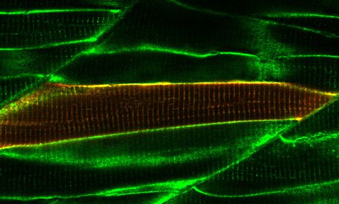
Tools and resources Cell Biology
A actb2: mKate2-P2A-Myc-TurboID-dGBP GFP-tagged fish lines B
GFP NeutrAvidin-DyLight 405 mKate2 Myc-TbID-dGBP: + + + − −
Cavin1a-Clover: + + − + +
Biotin: + − + + −
kDa 180
135
kdrl: GFP
100
75
63
Streptavidin-HRP
48
35
MotoN: GFP
25
IB: Myc
IB: GFP
IB: Actin
Cavin1a-Clover
C Streptavidin
Lysate pulldown
Biotin +
Myc-TbID-dGBP: + + + +
Cavin1a-Clover: + − + −
Cavin4a-Clover
Biotin: + + + +
kDa
180
135
100
75
Streptavidin-HRP
63
48
GFP-negative
TurboID only
35
25
17
mKate2-negative IB: Cavin4b
Cavin1a-Clover only
IB: Myc
Short exposure
IB: Myc
Long exposure
IB: GFP
IB: Actin
Cavin1a-Clover
Biotin –
40 µm
Figure 4. GFP-directed in vivo biotin labelling. (A) Representative images of TurboID-dGBP catalysing GFP-dependent biotinylation in transgenic
zebrafish embryos at 3 dpf. The TurboID-dGBP line was crossed with different GFP-tagged zebrafish lines: Cavin1a-Clover (plasma membrane),
Cavin4a-Clover (sarcolemma and T-tubules), kdrl:eGFP (vasculature), and MotoN:eGFP (motor neurons). After biotin incubation, embryos were fixed,
permeabilised, and stained with NeutrAvidin to visualise the biotinylated protein. mKate2 is a fluorescent indicator for expression of TurboID-dGBP.
Figure 4 continued on next page
Xiong et al. eLife 2021;10:e64631. DOI: https://doi.org/10.7554/eLife.64631 9 of 24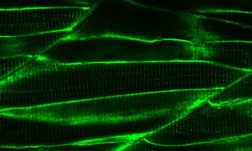
Tools and resources                                                                                                             Cell Biology

Figure 4 continued
Controls were carried out by using siblings from the same clutch without GFP expression (TurboID only) and siblings without TurboID expression
(Cavin1a-Clover only), as well as omitting biotin incubation. The scale bar denotes 40 mm; n = 3. (B) Western blot analysis showing the biotinylated
proteins in 3 dpf zebrafish embryos from TurboID-dGBP outcrossing with Cavin1a-Clover line. Each sample is a pool of 30 embryos. (C) Western blot
analysis of fish lysates and streptavidin pulldown with embryos from TurboID-dGBP line outcrossing with Cavin1a-Clover line. Each pulldown sample is
a pool of 200 embryos. For confocal images comparing the biotin labelling specificity in zebrafish embryos with different expression level of TurboID-
dGBP see Figure 4—figure supplement 1. For table summarising proteins identified in control embryos expressing only TurboID-dGBP, see
Supplementary file 3. For original western blot images see Figure 4—source data 1.
The online version of this article includes the following source data and figure supplement(s) for figure 4:
Source data 1. Raw images of blots.
Figure supplement 1. The specificity of TurboID-dGBP biotin labelling is independent of its expression level in zebrafish embryos.

                                     (Supplementary file 2) but were not classified as significant hits based on the 1% global FDR analy-
                                     sis. This may reflect the poor accessibility of caveolins, as a large proportion of caveolins are buried
                                     in the plasma membrane (Ariotti et al., 2015a). Individual hits and general properties of putative
                                     interacting proteins are further discussed below.

                                     Discussion
                                     Advantages of BLITZ in proteomic mapping
                                     In this study, we have developed BLITZ (Biotin Labelling In Tagged Zebrafish): a modular system for
                                     in vivo proteomic mapping (Figure 6). This system utilises the advantages of BioID at capturing
                                     weak or transient interactions in living cells, but extends its application to an in vivo setting, enabling
                                     interactome investigation at specific developmental stages under physiological conditions, and
                                     potentially in disease models. The system also has several advantages over conventional BioID meth-
                                     ods for studying the proteome and interactome.
                                         Firstly, BLITZ does not require extensive molecular biology steps to produce numerous expres-
                                     sion constructs, or laborious embryonic manipulation. It instead relies on simple crossing of a Tur-
                                     boID-dGBP line with an existing GFP-tagged fish line of choice; a plethora of such lines currently
                                     exist in stock centres globally and, with the advent of nuclease directed genome editing, this num-
                                     ber is rapidly increasing. Secondly, BLITZ enables cell- and tissue-specific proteomic studies, since
                                     the stability of TurboID-dGBP is dependent on its binding to GFP targets. Non-specific biotin label-
                                     ling in tissues that do not express GFP-tagged constructs is avoided. Thirdly, our TurboID-dGBP sys-
                                     tem has the potential to be used with knock-in fish lines carrying a GFP fusion protein at the
                                     endogenous locus of a POI. This will enable the application of BioID to study proteomic associations
                                     with endogenous proteins, which, to our knowledge, has not been achieved by conventional BioID
                                     methods. Finally, the modularity of the BLITZ system could be advantageous for use in established
                                     tissue culture systems using existing GFP expression vectors or/and knock-in cell lines, as well as
                                     extended to other organisms.
                                         The use of the BLITZ system also comes with some caveats. Unlike the traditional BioID approach
                                     using a direct fusion of the biotin ligase with the bait protein, our system targets TurboID to the POI
                                     through the binding of dGBP nanobody to GFP. In this case, the indirect binding increases the physi-
                                     cal distance between biotin ligase and the POI, which could potentially enlarge the effective label-
                                     ling radius and include more non-interacting neighbouring proteins. However, we have previously
                                     shown that the use of a GFP-directed nanobody to target a genetically encoded peroxidase (APEX2)
                                     for protein localisation does not appear to compromise the fidelity of labelling: APEX2 staining was
                                     rarely observed beyond 25 nm from the site of POI (Ariotti et al., 2015a). It is also possible that the
                                     binding of the biotin ligase-nanobody with the GFP-tagged POI could perturb the localisation of the
                                     POI, either by masking interacting surfaces or simply due to the larger size of a complex. For this
                                     reason, we routinely examine the distribution of the GFP-tagged POI both with and without biotin
                                     ligase-dGBP expression as well as the distribution of biotinylated proteins using fluorescent neutravi-
                                     din staining. Since our in vivo system is based on the simple crossing of heterozygous transgenic
                                     lines, every new clutch contains offspring with every possible combination of alleles, and the appro-
                                     priate internal controls can be sorted by fluorescence. As BLITZ uses biotin as a label, the method

Xiong et al. eLife 2021;10:e64631. DOI: https://doi.org/10.7554/eLife.64631                                                                    10 of 24
Tools and resources Cell Biology
A TurboID-dGBP cavin-Clover B Cavin1a
15
1 4
TurboID-dGBP
+ cavin-Clover
TurboID-dGBP
WT
6
control
cavin-Clover
12 3 12
streptavidin purification
MS sequencing
subtract
identified proteins background proteins Cavin-associated protein Cavin4a Cavin4b
Cavin1a Cavin4a Cavin4b Cellular Component Percentile
C D
cavin4a
limch1a
cavin1a
dmd Plasma membrane
camk2b1
dusp27 1000001 - 5000000
pvalb2
ank3a
gspt1l Nucleus
sptan1
cavin4b
ehbp1l1b
cmya5
palm2
gspt1 Cytoplasm
top2b
stx4 10001 - 1000000
myhz2
mdh1ab
jade3 Cytoskeleton
gapdh
camk2g1
prx
naca
rpl24
Endosome
hspa8
spegb
gnl3
201 - 10000
hp1bp3
si:dkeyp-77c8.3
ldb3a Z-lin e
dhx37
nop58
hpcal1
setdb1a
si:dkeyp-77c8.2 T-tubule
adamts3
grin2ab 1 - 200
hpca
ahnak
si:dkey-78l4.8
actc1b Sarcoplasmic Reculum
rsl1d1
bin1b
abcf1
zgc:123289
0% 10% 20% 30% 40% 50% 60%
smap1
senp6b
Cavin1a Cavin4a Cavin4b
dkc1 0
lbr
zgc:91999
ttn.1
eef1a1l2
dusp27
Figure 5. Proteomes identified by BLITZ system in Clover-tagged cavin zebrafish. (A) A schematic overview of applying TurboID-dGBP fish to identify
cavin-associated proteins. The TurboID-dGBP zebrafish was crossed with Clover-tagged cavin fish lines. The embryos carrying both transgenes were
selected for subsequent biotin incubation and biotin affinity purification coupled MS sequencing. Identified proteins were refined by subtracting
proteins identified in control embryos expressing only TurboID-dGBP. (B) Venn diagram showing the overlap of identified proteins in Cavin1a, Cavin4a,
and Cavin4b samples. (C) Heatmap showing relative abundance of identified proteins based on normalised MS2Count in Cavin1a, Cavin4a, and
Cavin4b proteomes. (D) Bar graph showing the distribution of proteins at subcellular level. The cellular component information was curated from
Uniport database and literature. For table summarising all identified and enriched proteins, see Supplementary file 1 – Tables 1-3. For table
annotating all identified and enriched protein with subcellular localisation and functions, see Supplementary file 1 – Table 4. For table showing all
Figure 5 continued on next page
Xiong et al. eLife 2021;10:e64631. DOI: https://doi.org/10.7554/eLife.64631 11 of 24Tools and resources Cell Biology
Figure 5 continued
identified peptides in Cavin1a sample and sibling control sample see Supplementary file 2 – Tables 1-2. For protein identification report generated by
ProteinPilot, see Figure 5—source datas 1–6.
The online version of this article includes the following source data for figure 5:
Source data 1. Protein identification report for Cavin1a sample generated by ProteinPilot.
Source data 2. Protein identification report for Cavin1a control sample generated by ProteinPilot.
Source data 3. Protein identification report for Cavin4a sample generated by ProteinPilot.
Source data 4. Protein identification report for Cavin4a control sample generated by ProteinPilot.
Source data 5. Protein identification report for Cavin4b sample generated by ProteinPilot.
Source data 6. Protein identification report for Cavin4b control sample generated by ProteinPilot.
(like most BioID methods) is problematic for the identification of interactors that are endogenously
biotinylated, such as carboxylases. In addition, as all BioID methods will label proteins within a small
number of nanometres, non-interacting proteins could be detected simply due to close proximity.
Thus, subsequent validation using other independent approaches such as biomolecular fluorescence
complementation and affinity pulldown is essential to distinguish bona fide interactors from non-
interacting neighbouring proteins.
Schematic representation of BLITZ system
Tg(actb:mKate2-P2A-TurboID-dGBP) Crossing Existing GFP-tagged fish lines
Fish embryos co-expressing
both constructs
Proximity-dependent biotin labelling
in vivo
dGBP-GFP binding
Biotin:
TurboID-dGBP GFP POI
Degradation of
unbound TurboID-dGBP Harsh lysis and
protein extraction
ex vivo
Biotin affinity
purification
Trypsin digestion
and MS Analysis
Figure 6. A schematic overview of the BLITZ system. The TurboID-dGBP lines can be crossed with existing GFP-tagged lines. In the embryos carrying
both transgenes, the binding between dGBP and GFP stabilise TurboID-dGBP, which leads to proximity biotinylation around the GFP-tagged POIs.
The unbound TurboID-dGBP will be rapidly degraded by the ubiquitin proteasome system, which minimises non-specific labelling when dGBP-GFP
binding saturates, as well as achieving tissue specificity by averting labelling in cells/tissues that do not express GFP. The biotin-labelled proteins can
be isolated by biotin affinity purification and identified by MS analysis.
Xiong et al. eLife 2021;10:e64631. DOI: https://doi.org/10.7554/eLife.64631 12 of 24Tools and resources Cell Biology
Application of BLITZ to the identification of cavin-association networks
in muscle
Cavin family proteins are key components of the caveolar coat complex associated with the inner
leaflet of the plasma membrane. Cavin1 is present in all tissues and is essential for caveolar forma-
tion and function. Cavin2, 3, and 4 show more restricted tissue distributions with Cavin4 being spe-
cific to skeletal and cardiac muscle (reviewed in Parton et al., 2018). In the zebrafish, Cavin1 and 4
are each duplicated such that four loci exist; Cavin1a/b and Cavin4a/b. Cavin1a and b show spatially
distinct expression patterns with Cavin1b being largely restricted to the developing notochord
whereas Cavin1a, 4a, and 4b are all highly expressed in skeletal muscle (Hill et al., 2008; Lo et al.,
2015; Housley et al., 2016). In this study we used the BLITZ system to identify putative interactors
for all three skeletal muscle cavins, and identified sets of putative interactors both unique and com-
mon to all three proteins.
The majority of proteins identified for all cavin proteomes were muscle-enriched factors and
plasma membrane proteins. We also saw a specific enrichment of known caveola-associated proteins
consistent with initial expectations. Interestingly, the cavin proteomes also contained a dispropor-
tionate number of nuclear proteins, such as Gnl3, Naca, and Lbr. In cultured cells, cavins have been
shown to be released from the plasma membrane in response to external stimuli (such as mechanical
stress) and are able to bind intracellular targets in variety of subcellular locations to regulate pro-
cesses such as ribosomal RNA transcription and apoptosis (Liu and Pilch, 2016; McMahon et al.,
2019). In addition, in the absence of Cavin1, in knockout mouse muscle, Cavin4 has been shown to
localise predominantly to the nucleus rather than the sarcolemma (Lo et al., 2015).
What processes might Cavin1 and Cavin4 be regulating? We know that loss of Cavin1 causes lipo-
dystrophy and muscular dystrophy in humans. Patient and animal muscle shows hypertrophied mus-
cle fibres (Hayashi et al., 2009; Rajab et al., 2010; Ding et al., 2017). Cavin4 mutations have been
described in dilated cardiomyopathy patients and there is evidence that Cavin4 recruits ERK in cardi-
omyocytes (Rodriguez et al., 2011; Ogata et al., 2014). Thus, there is supporting data for the posi-
tive regulation of hypertrophy in skeletal muscle fibres by Cavin1, and in cardiomyocytes by Cavin4.
The cavin proteome showed an enrichment of protein kinases, such as calcium/calmodulin-depen-
dent protein kinase II (CaMKII). CaMKII regulates Ca2+ signalling and plays an important role in the
development of cardiac hypertrophy through the ERK signalling pathway (Illario et al., 2003;
Cipolletta et al., 2010; Cipolletta et al., 2015). The activation of CaMKII can be induced by exer-
cise in skeletal muscle, with the activation level proportional to the intensity of exercise (Rose et al.,
2006).
In this study, BLITZ revealed several putative cavin interactors that have also been shown to be
involved in cardiomyopathies and/or skeletal myopathies, including the membrane protein Dystro-
phin (Deconinck and Dan, 2007), the triad-associated proteins Bin1 (Nicot et al., 2007), Cypher/
ZASP (Selcen and Engel, 2005), and SPEG (Agrawal et al., 2014). Genetic ablation of zebrafish
Cavin4b causes aberrant T-tubules in skeletal muscle (Housley et al., 2016). It is possible that
Cavin4 may be involved in T-tubule formation through interaction with triad associated proteins,
such as Bin1.
Overall, our BLITZ system enables the in vivo identification of protein interactors in a cell- and tis-
sue-specific manner, with high precision. We demonstrated the applicability of this approach in
diverse cell types including neurons and vascular endothelial cells and applied the BLITZ system to
identify factors associated with cavin family proteins in differentiated skeletal muscle. BLITZ provides
a versatile and valuable tool for proteomic discovery using the zebrafish model, but also has the
potential for application in other in vivo contexts that to date have been challenging or intractable.
Materials and methods
Key resources table
Reagent type
(species) or resource Designation Source or reference Identifiers Additional information
Gene BirA* Roux et al., 2012; R118G mutant of WT BirA
(E. coli- modified) DOI: 10.1083/jcb.201112098
Continued on next page
Xiong et al. eLife 2021;10:e64631. DOI: https://doi.org/10.7554/eLife.64631 13 of 24Tools and resources Cell Biology
Continued
Reagent type
(species) or resource Designation Source or reference Identifiers Additional information
Gene BASU Ramanathan et al., 2018; R124G, E323S, G325R mutation
(Bacillus subtilis - DOI: 10.1038/NMETH.4601 and N-terminus deletion of WT
modified) biotin ligase from B. subtilis
Gene BioID2 Kim et al., 2016; R40G mutation of WT biotin
(Aquifex aeolicus - DOI: 10.1091/mbc.E15-12-0844 ligase from A. aeolicus
modified)
Gene miniTurbo Branon et al., 2018; 13 point mutations and
(E. coli- modified) DOI: 10.1038/nbt.4201 N-terminal deletion of WT BriA
Gene TurboID Branon et al., 2018; 15 point mutations of WT BirA
(E. coli- modified) DOI: 10.1038/nbt.4201
Strain, strain TAB University of Queensland (UQ) Wild-type (TAB), an AB/TU line
background Biological Resources Aquatics generated in UQBR Aquatics
(Danio rerio) (UQ Biological Resources)
Strain, strain TurboID-dGBP Generated in this paper Tg(actb2:mKate2
background -P2A-TurboID-dGBP)
(Danio rerio)
Strain, strain Cavin1a-Clover Generated in this paper Tg(actc1b:Cavin1a-Clover)
background
(Danio rerio)
Strain, strain Cavin4a-Clover Generated in this paper Tg(actc1b:Cavin4a-Clover)
background
(Danio rerio)
Strain, strain Cavin4b-Clover Generated in this paper Tg(actc1b:Cavin4b-Clover)
background
(Danio rerio)
Strain, strain Kdrl:GFP (Beis et al., 2005); Tg(kdrl:EGFP)
background DOI: 10.1242/dev.01970
(Danio rerio)
Strain, strain MotoN:GFP (Punnamoottil et al., 2015); Tg(miR218:EGFP)
background DOI: 10.1002/dvg.22852
(Danio rerio)
Genetic reagent actb2:mKate2-P2A-TurboID-dGBP Generated in this paper Addgene: 163857 Construct for generating stable
(Danio rerio) transgenic fish line; see Materials
and methods for line generation
Genetic reagent actc1b:Cavin1a-Clover Generated in this paper Addgene: 163852 Construct for generating stable
(Danio rerio) transgenic fish line; see Materials
and methods for line generation
Genetic reagent actc1b:Cavin4a-Clover Generated in this paper Addgene: 163853 Construct for generating stable
(Danio rerio) transgenic fish line; see Materials
and methods for line generation
Genetic reagent actc1b:Cavin4b-Clover Generated in this paper Addgene: 163854 Construct for generating stable
(Danio rerio) transgenic fish line; see Materials
and methods for line generation
Genetic reagent pT3TS-BASU-EGFP Generated in this paper Addgene: 163845 Construct for in vitro RNA
(Danio rerio) synthesis and RNA injection
Genetic reagent pT3TS-TurboID-EGFP Generated in this paper Addgene: 163846 Construct for in vitro RNA
(Danio rerio) synthesis and RNA injection
Genetic reagent pT3TS-miniTurbo-EGFP Generated in this paper Addgene: 163847 Construct for in vitro RNA
(Danio rerio) synthesis and RNA injection
Genetic reagent pT3TS-TurboID-CaaX Generated in this paper Addgene: 163848 Construct for in vitro RNA
(Danio rerio) synthesis and RNA injection
Genetic reagent pT3TS-nls-TurboID Generated in this paper Addgene: 163849 Construct for in vitro RNA
(Danio rerio) synthesis and RNA injection
Genetic reagent pT3TS-CD44b-TurboID Generated in this paper Addgene: 163850 Construct for in vitro RNA
(Danio rerio) synthesis and RNA injection
Continued on next page
Xiong et al. eLife 2021;10:e64631. DOI: https://doi.org/10.7554/eLife.64631 14 of 24Tools and resources Cell Biology
Continued
Reagent type
(species) or resource Designation Source or reference Identifiers Additional information
Genetic reagent pT3TS-Cavin4b-TurboID Generated in this paper Addgene: 163851 Construct for in vitro RNA
(Danio rerio) synthesis and RNA injection
Genetic reagent actc1b:mRuby2-GBP Generated in this paper Addgene: 163856 Construct for transient
(Danio rerio) expression in zebrafish
Genetic reagent actc1b:mRuby2-dGBP Generated in this paper Addgene: 163855 Construct for transient
(Danio rerio) expression in zebrafish
Antibody Anti-Myc Cell Signaling Technology 2276S (1:2000) dilution with 5%
(Mouse monoclonal) skim milk in PBST
Antibody Anti-Actin EMD Millipore MAB1501 (1:5000) dilution with 5%
(Mouse monoclonal) skim milk in PBST
Antibody Anti-Cavin4b Boster Biological DZ33949 Customised antibody against
(Rabbit polyclonal) Technology zebrafish Cavin4b; (1:1000) dilution
with 3% skim milk in PBST
Recombinant p5E-actb2 Kwan et al., 2007; N/A
DNA reagent DOI: 10.1002/dvdy.21343
Recombinant p5E-actc1b Ariotti et al., 2018b; N/A
DNA reagent DOI: 10.1242/dev.034561
Recombinant pME-BASU-NS generated in this paper Addgene: 166565 Gateway Entry clone contains
DNA reagent BASU without a stop codon;
see Materials and methods for cloning
and Addgene for vector map
Recombinant pME-TurboID-NS generated in this paper Addgene: 166566 Gateway Entry clone contains
DNA reagent TurboID without a stop codon;
see Materials and methods for cloning
and Addgene for vector map
Recombinant pME-nls Ariotti et al., 2018a; Addgene: 108882
DNA reagent DOI: 10.1371/journal.pbio.2005473
Recombinant pME-CD44b Hall et al., 2020 Addgene: 109576
DNA reagent DOI: 10.1038/s41467-020-17486-w
Recombinant pME-miniTurbo-NS generated in this paper Addgene: 166567 Gateway Entry clone contains
DNA reagent miniTurbo without a stop codon;
see Materials and methods for
cloning and Addgene for vector map
Recombinant pME-Cavin1a Hall et al., 2020; Addgene: 126927
DNA reagent DOI: 10.1038/s41467-020-17486-w
Recombinant pME-Cavin4a Hall et al., 2020; Addgene: 109562
DNA reagent DOI: 10.1038/s41467-020-17486-w
Recombinant pME-Cavin4b Hall et al., 2020; Addgene: 109563
DNA reagent DOI: 10.1038/s41467-020-17486-w
Recombinant pME-mKate2-P2A-TurboID-NS Generated in this paper Addgene: 166568 Gateway Entry clone contains
DNA reagent mKate2-P2A-TurboID
without a stop codon;
see Materials and methods for
cloning and Addgene for vector map
Recombinant pME-mRuby2-NS Generated in this paper Addgene: 166569 Gateway Entry clone contains
DNA reagent mRuby2 without a stop codon;
see Materials and methods for
cloning and Addgene for vector map
Recombinant p3E-TurboID Generated in this paper Addgene: 166570 Gateway Entry clone contains
DNA reagent TurboID with a stop codon; see
Materials and methods for cloning
and Addgene for vector map
Recombinant p3E-Clover Generated in this paper Addgene: 126572 Gateway Entry clone contains
DNA reagent Clover with a stop codon; see
Materials and methods for cloning
and Addgene for vector map
Continued on next page
Xiong et al. eLife 2021;10:e64631. DOI: https://doi.org/10.7554/eLife.64631 15 of 24Tools and resources Cell Biology
Continued
Reagent type
(species) or resource Designation Source or reference Identifiers Additional information
Recombinant p3E-EGFP Generated in this paper Addgene: 126573 Gateway Entry clone contains
DNA reagent EGFP with a stop codon; see
Materials and methods for cloning
and Addgene for vector map
Recombinant p3E-csGBP (dGBP) Ariotti et al., 2018a; Addgene: 108891 Gateway Entry clone contains
DNA reagent DOI: 10.1371/journal.pbio.2005473 csGBP with a stop codon; see
Materials and methods for
cloning and Addgene for vector map
Recombinant p3E-GBP Ariotti et al., 2015a; Addgene: 67672 Gateway Entry clone contains
DNA reagent DOI: 10.1016/j.devcel. GBP with a stop codon; see
2015.10.016 Materials and methods for cloning
and Addgene for vector map
Recombinant p3E-CaaX (tH) Hall et al., 2020; Addgene: 109539
DNA reagent DOI: 10.1038/
s41467-020-17486-w
Recombinant pT3TS-DEST Generated in this paper Addgene: 166571 Gateway Destination vector
DNA reagent contains T3 and T7 promoters
for in vitro RNA synthesis;
see Materials and methods
for cloning and Addgene
for vector map
Peptide, Streptavidin-HRP Abcam Ab7403 (1:5000) dilution
recombinant protein with 5% BSA in PBST
Peptide, Proteinase K Invitrogen 25530015
recombinant protein
Peptide, Pronase Roche 10165921001
recombinant protein
Peptide, Trypsin/Lys-C Mix, Promega V5073
recombinant protein Mass Spec Grade
Commercial Pierce BCA protein Thermo Scientific 23225
assay or kit assay kit
Commercial Clarity Western Bio-Rad 1705061
assay or kit ECL Substrate
Commercial InstantBlue Expedeon ISB1L-1L
assay or kit
Commercial SYPRO Ruby Protein Invitrogen S12000
assay or kit Gel Stain
Chemical Biotin Sigma-Aldrich B4639-1G
compound, drug
Chemical Phenol Red Sigma-Aldrich P0290-100ML
compound, drug
Chemical NeutrAvidin-DyLight 405 Invitrogen 22831
compound, drug
Chemical NeutrAvidin-DyLight 550 Invitrogen 84606
compound, drug
Chemical Sodium deoxycholate Sigma-Aldrich D6750-10G
compound, drug
Chemical NP-40 Sigma-Aldrich 18896–50 ML
compound, drug
Chemical EDTA Astral Scientific BIOEB0185-500G
compound, drug
Chemical Complete Protease Sigma-Aldrich 11836145001
compound, drug Inhibitor Cocktail
Chemical Paraformaldehyde Sigma-Aldrich P6148-500G
compound, drug
Continued on next page
Xiong et al. eLife 2021;10:e64631. DOI: https://doi.org/10.7554/eLife.64631 16 of 24Tools and resources Cell Biology
Continued
Reagent type
(species) or resource Designation Source or reference Identifiers Additional information
Chemical PBS tablets Medicago 09-8912-100
compound, drug
Chemical Triton-X100 Sigma-Aldrich T9284-500ML
compound, drug
Chemical Tween 20 Sigma-Aldrich P1379-500ML
compound, drug
Chemical DAPI Sigma-Aldrich D9542-5MG
compound, drug
Chemical Bolt LDS sample Invitrogen B0008
compound, drug buffer (4X)
Chemical Dynabeads Invitrogen 65001
compound, drug MyOne Streptavidin C1
Chemical Agarose, low Sigma-Aldrich A9414-100G
compound, drug gelling temperature
Software, algorithm ProteinPilot SCIEX Version 5.0.1
Software, algorithm Analyst TF SCIEX Version 1.7
Software, algorithm Excel Microsoft Version 16.45
Software, algorithm Prism8 GraphPad Version 8.0.2
Software, algorithm Fiji ImageJ Version 2.0.0-
rc-69/1.52 p
Software, algorithm Illustrator Adobe Version 23.1.1
Other PD-10 desalting GE Healthcare 17-0851-01
column
Other LoBind tube Eppendorf 022431048
Other Blot 4–12% Bis-Tris Invitrogen NW04120BOX
Plus precast gels
Zebrafish husbandry
Zebrafish were raised and maintained according to institutional guidelines (Techniplast recirculating
system, 14 hr light/10 hr dark cycle, University of Queensland, UQ). Adults (90 dpf above) were
housed in 3 or 8 L tanks with flow at 28.5˚C and embryos up to five dpf were housed in 8 cm Petri
dishes in standard E3 media (5 mM NaCl, 0.17 mM KCl, 0.33 mM CaCl2, and MgSO4) at 28˚C (incu-
bated in the dark) (Westerfield, 2007). All experiments were approved by the University of Queens-
land Animal Ethics Committee. The following zebrafish strains were used in this study: wild-type
(TAB), an AB/TU line generated in UQBR Aquatics (UQ Biological Resources), Tg(actc1b:cavin1a-Clo-
ver), Tg(actc1b:cavin4a-Clover), Tg(actc1b:Cavin4b-Clover), Tg(actb2:mKate2-P2A-TurboID-dGBP),
Tg(kdrl:eGFP) and Tg(MotoN:GFP). The developmental stages of zebrafish used in experiments are
prior to specific sex determination. All zebrafish used in experiment were healthy, not involved in
previous procedures and drug or test naive.
DNA constructs and transgenic fish lines
The protein sequence of TurboID and MiniTurbo was constructed according to Branon et al., 2018
while the protein sequence of BASU was designed according to Ramanathan et al., 2018. The cod-
ing sequences of TurboID, MiniTurbo, and BASU were ordered from IDT as gene fragment with
codon optimised for zebrafish expression (https://sg.idtdna.com). The expression of biotin ligases
was driven by a ubiquitous promoter of actb2 (Higashijima et al., 1997; Casadei et al., 2011). A
red fluorescent reporter, mKate2, was indirectly linked into the N-terminus of biotin ligase through a
self-cleaving P2A sequence (Shcherbo et al., 2009; Kim et al., 2011). Promoter, fluorescent report
and biotin ligase were cloned into destination vector using Gateway cloning system. All fish lines
were generated by using Tol2kit system according to established methods (Kawakami, 2004;
Kwan et al., 2007). In brief, plasmid constructs for generating transgenic lines were co-injected with
Xiong et al. eLife 2021;10:e64631. DOI: https://doi.org/10.7554/eLife.64631 17 of 24Tools and resources Cell Biology
tol2 mRNA into one-cell-stage WT zebrafish embryos (Nusslein-Volhard and Dahm, 2002). Injected
F0s were raised and screened for founders producing positive F1s with Mendelian frequencies, indic-
ative of single genomic integration. Positive F1s grown to reproductive age were used for our experi-
ments. Stable lines were maintained as heterozygotes. All stable lines used are given in the Key
Resource Table.
Transient expression by DNA/RNA microinjection
DNA plasmid and RNA transcript for injection were diluted to final concentration of 30 ng/ml and
200 ng/ml, respectively, with addition of Phenol Red (Sigma-Aldrich) as injection tracer. A bolus of
approximately 1/5 of the total cell diameter was injected into each embryo. For DNA injection, the
bolus was injected into the cell of embryos at single cell stage (5–25 min-post-fertilisation). For RNA
injection, the bolus was injected into the yolk of the embryos up until two-cell stage. The RNA tran-
script was synthesised by mMESSAGE mMACHINE T3 (Invitrogen) according to manufacturer’s
instruction. The RNA transcripts were tagged with poly(A) tail using Poly(A) Tailing Kit (Invitrogen) to
extend the stability of mRNA in zebrafish embryos.
In vivo biotin labelling
Embryos at indicated developmental stage were incubated in the E3 media supplemented with 500
mM biotin for 18 hr to initiate biotinylation in vivo. For embryos before hatching, a dechorionation
step was carried out by using Pronase (Roche, 100 mg/ml in E3 media for 40 min at 28˚C) prior to the
biotin incubation. After biotin incubation, embryos were washed for 40 min with two changes of
standard E3 media to remove unincorporated biotin before subsequent immunostaining or protein
extraction.
Zebrafish embryos protein extraction
Fish embryos after in vivo biotin labelling were deyolked by mechanical disruption through a 200 ml
pipette tips in calcium-free Ringer’s solution followed by two changes of solution at 4˚C. The
deyolked embryos was lysed by brief sonication in RIPA buffer (50 mM Tris-HCl, pH 7.5; 150 mM
NaCl; 1% NP-40; 0.1% SDS; 5 mM EDTA; 0.5% Na-deoxycholate,) with freshly added cOmplele Pro-
tease Inhibitor. Lysates were further solubilised at 4˚C with rotation for 30 min. Insoluble material
was removed by centrifugation at 14,000 g for 10 min at 4˚C, and supernatant were collected for
BCA protein assay determining protein concentration. For western blot analysis, 25 fish embryos per
group were used for protein extraction, whilst, for streptavidin affinity purification, approximately
350 embryos were used for each group.
Western blotting
Western blot analysis was performed largely as described previously Lo et al., 2015. Briefly, zebra-
fish samples from protein extraction were mixed with NuPAGE LDS sample buffer (4X) and 10 mM
DTT. Protein samples were analysed by Western blotting with following antibodies: mouse anti-Myc
(dilution 1:2000), mouse anti-Actin (dilution 1:5000), rabbit anti-Cavin4b (dilution 1:2000), anti-mouse
and anti-rabbit HRP-conjugated antibodies (dilution 1:5000), streptavidin-HRP (dilution 1:5000). ECL
blotting reagent was used to visualise HRP and chemiluminescent signal was detected using the
ChemiDoc MP system (BioRad) as per the manufacture’s instruction.
Streptavidin beads pulldowns
Fresh embryo protein extracts (4 mg in 2.5 ml RIPA buffer) was passed through PD-10 desalting col-
umn (GE Healthcare) to remove excess free biotin using the gravity protocol according to manufac-
turer’s instruction. Protein extracts were then mixed with Dynabeads MyOne Streptavidin C1
(Invitrogen) from 200 ml bead slurry that were pre-washed with RIPA buffer, and incubated on a rotor
wheel at 4˚C overnight (16 hr). The next day, the beads were separated from the protein extracts on
a magnetic rack and transferred to a new 2 ml LoBind tube (Eppendorf). The beads were washed
with 1 ml of each following solution: twice with RIPA buffer, once with 2% SDS in 50 mM Tris-HCl
pH7.5, once with 2 M urea in 10 mM Tris-HCl pH8.0 and twice again in RIPA buffer without cOm-
plete Protease Inhibitor. Washed beads were boiled in 60 ml of 2X Blot LDS sample buffer (4X
diluted to 2X with RIPA buffer) containing 2 mM biotin and 20 mM DTT at 95˚C for 10 min with 10 s
Xiong et al. eLife 2021;10:e64631. DOI: https://doi.org/10.7554/eLife.64631 18 of 24You can also read