Carbon dots: Discovery, structure, fluorescent properties, and applications
←
→
Page content transcription
If your browser does not render page correctly, please read the page content below
Green Processing and Synthesis 2021; 10: 134–156
Review Article
Asmaa M. El-Shafey*
Carbon dots: Discovery, structure, fluorescent
properties, and applications
https://doi.org/10.1515/gps-2021-0006 and huge challenge [1–3]. Agriculture and industry outputs
received June 16, 2020; accepted November 19, 2020 in food production increased by science and technology
Abstract: Nanotechnology has become one of the most support, but the statistical studies proved a marked increase
important topics since the beginning of the twenty-first in food waste by both the consumer and production sides
century in numerous fields including drug synthesis and [4]. It was essential to think of a solution to convert wastes
delivery, environmental protection, electronics manufac- into useful materials to achieve the sustainability principle
ture, and astronomy due to their nanoscale particles and [5], so scientists worked on nanomaterials synthesis from
their properties. The traditional semi-quantum dots are natural sources and products for biological and environ-
replaced by a new category of fluorescent carbon nano- mental sensing applications [6–8]. Xu and colleagues
materials. Carbon dots (CDs) have been explored in the were the first team to discover carbon nanodots (CDs)
last few years for their simple synthetic accession, good which are a category of photo luminescent nanoparticles
bio-consonance, and several revelation applications. This (NPs) [9]. CDs and its derivatives have been used for
review explains the fluorescent properties of CDs in brief, numerous applications which are considered bioconve-
giving also a background on CDs discovery, structure, and nient and eco-friendly as CDs can be prepared from any
composition, as well as on nanocomposites, green synth- starting material including carbon; therefore, food wastes
esis, and their applications. Resources conservation can be were reused because of the majority of by-products of the
achieved by using recycled substances for sustainable devel- food; food wastes from vegetables, livestock, fruits, and
opment which lead to a new technology. Fluorescent CDs food industry enhance the progress and sustainability of
synthesized from food wastes like bananas, orange peel nanotechnology [10–14]. Figure 1 explains sustainable
waste, sugarcane bagasse, Trapa bispinosa peels, bread, development of CDs technology [15].
and jaggery have several applications such as sensing, New category of fluorescent carbon nanomaterials
drug delivery, gene transfer, biological imaging, and has size below 10 nm, which means it possesses ultrafine
food safety. In this study, we concentrate on CDs greener size which is referred to as carbon quantum dots (CDs). The
methods to prepare effective and biocompatible CDs. separation and purification of single-walled carbon nano-
tubes were the main reason for its first discovery in 2004
Keywords: C-dots, fluorescence, nanocomposites, green [9,16]. CDs are perfect for bioimaging [17–19] by shifting
synthesis, bioimaging the emission wavelength to the red region as CDs emit
blue fluorescence under UV excitation and they also have
different properties such as excitation-/pH-dependent pro-
perty, chemical stability, and resistance to photobleaching
1 Introduction as well as up-conversion fluorescence so that the CD
synthesis and its numerous applications have been stu-
The important environmental matter that occupied our died (Figure 2) [20].
minds is that the world consists of 7.2 billion people Renewable resources and small molecules are used
and tolerating the waste resulted from food production as as precursors with the aid of heating, microwave irradia-
well as consumption of this population is considered a big tion, and ultrasound. Depending on the fluorescence
disaster in our universe, a fundamental environmental issue changes, different sensing protocols will be achieved
especially plenty of sensing strategies have been devel-
oped by variations in the CD synthesis methods [21–23].
* Corresponding author: Asmaa M. El-Shafey, Chemistry
CDs have several applications [24–26], such as
Department, Faculty of Science and Arts, King Khalid University, CD probes in food safety [27]. Various types of contami-
Sarat Ebida, Saudi Arabia, e-mail: sommy_28@yahoo.com nants, involving pesticides [28], veterinary drugs [29],
Open Access. © 2021 Asmaa M. El-Shafey, published by De Gruyter. This work is licensed under the Creative Commons Attribution 4.0
International License.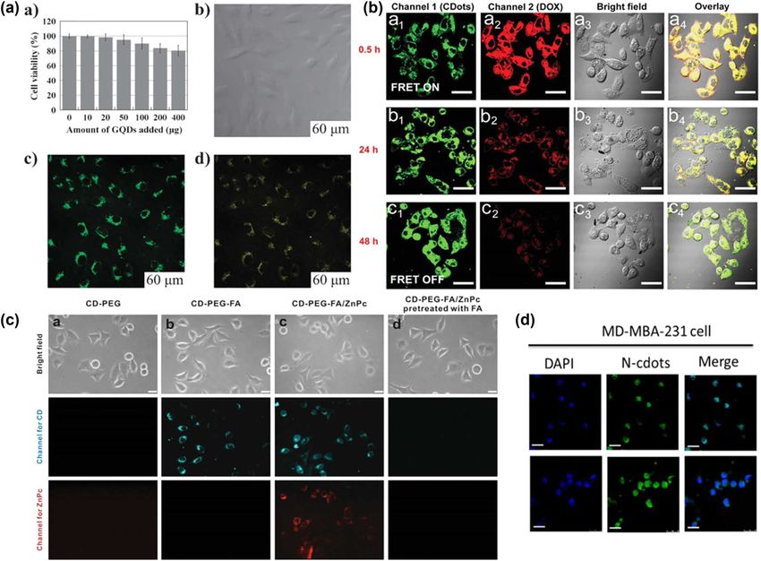
Carbon dots: A review 135
discovery, structure, and composition as well as about
the green synthesis of CDs, its fluorescent properties,
and its several applications. We wish that the review pre-
sents worth information and easy induction of CD-based
sensing.
2 CDs discovery
Surface defects in single- and multiwalled nanotubes
were the provenance of fluorescent carbon observation.
Luminescence is promoted by the carbon nanotube sur-
face passivation [32,33], which led to the CD discovery as
we mentioned before; the first time of discovery in 2004
was fortuitously through electrophoretic single-walled
nanotube purification. Under UV light, different colors
have been exhibited by the fluorescent material of the
Figure 1: Sustainable development of carbon nanodot tech-
fast moving band. The result of characterization indi-
nology [15].
cated that the material was completely metal free and
consisted of carboxyl groups, and the composition deter-
bacteria [30], and banned additives [31] can be deter- mined was C 53.93%, H 2.56%, O 40.33%, and N 1.20% as
mined by using CD probes beside food analysis applica- indicated in Figure 3 [9]. Since then, numerous methods
tion. Therefore, it is fundamental to gather an inclusive of CDs synthesis have been reported by many groups of
literature review to offer great knowledge about the CD researchers.
Figure 2: The importance of CDs in fluorescence properties, synthesis, and detection implementations in foods. (a) Fluorescent properties
involve excitation-dependent, pH-dependent, antiphotobleaching, and up-conversion fluorescence. (b) Synthetic methods include
hydrothermal/solvothermal, microwave-assisted, and ultrasonic-assisted methods. (c) The principles of CD-based biosensor can be
divided into two types: fluorescence quenching and fluorescence recovery [20].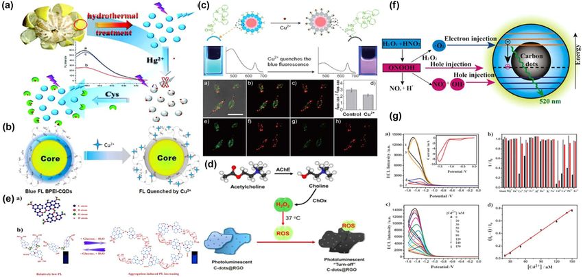
136 Asmaa M. El-Shafey
Figure 3: Electrophoretic profile in 1% agarose gel under 365 nm UV
light: (a) crude SWNT suspension, (b) fluorescent carbon, (c) short
tubular carbon, (d and e) further separation of (c), and (f) cut
SWNTs [9].
Figure 4: Capillary electrophoretic CD separation monitored at
(a) absorption wavelength of 250 nm and (b) laser-induced fluores-
2.1 Capillary zone electrophoresis (CZE)
cence at λex/λem of 488/550 nm. Peaks (1), (2), and (3) are positively
charged, neutral, and negatively charged CDs, respectively [34].
CZE with featured separation qualification possesses
an important speed separation approach for NPs due to
their different electrophoretic mobilities (on the basis of the reaction time-associated kinetics of CD consistence
charge/size ratio) through an electrolyte solution con- and identify the functional groups-associated charge states,
tained in a fused silica capillary under the impact of an the CE method was used. Applied CE coupled with UV detec-
electric field. The CZE utilization in conjunction with a tion (250 nm) and LIF detection (λex/λem 488/550 nm) for the
diode array detector (230 nm) for CD separation attained separation of hollow CD separation synthesized from glacial
from the flame of an oil lamp. Buffer composition effect acetic acid and diphosphorus pentoxide without external
tested on the negatively charged CD electropheric pattern heating. The separation was acheived utilizing 40.0 cm
by using various pattern types, pH, and concentrations as capillary (50 µm i.d. and 325 µm o.d.) with 10 mM sodium
capillary electrophoresis (CE) is a perfect method conve- dodecyl sulfate and 30 mM phosphate (pH 9.0) as run buffer
nient to implement favorable separation qualification, the at an applied voltage of 15 kV. By utilizing UV-Vis spectro-
advantages provided by the theoretical plates as high scopy, photoluminescence (PL) spectroscopy, and transmis-
number obtained with CE for CD separation are oversha- sion electron microscopy (TEM), the separated fractions
dowed by its UV detection systems, and low sensitivity due were also gathered and analyzed. The CZE is confirmly
to the sample volumes of small injection. To overcome the utilized in identifying the different charge states of CDs spe-
low-sensitivity limitation of the CDs’ analysis accuracy, cies existed in a complex CDs mixture by using individual
CDs can be separated by the utilization of CZE apparatus CE-fractionated. CDs fractions enough amounts collection
coupled with a diode array detector and a laser-induced owing to the low sample injection volume of CZE is time-
fluorescence (LIF) detector. The CD absorption electro- consumption. The neutral CD species eluted out as one
grams were obtained at 250 nm and the fluorescence single strong peak, but the charged CDs can only be sepa-
was observed at an excitation wavelength (λex/λem) of rated by CE. Isoelectric focusing (IEF) is used for isoelectric
488/550 nm. The separation was achieved using 40.0 cm point detection of macromolecules and bioparticles. The dif-
capillary (50 µm i.d. and 325 µm o.d.) with 30 mM sodium ferent sizes of colloidal NPs can selectively be separated by
acetate–acetic acid (NaAc–HAc) at pH 3.6 as run buffer size from their mixture in a homemade miniscale IEF unit as
and applied voltage of 15 kV. Figure 4 described that the it is a fast, sensitive, and low-cost-effective method [34].
UV (curve (a) and LIF (curve (b)) detection combination
detects boosting as well as complex CD mixture supple-
mental information. For example, positively charged, neu-
tral, and negatively charged CDs gained with two detection 3 CD structure and composition
processes are the same. While peaks (1) and (2) show
intense UV and fluorescence signals, peak (3) exhibits CDs obtained from different structures may be either gra-
strong absorption but weak emission signals. To explore phitic or amorphous. The size of CDs can be tuned by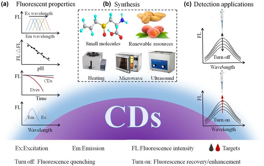
Carbon dots: A review 137
different nanocomposites. CDs have been studied as a
material with a multitude of applications. In terms of
analytical attributes (Figure 5), transmission electron
microscopy (TEM) and X-ray diffraction (XRD) are com-
monly utilized to characterize CDs as new material. IR
spectroscopy and elementary analysis help in recog-
nizing the surface functional groups which mark CDs’
affinity properties and signify chemical mechanisms
to be utilized in further expansion. Another attribute of Figure 6: TEM image (left) and size distribution (right) of
CDs is absorption in UV-Vis spectrum to assess the exci- C-dots [36].
tation band for fluorescence spectrum and observe the
change in surface properties of CDs because of inter-
action with another material or derivatization by subor-
dinating fluctuations in the fluorescence intensity of That means if the size of the particle remains under
C-dots. CDs’ fluorescence emission is used for targets 10 nm even after surface passivation to promote optical
observed under microscope as it makes NPs as label- properties, CDs would have the best performance in cell
free probes [11,12]. imaging.
The reported average size of CDs of less than 10 nm Another group of CDs prepared from bananas as a
facilitated its use in biological applications. Fluorescence starting material had reported a mean size of 3 nm; is
emission can be enhanced by surface passivation of CDs. spherical in shape; and includes carbon, oxygen, and
When CDs were coated by the hydroxyl groups, we potassium, though the percentage of potassium is very
noticed the following observations: less and the majority are carbon and oxygen. The inter-
1. The reported CD diameter was 3.1 ± 0.5 nm. layer spacing of CDs that involved oxygen-containing
2. TEM showed that the particles were uniformly dis- groups with sp3 carbon and hydroxyl connected carbon
persed and spherical in shape as shown in Figure 6. groups was established to be 0.42 nm which is higher
3. The quantum yield (QY) increased and was about 5.5%. than the graphitic interlayer spacing of 0.33 nm [37],
4. Better fluorescence emission and the particles were which denotes that CDs are crystalline in nature in com-
photostable [35,36]. parison to graphite as indicated in Figure 7. The third
group of CDs synthesized from food caramels and orange
peel waste is amorphous in nature and has no crystalline
nature as the XRD displayed a broad amorphous peak
[38]. The fourth group of CDs prepared from graphite
had a diamond-like structure. The percentage of squares
of the ring radius was reported to be 3:8:11:16:19, where
the rings correspond to the planes {111}, {220}, {311},
{400}, and {331} that belong to diamond [39]. The fifth
group of CDs derived from multiwalled carbon nanotubes
by electrochemical oxidation also showed graphitic nature.
The high-resolution TEM displayed the lattice spacing
to be 3.3°A, which is close to the (002) facet of graphite.
The sixth group of CDs attained from candle soot is high
in oxygen content and different functional groups such
as hydroxyl, carbonyl, carboxyl, and epoxy groups were
attached to CDs [40]. The reported structure of soot
was C 91.69%, H 1.75%, N 0.12%, and O (calculated)
44.66% [41]. The size distribution of CDs was narrow,
i.e., between 2 and 7 nm. CDs have a larger area due
to their smaller size, which led to greater absorption
to boost CDs’ applicability to optoabsorption. CDs also
Figure 5: Characterization of C-dots: spectra and images [15]. have the susceptibility to allow drug connection to the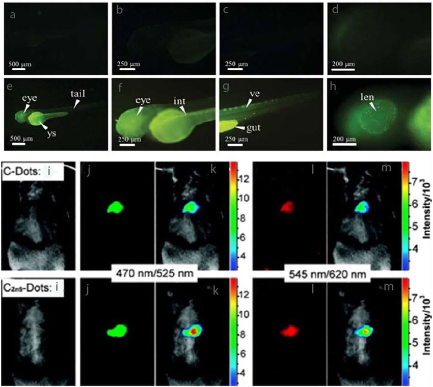
138 Asmaa M. El-Shafey
Figure 7: (a) EDX spectrum, (b) XRD pattern, and (c) FTIR spectrum of the carbon dots [37].
surface for drug delivery systems. Finally, the seventh 4.1 Excitation-/pH-dependent property
group of CDs, CD/monoclinic bismuth vanadate (BiVO4)
nanocomposites, formed two various structures by attaching By adjusting the excitation wavelength, different fluores-
to nanospherical and nanoplatelet-shaped BiVO4. The com- cence emissions can be achieved especially when the
plex size increased to 350–400 nm for nanospheres and excitation wavelength changed from 340 to 480 nm, Liu
500 nm for nanoplatelets [42]. and coworkers observed that the maximum emission
wavelength increased from 450 to 550 nm [49] due to
the distribution of CDs and the surface states [50–53].
CD fluorescence is affected by pH [54] due to functional
4 Fluorescent properties group protonation and deprotonation on CDs’ surface
[55–57]. One of the fluorescence pH sensors has been
CDs’ essential optical property is fluorescence emission. manufactured by displayed good linearity between fluo-
However, the mechanism of fluorescence emergence rescent intensity and pH in the range of 3.5–10.00 [56].
still remains detectable. There are several reported Multicolor imaging, pH sensing, and the fluorescence
mechanism, including quantum size effect [16,43], sur- mechanism are controlled by excitation-/pH-dependent
face defect states [44,54], molecular, molecule like states properties which play the main role [58–60].
[44–46]. The surface defect states allude to the formed
surface defects by oxidation whereas the quantum size
effect points to the size effect on radiative recombina-
tion of electron–hole pair [47,48]. The CD fluorescence 4.2 Chemical stability, antibleaching, and
radix in some cases is due to the molecular and mole- photobleaching properties
cular-like states, especially for the citric acid-derived
CDs [46]. These mechanisms could give explanation Fluorescence sensing/imaging in detection applications
for the different fluorescent properties involving excita- needs stable fluorescence signal and long emission life-
tion- and pH-dependent properties. times. CDs emitted strong fluorescence in aqueous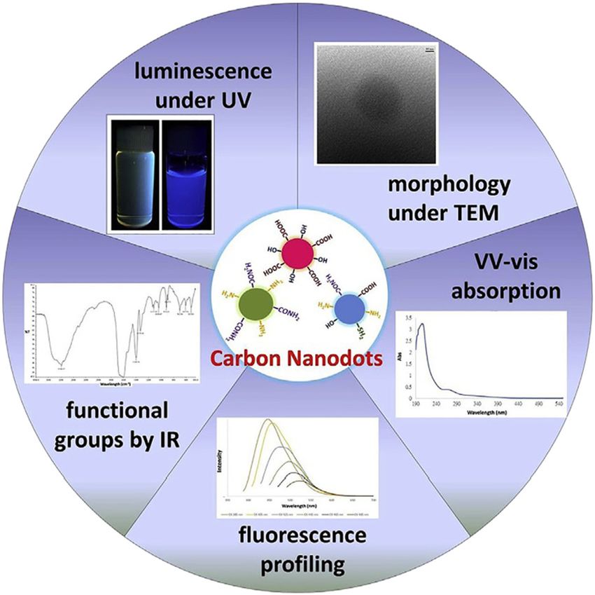
Carbon dots: A review 139
solution for 6 h [61,62] or even 1 year [63] as CDs are system, the CDs with red fluorescence have been easily
stable when maintained in aqueous solution and high- prepared. Preparation of red-fluorescencing CDs with
salinity solutions for long period. By dissolving in 4 NaCl high QY can be executed by using p-phenylenediamine
solution as well as in high concentrations (400 µM) of [77] and 1,3-di-hydroxynaphthalene [78] and using the
H2O2 solution, Liu’s group noticed a perfect signal of oxi- threefold symmetric phloroglucinol as precursor, Yuan
dation impedance when the prepared CDs were quite et al. [79] worked on expanding the conjugated π system
stable [64]. CDs usually display excellent impedance to under controlled conditions to prepare highly fluorescent
photobleaching as CDs have the ability to resist changes CDs from blue to red to decrease the spectral interference
in pH over the wide range of 3–12 [65] that is, photo- fluorescent signals and protect tissues from potential
bleaching impedance is more important than chemical damage in addition to CDs with longer emission wave-
stability due to the long observation times reported in lengths’ preparation shares in the explanation of the
other essays. For instance, various CDs fluorescent inten- fluorescence mechanism.
sity reduced slightly after UV irradiation for 12 h [66] and
24 h [67].
4.5 Toxicity
4.3 Up-conversion fluorescence Material toxicity is a significant measure of the material’s
potential manipulate environment and its denizens in
The emission wavelength is shorter than the excitation various applications. In the last few years, the toxicity
wavelength which is referred to as up-conversion fluo- of CDs was discovered. 3-(4,5-Dimethyl thiazol-2-yl)-2,5
rescence. Decrease in the background autofluorescence diphenyltetrazoliumbromide (MTT) assay is a common
is due to the longer excitation wavelengths and various in vitro toxicity analysis and is assessed by multiple
labeling with different emission wavelengths’ sand groups. Zhang et al. explored whether the assigned CDs
photon tissue permeation improvement as a result of in HeLa cells would have frivolous toxicity in the con-
up-conversion fluorescence, which makes it an essential centration range of 50–400 µg/mL [80–82]. Sahu and
role in imaging [68]. CDs prepared by ultrasonic treat- coworkers found that CDs were acceptable at high dose
ment has the property of up-conversion fluorescence (200 µg/mL) for prolonged incubation time (72 h) and
[62,69,70]. CDs prepared from glucose and ammonium nontoxicity to L929 cell lines when the CD concentration
hydroxide displayed an up-conversion emission in the is elevated as 1 mg/mL [83]. Zhou et al. show that the
range of 300–600 nm with excitation at 650–1,000 nm viabilities in MCF-7 cells minimized by 20% [84]. CD toxi-
[62]. Two photon absorption may generate up-conversion city is connected with the surface passivating agents [85]
fluorescence [71]. and modulated materials, such as polyethylene glycol
(PEG) [80] and polyacrylic acid [86]. The attained results
indicated that these CDs are still characterized by the low
toxicity in vitro. Yang and coworkers estimated the toxi-
4.4 Long wavelength attributes city of CDs in male CD-1 mice even at elevated dosage of
40 mg/kg (CD/body weight) according to the serum bio-
When the CDs reported were illuminated with UV light, it chemistry assays and histopathological analyses [87].
emits blue fluorescence [21,72,73]. The biological analysis Also, in vitro toxicity of CDs and by taking their surface
utilizes blue-emitting CDs which is not veiled since bio- charge into consideration, mouse fibroblasts NH/3T3
tissues and cells also emit blue fluorescence, which will cells [88] showed that CDs with negative charge due to
lead to spectral interference, so the perfect solution to the carboxylic groups arrested the G2/M phase of the cell
this problem was to use long wavelength emission to cycle. It is clear that CDs’ fabricated reproduction led
conquer this matter. Numerous treatments such as sur- to higher active oxygen species index without emanating
face modification [74], size control [75], and pH control the cell nucleus. Polyethylene glycol (PEG)-modified CDs
[60] were used to prepare CDs with longer wavelength having neutral charge did not stimulate any abnormal-
fluorescence. The introduction of heteroatoms such as ities in cell morphology, neither in intracellular traf-
nitrogen [76] and oxygen [71] led to the red shift in the ficking nor in cell up to a dose of 300 mg/mL. If CDs
emission wavelength in spite of the fluorescence from were coated by polyethylenimine (PEI) with positive
blue to green/yellow. By forming a large conjugated π charge, they will be the most toxic, being capable of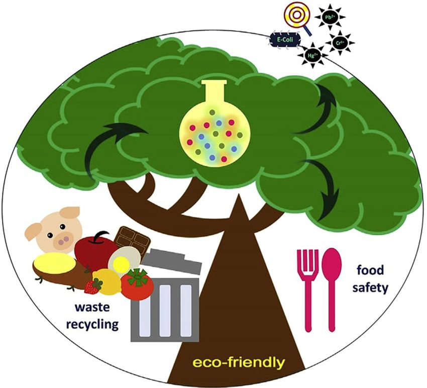
140 Asmaa M. El-Shafey
getting in the nucleus and boosting changes in the G0/G1
phase of the cell cycle at concentration as low as 100 mg/mL.
Cytotoxicity profiling CDs from fresh tender ginger juice
quelled the growth of human hepatocellularcarcinoma
cells with an IC50 of 0.35 mg/mL and accompanying
18-fold rise in cellular ROS production [89]. Furthermore,
CDs arrested the growth of liver tumors in a BALB/c mouse
paradigm, and these CDs were intoxic to MCF-10A cells
(noncancerous mammary epithelial cells), FL83B cells
(normal liver cells), a 549-cell (human lung cancer), MDA-
MB-231 cells (human breast cancer), and HeLa cells (human
Figure 8: Absorption intensity differentiations of NBB azodye at
cervical cancer) [90]. CDs synthesized from spent green tea different irradiation intervals in the existence of CDs/ZnO [40].
were water soluble and arrested MCF-7 (IC50 = 0.155 mg/mL)
and MDA-MB-231 (IC50 = 0.072 mg/mL) cell growth with lower
toxicity to HeLa, LLC-PKI (renal proximal tubular), and MCF- Waste water treatment can be executed by using CDs with
10A cells. ROS generation occurred in the inhibitory activity. other photocatalytic materials. Excellent photocatalytic char-
CDs had a little impact on the thrombin activity by fibri- acterization of silver orthophosphate (Ag3PO4) was
nogen conversion to fibrin, thus posing no hazard on blood explained by many reports. It can be used to degrade
coagulation. These results showed that CDs with low toxicity organic contaminants and water oxidation in spite of
can be considered the gold standard in the context of sus- less tendency to be water soluble. CDs/Ag/Ag3PO4 and
tainability. According to the CDs’ impact in air, rats, and fish CDs/Ag3PO4 photocatalytic systems were prepared as
[91], many scientists evaluated the low [92] environmental mentioned by different studies by simple dispersion of
CD toxicity when the CD concentration is not so high. CH3COOAg and polyvinyl pyrrolidone in a CD solution,
and Na2HPO4 was added drop wise, and the former was
stirred at 25°C for 4 h in dark and the resultant material
was dried in an oven at 50°C for 12 h [95]. After complex
5 CD nanocomposites formation, characterization was examined by various
techniques – as seen in Figure 9, SEM image detected
CDs offer an appropriate photo charge carrier flow as it the rhombic dodecahedral morphology of the complex
both accepts electron and transports electron, thus boosting with a size of 800–900 nm; and the UV-Vis spectroscopy
the photocatalytic characterization. Semiconductor carbon explained that the complex absorbs in the range of
nanomaterials’ preparation led to enhanced catalytic qua- 530–1,000 nm led to photocatalytic activity estimation
lification. A lot of reports discussed CDs photocatalytic and also its essential role in absorbing sunlight. Under
nature without complex formation as CDs were prepared visible light irradiation, methyl orange (MO) photocata-
by using alkali-assisted electrochemical method. C-dot irra- lytic activity was shown by CDs/Ag/Ag3PO4 complex as
diation by near-infrared (NIR) light was used to test the MO dye was completely degraded within 10 min, on the
photocatalytic activity led to benzyl alcohol oxidation contrary to CDs/Ag3PO4 complex which needed at least
to form benzaldehyde, which means the photocatalytic 25 min for degradation while pure Ag3PO4 requires 55 min
activity of CDs has the ability to transform alcohols to their to degrade, which means the prepared complex has the
corresponding aldehyde [93]. When ZnO is excited by highest photostability and catalytic activity in addition
UV light, electron–hole pair results plus initiation of to up-conversion PL exhibition. Photostability improve-
hydroxyl formation in water [94] making it a stable ment, photocatalytic activity, and photocorrosion dodge
visible-light-driven photocatalyst [36]. Carbon NPs’ union of metal complex perform as an electron reservoir and
with ZnO aids in enhancing the optical properties. A report take advantage of the entire spectrum of sunlight. By
explained the solution dispersion method of CDs loaded mixing the as-prepared mSiO2 with glycerol and PEG-
with ZnO and also naphthol blue black (NBB) azodye was NH2 at 230°C for 30 min, a C-dots@miSiO2-PEG nanocom-
tested using ZnO doped on CDs, as seen in Figure 8. Com- posite resulted, which was tested for drug delivery as it
plete degradation of CDs occurs after 45 min whereas using offered enough space for storing drug molecules, thereby
pure CDs led to the degradation of 4.4% NBB was within the performing as an efficient carrier. For example, controlled
same period [40]. This reflects the fact that photocatalytic drug delivery was prepared using the water-soluble antic-
nature of CDs makes it useful for harmful dye degradation. ancer drug doxorubicin (DOX) was loaded into the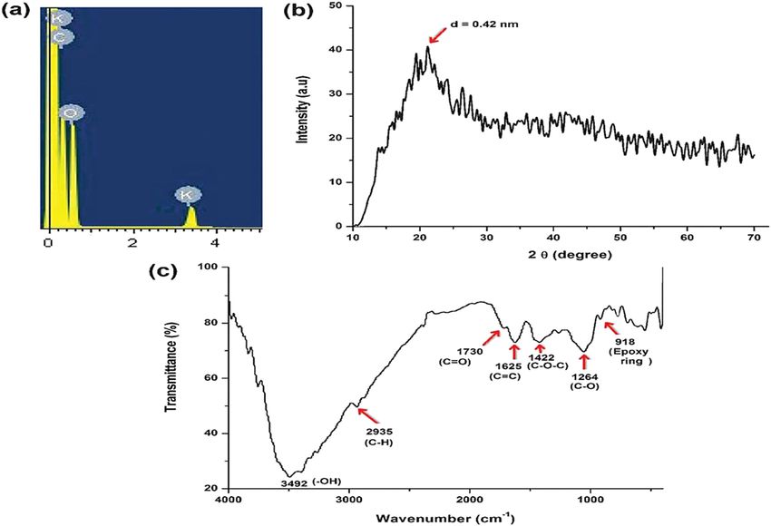
Carbon dots: A review 141
6 CD green synthesis
New research discussed green synthesis and found good
salience. Preparation of value-added products from bio-
degradable waste is an important topic in the green
chemistry field. The affirmation on green chemistry to
decrease the use of high-end chemicals due to the toxicity
concerns. The materials used in the green synthesis
should be eco-friendly and cheap so they are produced
from plants to be used as the starting materials. Green
synthesis has many advantages, that is, it is biocompa-
Figure 9: (a) SEM and (b) HRTEM images of CDs/Ag3PO4 complex tible, non-time-consuming, less toxic, economical, and
photocatalyst. (c) SEM and (d) HRTEM images of CDs/Ag/Ag3PO4 requires lower temperatures, so it is considered an inter-
complex photocatalyst [22]. esting topic for researchers. Carbon can be extracted by
green synthesis as it is available in all organic materials.
nanocomposite by reducing the drug pre-release before One of the reports discussed CDs’ green synthesis from
drug absorption by the target cell [96]. BiVO4, a semicon- orange peels by hydrothermal treatment [40], as it is a
ductor material, was used to improve the photocatalytic cheap renewable method to prepare chemical compounds
activity of complex formation with CDs. Degradation of from eco-friendly starting material. Hydrothermal synth-
methyl blue dye under visible light at room temperature esis uses high-pressure and high-temperature condi-
was used to examine the photocatalytic ability of this com- tions, leading to perfect morphology of the structures
plex [42]. Besides, semiconductor complexes considered an and considered economic method because of using water
important example of photocatalytic activity of CD com- as a solvent. First, washing the peels and then exposing it
plexes, especially formation of semiconductor/CD com- to sunlight for drying followed by oven drying at 150°C
plexes gives the maximum light energy [97]. The essential for 10 h followed by the addition of H2SO4 to wash the
requirement for photocatalytic systems is improving the carbonized peels rinsed with water, soaked with sodium
power conversion efficiency. Rhodamine B (RhB)/TiO2 com- hypochlorite solution for 4 h, and after that washed with
plex was doped on CDs to give an example of CD complex water until the pH of the washed water reached 7; the
synthesis to perform a semiconductor photoelectric conver- preheated oxidized orange peels were hydrothermally
sion system that led to improved photocurrent density. By treated in a Teflon lined autoclave at 180°C for 12 h and
CDs’ addition to the aqueous RhB solution, there is an then dichloromethane was added to the resulting solu-
increase in the UV-Vis absorption. Due to the presence of tion and the separating solution centrifuged at 5,000 rpm
oxygen-bearing functional groups, the doped CDs were of for 20 min to obtain a brown solution of an aqueous sus-
greater solubility, and UV-Vis absorption becomes greater pension of CDs as shown in Figure 10. The QY was 12.7%
when CD was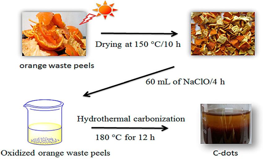
142 Asmaa M. El-Shafey
starting materials are nontoxic and biocompatible and
have fluorescence properties to be applicable in bioima-
ging technique.
7 CDs synthesis from commercially
available food products
The commercial food products consumed in our day-
to-day life can be discussed in many recent research.
Figure 10: Formation of C-dots by using waste orange peels’ Amorphous CD synthesis is discussed in many recent
hydrothermal treatment [40].
research. Amorphous CDs are present in different car-
amel-containing food products like jaggery, sugar, bread,
Another group prepared biocompatible CDs from aqu- biscuits, and corn flakes; and CDs were extracted from
eous extracts of Trapa bispinosa peels which is consid- the browner part of the bread, caramelized sugar, and
ered simple, rapid, and effective method. First, T. bispi- jaggery. The materials were of caramel color under white
nosa peels were washed in water, soaked in cold water for light and blue fluorescence under UV light as shown in
30 min, the peels were crushed in distilled water, centri- Figure 11. The lowest QY was from jaggery observed at
fuged, and suspended in NaOH solution to produce a 0.55% and the highest bone was from bread observed at
clear yellow CD suspension [101]. CDs were prepared 1.2%. CD morphology was spherical according to TEM
by centrifugal separation from sugarcane juice; a facile images. Bread-extracted CDs possess the highest particle
method to prepare crystalline CDs from highly alkaline size while those extracted from sugar caramel have the
sugarcane juice. A few milliliters of NaOH solution were lowest one. The amorphous nature of CDs was obvious
added drop wise into the sugarcane juice, which is stirred by XRD analysis. Due to the presence of carboxyl and
until a reddish brown solution is obtained, centrifuged at alcohol functional groups, the synthesized CDs were
5,000 rpm for 15 min, and checked under the UV light to hydrophilic in nature [38]. All the pervious perceptions
have a dark green fluorescence. Sucrose density gradient led to the affirmation of CDs’ existence in these food pro-
centrifugation was executed to separate CDs from the ducts. Coffee grounds can be heated, dried, and sepa-
mixture. As banana juice contains glucose, sucrose, fruc- rated to synthesize CDs. CDs’ preparation from coffee
tose, and ascorbic acid, some recent study synthesized grounds is a simple method as it includes the following
CDs from banana juice. Banana juice was extracted by four stages: dehydration, polymerization, carbonization,
crushing banana with a small amount of water, the and passivation. As a result of dehydration, polymeriza-
pulp-free juice was extracted, mixed with ethanol, heated tion and nucleation occurred in addition to carbonization
in a glass bottle closed with a cotton cork, then kept in an due to heating. Nuclei growth occurred by solute dif-
oven at 150°C for 4 h. When the solution cooled to room fusion toward particle surfaces. Prepared CDs were char-
temperature, it turned into a dark brown product, which acterized by using different techniques. The average
was then mixed with water and filtered to separate the diameter of the particles was ±2 nm according to TEM
residue. The aqueous solution was mixed with ethanol results. The existence of carbon atoms was confirmed
and centrifuged at 3,000 rpm for 15 min at room tempera- by XRD as well as EDX data as showed in Figure 12.
ture, and the ethanol was evaporated completely to obtain The amorphous nature of CDs was detected by Raman
the fluorescent CDs whose yield was 600 mg [37]. Accord- analysis. The fluorescence emission spectra exhibited a
ing to all the previous reports, CDs’ green synthesis is of broad range from blue (400 nm) to red (600 nm). The
high prominence in the present-day script. These methods emission peak position shifted to longer wavelengths
have many advantages as it uses very cheap starting mate- when the excitation wavelength was increased and the
rials so it considered cost-effective in addition to high yield intensity decreased. When excited at 365 nm, the stron-
of CDs [102]. All the previous methods in the abovemen- gest PL of 3.8% occurred at 400 nm and the CDs’ fluo-
tioned reports facilitate the discovery, and many other rescence could only be noticed when they were surface
research of CDs synthesis explained the simple and cost- passivated [103]. CD synthesis aids in the management of
effective methods. The CDs obtained from eco-friendly wastes as the mentioned in the previous reports. CDs can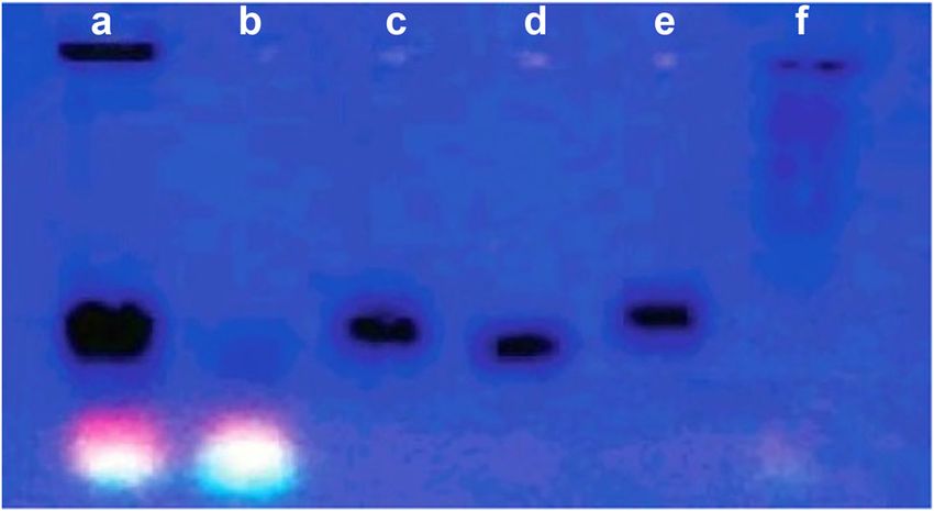
Carbon dots: A review 143
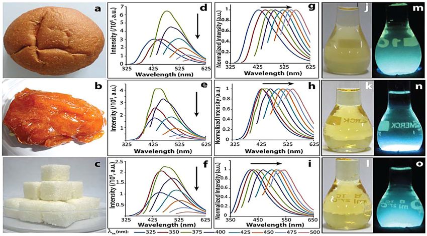
Figure 11: (a–c) Photographs of commercial bread, jaggery, and sugar. (d–i) Excitation-wavelength-dependent emission spectra of CDs from
bread, jaggery, and sugar caramel. (j–l) Photographs of CDs dispersion from bread, jaggery, and sugar caramel observed under white light,
and (m–o) the same under UV light [38].
be produced from many carbohydrate-rich food pro- group and Cu2+, Dong et al. [105,106] synthesized a
ducts. Waste management participation and green branched PEI-modified CDs for Cu2+ detection in a river
chemistry help in promoting scientific research. Recent samples with a 6-nM detection limit of 9 (Figure 13b).
improvement in CD synthesis was achieved using greener Quenching effect of Cu2+ on the fluorescence CDs enhances
methods. Green chemistry use compared to the physical image detection of cellular Cu2+, and probing the Cu2+
methods facilitates the process and offers an eco-friendly position in cells (Figure 13c) was attained by using AE-
synthesis. TPEA-CDs-CdSe/ZnS nanocomposite [107]. Cellular Cu2+
was detected by Vedamalai et al. [108] who used synthe-
sized Cu2+-sensitive CDs. Acetyl choline (ACh), which
transformed to choline by acetylcholinesterase by using
CDs reduced graphite oxide (CDs@RGO) composites whereas
8 Applications of C-dots choline could beget H2O2 in the existence of choline oxidase
(Figure 13d) [109]. The quenching C-dots@RGO fluorescence
8.1 Sensing
The most important CD application is the sensing field.
On the basis of the fluorescence properties and the sur-
face of CDs’ functional groups, such as Hg2+ detection
and biological thiols (Figure 13a) [104] the electrons and
the holes of CDs can be restructured and the fluorescence
of CDs can be quenched by Hg2+. The fluorescence of CDs
can be restored as Hg2+ has strong coordination ability
with thiols and recognize the “turn-off” detection of Hg2+
and “turn-on” detection of thiols. By using fluorescence
quenching from the coordination between CDs’ amino Figure 12: (a) TEM and (b) HRTEM images of CDs [103].144 Asmaa M. El-Shafey ability of H2O2 assists the ACh quantitative detection with limit of 13 nM was achieved by Li et al. [117] who constructed 30 PM detection limit. Biological active substances were a graphene nanodots-based ECL sensor (Figure 13g). detected by CDs-Ag/CDs-Au nanocomposite which has another trial in biosensing field [110–112]. The detection limit of glutathinone by using fluorometric/colorimetric bimodal sensor based on CDs-Au composites was 50 nM 8.2 Drug delivery and gene transfer [111]. In colorimetric method, the detection limit was 0.18 and 1.6 µM for H2O2 and glucose, respectively [110]. A One of the interesting fields of CDs application is drug boric acid-modified CDs were prepared by Zhang et al. delivery and gene transfer. Recognition of cancer cells [113] for the glucose detection with the limit of 0.03 nM, can be achieved by using folic acid-modified CDs by taking into consideration the strong affinity between amide condensation reaction which offered a good glucose and boric acid (Figure 13e). Glucose was used method to improve cell screening and disease diagnosis as a precursor to synthesize a CD via neutralizing the [118]. PEI-modified CDs have a positive surface potential, heat process [114]. Because the adjacent hydroxyl groups so it could absorb the negative DNA to transfer gene [119]. from glucose remained on the CDs’ surface, these CDs Liu et al. [119] discovered that CDs not only had fluores- exhibited a perfect affinity to boric acid, so CDs had an cence characterization to display the plasmid DNA dis- ability to detect glycoprotein. Chemiluminescence (CL) tribution during the transfer process but also offered and electrochemiluminescence (ECL) biosensors were well detailed information for plasmid DNA physiological func- constructed by using CDs. Lin et al. invented a CD-based CL tion research. After transfection for 3 h, CD DNA compo- sensor for nitride detection as they discovered that CDs had sites could enter the cell. During the transport process, CL signal in the presence of nitrous acid peroxide (Figure different wavelength excitation CDs preserved multicolor 13f) [115]. To track Cu2+ in the mice brain, Shao et al. [116] fluorescence property. utilized CDs and Tri prime and amplification (TPEA), which Lai et al. [120] synthesized a PEG-modified CDs and showed electrochemical response. Cd2+ detection with a fulfilled DOX loading and delivery. DOX in cells’ release Figure 13: Bio-/chemsensing applications of carbon dots. (a) Schematic representation of an Hg2+/cys (cysteine) biosensor based on CDs prepared from pomelo peel [104]. (b) Schematic representation of a Cu2+ biosensor based on CDs-BPEI (branched polyethylenimine) [105]. (c) Fabrication of a ratiometric fluorescent sensor based on a CDs-CdSe/ZnS hybrid material for detection of copper ions, and in vivo imaging and monitoring of cellular copper ions [107]. (d) Schematic representation of detection of ACh by C-dots@RGO in a TURN-OFF Strategy [109]. (e) Schematic representation of boron-doped graphene quantum dots, and mechanism for glucose sensing by boron-doped graphene quantum dots [113]. (f) Mechanism for nitrite sensing based on CL of CDs induced by peroxynitrous acid [115]. (g) ECL sensing for Cd2+ based on graphene quantum dots [117].
Carbon dots: A review 145
Figure 14: (a) Multicolor images of cells treated with CDs [16]. (b) Fluorescence images of cells treated with CDs-DOX at 0.5, 24, and 48 h
[133]. (c) Fluorescence images of (a) CDs-PEG, (b) CDs-PEG-FA, (c) CDs-PEG-FA/ZnPc, and (d) CDs-PEG-FA/ZnPc pretreated with FA [123].
(d) Fluorescence images of MD-MBA-231 cells pretreated with “biomolecule-mimicking” CDs [134].
process is exhibited by fluorescence images. CDs acquired transfection such as HeLa [123–129], human neutral
cytoplasm green fluorescence. At the time of treatment, stem cells [130] 4-T1 [131], NIH-3T3 [132], A549 [108,133],
DOX releases into the nucleus after entering the cells, HepG-2 [126], etc. Through endocytosis, CDs enter into
causing the appearance of the red fluorescence. To observe the cells and concentrate in the cytoplasm [126]. CDs
drug delivery, Chowdhuri et al. [121] joined CDs with metal may enter cell nucleus but only a few reports were dis-
organic frameworks. Wang et al. [122] used the mixture of cussed this way. Zhu et al. [16] successfully used CDs
chitosan, PEG, and CDs to attain a NIR light/pH dual-respon- in cell imaging as they synthesized fluorescent CDs with
sive hybrid gel. All the previously mentioned research indi- low cytotoxicity in solvothermal method (Figure 14a).
cate CDs’ application in drug delivery and gene transfer and Nitrogen-doped CDs displayed an excitation which was
corroborate CDs’ clinical applications. dependent on fluorescence emission for multicolor fluo-
rescence imaging [126] by carbonization extraction. Multi-
functional tumor targeting probe preparation can be
8.3 Biological imaging prepared by CDs’ surface modification. Tang et al. [133]
discussed cancer cell recognition, drug transport, and
8.3.1 In vitro imaging fluorescence imaging by using CDs with folic acids and
DOX (Figure 14b). Bhunia et al. [129] explained surface
In vitro imaging is executed to offer plentiful information modification of CDs with folic acid by preparing a series
on the imaging ability, distribution and cytotoxicity of of CDs with different fluorescence and observed target
probes in cells. CDs have many effective uses in cell recognition. To make the tumor abilities obvious and146 Asmaa M. El-Shafey
Figure 15: (a–h) Fluorescence image of zebrafish with CDs [139] and (i–m) fluorescence image of rat injected with CDs [87].
execute photothermal therapy (Figure 14c), Choi et al. developmental stages and optical imaging amenability
[123] used CDs’ altered with zinc phthalocyanine and folic [137]. The fluorescence imaging model was the reason
acid. Dopamine-neutralization heat treatment [134] was to choose zebrafish; CDs were found to heap up in eyes
used to synthesize a biomolecule mimicking CDs. The and yolk sac selectively (Figure 15) [114,138,139]. CDs
dopamine-mimicking CDs could “hoax” nuclear mem- were preserved over 60 h, so they are suitable for obser-
branes to fulfill nuclear localization and imaging [134] ving the long-term zebrafish developmental stages. PEG-
because the functional groups of dopamine were main- passivated CDs and Zns-doped CDs (CZns-dots-PEG) [80]
tained in this CDs (Figure 14d). Elimination of autofluo- were used for vivo imaging in spite of their blue and
rescence interference can be achieved by CDs’ two-photon, green emission, which were not suitable for in vivo ima-
cell imaging upon NIR light excitation (800–900 nm) ging. Under different excitations, no apparent toxicity
[134]. Under the 880 nm laser excitation, Yang et al. [87] was noticed in tissues and organs [80], green and red
attained green fluorescence images. Cell nucleus two- emission fluorescence images were obtained. Using CD
photon imaging was fulfilled by Zhang et al. [135] who probes by administering interdermal injection, the lymph
utilized C3N4 nanodot as a probe. For living cells and tissue of mice was labeled. The metabolism speed of the non-
imaging, Kong et al. [136] synthesized pH-sensitive CDs modified CDs was faster than the metabolism speed of
because of its two-photon fluorescence property. PEG-modified CDs. CD fluorescence was seen at the sto-
mach past intravenous injection and heaped up in the
bladder after 1 h. Four h post injection, the fluorescence
8.3.2 In vivo imaging signal decreased and heaped up in the kidney, explaining
that CDs are excreted from mice by urine [80]. Under dif-
Many essential medicine science such as disease progres- ferent excitations (455–704 nm), Tao et al. [140] obtained
sion, improved mechanism, and pattern formation CD in vivo images. When CDs were used in vivo imaging
were achieved using zebrafish due to its well-defined [141], CDs could enter into brain and appeared blue.Carbon dots: A review 147
8.4 CDs food safety applications synthesis and gave a brief summary of the latest research
development of CDs in fluorescent properties and their
Chemical contaminations such as metal ions, anions, pes- applications that can give explanation of the optical
ticides, veterinary drugs, and banned additive are the properties among the fluorescent properties. It is obvious
main reasons for many health problems. This demands that understanding these properties has a long way to go.
very effective and actual methods for guaranteeing food We focused on the CDs’ greener methods which are more
safety. In the food quality and safety area, a number of economical, less complicated, and less consuming, have
sophisticated CD-based biosensors are required. The main lower toxicity, better compatibility, nondestructive nature,
concepts of these CDs-based biosensors can be divided and improved optical properties. High QY of CDs by using
into four kinds: green synthesis methods is not difficult any more. CDs’
1. Direct fluorescence quenching (turn-off), research emphasizes on the development of optical proper-
2. Direct fluorescence boost (turn-on), ties and discovery of red fluorescence of CDs. Moreover,
3. Fluorescence quenching again (off-on), and functional modifications play another essential role in CD
4. Fluorescence quenching again (off-in-off). improvement: CD fluorescence focuses on blue-green emis-
sion and few red-emitting CDs were discussed [145]. Fluor-
The fluorescence quenching mechanisms implicate: escent CDs can be as follows:
1. Dynamic quenching, 1. CDs’ optical properties could be enhanced by tuning
2. Static quenching, carbon sources.
3. Fluorescence resonance energy transfer (FRET), and 2. Red-emitting CDs can be obtained from phenylene-
4. Inner filter effect (IFE) [142]. diamines as carbon source as Jiang et al. discussed
[146]. Polythiophene as a carbon source exhibited
CDs’ excited states return to ground state by collision CDs’ fluorescence emission shifting to 650–700 nm
with quencher and produces energy transfer/charge transfer as Ge et al. explained [147].
(ET/CT), which is known as dynamic quenching while the 3. CDs’ fluorescence may be improved by using appro-
static quenching results when CDs form nonfluorescent priate passivated agents.
ground state complex with quencher. FRET and IFE are on 4. The bandgap spacing of CDs’ adjusting fluorescence
the contrary mechanisms that demand two essential factors, yield and emission range can be done by introducing
namely, spectrum overlap and the distance between CDs heteroatoms.
and quencher (receptors). At less than 10 nm distance, 5. Functional molecules can be used to modify carboxyl
FRET occurs while at more than 20 nm distance, IFE takes and amino groups on the surface of CDs by amide
place. Some targets’ quench lead to improve the CDs’ fluo- condensation [39].
rescence immediately, permitting merely sensors’ design for 6. CDs’ blue shift emission and fluorescence quenching can
some targets (metal ions, veterinary drugs, bacteria, and be achieved by fluorescence resonance energy transfer
some functional components in foods) [29,56,143,144]. between CDs and modifications which must be taken
Restoring CDs’ fluorescence after supplementing the target into consideration through the following points:
into quencher-containing fluorescence quenching system a. The distance between CDs and modifications could be
is known as fluorescence quenching (off-on-off) strategy, increased by modified method choice such as choosing
which depends on quencher, restorer, and target. Where appropriate bridging content (such as silicon beads,
the fluorescence recovers due to the interaction between inorganic clay, etc.) led to resonance energy transfer
quencher and restorer and then quench again; to enable reduction.
the interaction of target and restorer, the “off-on” and “off- b. Building novel CDs for multimodal imaging can be
on-off” strategies can be utilized to determine other targets executed by multimodal functionalization and multi-
(such as anions, pesticides, veterinary drugs, and some modal imaging factor introduction.
functional components in foods) [21,23].
9 Conclusion 10 Example
This review discussed the discovery, structure, and com- One pot hydrothermal treatment of gadopentetic acid
position of CDs as well as CDs’ nanocomposites and green mixture, trihydroxy methyl aminomethane, and betaine148 Asmaa M. El-Shafey
[148] to attain Gd-doped CDs with a size of 3–4 nm was TiO2 – water - soluble CDs composite was inspected
explained by Bourlinos et al. [148,149]. utilizing methylene blue dye as a model pollutant
Heteroatom doping [150], large conjugated system because CDs have been immobilized over electrospan.
formation [78], and change the reaction medium [151] TiO 2 nanofibers and the emanating photocatalytic
have been executed to enhance QY or red-shift degree. activity being about 2.5 times more than that of titanium
Due to the surface groups (such as carboxylic acids dioxide nanofibers [161]. Water-soluble fluorescent carbon
and hydrolysis), most of the synthesized CDs are water quantum dots (∼260–400 nm) were reported via a simple
soluble. Further surface functionalization of CDs can be one-step hydrothermal treatment utilizing Tamarindus
achieved by doping with heteroatoms (such as N and S) indica leaves. These bioconvenient CDs can be potentially
conferring specific chemical reactivity to CDs permitt- executed in sensing, bioimaging, disease diagnostics, and
ing CDs to be specific probes/bioconjugates in detection other analytical implementations [162]. Another fluorescent
application which is very critical toward surface functio- CD synthesis facile approach requires the hydrothermal
nalization [54,152]. Surface groups’ significance in linkage treatment of pineapples (Ananas comosus) and calamansi
with targets is to enhance the CD-based sensors selectivity (Citrofortunella microcarpa) wastes [163]. Escherichia coli,
in food safety applications [153–159]. Toxic elements’ fluoresced with CDs as operative probe, has been used for
detection by CDs depends on energy-transfer capability. bioimaging applications. Generation of hydrogen bonds
CDs can be used in biological sample analyses due to its between the bacterial cells and CDs eases the quantum
biocompatible nature. There is still a lot of areas for dis- dots’ attachment onto the bacteria [163]. Greener ozone
covering CDs’ functionality with various elements. In the oxidation of ignite coal was utilized for well-dispersed CDs
near future, CDs will lighten human lives by slapping the was utilized for well-dispersed CDs (∼2–4 nm) which is
way for more advanced techniques. copious, cost-effective, and involves oxygen-rich functional
groups with suitable water-solubility, optical characteristics,
and yield reaching 35% [164]. Synthesized quantum dots
11 Future perspectives utilized for Fe3+ detection with remarkable sensitivity and
selectivity as well as desirable anti-interference had quench-
ing effects but the CDs’ intensity showed clear linear
11.1 Green chemistry as an essential responses to the Fe3+ concentrations (10–150 µmol L−1).
approach to attain heteromeric With the detection limit of 0.26 µmol L−1 [164], in one
superstructures pot by using cydonia oblonga powder as carbon precur-
sors via microwave irradiation which shows maximum
11.1.1 CDs eco-friendly and sustainable synthesis emission at 450 nm if excited at 350 nm with a QY
of 85% highly stable and luminescent multi-color CDs
Green chemistry is a linkage of important concepts that would generate. Also CDs were prepared through micro-
minimizes or eliminates the implementation or genera- wave heating technique in 30 min in comparison to those
tion of hazardous substances in the design, manufacture, produced hydrothermally in a Teflon-linen stainless steel
and chemical product implementations. Green chemistry body autoclave at 200°C [165]. Fluorescent CDs’ greener
involves using none or less dangerous chemical synthesis, synthesis by using citrus lemon juice (∼2–10 nm) was
applying safer and nontoxic chemicals, solvents, and pro- achieved utilizing hydrothermal approach [166] which
cesses [160]; for example, biowastes can be utilized as showed high PL intensity was obtained at pH of 6 in
sustainable and cheap carbon sources for CD synthesis. cell imaging [166]. CDs can be utilized as a fluorescent
Spherical water-soluble CDs (about 1–3 nm) have been probe. Amorphous fluorescent CDs can be obtained from
prepared from lemon peel waste, which is the prevailing orange peel waste by utilizing hydrothermal carboniza-
cost-effective hydrothermal strategy, and the ensuring tion approach at mild temperature [40]. Bright green
stable CDs were discovered to be oxygen rich in surface luminescent graphitic carbon nitride doped with oxygen
functionalities and displayed the water-soluble and and sulfur were prepared using microwave treatment of
unique PL properties with QY of about 14%. The synthe- citric acid and thiourea. They exhibited excitation wave-
sized CDs were used to design an economic, green, and length and pH-dependent luminescence behaviors in the
highly sensitive fluorescent probe for Cr6+ ions’ detection visible range. Moreover, the QY of 31.67% shows a strong
limit of about 73 nM. Water-soluble CDs considered impedance to the interference of high ionic strength
simple, fast sensitive, and selective Cr6+ detection is environment, and perfect biocompatability as estimated
suitable technique in operations of water purification. by the cell viability assay [167]. Prunus avium fruitCarbon dots: A review 149
mechanisms in which fluorescence is executed without
external high-power source, for instance, CL resonance
energy transfer (CRET) and bioluminescence resonance
energy transfer (BRET) as they include a nonradiative energy
transfer between a light energy donor and a lumines-
cence acceptor. The donor molecular emission enables
covering the acceptor molecular excitation or absorption
when the distance between donor and acceptor mole-
cules is close, i.e.,150 Asmaa M. El-Shafey
Research funding: Author states no funding involved. [10] Jiang C, Wu H, Song X, Ma X, Wang J, Tan M. Presence of
photoluminescent carbon dots in Nescafe® original instant
Author contributions: Asmaa Mohamed El-Shafey was coffee: applications to bioimaging. Talanta. 2014;127:68–74.
PMID: 24913858. doi: 10.1016/j.talanta.2014.01.046.
involved in writing – original draft, writing – review and
[11] Sharma V, Tiwari P, Mobin SM. Sustainable carbon-dots:
editing, conception, design, and interpretation of the recent advances in green carbon dots for sensing and bio-
reported review. imaging. J Mater Chem B. 2017;5(45):8904–24. doi: 10.1039/
C7TB02484C.
Conflict of interest: Author states no conflict of interest. [12] Zhang X, Jiang M, Niu N, Chen Z, Li S, Liu S, et al. Natural-
product derived carbon dots: from natural products to func-
tional materials. ChemSusChem. 2018;11(1):11–24. PMID:
Data availability statement: All the data generated or 29072348. doi: 10.1002/cssc.201701847.
analyzed during this review are included in this pub- [13] Zhang J, Wang H, Xiao Y, Tang J, Liang C, Li F, et al. A simple
lished article. approach for synthesizing of fluorescent carbon quantum
dots from Tofu wastewater. Nanoscale Res Lett.
2017;12(1):611–8. PMID: 29188541, PMCID: PMC5707215.
doi: 10.1186/s11671-017-2369-1.
[14] Zhou Y, Liu Y, Li Y, He Z, Xu Q, Chen Y, et al. Multicolor carbon
References nanodots from food waste and their heavy metal ion detec-
tion application. RSC Adv. 2018;8(42):23657–62.
[1] Xie Y, Filchakova O, Yang Q, Yesbolatov Y, Tursynkhan D, doi: 10.1039/C8RA03272F.
Kassymbek A, et al. Inhibition of cancer cell proliferation by [15] Huang CC, Hung YS, Weng YM, Chen W, Lai YS. Review:
carbon dots derived from date pits at low-dose. sustainable development of carbon nanodots technology:
ChemistrySelect. 2017;2:4079–83. doi: 10.1002/ natural products as a carbon source and applications to food
slct.201700575. safety. Trends Food Sci Tech. 2019;86:144–52. doi: 10.1016/
[2] Baig MB, Al-Zahrani KH, Schneider F, Straquadine GS, j.tifs.2019.02.016.
Mourad M. Food waste posing a serious threat to sustain- [16] Zhu S, Zhang J, Qiao C, Tang S, Li Y, Yuan W, et al. Strongly
ability in the Kingdom of Saudi Arabia – a systematic review. green-photoluminescent graphene quantum dots for bio-
Saudi J Biol Sci. 2019;26(7):1743–52. doi: 10.1016/ imaging applications. Chem Commun. 2011;47:6858–60.
j.sjbs.2018.06.004. [17] Ding H, Ji Y, Wei JS, Gao QY, Zhou ZY, Xiong HM. Facile
[3] De Menna F, Dietershagen J, Loubiere M, Vittuari M. Life cycle synthesis of red-emitting carbon dots from pulp-free lemon
costing of food waste: a review of methodological juice for bioimaging. J Mater Chem B. 2017;5(26):5272–7.
approaches. J Waste Manag. 2018;73:1–13. doi: 10.1016/ doi: 10.1039/C7TB01130J.
j.wasman.2017.12.032. [18] Miao X, Yan X, Qu D, Li D, Tao FF, Sun Z. Red emissive sulfur,
[4] Galanakis CM. Food waste recovery: prospects and oppor- nitrogen codoped carbon dots and their application in ion
tunities. Sustainable food systems from agriculture to detection and theraonostics. ACS Appl Mater Interfaces.
industry, improving production and processing. Cambridge: 2017;9(29):18549–56. PMID: 28508626. doi: 10.1021/
Academic Press; 2018. p. 1–442. Paperback ISBN: acsami.7b04514.
9780128119358, eBook ISBN: 9780128119617. [19] Gao W, Song H, Wang X, Liu X, Pang X, Zhou Y, et al. Carbon
[5] Rafiq S, Kaul R, Sofi SA, Bashir N, Nazir F, Nayik GA. Citrus dots with red emission for sensing of Pt2+, Au3+, and Pd2+
peel as a source of functional ingredient: a review. J Saudi and their bioapplications in vitro and in vivo. ACS Appl Mater
Soc Agric Sci. 2016;17(4):351–8. doi: 10.1016/ Interfaces. 2018;10(1):1147–54. PMID: 29235348.
j.jssas.2016.07.006. doi: 10.1021/acsami.7b16991.
[6] Hu Q, Gong X, Liu L, Choi MMF. Characterization and [20] Shi X, Wei W, Fu Z, Gao W, Zhang C, Zhao Q, et al. Review on
analytical separation of fluorescent carbon nanodots. carbon dots in food safety applications. Talanta.
J Nanomater. 2017;2017:1–23. doi: 10.1155/2017/1804178. 2019;194:809–21. doi: 10.1016/j.talanta.2018.11.005.
Article ID 1804178. [21] Borse V, Thakur M, Sengupta S, Srivastava R. N-doped multi-
[7] Mishra RK, Ha SK, Verma K, Tiwari SK. Recent progress in fluorescent carbon dots for ‘turn off-on’ silver-biothiol dual
selected bionanomaterials and their engineering applica- sensing and mammalian cell imaging application. Sens
tions: an overview. J Sci Adv Mater Devices. Actuators B Chem. 2017;248:481–92. doi: 10.1016/
2018;3(3):263–88. doi: 10.1016/j.jsamd.2018.05.003. j.snb.2017.03.158.
[8] Safarik I, Baldikova E, Prochazkova J, Safarikova M, [22] Fong JFY, Chin SF, Ng SM. A unique “turn-on” fluorescence
Pospiskova K. Magnetically modified agricultural and food signalling strategy for highly specific detection of ascorbic
waste: preparation and application. J Agric Food Chem. acid using carbon dots as sensing probe. Biosens
2018;66(11):2538–52. doi: 10.1021/acs.jafc.7b06105. Bioelectron. 2016;85:844–52. PMID: 27290666.
[9] Xu X, Ray R, Gu Y, Ploehn HJ, Gearheart L, Rakeret K, et al. doi: 10.1016/j.bios.2016.05.087.
Electrophoretic analysis and purification of fluorescent [23] Gao Z, Wang L, Su R, Huang R, Qi W, He Z. A carbon dot-based
single-walled carbon nanotube fragments. J Am Chem Soc. “off-on” fluorescent probe for highly selective and sensitive
2004;126(40):12736–7. PMID: 15469243. doi: 10.1021/ detection of phytic acid. Biosens Bioelectron. 2015;70:232–8.
ja040082h. PMID: 25829220. doi: 10.1016/j.bios.2015.03.043.You can also read