Lipid Dependence of Xanthophyll Cycling in Higher Plants and Algae - Frontiers
←
→
Page content transcription
If your browser does not render page correctly, please read the page content below
REVIEW
                                                                                                                                                  published: 21 April 2020
                                                                                                                                            doi: 10.3389/fpls.2020.00455

                                             Lipid Dependence of Xanthophyll
                                             Cycling in Higher Plants and Algae
                                             Reimund Goss 1* and Dariusz Latowski 2
                                             1
                                              Department of Plant Physiology, Institute of Biology, Leipzig University, Leipzig, Germany, 2 Department of Plant Physiology
                                             and Biochemistry, Faculty of Biochemistry, Biophysics and Biotechnology, Jagiellonian University, Kraków, Poland

                                             The xanthophyll cycles of higher plants and algae represent an important
                                             photoprotection mechanism. Two main xanthophyll cycles are known, the violaxanthin
                                             cycle of higher plants, green and brown algae and the diadinoxanthin cycle of
                                             Bacillariophyceae, Xanthophyceae, Haptophyceae, and Dinophyceae. The forward
                                             reaction of the xanthophyll cycles consists of the enzymatic de-epoxidation of
                                             violaxanthin to antheraxanthin and zeaxanthin or diadinoxanthin to diatoxanthin during
                                             periods of high light illumination. It is catalyzed by the enzymes violaxanthin or
                                             diadinoxanthin de-epoxidase. During low light or darkness the back reaction of the
                                             cycle, which is catalyzed by the enzymes zeaxanthin or diatoxanthin epoxidase,
                                             restores the epoxidized xanthophylls by a re-introduction of the epoxy groups. The
                           Edited by:
                 Yoshitaka Nishiyama,        de-epoxidation reaction takes place in the lipid phase of the thylakoid membrane
            Saitama University, Japan        and thus, depends on the nature, three dimensional structure and function of the
                     Reviewed by:            thylakoid lipids. As the xanthophyll cycle pigments are usually associated with the
                  Koichi Kobayashi,
                                             photosynthetic light-harvesting proteins, structural re-arrangements of the proteins and
  Osaka Prefecture University, Japan
             Alexei E. Solovchenko,          changes in the protein-lipid interactions play an additional role for the operation of the
Lomonosov Moscow State University,           xanthophyll cycles. In the present review we give an introduction to the lipid and fatty
                              Russia
                                             acid composition of thylakoid membranes of higher plants and algae. We introduce the
                  *Correspondence:
                      Reimund Goss           readers to the reaction sequences, enzymes and function of the different xanthophyll
               rgoss@rz.uni-leipzig.de       cycles. The main focus of the review lies on the lipid dependence of xanthophyll cycling.
                                             We summarize the current knowledge about the role of lipids in the solubilization
                   Specialty section:
         This article was submitted to       of xanthophyll cycle pigments. We address the importance of the three-dimensional
                      Plant Physiology,      lipid structures for the enzymatic xanthophyll conversion, with a special focus on
               a section of the journal
            Frontiers in Plant Science
                                             non-bilayer lipid phases which are formed by the main thylakoid membrane lipid
        Received: 25 February 2020
                                             monogalactosyldiacylglycerol. We additionally describe how lipids and light-harvesting
         Accepted: 27 March 2020             complexes interact in the thylakoid membrane and how these interactions can affect the
           Published: 21 April 2020          structure of the thylakoids. In a dedicated chapter we offer a short overview of current
                             Citation:       membrane models, including the concept of membrane domains. We then use these
       Goss R and Latowski D (2020)
    Lipid Dependence of Xanthophyll          concepts to present a model of the operative xanthophyll cycle as a transient thylakoid
   Cycling in Higher Plants and Algae.       membrane domain which is formed during high light illumination of plants or algal cells.
              Front. Plant Sci. 11:455.
        doi: 10.3389/fpls.2020.00455         Keywords: fatty acid, lipid, MGDG, thylakoid membrane domain, violaxanthin de-epoxidase, xanthophyll cycle

Frontiers in Plant Science | www.frontiersin.org                                    1                                                  April 2020 | Volume 11 | Article 455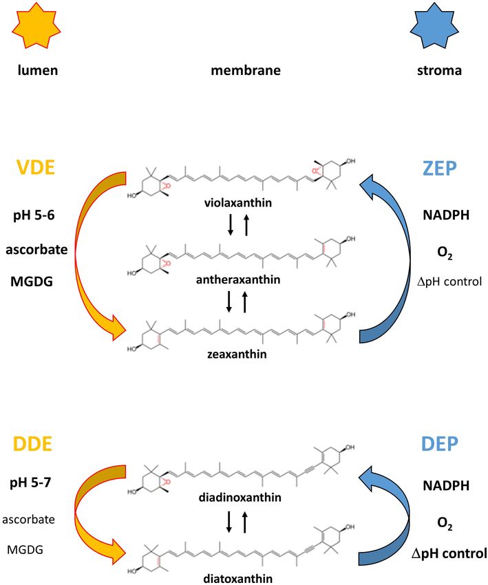
Goss and Latowski                                                                                             Lipid Dependece of Xanthophyll Cycling

INTRODUCTION                                                               2014; Ruban, 2016) and even today work on the molecular
                                                                           mechanism of NPQ in higher plants and algae represents an
Xanthophyll cycles, which consist of the de-epoxidation of                 important research topic. With respect to the localization of the
epoxy-xanthophylls during high light and the epoxidation of                xanthophyll cycle pigments it was clear from the beginning that
epoxy-free xanthophylls during low light or darkness, are found            the pigments are located within the chloroplast. Later studies
in all eukaryotic photoautotrophs (for reviews see Goss and                have presented evidence that within the plastidic thylakoid
Jakob, 2010; Niyogi and Truong, 2013; Goss and Lepetit,                    membranes the xanthophyll cycle pigments are associated with
2015). The xanthophyll cycles act as an important protection               the light-harvesting complexes of the photosystems (Bassi et al.,
mechanism against damage of the photosynthetic apparatus by                1993; Ruban et al., 1994, section Localization of Xanthophyll
supersaturating light conditions (for reviews see Horton, 2014;            Cycle Pigments in the Thylakoid Membrane). Regarding the
Ruban, 2016). The main part of photoprotection provided by                 main topic of the present review, i.e., the influence of lipids
the xanthophyll cycles operates on the time-scale of minutes and           on the operation of the xanthophyll cycles, first evidence
thus allows the plants to react to short-term changes of the light         that the main thylakoid membrane monogalactosyldiacylglycerol
intensities in their natural environment (Demmig-Adams and                 (MGDG) plays an important role in the conversion of Vx to Ax
Adams, 2006; Demmig-Adams et al., 2014). For higher plants                 and Zx dates back to the 1970s (Yamamoto and Higashi, 1978,
fast fluctuations of the light intensity can be induced by clouds          sections Lipid Classes and Lipids as Solvents for Xanthophyll
or by rapid changes of the leaf coverage in shaded environments            Cycle Pigments). Later measurements demonstrated that the
such as the tropical rainforests. For algae even moderate water            thylakoid membrane lipids, and especially MGDG, act as solvents
mixing can result in a rapid change of the light intensity from full       of the xanthophyll cycle pigments (Latowski et al., 2004; Goss
sunlight to almost complete darkness. In addition, tidal changes           et al., 2005, section Lipids as Solvents for Xanthophyll Cycle
affect the light exposure of those species inhabiting the coastal          Pigments) and that special three-dimensional lipid structures or
regions. Besides the fast photoprotection the xanthophyll cycles           phases, i.e., non-bilayer lipid phases, are needed for the efficient
provide long-term protection which lasts for days, weeks or even           operation of the xanthophyll cycles of higher plants and algae
months (Demmig-Adams and Adams, 2006; Demmig-Adams                         (Latowski et al., 2002, 2004; Goss et al., 2005, 2007, sections Three
et al., 2014). These long-lasting photoprotection components               Dimensional Structures of Lipids and Role of Non-bilayer Lipid
can be observed in evergreen plant species which are exposed               Phases for Xanthophyll Cycling). Additional investigations could
to prolonged environmental stress like the combination of cold             show that the xanthophyll cycle pigments are not only associated
temperatures and high light intensities during the winter months.          with the light-harvesting proteins via special protein binding
   The observation that changes in the light intensities lead              sites but also exist in lipid shields surrounding the complexes
to inter-conversions of specific leaf xanthophylls dates back to           (Lepetit et al., 2010; Schaller et al., 2010; section Localization of
the late 1950s/early 1960s (Hager, 1957; Saphozhnikov et al.,              Non-bilayer Lipid Phases in the Thylakoid Membrane). Recent
1957; Yamamoto et al., 1962). These first experiments revealed             research on the lipid dependence of xanthophyll cycling has
that the xanthophyll violaxanthin (Vx) is converted to the                 focused on the localization of the non-bilayer lipid phases
intermediate xanthophyll antheraxanthin (Ax) and then finally              within the thylakoid membrane (Garab et al., 2017, section
to zeaxanthin (Zx) during high light illumination. Later, it was           Localization of Non-bilayer Phases Involved in Xanthophyll
demonstrated that darkness or low light lead to the reversal of            Cycling) and how these structures are formed during high
the light-driven xanthophyll inter-conversions (Hager, 1967a)              light illumination (Goss et al., 2007; Jahns et al., 2009, section
and the term Vx cycle was introduced (section Types of                     Formation of Non-bilayer Lipid Phases by Structural Changes of
Xanthophyll Cycles). In addition, the existence of a second                Light-Harvesting Proteins). Models describing the operation of
xanthophyll cycle, namely the diadinoxanthin (Ddx) cycle, in               the xanthophyll cycle within the thylakoid membrane have also
several groups of algae was reported (Hager and Stransky, 1970;            included information from the newest concepts on the structure
Stransky and Hager, 1970). Newer measurements have shown                   and function of biological membranes (Goni, 2014; Nicholson,
that, besides the dominant Vx- and Ddx cycles, further less                2014, section The Xanthophyll Cycle Membrane Domain in the
common light-driven cyclic inter-conversions of xanthophylls               Light of Recent Membrane Models).
exist (Rabinowitch et al., 1975; Goss et al., 1998; Bungard
et al., 1999). The following investigations were concerned with
the characteristics of the enzymes which carry out the de-
                                                                           LIPID COMPOSITION OF THYLAKOID
epoxidation and epoxidation reactions such as pH-optimum
and co-substrate requirements (Hager, 1967a,b; section Reaction            MEMBRANES OF HIGHER PLANTS AND
Sequences and Xanthophyll Cycle Enzymes). With regard to the               ALGAE
function of the xanthophyll cycles it took until the late 1980s
that a connection between the conversion of Vx to Zx and                   Lipid Classes
the quenching of chlorophyll a fluorescence, which indicates               One of the most important biological functions of lipids is the
a thermal dissipation of excitation energy, could be obtained              formation of membranes within the cell and the cell organelles.
and described (Demmig et al., 1987, 1988, section Function of              In plant cells the largest membrane system is the thylakoid
Xanthophyll Cycles). Since then numerous studies have dealt                membrane within the chloroplast. Thylakoid membranes, like
with this process which was called NPQ (reviewed in Horton,                the inner membranes of mitochondria, are characterized by a

Frontiers in Plant Science | www.frontiersin.org                       2                                          April 2020 | Volume 11 | Article 455
Goss and Latowski Lipid Dependece of Xanthophyll Cycling
high protein per lipid ratio which already indicates that the (DGDG) and monogalactosyldiacylglycerol (MGDG),
thylakoid lipids play an important role not only as membrane contain a galactopyranosyl residue, whereas the sulfolipid
building blocks, but also as regulators of the structure and sulfoquinovosyldiacylglycerol (SQDG) carries a glucopyranosyl
function of both integral and peripheral membrane proteins, residue with a sulfonic group at the C6 position of the glucose
including the photosynthetic complexes or the xanthophyll cycle residue. The first galactose residues of MGDG and DGDG are
enzymes (Krauss, 2001; Kirchhoff et al., 2002; Kobayashi et al., β-anomeric forms, while the second galactose of DGDG, as
2016, see also section Xanthophyll Cycles of Higher Plants well as the glucose residue of SQDG are α-sugars (Hölzl and
and Algae). In the thylakoid membranes representatives of the Dörmann, 2019). In addition to MGDG, DGDG, and SQDG
following three lipid classes are present: glycosylglycerol lipids phosphatidylglycerol (PG), a lipid belonging to the class of GPs,
(GGLs), glycerophospholipids (GPs), and prenol lipids (PRs). is present in the thylakoid membrane (Harwood and Guschina,
Both GGL and GP molecules are characterized by the presence 2009; Figure 1). While DGDG and MGDG are neutral lipids,
of two fatty acyl residues and one trihydric alcohol, namely SQDG and PG are negatively charged due to the presence of
glycerol. The main difference between these two lipid groups the sulfate and phosphate residues, respectively (Boudiere et al.,
is the presence of a phosphate residue attached to glycerol in 2014). The presence of three other representatives of the GPs,
GPs, whereas in GGL molecules one or more sugar residues are namely phosphatidylinositol (PI), phosphatidylcholine (PC),
directly associated with glycerol through glycosidic bonds (Li- and in the case of the green alga Chlamydomonas reinhardii
Beisson et al., 2016). PRs also represent an important group of phosphatidylethanolamine (PE), in thylakoid membranes has
thylakoid lipids. They include, among others, quinones such as been proposed. However, it is still under discussion if these
plastoquinone, phylloquinone (vitamin K1 ) or tocopherols, as lipids really represent specific components of the inner thylakoid
well as isoprenoids such as the carotenoids or the phytol chains membrane. While in all thylakoids isolated from both plants
of the chlorophyll molecules (Li-Beisson et al., 2016). PRs, and and algae PI was commonly detected with concentrations up to
especially carotenoids, play an important role for the variability 5% of the total membrane lipid, it is not clear whether PC and
of the thylakoid lipid composition and can be used for the PE simply represent contaminations of thylakoid preparations
classification of various groups of algae. with lipids from membranes rich in GPs, such as cellular or
While the thylakoid membranes of different photoautotrophs mitochondrial membranes. In addition, nothing is known so
may differ in the nature of their prenol lipids, the other far about the putative role of these three GPs in photosynthesis
lipid classes appear to be comparable for different types (Boudiere et al., 2014).
of thylakoids. Differences are not observed in the general Whereas the presence of PI, and especially that of PC or
structure of the different lipid classes but rather in their PE, in thylakoid membranes still needs verification, MGDG,
fatty acid (FA) composition and their contribution to the DGDG, SQDG, and PG unequivocally represent the typical
total thylakoid membrane lipid. The most common lipids lipids of all oxygenic photosynthetic membranes, ranging from
of thylakoid membranes are three types of GGLs and one cyanobacteria to thylakoids of algae and plants (Boudiere et al.,
representative of the GPs (Figure 1). Among the lipids belonging 2014; Kobayashi et al., 2016). Among these four thylakoid
to the GGLs two lipids, namely digalactosyldiacylglycerol lipids, MGDG contributes up to 50% to the total higher
FIGURE 1 | Schematic structures of the four main thylakoid membrane lipids, i.e., the three glycosylglycerol lipids (GGLs) monogalactosyldiacylglycerol (MGDG),
digalactosyldiacylglycerol (DGDG) and sulfoquinovosyldiacylglycerol (SQDG), and the one glycerophospholipid (GP) phosphatidylglycerol (PG).
Frontiers in Plant Science | www.frontiersin.org 3 April 2020 | Volume 11 | Article 455Goss and Latowski Lipid Dependece of Xanthophyll Cycling
plant membrane lipid, DGDG contributes approximately 25%, charged membranes for the diatom physiology is, however, still
whereas the content of PG reaches up to 10% and SQDG unknown (Vieler et al., 2007; Goss and Wilhelm, 2009).
oscillates in the range from about 2 to 20%. Despite the fact Whereas SQDG and MGDG are almost undetectable in
that SQDG is widely considered as the least concentrated of extraplastidic membranes of algae and plant cells, DGDG
the four main thylakoid lipids, many results have shown that was shown to be able to substitute phospholipids in non-
especially the SQDG levels are highly variable in response photosynthetic membranes, like the tonoplast (Andersson
to different environmental conditions. Thylakoid membranes et al., 2005), the mitochondrial (Jouhet et al., 2004) or other
of higher plants or marine cyanobacteria under phosphate plasma membranes (Andersson et al., 2003). Replacement of
depletion can show maximum levels of SQDG of up to 50 phospholipids by DGDG usually takes place under phosphate-
or even 70%, respectively. Additionally, under conditions of deficient conditions (Härtel and Benning, 2000; Li et al.,
phosphate limitation, PG seems to be the only representative 2006). Under these limitations DGDG can amount to high
of the GP lipids with a content of solely 2% of the total concentrations and DGDG concentrations of up to 25% of the
membrane lipid of marine cyanobacteria (van Mooy et al., total membrane lipid were found in the plasma membranes
2006; Shimojima, 2011; Boudiere et al., 2014). A replacement and tonoplasts of oat root cells (Andersson et al., 2005).
of PG by SQDG is commonly observed and is considered DGDG together with MGDG, seems to play a key role
to be a ubiquitous phenomenon in photosynthetic organisms. in the stabilization of chloroplast membranes under various
Keeping constant the concentrations of the negatively charged kinds of environmental stresses such as drought, exposure
lipids most likely ensures the stability of an anionic lipid to ozone, cold or heat stress, which generally result in an
environment within the photosynthetic membranes under increase of the DGDG to MGDG ratio in the photosynthetic
phosphate limitation (Boudiere et al., 2014). With respect to membrane (van Besouw and Wintermans, 1978; Heinz and
function, SQDG appears to be important for the protection of Roughan, 1983; Heemskerk et al., 1986; Chen et al., 2006;
PSII in halophytes. In general, the level of SQDG was found Moellering et al., 2010; Moellering and Benning, 2011). This
to be considerably higher in many salt treated halophytes, is not surprising, considering that MGDG is a substrate for
e.g., in Aster tripolium, Sesuvium portulacastrum, or Crithmum DGDG synthases which belong to the glycosyltransferases and
maritimum, although no changes in the sulfolipid contents were are upregulated by environmental stress like e.g., phosphate
observed in glycophytes which were treated with different salt limitation. With respect to the role of MGDG, reduction of
concentration (Ben Hamed et al., 2005; Ramani et al., 2006). the MGDG concentrations of the thylakoid membrane by a
In the light of these results it is possible that the high levels of down-regulation of MGDG synthases led to an impairment of
SQDQ reported for various marine photoautotrophs represent the photosynthetic performance and photoautotrophic growth
an adaptation to the high salt concentration in their natural (Kobayashi et al., 2007). Moreover, in one of two MGDG-
environment. In 2013 a small amount of a new negatively charged deficient mutants of Arabidopsis thaliana, mgd1-1, with about
glycolipid was detected in several plant species under phosphorus 40% less MGDG compared to the wild-type plant, a limitation
depletion. The lipid was identified as glucuronosyldiacylglycerol of the Vx de-epoxidation to Ax and Zx was observed (see
(GlcADG), which contains a glucuronic acid instead of the section Xanthophyll Cycles of Higher Plants and Algae). The
glucose residue (Okazaki et al., 2013). GlcADG is synthesized by reason for this limitation is an almost 40% reduction of
sulfoquinovosyltransferase SQD2, the same glycosyltransferase the proton conductivity of the thylakoid membrane of the
which is located in the inner envelope membrane of the mgd1-1 mutant under light stress (more than 1000 µmol
chloroplast and is responsible for SQDG synthesis in plants and photons m−2 s−1 , Aronsson et al., 2008). The lower MGDG
eukaryotic algae (Yu et al., 2002). concentrations of the mgd1-1 mutant thylakoid membrane led
In general, the level of SQDG is significantly higher in to an increased permeability of the membrane for protons and
cyanobacteria and algae than in plants. Within the algae diatoms, thus a decreased lumen acidification, resulting in a decreased
brown and red algae seem to be characterized by higher SQDG pH-dependent activation of the xanthophyll cycle enzyme Vx
contents compared with the green algae. High concentrations of de-epoxidase (VDE, see section Xanthophyll Cycles of Higher
SQDG have been reported for the pennate diatom Phaeodactylum Plants and Algae).
tricornutum and the centric diatoms Cyclotella meneghiniana Besides the lipids described above, two further lipids were
and Skeletonema sp. (Goss et al., 2009; Yan et al., 2011; Lepetit identified in the thylakoid membranes of the green alga
et al., 2012). The high SQDG contents were especially obvious in C. reinhardtii. One of them, acylsulfoquinovosyldiacylglycerol
thylakoid membranes isolated from cultures of P. tricornutum or belongs to the GGLs whereas the other lipid, diacylgyceryl-
C. meneghiniana which were grown under high light intensities N-trimethylhomoserine (DGTS), is a representative of the
(Lepetit et al., 2012). In these membranes SQDG represented the betaine lipids. DGTS was also detected in several other
most abundant lipid and SQDG and PG contributed to more than species of green algae as well as in ferns and mosses, but
50% to the total membrane lipid. The ratio of neutral to negatively it is unknown if this lipid really represents a genuine
charged lipids was in the range between 1 and 2 whereas in thylakoid membrane lipid (Goss and Wilhelm, 2009).
higher plants and green algae values between 3 and 4 are typically Similarly, other exotic lipids, which are often characteristic
observed. The data suggest a significantly higher negative charge for only one algal species, such as the betaine lipids DGTA
of diatom thylakoid membranes compared to the thylakoids of (diacylglycerylhydroxymethyl-N,N,N-trimethyl-β-alanine) and
higher plants or green algae. The significance of these extremely DGCC (diacylglycerylcarboxyhydroxymethylcholine) are found
Frontiers in Plant Science | www.frontiersin.org 4 April 2020 | Volume 11 | Article 455Goss and Latowski Lipid Dependece of Xanthophyll Cycling
in brown algae or diatoms (Guschina and Harwood, 2006; prokaryotic pathway and the other half is synthesized within
Goss and Wilhelm, 2009). the eukaryotic pathway. However, the main representative of the
However, in the case of C. reinhardtii it was not only thylakoid GPs, namely PG, is predominantly derived from the
demonstrated that DGTS represents a thylakoid lipid, but also prokaryotic pathway. Moreover, A. thaliana, like about 12% of
that thylakoid DGTS is richer in trienes and C20 FAs than DGTS the Angiosperm species, belongs to the so-called 16:3 plants,
of other cellular membranes, which contains equal amounts i.e., plants, which under physiological growth conditions, contain
of saturated and tetraene FAs (Janero and Barrnett, 1982). more than 10% of the total MGDG pool with 16:3 and 18:3
Interestingly, not only the composition of C. reinhardtii thylakoid FA residues (Higashi and Saito, 2019). Plant mutants, with
membranes involves untypical lipids, but also the VDE of this decreased levels of 16:3 or 16:3 and 18:3 FAs, appeared to
algae seems to be unique (see also section Xanthophyll Cycles be more sensitive to low temperatures and expressed growth
of Higher Plants and Algae). The enzyme is located at the inhibition and leaf chlorosis at 6 but not at 22◦ C (Hölzl and
stromal side of the thylakoid membrane and is related to a Dörmann, 2019). Numerous plant species, including wheat and
lycopene cyclase of photosynthetic bacteria but not to the typical turf grasses, in which the prokaryotic pathway is not used for
VDE of plants or other algae (Li et al., 2016). Interestingly, the the synthesis of GGLs, are termed 18:3 plants. In the thylakoids
unique lipid and VDE composition of C. reinhardtii may be of these plants GGLs are synthesized via the eukaryotic pathway
seen as one of the indicators of a strong relationship between and thus contain 18:3 at the sn-2 position, while GGLs with
the xanthophyll de-epoxidases and the lipid composition of the 16:3 FAs can only be found in trace amounts (Roughan and
thylakoid membrane. Slack, 1984; Mongrand et al., 1998; Higashi and Saito, 2019).
Additionally, nowadays 468 plant species are known, whose leaf
Fatty Acids FA profile suggests a loss of the prokaryotic pathway during
Both the cross-species acclimation and the species-specific evolution (Mongrand et al., 1998). Moreover, in plants possessing
adaptation of biological membranes to various environmental both active pathways of PA acylation, the temperature was
conditions are based not only on changes of the stoichiometry of shown to play an important role in the balance between the
the individual lipid classes but also on changes of the respective prokaryotic and eukaryotic pathways. Decreases of the expression
FA residues of the lipids. of important genes of the prokaryotic pathway during heat
In plant cells, plastids, including chloroplasts, play the most stress lead to reduced levels of 16:3 FAs in MGDG and DGDG
important role in these adaptation processes since they represent at the sn-2 position (Higashi et al., 2015; Li et al., 2015). In
the organelles where about 95% of the total plant FAs are addition, FAD8, which introduces double bonds at the ω-3
produced (Ohlrogge et al., 1979). The fundamental importance position of the saturated acyl chains of the MGDG molecule, is
of FAs was shown by null mutations in many single locus degraded under high temperatures (Matsuda et al., 2005). Heat-
genes of FA synthesis which resulted in plant death during stress also leads to a hydrolization of the 18:3 FAs of MGDG
gamete or embryo development. Mutations in genes responsible by a chloroplast heat-inducible lipase (HIL1), which in this
for the later steps of the GP lipid synthesis seem to be way initiates and contributes to the MGDG degradation under
not as deleterious for the establishment of photosynthetically high temperatures. The concerted action of these mechanisms
active chloroplasts like mutations in the genes for FA synthesis explains the decreased levels of MGDG and DGDG-bound
(Hölzl and Dörmann, 2019). polyunsaturated FAs (PUFAs) during heat stress (Higashi et al.,
The first products of phosphatidic acid (PA) acylation in 2018). Interestingly, in chloroplasts, the decrease of 18:3 FAs is
chloroplasts are mainly PAs with residues of oleic acid (18:1) accompanied by an increased concentration 18:2 acyl chains in
at the sn-1 and palmitic acid (16:0) at the sn-2 position, the GGLs (Higashi and Saito, 2019).
because the plastidic acyltransferases are specific for 16:0 and Temperature is not the sole environmental factor resulting
18:1 acyl groups (Frentzen et al., 1983). Subsequently, after in modification of the FA composition of the thylakoid lipids.
dephosphorylation of PA to diacylglycerol (DAG), FA specific Thylakoid membrane lipids and the FAs bound to the lipids
desaturases (FADs) form double bonds in the acyl groups. This are involved in the protection against a great number of biotic
results in the transformation of 18:1 into linoleic (18:2) and α- and abiotic stresses generated by various environmental factors
linolenic (18:3) acid and the conversion of 16:0 into hexadecenoic (Wang et al., 2014). Among the thylakoid lipids, MGDG plays
(16:1) and hexadecatrienoic (16:3) FA residues. These FAs are a central role in these protection mechanisms. MGDG is highly
commonly found in thylakoid PG (especially 16:1) and MGDG enriched in 18:3 and 16:3 PUFAs whereas the other membrane
(especially 16:3), as a result of the so-called prokaryotic pathway lipids DGDG, SQDG, and PG also contain saturated FAs in the
of FA incorporation into glycerolipids (Higashi and Saito, 2019). form of 16:0. MGDG seems to be involved in the protection
On this pathway FAs are directly integrated into the glycerol against reactive oxygen species (ROS) and it has been suggested
backbones within the thylakoid membranes. Another way to that MGDG molecules surrounding photosystem (PS) I and II
combine FAs with PA, the so-called eukaryotic pathway, takes act as efficient scavengers of 1 O2 as well as hydroxyl radicals
place in the membranes of the endoplasmic reticulum (ER). which are mainly created within PSII (Schmid-Siegert et al.,
DAGs synthesized within the ER are subsequently transported 2016). MGDG is furthermore engaged in a cyclic mechanism
to the chloroplast to be converted into GGLs. Recently it was with antioxidative function. In this process lipid peroxidation
determined that under physiological conditions in A. thaliana products such as malondialdehyde (MDA) are formed within
approximately one half of the plastidic GGLs is formed via the the GGL molecules, which, after self-regeneration, can again
Frontiers in Plant Science | www.frontiersin.org 5 April 2020 | Volume 11 | Article 455Goss and Latowski Lipid Dependece of Xanthophyll Cycling
be fragmented into MDA. This way, the PUFAs bound to the exhibits a comparable FA composition to MGDG and is also
thylakoid membrane GGLs act as a sink for various types of enriched in EPA (Yongmanitchai and Ward, 1993; Yan et al.,
ROS (Mène-Saffrané et al., 2009; Schmid-Siegert et al., 2016). 2011; Dodson et al., 2013, 2014). Like in the MGDG molecule
Moreover, 18:3 FAs are postulated to act as direct non-enzymatic EPA usually occupies the sn-1 position of DGDG while at the sn-
scavengers of ROS (Mène-Saffrané et al., 2009). 2 position C16 FAs are observed. As it has been demonstrated
Besides the formation of MDA, oxidative stresses, including for MGDG, DGDG forms with C16 FAs at both the sn-1 and
heat and high light stress, lead to a conversion of the lipid-bound sn-2 positions can be found and DGDG with C20:5 and C16:3
PUFAs to lipid peroxides (LOOHs). This lipid peroxidation seems to represent the most abundant diatom DGDG molecule.
generates different types of harmful chemical components such Earlier studies have reported that a difference exists in the FA
as the secondary products of lipid peroxidation and ROS. composition of centric and pennate diatoms and that in the
Lipid peroxidation products subsequently cause protein cleavage, pennate diatoms EPA is replaced by C18 FAs in the MGDG and
protein oxidation or crosslinking between proteins, lipids and DGDG molecules (Dodson et al., 2013). However, more recent
proteins with lipids. The numerous molecules which provide studies have indicated that the differences in the FA composition
protection against or are formed by the action of ROS and of MGDG and DGDG of centric and pennate diatoms may
lipid peroxidation products within the thylakoid membrane be related to differences in their reaction to environmental
include (i) large complexes, like the central PSII dimers which temperatures and that EPA is present at lower temperatures and
dissociate from the surrounding LHCs, (ii) smaller molecules, may be replaced by C18 FAs at higher temperatures (Dodson
like the photodamaged D1 protein together with degraded et al., 2014). The anionic membrane lipid SQDG of diatoms
zexanthin expoxidase (ZEP, see section Xanthophyll Cycles of seems to be enriched in FAs with a shorter chain length and
Higher Plants and Algae), and finally (iii) small molecules, like C14 and C16 FAs are usually observed in both the sn-1 and sn-
the de-epoxidized xanthophylls of the xanthophyll cycle (see 2 positions (Yongmanitchai and Ward, 1993; Yan et al., 2011).
section Xanthophyll Cycles of Higher Plants and Algae). All of The second anionic lipid PG, on the other hand, seems to contain
these molecules and processes require a free movement in the C18:1 FAs as the main molecular species (Yan et al., 2011).
thylakoid membrane to exert their protective function (Jahns The FA composition of MGDG also influences the three-
et al., 2009; Kirchhoff, 2014; Yamamoto, 2016). Free movement in dimensional structure of the MGDG molecule (see section Three
the thylakoid membrane depends on the fluidity of the membrane Dimensional Structures of Lipids). Among the thylakoid lipids,
which itself is determined by the structure and composition MGDG represents the sole non-bilayer lipid and forms the
of the FAs bound to the thylakoid membrane lipids. Highly so-called inverted hexagonal phases (HII -phases) in aqueous
unsaturated FAs lead to a membrane in a more fluid state whereas solutions. The ability of MGDG to form HII -phases as well as
a high concentration of saturated FAs increases the rigidity of the properties of the HII -phase, such as the flexibility, strongly
the thylakoids. Thus, changes of the FA composition of thylakoid depend both on the proportion of MGDG in a lipid mixture and
membrane lipids in response to various environmental stresses the FA composition of the MGDG molecule. High concentrations
seem to play an important role in the remodeling and functional of MGDG-bound PUFAs seem to facilitate the HII formation
stabilization of the membranes and the integrated protection (Kobayashi et al., 2016).
mechanisms. With regard to the FA composition it has become
clear that photoautotrophs inhabiting different ecosystems show Three Dimensional Structures of Lipids
pronounced differences in the FA profiles of both MGDG and All lipids can be divided into two groups depending on the
the other thylakoid lipids. While in general the FA composition type of lipid phases they create in aqueous systems. These
of the thylakoid lipids of the majority of freshwater green algae groups are the bilayer-forming and the non-bilayer-forming
is comparable to that of 16:3 plants, the algal lipids seem to lipids. Lipids of the first group aggregate to bilayers, which
be enriched in 16C PUFAs while the C18 PUFAs exhibit lower form lamellar phases in one of the two main states: (i) “fluid”
concentrations. On the other hand, marine green algae are rich lamellar liquid crystalline (Lα ) phases, also referred to as liquid-
in C18 PUFAs, whereas red algae show high contents of C20 disordered (Ld , Ll/d ) phases, or (ii) “solid” lamellar gel (Lβ )
PUFAs, such as the 20:5 eicosapentaenoic acid (EPA) or the phases, which are also designated as ordered solid (So ) phases.
20:4 arachidonic acid (AA). High levels of both C18 and C20 The second group of lipids includes non-bilayer-forming lipids
PUFAs are also typical for brown algae (Goss and Wilhelm, 2009; which can aggregate to normal or inverted (reversed) phases
Kumari et al., 2013). which are denoted with subscripts I and II, respectively. Normal
In diatoms MGDG contains the main long-chain FA of phases, which are commonly observed in neutral lipid/water
diatoms, i.e., eicosapentaenoic acid (EPA, 20:5, Yongmanitchai systems, are micellas (L1 ), normal discontinuous cubic phases
and Ward, 1993; Yan et al., 2011; Dodson et al., 2013; Dodson (II ), normal hexagonal phases (HI ) and the normal bicontinuous
et al., 2014). EPA is preferentially bound to the sn-1 position cubic phases (QI ). The reversed phases include inverted micellas
of the glycerol backbone whereas the sn-2 position is usually (L2 ), reversed bicontinuous cubic phases (QII ), the reversed
occupied by C16 FAs with varying degrees of unsaturation discontinuous cubic phases (QIII ), and the inverted hexagonal
(16:1, 16:2, 16:3, 16:4). MGDG with C20:5 and C16:3 seems to phases (HII ) (Huang and Gui, 2018). Types of aggregates, as well
represent the most abundant form of the diatom GGL. Besides as types of phases created by the lipids, are determined both
the C20/C16 forms of MGDG, MGDG molecules with C16 FAs by geometry of the lipid molecule and several physicochemical
at both the sn-1 and sn-2 position can be observed. DGDG parameters of its surroundings. The chemical geometry of the
Frontiers in Plant Science | www.frontiersin.org 6 April 2020 | Volume 11 | Article 455Goss and Latowski Lipid Dependece of Xanthophyll Cycling
lipid molecule is described by the critical packing parameter differentiated chloroplasts (Zhou et al., 2009; Schaller et al., 2011).
(CPP) which denotes the ratio of the maximum volume, which On a molecular level MGDG molecules have been shown to be
can be occupied by the FA residues (V), and the product of integral parts of the PSI and PSII core complexes (van Eerden
the length of these residues (l) and the cross-sectional area of et al., 2017) where they promote the PSII dimerization or form
the hydrophilic lipid headgroups (a) (Table 1; Yamashita, 2018). a cavity for the docking of plastoquinone QB (Guskov et al.,
The relatively small polar headgroup of the MGDG molecule, 2009; Kansy et al., 2014). However, the importance of the three-
accompanied by the large area occupied by the expanded PUFA dimensional structures formed by MGDG for the function of the
tails, results in a cone-like geometry of the molecule. This MGDG molecules located within the PSI and PSII core complexes
structure enables MGDG to spontaneously form HII -phases in remains to be clarified.
both aqueous systems and model or natural lipid membranes Recently, the results of an in silico study using the
(Shipley et al., 1973). HII -phases consist of a great number PSII x-ray structure of the thermophilic cyanobacterium
of tightly packed cylindrical micelles with a diameter between Thermosynechococcus vulcanus showed that lipid domains
1–2 nm and contain 30–60 weight percent of water. DGDG, surrounding the PSII core complex may be also enriched
SQDG and PG possess a cylindrical shape due to the large in SQDG. Like it was discussed above for MGDG, it
headgroup areas and the lower content of longer-chain PUFAs is not clear if and how the three-dimensional phases
and thus belong to the bilayer-forming lipids (Jouhet, 2013). formed by SQDG influence the PSII structure and function
Besides the influence of the inherent chemical structures of the (van Eerden et al., 2017).
lipid molecules, the formation of lipid phases is strongly affected With respect to the HII -phases formed by MGDG it was
by the neighboring molecules like proteins, pigments, other demonstrated that physical factors, like a high temperature or
lipids or even ions. Divalent cations, for example, facilitate the dehydration, can also induce the formation of non-lamellar
formation of HII -phases. With respect to the lipid phases formed phases. Low pH-values, as they are typical for the thylakoid
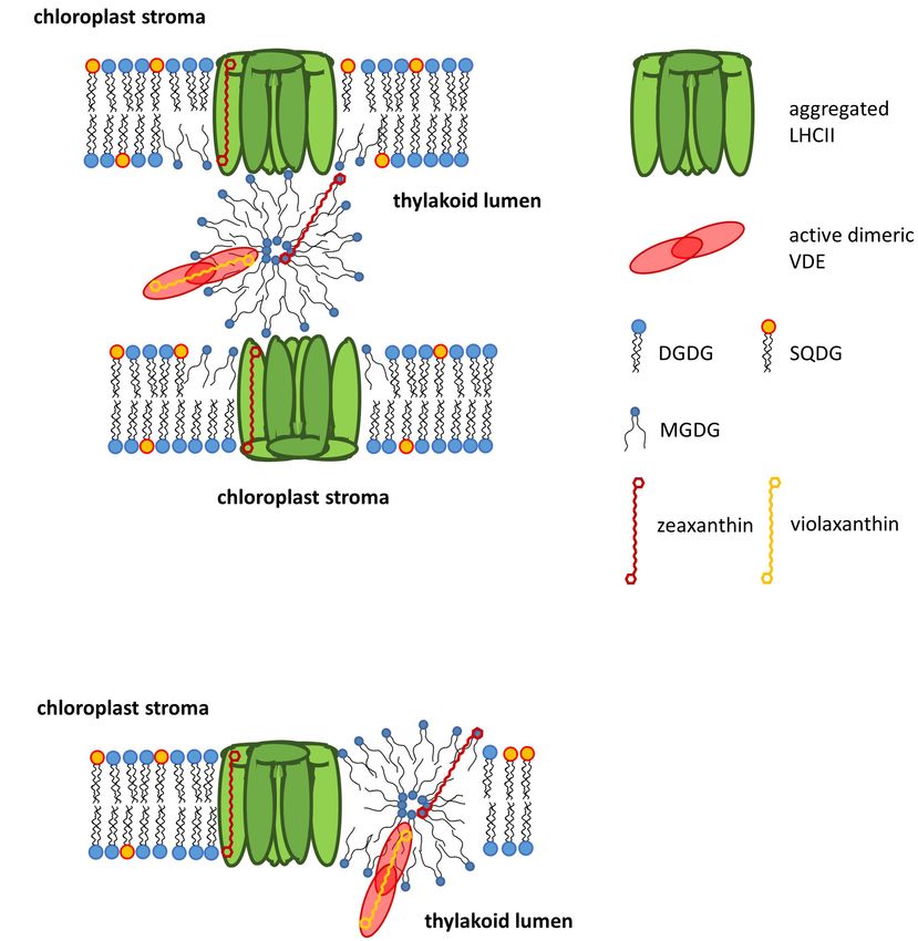
by MGDG it has been demonstrated that the proteins involved lumen during high light illumination have also been shown to
in the phototransformation of protochlorophylls support the promote the formation of HII -phases by MGDG (Garab et al.,
formation of a cubic phase (Selstam, 1998). For the function 2017, see also section Localization of Non-bilayer Lipid Phases
of the thylakoid membrane it is of high importance that the in the Thylakoid Membrane).
LHCII strongly interacts with MGDG and forces the MGDG
molecules into a membrane bilayer structure (Simidjiev et al.,
2000). In thylakoid membranes, MGDG seems to play a key role XANTHOPHYLL CYCLES OF HIGHER
in providing the membrane fluidity which is essential for the
PLANTS AND ALGAE
efficient diffusion of the xanthophll cycle pigments (see section
Xanthophyll Cycles of Higher Plants and Algae), LHC proteins
or proteins involved in the turnover and repair of PSII and ZEP.
Types of Xanthophyll Cycles
The main xanthophyll cycles that are known today are the
Thus, MGDG and the HII -phases created by MGDG are not
violaxanthin (Vx) cycle of higher plants, green and brown algae
only important for chlorophyll biosynthesis during chloroplast
and the diadinoxanthin (Ddx) cycle of diatoms, haptophytes and
development (Jarvis et al., 2000), but also play significant role
dinophytes (Figure 2, for a review see Goss and Jakob, 2010).
in the proper functioning of the photosynthetic machinery in
Algae containing the Ddx cycle also contain the pigments of the
Vx cycle because Vx is a precursor in the biosynthesis pathway
of the Ddx cycle pigments (Lohr and Wilhelm, 1998, 2001). The
TABLE 1 | Dependence of the lipid self-assembly structures on the value of the presence of Vx cycle pigments in Ddx cycle containing algae is
critical packing parameter (CPP). especially visible during longer periods of high light exposure
Critical packing parameter Type of structure
when a pronounced de novo synthesis of Ddx cycle pigments
value (CPP) is taking place. In addition to these two xanthophyll cycles, the
CPP = V/a*l existence of a lutein epoxide (Lx) cycle has been shown which is,
a
however, restricted to some families of higher plants (Esteban and
v l Garcia-Plazaola, 2014). Some members of the Prasinophyceae,
which represent a class of unicellular green algae, or some species
≤1/3 Normal micelle (L1 ), (II ) within the genus Gracilaria belonging to the Rodophyta are
[1/3 − 1/2] Normal hexagonal phase (HI ) characterized by a modified Vx cycle (Goss et al., 1998; Bertrand
[1/2 − 1] Normal bicontinuous cubic phase (QI ) and Schoefs, 1999; Cardol et al., 2008).
≈1 Lamellar phases (Lα , Ld , Lβ )
≥1 Reversed bicontinuous cubic phase (QII ) Reaction Sequences and Xanthophyll
>1 Reversed micelle (L2 ) Reversed
discontinuous cubic (QIII ) Reversed
Cycle Enzymes
hexagonal phase (HII ) The xanthophyll cycles consist of forward reactions which
take place during illumination of plants or algae with high
V, volume, which can be occupied by the FA residues; l, length of the FA residues;
a, the cross-sectional area of the hydrophilic lipid headgroups. For further detail light illumination and back reactions which revert the forward
see Yamashita (2018). reaction during periods of low light exposure or darkness.
Frontiers in Plant Science | www.frontiersin.org 7 April 2020 | Volume 11 | Article 455Goss and Latowski Lipid Dependece of Xanthophyll Cycling
study has presented clear evidence that at least a Zx epoxidase,
which is able to convert Zx to Ax, is present in the red algae
(Dautermann and Lohr, 2017).
Thus, these two modifications of the Vx cycle, like the Ddx
and the Lx cycles, consist of only one de-epoxidation and one
epoxidation step. In the Vx cycle of the Prasinophyceae the
xanthophyll di-epoxide Vx is converted to the mono-epoxide
Ax whereas in all other one-step xanthophyll cycles mono-
epoxides such as Ax, Ddx and Lx are converted to the epoxy-free
xanthophylls such as Zx, diatoxanthin (Dtx), and lutein (L),
respectively (Hager and Stransky, 1970; Stransky and Hager,
1970; Rabinowitch et al., 1975; Bungard et al., 1999; Bertrand
and Schoefs, 1999). In the back reaction one epoxy-group is
re-introduced into the epoxy-free xanthophylls Dtx or L and
Ddx or Lx are regenerated. The forward reaction of the Vx
cycle is catalyzed by the enzyme Vx de-epoxidase (VDE), the
back reaction by the enzyme Zx epoxidase (ZEP). Both the
VDE and the ZEP belong to a family of diverse proteins, the
so-called lipocalins (Hieber et al., 2000; Grzyb et al., 2006).
Lipocalins usually bind hydrophobic molecules and act as carrier
proteins for their substrates. VDE is localized in the thylakoid
lumen and has a pH-optimum of around pH 5.2 (Hager, 1969;
Pfündel et al., 1994). During high light illumination the light-
driven proton gradient leads to a decrease of the pH-value of
the thylakoid lumen, thereby activating the VDE. Activation of
VDE is probably driven by a dimerization of the water-soluble
FIGURE 2 | Reaction sequences and enzymes of the violaxanthin (A) and
monomeric VDE, followed by the binding of the active, dimeric
diadinoxanthin (B) cycle. The violaxanthin cycle is present in higher plants and
green and brown algae, the diadinoxanthin cycle is found in diatoms,
VDE to the lumen side of the thylakoid membrane (Hager
haptophytes and dinophytes. Figure 2 also shows the cofactor requirements and Holocher, 1994; Arnoux et al., 2009; Saga et al., 2010).
of the enzymes catalyzing the de-epoxidation reaction (violaxanthin With regard to the dimerization it has been proposed that the
de-epoxidase, VDE and diadinoxanthin de-epoxidase, DDE) and the C-terminus of the VDE plays a role in the interaction of the
epoxidation reaction (zeaxanthin epoxidase, ZEP and diatoxanthin epoxidase,
VDE monomers (Hallin et al., 2016) and that four specific amino
DEP), respectively. Representation of the cofactors in bold or normal type
indicates whether high or low concentrations of the respective cofactors are acid residues are important for the pH-dependent activation
needed for high enzyme activity. The establishment of a proton gradient (Fufezan et al., 2012). In addition, it has been suggested that
inhibits diatoxanthin epoxidation (high 1pH control) and is thus presented in the conserved cysteine residues and the disulfide bridges formed
bold type whereas zeaxanthin epoxidation is unaffected by the presence of by the cysteines are sensitive to redox changes of the thylakoid
the transmembrane 1pH (1pH control depicted in normal type). The pH value
of the thylakoid lumen which leads to VDE and DDE activation (possibly by
membrane induced by the photosynthetic electron transport
VDE or DDE dimerization) and membrane binding is also indicated for the two (Hallin et al., 2015; Simionato et al., 2015). Changes of the
xanthophyll cycles. thylakoid redox potential may play a role in the regulation and
activation of the VDE via dithiol/disulfide exchange reactions.
Reduced ascorbate has been identified as the co-substrate of the
de-epoxidation reaction and is important for the reduction of
In the Vx cycle the forward reaction consists of a two-step the epoxy group and the subsequent abstraction as H2 O (Hager,
de-epoxidation of the di-epoxy xanthophyll Vx to the mono- 1969). Interestingly, a recent study has reported that the atypical
epoxide Ax and finally to the epoxy-free Zx (Yamamoto et al., VDE of the green algae Chlamydomonas reinhardtii (Li et al.,
1962; Hager, 1967a). The back reaction re-introduces the two 2016) does not require the presence of ascorbate (Vidal-Meireles
epoxy groups into the Zx molecule and regenerates Vx via et al., 2020). In contrast to the VDE the ZEP seems to be
the intermediate reaction product Ax (Hager, 1967a). The Vx permanently associated with the stromal side of the thylakoid
cycle of the Prasinophyceae and of some species of the genus membrane as a peripheral membrane protein (Schaller et al.,
Gracilaria is incomplete and during high light illumination 2012b). ZEP uses O2 and NADPH + H+ as co-substrates to
an accumulation of the intermediate de-epoxidation product re-introduce the epoxy group into the Zx and Ax molecule,
Ax is observed in Prasinophycean cells (Goss et al., 1998; respectively (Hager, 1967a). The VDE of higher plants and
Cardol et al., 2008). In the red algae Graciliaria gracilis or G. green algae is characterized by a higher substrate affinity for Ax
multipartite an absence of Vx was detected and Ax seems to compared with Vx which results in faster kinetics of the second
be the only substrate which can be converted to Zx (Bertrand de-epoxidation step from Ax to Zx compared with the first de-
and Schoefs, 1999). Despite the ongoing uncertainties about epoxidation step from Vx to Ax (Frommolt et al., 2001; Goss,
the presence of a xanthophyll cycle in Rodophyta, a recent 2003). However, the VDE of the Prasinophyceae differs from
Frontiers in Plant Science | www.frontiersin.org 8 April 2020 | Volume 11 | Article 455Goss and Latowski Lipid Dependece of Xanthophyll Cycling
the VDE of higher plants and other green algae and exhibits a PtVDL2 (Coesel et al., 2008). Important differences can also be
higher substrate affinity for Vx compared with Ax (Goss, 2003). observed for the epoxidation reaction of the Ddx cycle. Here
This results in a very slow second de-epoxidation step from Ax it has been demonstrated that the Dtx epoxidase (DEP) shows
to Zx and together with the simultaneous epoxidation reaction significantly higher Dtx epoxidation rates than the ZEP of higher
results in the accumulation of Ax during high light illumination plants and green algae (Mewes and Richter, 2002; Goss et al.,
in these green algae (Frommolt et al., 2001). Interestingly, the 2006). The kinetics of the conversion of Dtx to Ddx are almost
decreased substrate affinity of the VDE of the Prasinophyceae comparable to the very fast de-epoxidation of Ddx to Dtx by the
is not restricted to the mono-epoxide Ax but the enzyme is DDE. To avoid a futile cycle and to enable a fast accumulation
characterized by a generally low substrate affinity for xanthophyll of Dtx during illumination with high light intensities the DEP
mono-epoxides whereas xanthophyll di-epoxides like Vx are underlies a strict light-dependent control (Mewes and Richter,
converted with high efficiency. For the Lx cycle which occurs 2002; Goss et al., 2006). DEP activity is completely suppressed
in some families of higher plants it has been suggested that during high light illumination by the build-up of the light-driven
the VDE and ZEP also carry out the additional de-epoxidation proton gradient. During low light illumination or during periods
of Lx to L and from L back to Lx (Esteban et al., 2009). The of darkness when no or only a weak trans-membrane proton
enzymes of the Ddx cycle in diatoms show some differences to gradient is present, DEP is fully activated and Dtx is rapidly
the respective enzymes in higher plants and green algae. VDEs converted back to Ddx.
in diatoms and some other groups of algae, like dinophytes,
haptophytes or phaeophytes, are also denoted as diadinoxanthin
de-epoxidases (DDEs). Before 2007 the presence of different Localization of Xanthophyll Cycle
VDE genes and thus the presence of different VDEs/DDEs in Pigments in the Thylakoid Membrane
organisms containing the Ddx cycle was not known and the With respect to the localization of the xanthophyll cycle pigments
measurements dealing with the properties of the DDE were in the thylakoid membrane two main pools can be differentiated.
ascribed to one single enzyme. It was shown that the DDE The first pool consists of xanthophyll cycle pigments which are
is characterized by a pH-optimum which is shifted toward bound to the light-harvesting proteins. In higher plants and green
higher pH-values (Jakob et al., 2001). In addition, it has been algae the majority of the protein-bound Vx cycle pigments is
reported that DDE activity and thus the de-epoxidation of Ddx associated with the light-harvesting complex of photosystem II
to Dtx can take place at neutral pH-values. In addition, DDE (LHCII), which represents the main thylakoid membrane protein
is able to operate efficiently with lower concentrations of the (Ruban et al., 1999), although proteins of the light-harvesting
co-substrate ascorbate compared with the VDE of higher plants complex of photosystem I (LHCI) also bind Vx, as well as L and
(Grouneva et al., 2006). β-carotene (Croce et al., 2002, 2007). Interestingly, recent data
It might be possible that a relationship exists between the have shown that the binding of the xanthophyll cycle pigments
higher SQDG and lower MGDG concentrations of the diatom can have an impact on the supramolecular structure of LHCII
thylakoid membrane (see section Lipid Classes) and the broad (Zhou et al., 2020). LHCII contains a special binding site for Vx
pH-optimum of the diatom DDE. Taking into account that the which has been termed V1. V1 is located at the periphery of each
thylakoid membranes of the A. thaliana mgd1-1 mutant, which LHCII apoprotein and contains a loosely bound Vx molecule
are strongly reduced in their MGDG content, are impaired (Morosinotto et al., 2003). In the minor PSII antenna proteins
in their ability to build-up a strong proton gradient during CP29, CP26, and CP24, which contain higher concentrations of
illumination (Aronsson et al., 2008, see also section Lipid Vx cycle pigments compared to the LHCII, Vx seems to occupy
Classes), a comparable situation might occur in the diatom the L2 binding site (Morosinotto et al., 2003). This binding
thylakoids with their low MGDG concentration. The possible site is usually responsible for the association of one of the two
inability of the diatom thylakoid membranes to generate a very lutein molecules in the LHCII. In contrast to the V1 site the L2
strong 1pH would then require the onset of DDE activity at site is not located at the periphery but represents an internal
a weaker pH-gradient across the membrane and thus at higher pigment binding site. Experiments with recombinant LHCII and
luminal pH-values. Such a behavior, i.e., DDE activity at almost minor PSII antenna proteins have shown that only Vx, which is
neutral pH-values, has been observed in in vitro experiments associated with the peripheral V1 binding site, can be efficiently
where the pH-activity and pH-optimum of the DDE were converted to Ax and Zx (Jahns et al., 2001; Wehner et al., 2006).
determined (Jakob et al., 2001). Vx bound to the internal L2 binding site is not easily accessible
Today, it is clear that the diatom genome codes for more by the VDE and no or only a very limited de-epoxidation can be
than one de-epoxidase. Two DDE-encoding genes were shown observed. The loose association of Vx with the LHCII apoprotein
to be present in the centric diatom Thalassiosira pseudonana at the peripheral binding site is thought to enable the efficient
(Montsant et al., 2007). One of these genes (labeled as TpVDE) detachment of Vx from the protein during high light illumination
is similar to the genes encoding the typical plant VDEs while followed by the diffusion into the lipid phase of the thylakoid
the other (designated as violaxanthin de-epoxidase-like; TpVDL) membrane where the actual enzymatic conversion to Ax and Zx
is more distantly related. Later, it was demonstrated that the by the enzyme VDE is taking place (Latowski et al., 2002; Goss
genome of the pennate diatom Phaeodactylum tricornutum et al., 2007). Recently, it has been shown that the binding of
contains one gene similar to the genes of “typical VDEs” (termed neoxanthin (Nx) to the LHCII affects the binding affinity of Vx
PtVDE) and two VDE-like genes, designated as PtVDL1 and (Wang et al., 2017). In the presence of Nx Vx is only weakly
Frontiers in Plant Science | www.frontiersin.org 9 April 2020 | Volume 11 | Article 455Goss and Latowski Lipid Dependece of Xanthophyll Cycling
bound to the LHCII, easily dissociates into the lipid phase of the and DGDG between the green lineage and diatoms/brown
membrane, thereby enhancing the first step of the de-epoxidation algae are related to differences in the structures of LHCs and
reaction. In contrast to the general assumption that Zx rebinds to FCPs. Furthermore, these differences may represent an optimal
the Vx binding sites at the LHCII and the minor PSII antenna harmonization of the thylakoid membranes to allow the best
proteins after the de-epoxidation of Vx, Xu et al. (2015) provided possible LHC/FCP structure and function, i.e., light-harvesting
evidence that Zx does not necessarily exchange for Vx in the or non-photochemical quenching of Chl a fluorescence (NPQ,
internal binding sites. It may be located in the periphery of see section Function of Xanthophyll Cycles). With respect to
the complexes and exert its quenching capacity in a position the different xanthophyll cycles in higher plants/green algae
between the LHCs. and diatoms the differences in the lipid and FA composition
In diatoms the protein bound Ddx cycle pigments are may even influence the interplay between the LHCs/FCPs
located in the different fucoxanthin chlorophyll protein (FCP) and the xanthophyll cycle enzymes. In this regard, it is
complexes. The recent elucidation of the molecular structure interesting that the Vx cycle enzymes of brown algae, which
of an FCP complex composed of Lhcf3 and Lhcf4 by x-ray contain FCP complexes and are enriched in C18 and C20
crystallography (Wang et al., 2019) showed that, like Vx in the FAs, show some of the typical features of the Ddx cycle
LHCII, Ddx is located at the periphery of the apoprotein and enzymes of diatoms, like e.g., a fast epoxidation reaction
thus most likely also loosely bound. Like the easy detachment (Garcia-Mendoza and Colombo-Pallotta, 2007).
of Vx from the LHCII followed by the rebinding of Zx, the The second main pool of xanthophyll cycle pigments consists
peripheral binding of Ddx is thought to facilitate the exchange of Vx or Ddx cycle pigments which are not protein bound
with Dtx during the operation of the Ddx cycle. Interestingly, but localized as free pigments in the lipid phase of the
the binding site of the Ddx molecule seems to be located at the thylakoid membrane. First evidence for the existence of free
opposite side of the apoprotein compared with the Vx binding Zx molecules was obtained from studies on the fluidity of
site in the LHCII. Besides the Lhcf proteins which build-up the thylakoid membranes which showed that the conversion of Vx
peripheral antenna system of diatoms, but may also be more to Zx leads to a rigidification of the membrane (Gruszecki
closely associated with the PSII core complex, the Lhcr proteins and Strzalka, 1991, 2005; Tardy and Havaux, 1997, see also
which form the PSI-specific antenna of diatoms, bind Ddx cycle section Function of Xanthophyll Cycles). Isolation of LHCII
pigments. According to Lepetit et al. (2008) the concentration of with different preparation methods led to the purification of
Ddx and Dtx seems to be even slightly higher in the PSI antenna LHCII with different concentrations of native lipids and Vx
compared to the peripheral FCP complex. Like higher plants cycle pigments (Schaller et al., 2010). Further analysis of the
and green algae diatoms show an increase of the xanthophyll LHCII preparations demonstrated that the concentration of
cycle pigment pool upon cultivation with higher light intensities LHCII-associated Vx was correlated with the amount of MGDG
(Lavaud et al., 2003; Schumann et al., 2007; Gundermann and which was isolated with the complexes. Decreases of the MGDG
Büchel, 2008; Lepetit et al., 2010; Gundermann et al., 2019). content led to a decrease of the Vx concentration, indicating
The increase of the Ddx cycle pigment pool size is significantly that a part of the Vx cycle pigment pool was protein-bound
more pronounced compared with the increase of the Vx cycle whereas another part was localized within an MGDG-shield
pigment concentrations. With respect to the additional Ddx and surrounding the LHCII. Comparable results were obtained for
Dtx synthesized during exposure to high light intensities it has the Prasinophyceae Mantoniella squamata where LHC could be
been proposed that a part of these additional Ddx cycle pigments isolated which contained high concentrations of MGDG and Vx
is bound by the photoprotective Lhcx proteins which also show (Schaller et al., 2012a). In diatoms which are characterized by
a stronger expression under high light conditions (Lepetit et al., a strong increase of the Ddx cycle pigment pool during high
2013). In the centric diatoms which are characterized by a more light exposure a comparable separation between a protein bound
complicated antenna system than the pennate diatoms, it could and lipid dissolved Ddx cycle pigments could be observed. The
be shown that both the FCPa and FCPb complexes bind Ddx first indication for a pool of Ddx cycle pigments, which are not
cycle pigments (Gundermann and Büchel, 2008). In the FCPa bound to FCP complexes, was obtained from measurements of
complex increased Ddx binding during high light cultivation NPQ of high light cultivated diatoms (Schumann et al., 2007).
was accompanied by an increased content of the Fcp6 and These measurements indicated that additional Dtx synthesized
Fcp7 proteins. Interestingly, Dtx-induced decreases of the FCP during high light treatment is not able to enhance NPQ and
fluorescence emission could only be observed in the FCPa. thus cannot be bound to the respective binding sites of the FCP
It is noteworthy that the differences in the main thylakoid complexes. Additional experiments with isolated FCP complexes
membrane proteins, i.e., the light-harvesting complexes (LHCs), from low and high light cultivated diatom cells demonstrated
go together with differences in the lipid and FA composition that the additional Ddx cycle pigments show the same absorption
of the thylakoids (see sections Lipid Classes and Fatty Acids). spectrum as purified Ddx which is dissolved in MGDG (Lepetit
In higher plants and green algae, which contain the LHCII et al., 2010). The enrichment of MGDG in the isolated FCP
and LHCI, the thylakoid lipids are rich in C16 and C18 complexes led to the conclusion that, like in higher plants,
FAs. Diatoms and brown algae, which are characterized by the diatom antenna complexes are surrounded by an MGDG
the presence of Fx, contain MGDG and DGDG molecules shield which incorporates a part of the Ddx cycle pigments.
with a high concentration of C18 and C20 FAs. It may be Also like in higher plants the free Ddx cycle pigments have
possible that the differences in the FA composition of MGDG been shown to play a role in the modulation of the thylakoid
Frontiers in Plant Science | www.frontiersin.org 10 April 2020 | Volume 11 | Article 455You can also read