Changing the Cortical Conductor's Tempo: Neuromodulation of the Claustrum - Caltech Authors
←
→
Page content transcription
If your browser does not render page correctly, please read the page content below
REVIEW
                                                                                                                                               published: 13 May 2021
                                                                                                                                       doi: 10.3389/fncir.2021.658228

                                            Changing the Cortical Conductor’s
                                            Tempo: Neuromodulation of the
                                            Claustrum
                                            Kelly L. L. Wong 1 , Aditya Nair 2,3 and George J. Augustine 1,2 *
                                            1
                                             Neuroscience and Mental Health Program, Lee Kong Chian School of Medicine, Nanyang Technological University,
                                            Singapore, Singapore, 2 Institute of Molecular and Cell Biology (IMCB), Agency for Science, Technology and Research
                                            (A∗ STAR), Singapore, Singapore, 3 Computation and Neural Systems, California Institute of Technology, Pasadena, CA,
                                            United States

                                            The claustrum is a thin sheet of neurons that is densely connected to many cortical
                                            regions and has been implicated in numerous high-order brain functions. Such brain
                                            functions arise from brain states that are influenced by neuromodulatory pathways from
                                            the cholinergic basal forebrain, dopaminergic substantia nigra and ventral tegmental
                                            area, and serotonergic raphe. Recent revelations that the claustrum receives dense
                                            input from these structures have inspired investigation of state-dependent control of
                                            the claustrum. Here, we review neuromodulation in the claustrum—from anatomical
                                            connectivity to behavioral manipulations—to inform future analyses of claustral function.
                                            Keywords: claustrum, acetylcholine, serotonin, dopamine, neuromodulation

                           Edited by:
                Edouard Pearlstein,         INTRODUCTION
  Independent Researcher, Marseille,
                           France           The claustrum is a long and irregular sheet of neurons nestled between the insula and striatum.
                                            As it is known to be heavily and bilaterally connected to many brain regions in organisms ranging
                      Reviewed by:
                            Ami Citri,
                                            from mice to humans (Sherk, 1986; Torgerson et al., 2015; Wang et al., 2017, 2019; Zingg et al.,
    Hebrew University of Jerusalem,         2018), the claustrum has been likened to a cortical conductor (Crick and Koch, 2005). Despite being
                                Israel      anatomically described since the late 1700s, even now little is known about claustral function. This
                     Brian N. Mathur,       is largely due to difficulties in reliably targeting the claustrum for experimental analysis. Recently,
University of Maryland, United States       application of molecular targeting approaches that permit reliable experimental interrogation of the
                *Correspondence:            claustrum has greatly advanced our understanding of the claustrum (Jackson et al., 2020). Current
               George J. Augustine          evidence points to the claustrum being involved in higher cognition. Hypothesized functions of
       george.augustine@ntu.edu.sg          the claustrum revolve around four main themes: consciousness (Crick and Koch, 2005; Koubeissi
                                            et al., 2014; Chau et al., 2015; Yin et al., 2016; Bickel and Parvizi, 2019), attention and salience
         Received: 25 January 2021          (Mathur, 2014; Chia et al., 2017; Atlan et al., 2018; Smith et al., 2019b), learning and memory
          Accepted: 29 March 2021
                                            (Grasby and Talk, 2013; Jankowski and O’Mara, 2015; Renouard et al., 2015; Liu et al., 2019;
           Published: 13 May 2021
                                            O’Mara and Aggleton, 2019; Reus-García et al., 2020), and sleep (Renouard et al., 2015; Narikiyo
                            Citation:       et al., 2020; Norimoto et al., 2020).
 Wong KLL, Nair A and Augustine GJ
                                                Brain states are coordinated changes in brain-wide activity observed during conditions
        (2021) Changing the Cortical
                Conductor’s Tempo:
                                            such as wakefulness, sleep and anesthesia. They are thought to result from the actions of
  Neuromodulation of the Claustrum.         neuromodulators, such as acetylcholine (ACh), dopamine (DA) and serotonin (5-HT), that
   Front. Neural Circuits 15:658228.        affect brain processes ranging from macroscopic networks down to subcellular signaling
     doi: 10.3389/fncir.2021.658228         (Kringelbach and Deco, 2020; McCormick et al., 2020). The claustrum, like every other brain

Frontiers in Neural Circuits | www.frontiersin.org                                 1                                          May 2021 | Volume 15 | Article 658228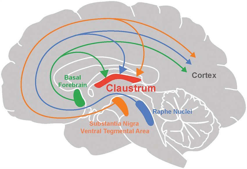
Wong et al.                                                                                                            Neuromodulation of the Claustrum

region, may be regulated by different combinations of
neuromodulators during different brain states. Thus, it is
difficult to determine the precise function of the claustrum
without considering how different brain states influence
the claustrum.
    Given that the claustrum is interconnected with many brain
regions, it is poised to participate in large-scale networks that
orchestrate wakefulness, sleep, anesthesia and other cognitive
functions. Yet it is entirely unclear how the claustrum operates
in an ever-changing neuromodulator landscape: How does the
claustrum conductor change its ‘‘tempo’’, namely the way it
controls the cortex? In this review article, we build upon on the
previous review of Baizer (2014) by providing an updated and
more comprehensive view of what is known about modulation
of the claustrum by ACh, DA and 5-HT. We also briefly consider
other neuromodulators and define important factors for thinking
                                                                            FIGURE 1 | Neuromodulation of the claustrum. The claustrum, as well as
about neuromodulation of the claustrum. Finally, we provide a               the cortex, receives neuromodulatory input from the basal forebrain
unified view of neuromodulator control of the claustrum, along              (acetylcholine), raphe nuclei (serotonin), substantia nigra and ventral
with a simplified model for the unique role of the claustrum in             tegmental area (dopamine).
brain function.
                                                                           cholinergic nucleus projecting to Egr2-expressing neurons in
ACh AND THE CLAUSTRUM                                                      the claustrum. This finding was corroborated in another
                                                                           mouse line, Tbx21-Cre, by Narikiyo et al. (2020), who also
Involvement of the cholinergic system in attention is well
                                                                           identified the SI as a major source of cholinergic input to
known: activation of cholinergic neurons in the basal forebrain
                                                                           the claustrum.
recapitulates many of the effects of attention on cortical firing
                                                                               The basal forebrain cholinergic system exerts its influence
rate and rate variability (Minces et al., 2017; Schmitz and
                                                                           throughout the brain by activating both of the main types of ACh
Duncan, 2018). Therefore, interplay between the basal forebrain
                                                                           receptors (AChRs; Ballinger et al., 2016). One type consists of
cholinergic system and the claustrum has also been hypothesized
                                                                           muscarinic ACh receptors (mAChRs), which are metabotropic
to enable the claustrum’s role in attention (Goll et al.,
                                                                           and thus activate G proteins to alter the gating and kinetics of K+ ,
2015). However, whether the cholinergic system implements
                                                                           Ca2+ and non-selective cation channels. The second type consists
its role in attention in concert with the claustrum has not
                                                                           of nicotinic ACh receptors (nAChRs), which are ionotropic and
been experimentally tested. Here, we review evidence for
                                                                           are non-selective cation channels that are opened by binding of
cholinergic modulation of the claustrum and highlight one recent
                                                                           ACh. AChRs are diverse, with at least 5 mAChR subtypes and
study from our group that addresses potential mechanisms of
                                                                           an even larger number of nAChR subunits that combinatorially
cholinergic modulation through a cell-type specific effect in
                                                                           assemble into pentameric receptors that vary in their location
the claustrum.
                                                                           and precise signaling roles (Luchicchi et al., 2014; Ballinger et al.,
Evidence of Cholinergic Input and                                          2016).
                                                                               Not all types of AChR have been found in the claustrum.
Receptors
                                                                           Studies have noted a particularly high expression of α4, α5 and
The cholinergic system emanating from the basal forebrain
                                                                           α7 nAChR subunits and M4 mAChRs in the claustrum of mice
sends diffuse projections throughout the brain (Ballinger et al.,
                                                                           (Gong et al., 2003; Han et al., 2003; Winzer-Serhan and Leslie,
2016). Many recent studies provide anatomical evidence for
                                                                           2005). There is also evidence for high expression of choline
cholinergic input to the claustrum (Figure 1; Atlan et al.,
                                                                           acetyltransferase (ChAT), an enzyme required for ACh synthesis,
2018; Zingg et al., 2018; Narikiyo et al., 2020). Monosynaptic
                                                                           in the primate claustrum (Sutoo et al., 1994). Thus, the molecular
retrograde tracing, using rabies viruses to target claustral neurons
                                                                           machinery for ACh signaling is present in the claustrum.
that project to the retrosplenial cortex, reveal that cholinergic
input is likely to be the largest source of neuromodulatory
input to the claustrum (Zingg et al., 2018). This study also               Cholinergic Modulation of the Claustrum
observed that although both cholinergic neurons and GABAergic              Microcircuit
neurons in the basal forebrain project to the claustrum, the               Despite the wealth of anatomical and histological evidence for
former constitute a larger percentage of claustrum-projecting              cholinergic modulation, little is known about how ACh affects
neurons. Studies based on claustrum-specific, Cre-based mouse              claustrum neuron function. The first study was done by Salerno
lines also find an abundance of cholinergic input to the                   et al. (1981), who investigated the actions of ACh within the
claustrum. Using anterograde and retrograde viral tracing in               claustrum by iontophoretically applying ACh onto neurons of
the Egr2-claustrum line, Atlan et al. (2018) identified the                the cat claustrum. These experiments showed that ACh exerted
substantia inominata (SI) of the basal forebrain as the major              heterogeneous effects, exciting 40% and inhibiting 47% of the

Frontiers in Neural Circuits | www.frontiersin.org                     2                                         May 2021 | Volume 15 | Article 658228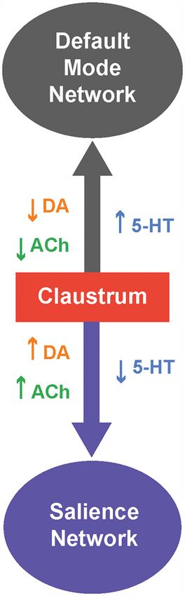
Wong et al.                                                                                                       Neuromodulation of the Claustrum

92 claustrum neurons examined. However, this pioneering work               These studies demonstrate that inactivation of the claustrum
did not distinguish between direct and polysynaptic effects of             impairs an animal’s performance in attention-related tasks and
ACh application.                                                           makes them more susceptible to distractors in their environment.
    The role of endogenous cholinergic signaling has recently              This may be mediated by indirect inhibition of the cortex,
been clarified in a study by our group that used optogenetics to           via claustrum neurons that excite cortical interneurons (Atlan
probe cholinergic modulation in mouse claustrum brain slices               et al., 2018; Jackson et al., 2018), although recent work by
(Nair et al., 2021). Optogenetic activation of cholinergic axons           Fodoulian et al. (2020) suggests that it could also be mediated,
indicated that claustral neurons receive cholinergic input in a            in part, by direct excitation of the cortex. The inhibition
cell-type specific manner: cortex-projecting claustrum neurons             observed by Atlan et al. (2018) reduced cortical gain and was
were primarily inhibited, while claustrum neurons projecting               suggested to suppress the representation of irrelevant stimuli in
to subcortical structures (including thalamus and habenula)                the environment.
and vasointestinal peptide (VIP) expressing interneurons in the                How might cholinergic modulation contribute to the
claustrum were excited. The excitation of VIP interneurons                 claustrum’s role in attention? We propose that cholinergic
in the claustrum mirrors observations in the cortex (Fu                    modulation is a permissive agent: in attentive states, when
et al., 2014), with the notable exception that cholinergic input           cholinergic input is high, the observations of Nair et al. (2021)
did not excite somatostatin (SST) expressing interneurons or               predicts that information flow will be biased towards the cortex-
parvalbumin (PV) expressing interneurons in the claustrum                  projecting claustrum population, allowing this projection to
(Nair et al., 2021) but does excite these interneurons in                  inhibit the cortex. In non-attentive states, when cholinergic levels
the cortex.                                                                are low, the claustrum will be unable to inhibit cortical targets
    The excitatory responses to cholinergic input observed by              as information flow is toggled towards the subcortex-projecting
Nair et al. (2021) were monosynaptic and mediated by nAChRs,               claustrum neurons (Nair et al., 2021). This proposal is consistent
in line with the high expression of nAChRs in the claustrum                with research that has focused on the role of claustrocortical
noted above (Winzer-Serhan and Leslie, 2005). Nair et al. (2021)           connections in attention (Atlan et al., 2018; Jackson et al., 2018;
observed that inhibitory cholinergic input received by cortex-             Fodoulian et al., 2020). In addition, regions such as the thalamic
projecting claustral neurons was also monosynaptic but was                 reticular nucleus and central thalamus have been reported to
produced by a surprising co-release of ACh and GABA by the                 contribute to aspects of attention, including sensory selection and
basal forebrain. Thus, depending on their projection targets,              attentional effort (Schiff et al., 2013; Wimmer et al., 2015). More
claustrum neurons receive opposing excitatory and inhibitory               research must be done to determine whether claustrothalamic
input from the basal forebrain.                                            projections and their modulation by cholinergic input also
    Cholinergic modulation is known to alter the gain of                   contribute to attention.
cortical neurons, which is defined as a change in neuronal                     Such a permissive role for cholinergic modulation may also
input-output properties (Silver, 2010; Polack et al., 2013;                explain the claustrum’s role in sleep. Studies by Narikiyo et al.
Dasilva et al., 2019). Similarly, cholinergic input also affects           (2020) and Norimoto et al. (2020) find that the claustrum
the gain of claustral neurons (Nair et al., 2021). The opposing            generates sharp ripples during slow-wave sleep. The application
excitatory and inhibitory effects observed on different projection         of carbachol (an agonist of both types of AChR) or 5-HT
neuron subtypes in the claustrum translates into opposing                  inhibit generation of sharp waves by the claustrum in brain
effects on the gain of these neurons: cholinergic input                    slices (Norimoto et al., 2020). Because ACh levels are low during
decreases the gain of cortex-projecting claustral neurons,                 slow-wave sleep and high during wake states, cholinergic input
while increasing the gain for subcortex-projecting neurons                 to the claustrum may play a restrictive role, preventing the
and VIP interneurons. This serves to enhance the ability of                generation of sharp ripples during wake states while allowing
cortex-projecting neurons to distinguish strong from weak                  such activity during sleep (Gais and Born, 2004; Nghiem et al.,
excitatory postsynaptic potentials, as well as strong postsynaptic         2020). Although Norimoto et al. (2020) used an exogenous
potentials from noise. A claustrum network model predicts                  AChR agonist, carbachol, endogenous cholinergic input may
that cholinergic input acts as a switch to toggle information              also abolish sharp wave ripple generation in the claustrum
processing between the cortex-projecting and subcortex-                    via co-release of ACh and GABA inhibiting cortex-projecting
projecting neurons of the claustrum. In a non-cholinergic state,           claustrum neurons (Nair et al., 2021). This underscores the
information flow in the claustrum is more efficient for the                importance of studying neuromodulation caused by endogenous
subcortex-projecting neurons, while cholinergic input switches             sources: precisely timed and highly local ACh release has specific
information flow towards the cortex-projecting neurons of                  effects that map onto only a subset of the phenomena observed
the claustrum.                                                             with widespread application of pharmacological agents (Urban-
                                                                           Ciecko et al., 2018).
                                                                               In summary, the opposing cholinergic gain mechanisms
Cholinergic Modulation as a Permissive                                     resulting from co-release of ACh and GABA may act as a
Gate for the Claustrum in Attention and                                    canonical motif, allowing the claustrum to inhibit the cortex
Sleep                                                                      during attention and to generate sharp waves during sleep. In
Recent findings have converged on a critical role for the                  both cases, cholinergic input would play a permissive role but
claustrum in attention (Atlan et al., 2018; Fodoulian et al., 2020).       permitting different forms of brain activity.

Frontiers in Neural Circuits | www.frontiersin.org                     3                                     May 2021 | Volume 15 | Article 658228
Wong et al. Neuromodulation of the Claustrum
Research Gaps system that is involved in aspects of higher cognition, while
Although we now have a potential mechanism and a hypothesis the SNpc afferents target subcortical regions and are involved
for cholinergic modulation in the claustrum, the behavioral role in motor-related functions and learning (Grace et al., 2007;
played by this permissive mechanism needs to be experimentally Luo and Huang, 2016; Klein et al., 2019). Although outputs
tested in vivo. In addition to actions within the claustrum, the of the VTA and SNpc are not exclusively dopaminergic (Beier
cholinergic system can also cause attention-like effects by acting et al., 2015; Morello and Partanen, 2015; Bouarab et al., 2019;
directly on the cortex. Thus, cholinergic actions on cortex and Nagaeva et al., 2020), additional support for DA input to
claustrum during attention will need to be disambiguated. the claustrum comes from other studies. Tyrosine hydroxylase,
Besides opposing cholinergic modulation of claustrum an enzyme involved in DA synthesis, has been detected in
projection neuron types, VIP interneurons also receive strong innervation of the claustrum in humans (Sutoo et al., 1994),
excitatory cholinergic input. In the claustrum, VIP interneurons monotremes (Ashwell et al., 2004), rodents (Khlghatyan et al.,
are known to disinhibit projection neurons via their inhibitory 2019; Borroto-Escuela and Fuxe, 2020) and pigs (Pirone et al.,
action on other interneurons such as PV and SST interneurons 2018). However, because this enzyme is also found in neurons
(Graf and Augustine, 2019). However, the exact consequences that release other types of catecholamines, such as epinephrine
of cholinergic modulation of VIP interneurons for claustrum and norepinephrine (Purves et al., 2018), it is encouraging
network function remains unknown; predicting and testing its that terminals expressing DA transporter—which is specific to
role will require detailed knowledge of claustral interneuron- dopaminergic neurons—is found in the claustrum of humans
projection neuron connectivity. (Ciliax et al., 1999), non-human primates (Al-Tikriti et al.,
Endogenous cholinergic input in the claustrum appears to be 1995) and rodents (Ciliax et al., 1995; Freed et al., 1995;
mediated entirely by nAChRs. While Nair et al. (2021) did not Delis et al., 2004).
observe mAChR activation, recent studies in the cortex indicate An additional indication of DA signaling within the claustrum
that endogenous acetylcholine activates mAChRs on dendrites comes from studies of DA receptors. All five types of DA
rather than at the soma (Williams and Fletcher, 2019). The receptors, D1R to D5R, are metabotropic receptors and can be
potential impact of cholinergic input on dendritic integration in grouped into two classes. These two different classes of DA
the claustrum remains unknown and awaits further scrutiny. receptors produce different types of downstream responses. The
first class, D1R-like, consists of D1R and D5R that predominantly
activate Gs/olf proteins and increase intracellular cAMP levels.
DA SIGNALING AND THE CLAUSTRUM The second class, D2R-like, consists of D2R, D3R, and D4R that
predominantly activate Gi/o proteins and decrease cAMP levels.
Dopaminergic modulation of the brain has received much D2R can undergo mRNA editing to yield to two variants: short
attention, particularly in relation to the well-established role of and long forms. The molecular biology of DA receptors is a rich
DA in brain reward (Montague et al., 1996; Robinson et al., research area that is nicely summarized in multiple review articles
2014). However, the role of DA in modulating the claustrum (Beaulieu and Gainetdinov, 2011; Beaulieu et al., 2015; Mishra
has received much less attention. Claustrum activity has been et al., 2018; Klein et al., 2019).
linked to rewarding, goal-directed behaviors such as the 5-choice Many studies have used in situ hybridization and radioligand
serial reaction time task (5-CSRTT) and pup retrieval behaviors assays to localize D1R and D2R and to quantify levels of
(Atlan et al., 2018; White et al., 2018, 2020), hinting at a these receptors within the claustrum. Most of these studies
possible role for DA signaling in the claustrum. To better agree that the claustrum has a high to moderate amount
understand dopaminergic regulation of the claustrum, we begin of D1R, albeit at levels much lower than found in the
by describing the dopaminergic input that the claustrum receives neighboring striatum. Such analyses have been done in
and the types of DA receptors that are found within the rodents (Dawson et al., 1986; Savasta et al., 1986; Fuxe
claustrum. We will then consider the possible functions of DA et al., 1987; Wamsley et al., 1989; Camps et al., 1990; Yoo
regulation of the claustrum, particularly in the contexts of reward et al., 2010; Borroto-Escuela and Fuxe, 2020; Hasbi et al.,
and brain disorders. 2020; Terem et al., 2020), as well as in cats and monkeys
(Camps et al., 1990). In contrast, studies of D2R receptor
Evidence of Dopaminergic Input and expression in the claustrum have yielded inconsistent results.
Receptors Some report high D2R expression (Meador-Woodruff et al.,
Several lines of evidence point toward dopaminergic input to 1991; Hall et al., 1996), while others indicate moderate to
the claustrum in a variety of organisms. First, anterograde very low expression of D2R (Wamsley et al., 1989; Weiner
and retrograde tracers have been used to identify projections and Brann, 1989; Camps et al., 1990; Mijnster et al., 1999;
to the claustrum from the ventral tegmental area (VTA) Khlghatyan et al., 2019; Borroto-Escuela and Fuxe, 2020;
and the substantia nigra pars compacta (SNpc), two of the Hasbi et al., 2020; Terem et al., 2020). These diverse results
main sources of dopaminergic modulation. Such evidence could arise from the use of different species in different
has been obtained in rodents (Lindvall et al., 1978; Zhang analyses, with D2R being more readily detected in primates
et al., 2001; Aransay et al., 2015; Beier et al., 2015; Zingg (Camps et al., 1990; Meador-Woodruff et al., 1991; Hall
et al., 2018) as well as in reptiles (Norimoto et al., 2020). et al., 1996). In addition, these results could be different
Conventionally, the VTA is part of the mesocorticolimbic due to isoform expression of D2R and type of radio-ligand
Frontiers in Neural Circuits | www.frontiersin.org 4 May 2021 | Volume 15 | Article 658228Wong et al. Neuromodulation of the Claustrum
tagged DA agonist/antagonist utilized (Camps et al., 1990). between claustral dopaminergic modulation—perhaps
Nonetheless, dopaminergic modulation of the claustrum is coming from the VTA—and reward acquisition (Smythies
likely to rely more on D1R than D2R as suggested by et al., 2012; Zingg et al., 2018). It also provides additional
many studies in rodents (Fuxe et al., 1987; Wamsley et al., support for a general role for the claustrum in salience
1989; Weiner and Brann, 1989; Camps et al., 1990; Mijnster (Graf et al., 2020b).
et al., 1999; Terem et al., 2020). The expression of other
DA receptors within the claustrum has been examined Clinical Relevance of DA Modulation of the
less thoroughly: D3R and D4R have been detected in the Claustrum
claustrum, whereas D5R is likely to be absent (Meador- There are hints that DA modulation of the claustrum may
Woodruff et al., 1992; Suzuki et al., 1998; Mijnster et al., 1999; also have clinical importance. In patients with Parkinson’s
Borroto-Escuela and Fuxe, 2020). disease, the claustrum has significantly lower DA levels,
presumably due to degeneration of SNpc (Sitte et al., 2017).
Possible Roles of Dopaminergic How the effects of Parkinson’s disease on the nigroclaustral
Modulation of the Claustrum pathway differ from those of the nigrostriatal pathway
At present, only a few studies have examined the physiological remains unclear: Sitte et al. (2017) hypothesized that the
actions of DA in the claustrum. Overall, DA seems to exert an nigroclaustral pathway relays sensorimotor information to the
inhibitory influence on the claustrum: Salerno et al. (1981) found cortex faster than the nigrostriatal pathway and thereby could
that DA application inhibited a majority of claustral neurons in be responsible for the non-motor symptoms of Parkinson’s
cats, while very few neurons were excited by DA. Unfortunately, disease. Other studies have suggested that delusions associated
neither the identity of the DA responsive neurons nor the DA with conditions such as bipolar disorder, dementia and
receptors involved were identified. Considering the strong bias depression occur due to hyperactivity of the mesolimbic
toward D1R expression in claustral neurons (Fuxe et al., 1987; pathway and the resultant aberration in salience, perhaps
Wamsley et al., 1989; Weiner and Brann, 1989; Camps et al., via recruitment of D2R in claustral neurons (Sitte et al.,
1990; Mijnster et al., 1999), and the propensity of this receptor 2017). Whether delusions are caused by D2R activation in
to increase neuronal excitability in other brain areas (Tritsch and the claustrum remains unknown. Nevertheless, a role for
Sabatini, 2012), the proportion of claustral neurons excited by claustrum DA signaling in delusions is plausible and consistent
DA is surprisingly low. More research is required to determine with the findings of Terem et al. (2020). More research,
the exact mechanism of this claustral inhibitory DA response, using analyses in both human and animal subjects, will be
be it through D1R/D2R homodimers or heterodimers (Hasbi needed to clarify the clinical impact of DA modulation of
et al., 2020), Gi/o-associated D2R-like receptors, or activation of the claustrum.
inhibitory interneurons.
A recent study by Terem et al. (2020) has examined the Research Gaps
role of D1R-expressing neurons in the claustrum in incentive The possible modulation of the claustrum by DA is beginning to
salience. Incentive salience, otherwise known as ‘‘wanting’’, come into focus. However, much more research will be required
is involved in reward learning as well as drug addiction to understand how DA affects claustral microcircuitry. As a
(Robinson et al., 2014). Terem et al. (2020) demonstrated that start, it will be valuable to define whether DA acts on claustrum
D1R-expressing claustrum neurons are activated by cocaine neurons by binding to D1R, D2R or D1R/D2R heterodimers.
administration, a condition known to involve DA signaling. The physiological consequences of activating these receptors
Incentive salience was measured with a cocaine conditioned also needs to be determined. It will also be important to
place preference (CPP) task: when the claustrum was inhibited differentiate the effects of tonic versus phasic DA release in the
during conditioning, cocaine CPP formation was disrupted. In claustrum: tonic DA release, which generates a relatively low and
a complementary experiment, CPP was produced when the sustained elevation of DA concentration, preferentially affects
claustrum was activated in a particular context. This action D2-like receptors, while phasic DA release, which produces
appeared to involve claustrum neurons projecting to frontal a higher and more transient rise in DA levels, preferentially
cortices, an area known to be involved in incentive salience affects D1-like receptors (Goto et al., 2007; Grace et al., 2007).
(Robinson et al., 2014). Thus, the activity of these DA receptor- Finally, possible differences in claustrum responses to DA
expressing projection neurons appears to be both necessary and released by the VTA versus the SNpc, during different tasks,
sufficient for incentive salience. must be examined to distinguish possible roles in reward versus
Because the actions of cocaine could also involve targets motor function.
beyond DA signaling (Carta et al., 2003; Filip et al., 2004), it
is important to note that the study by Terem et al. (2020) did
not directly examine the effects of DA on claustrum neurons. SEROTONERGIC REGULATION OF THE
Nevertheless, this study is the first to implicate D1R-expressing CLAUSTRUM
claustrum neurons in incentive salience and complements other
work that has indicated a role for the claustrum in rewarding, From an evolutionary perspective, 5-HT is one of the oldest
goal-directed behaviors (Atlan et al., 2018; Graf et al., 2020b; neuromodulators (Peroutka, 1995). In the mammalian brain,
White et al., 2020). This strengthens the potential relationship 5-HT is involved in a diverse range of processes, including sleep
Frontiers in Neural Circuits | www.frontiersin.org 5 May 2021 | Volume 15 | Article 658228Wong et al. Neuromodulation of the Claustrum
(Jouvet, 1999; Monti, 2010), learning and memory (Meneses, the claustrum. Subsequent work identified the 5-HTR types
2015; Zhang and Stackman, 2015), emotions (Altieri et al., 2013; that are present in the claustrum. These studies employed
Aznar and Klein, 2013; Bauer, 2015) and psychedelic drug action 5-HTR agonist and antagonist radioligands (e.g., ketanserin);
(Nichols, 2016; Canal, 2018). Although many of these processes one caveat of such analyses is the known off-target effects
overlap with the functions proposed for the claustrum, until of these agents (Aloyo and Harvey, 2000; Canal, 2018). The
recently serotonergic modulation of the claustrum was largely consistent conclusion of such studies is that 5-HTR-2A and 5-
ignored. However, serotonergic modulation of the claustrum HTR-2C are highly expressed in the claustrum of several species
now is becoming an exciting topic. In this section, we summarize (Dawson et al., 1986; Mengod et al., 1990; Pompeiano et al.,
what is known about serotonergic modulation of the claustrum 1994; Wright et al., 1995; Ward and Dorsa, 1996; Hamada
and the possible roles of such modulation in a variety of et al., 1998; Rioux et al., 1999; Kinsey et al., 2001; Olaghere
brain states. da Silva et al., 2010; Gawli ński et al., 2019). Remarkably, the
claustrum has been found to have the highest density of 5-
Evidence of Serotonergic Input and HTR-2A in the entire mouse brain (Rioux et al., 1999). Other
Receptors studies have found either low or high expression of 5-HTR-
Two midbrain nuclei are responsible for 5-HT release within 1A in the claustrum; the labeling efficiency of 5-HTR-1A
the brain: the dorsal and median raphe nuclei (DRN and antagonists is reportedly better than that of 5-HTR-1A agonists
MRN respectively; Figure 1). Both have divergent and (Mengod et al., 1990) and rats might have lower levels of
convergent targets throughout the brain (Hornung, 2003; 5-HTR-1A (Wright et al., 1995) in comparison to monkeys
Fernandez et al., 2016; Okaty et al., 2019). Anterograde and (Pazos et al., 1987), humans (Mengod et al., 1990) and tree
retrograde tracing experiments have established that both shrews (Palchaudhuri and Flügge, 2005). While 5-HTR-1F are
of these serotonergic nuclei project to the claustrum in cats also abundant in the guinea pig claustrum (Mengod et al.,
(Rahman and Baizer, 2007) and rodents (Vertes, 1991; Peyron 1990; Bruinvels et al., 1994), other metabotropic 5-HTRs—such
et al., 1998; Zhang et al., 2001; Zingg et al., 2018; Narikiyo as 5-HTR-1D, 5-HTR-2B, 5-HTR-4, 5-HTR-5A, 5-HTR-5B,
et al., 2020). Most claustral serotonergic fibers originate in 5-HTR-6, and 5-HTR-7—are expressed at low levels in the
the dorsal and rostral DRN (Peyron et al., 1998; Muzerelle claustra of a variety of species, including rodents (Mengod
et al., 2016; Zingg et al., 2018) and are distinct from those et al., 1990; Bruinvels et al., 1994; Wright et al., 1995; Ward
projecting to the cortex (Rahman and Baizer, 2007). The and Dorsa, 1996; Bonaventure et al., 1997; Gérard et al., 1997;
synaptic terminals of DRN input to the claustrum are smaller Kinsey et al., 2001), non-human primates (Mengod et al.,
and more spindle-shaped compared to terminals coming from 1990; Bruinvels et al., 1994), rabbits, and humans (Mengod
MRN input (Wojcik et al., 2006). Both claustral projection et al., 1990). 5-HTR-1B levels are low in rat and moderate in
neurons and interneurons receive serotonergic innervation guinea pig brains (Bruinvels et al., 1994; Bonaventure et al.,
(Baizer, 2001; Wojcik et al., 2006). As there are few claustral 1997). Some studies report a moderate to low expression
projections to the DRN and MRN (Peyron et al., 1998; Zhang of 5-HTR-3 in the claustrum (Gehlert et al., 1991; Carrillo
et al., 2001; Ogawa et al., 2014; Pollak Dorocic et al., 2014), it is et al., 2010). Sparse expression of 5-HTR-3 is expected
unlikely that the claustrum has a major reciprocal influence on because these receptors are found only in a subpopulation of
the serotonergic system. claustrum interneurons (Lee et al., 2010; Rudy et al., 2011;
The mammalian 5-HT receptor (5-HTR) family consists Koyama et al., 2017), including VIP interneurons that are
of seven subfamilies. 5-HTRs that activate Gi/o proteins approximately 4% of all claustrum neurons (Graf et al., 2020a).
include the 5-HTR-1 subfamily—subtypes 5-HTR-1A to No study has systematically characterized 5-HTR expression in
1F—as well as 5-HTR-5 subfamily members 5-HTR-5A claustral interneurons.
and 5-HTR-5B. The 5-HTR-2 subfamily includes three
receptors—5-HTR-2A, 5-HTR-2B, and 5-HTR-2C—that Actions of 5-HT and Its Agonists in the
activate Gq proteins. The remaining metabotropic 5-HTRs, Claustrum
which are subtypes 5-HTR-4S, 5-HTR-4L, 5-HTR-6, and At the microcircuit level, whole-cell patch clamp recordings in
5-HTR-7, activate Gs proteins. 5-HTR-3 subfamily members claustrum slices have established that 5-HT overall exerts an
are unique because they are ionotropic 5-HTRs that are inhibitory effect on the claustrum (Wong and Augustine, 2019).
structurally similar to nAChRs (Thompson et al., 2010). Unlike 5-HT produces a prolonged inhibition of claustrum projection
metabotropic 5-HTRs, 5-HTR3s are expressed exclusively in a neurons that lasts for seconds. Conversely, interneurons exhibit
subset of brain interneurons; indeed, expression of 5-HTR3s diverse responses to 5-HT: some are excited while others are
defines this interneuron type (Lee et al., 2010; Rudy et al., inhibited by an action of 5-HT. Together, the net effect of 5-HT
2011; Koyama et al., 2017). Comprehensive descriptions of responses from claustral projection neurons and interneurons
5-HTRs can be found in numerous reviews (Ciranna, 2006; reduces claustral output.
Nichols and Nichols, 2008). Psychedelic drugs—such as LSD and psilocybin—exert their
Altar et al. (1985) apparently were the first to identify actions by binding to 5-HTR-2 and other targets (Wacker
5-HTRs in the claustrum. They observed an enrichment of et al., 2017). Because the claustrum highly expresses 5-HTR-
5-HT binding to membrane fractions from the rat claustrum, 2A and sends dense projections throughout the brain, it is
establishing the presence of membrane-associated 5-HTR in ideally positioned as a potential target for psychedelic action
Frontiers in Neural Circuits | www.frontiersin.org 6 May 2021 | Volume 15 | Article 658228Wong et al. Neuromodulation of the Claustrum
and consequent cortical network destabilization (Martin and the claustrum (Koubeissi et al., 2014), though a follow-up
Nichols, 2016; Nichols, 2016). For these reasons, activation study was unable to replicate this intriguing finding in five
of the claustrum by 5-HTR-2A has been hypothesized to subjects (Bickel and Parvizi, 2019). Thus, we do not know
contribute to the actions of psychedelic drugs. Martin and what influences the claustral ‘‘consciousness conductor’’ in a
Nichols (2016) have shown that the psychedelic compound DOI, normal brain.
which is an agonist of 5-HTR-2A and 5-HTR-2C, activates Perhaps 5-HT actions within the claustrum play a role
claustrum neurons. Specifically, they found that c-Fos levels, a in maintaining consciousness. To identify loci associated with
surrogate of neuronal electrical activity, are sharply increased LOC, Snider et al. (2020) used lesion network mapping in
in claustrum neurons in response to DOI. They also found humans to uncover an anticorrelation of activity between the
an associated internalization of 5-HTR-2A, which is consistent DRN and claustrum that is strongly linked to LOC. Such an
with an action of DOI on these receptors. The effects of anticorrelation could be explained by evidence from the work
psilocybin, a 5-HTR-2A agonist, in the human claustrum of Wong and Augustine (2019) and Norimoto et al. (2020)
were recently studied by Barrett et al. (2020). Psilocybin clearly demonstrating that 5-HT inhibits the claustrum. Because
decreased the activity of the claustrum, but not the activity various phases of sleep are considered to represent different
of neighboring structures such as insula or putamen. This states of consciousness (Laureys, 2005; Kraehenmann, 2017),
effect was correlated both with self-reported measures of the it is possible that serotonergic inhibition of the claustrum
perceived strength of psilocybin and with measures of mystical is also involved in regulating consciousness. These studies
experiences, such as ineffability. Psilocybin was also found to suggest that LOC is caused by disinhibition of the claustrum
alter claustral functional connectivity within various cognition- resulting from loss of serotonergic inhibition from the DRN,
related networks. In particular, the connectivity of the fronto- and harkens back to the seminal proposal by Crick and Koch
parietal task network with both left and right claustra were (2005). More research clearly is needed to determine whether
altered; psilocybin also attenuated the connectivity of the the claustrum has an actual role in consciousness, as advocated
right claustrum with the default mode network. Collectively, by Crick and Koch (2005), and whether 5-HT participates in
these findings indicate that much more work is needed to such a role. Further, consciousness is a complex phenomenon
clarify the action of psychedelics on the claustrum and to and there undoubtedly are many other contributors beyond
reconcile measurements made on molecular, microscopic and serotonergic inhibition of the claustrum (Zhao et al., 2019;
macroscopic levels. Snider et al., 2020).
Previous work has suggested that the claustrum is involved
in sleep (Hong et al., 2009; Renouard et al., 2015; Jansen Link Between Neuropsychiatric Disorders
et al., 2019; Narikiyo et al., 2020). A recent landmark study and the Claustrum
by Norimoto et al. (2020) has advanced this suggestion Many neuropsychiatric disorders are associated with 5-HT
by providing key evidence that claustrum 5-HT signaling imbalances in the brain (Marek et al., 2003; Nordquist and
is important for sleep. They found that the reptilian Oreland, 2010; Lin et al., 2014). One of the main approaches to
claustrum is involved in the generation of slow waves manage such disorders is to administer 5-HT reuptake inhibitors,
during sleep. This effect is regulated by 5-HT, because drugs that target 5-HTR, and—more recently—psychedelics
uncaging of 5-HT in claustrum slices during sleep-like (Marek et al., 2003; Rucker et al., 2018). Similarly, the claustrum
states abolished slow-wave activity. Brain 5-HT levels has also been implicated in many neuropsychiatric disorders:
are lower during sleep than during wakefulness (Jouvet, claustral volume is smaller in humans with bipolar disorder
1999; Portas et al., 2000; Monti, 2010); thus, sleep may (Selvaraj et al., 2012), depression and schizophrenia (Bernstein
be enabled by the resulting claustrum slow-wave activity. et al., 2016). The human claustrum also expresses depression-
The inhibition of claustrum slow-wave activity by 5-HT related genes (Ibrahim et al., 2019) and its activity is altered
during the awake state is primarily mediated by 5-HTR- across multiple neuropsychiatric disorders (Farruggia et al.,
1D, with lesser contributions by 5-HTR-1A and 5-HTR-2C. 2020). Moreover, the claustrum has been implicated in anxiety
This corroborates claustral inhibition by 5-HT observed by (Smith et al., 2019b; Niu et al., 2020). Given that the claustrum
Wong and Augustine (2019). has a high density of 5-HTRs and is influenced by serotonergic
psychedelics, the claustrum can be considered as an emerging
Consciousness and Serotonergic target for these neuropsychiatric disorders.
Modulation of the Claustrum
Crick and Koch (2005) proposed that the claustrum serves as Research Gaps
the ‘‘seat of consciousness’’. Several lines of evidence supporting 5-HT is important for many complex and essential brain
the claustrum-consciousness connection have been derived functions. When attempting to map the known roles of 5-HT
from electrical stimulation studies. For instance, electrical onto the many proposed functions of the claustrum, it is hard to
stimulation of the claustrum induces sleep bouts and/or pinpoint a precise function for serotonergic modulation of the
unresponsiveness in animals (Gabor and Peele, 1964; Vakolyuk claustrum. The newly established link between the claustrum,
et al., 1983) and increases anesthetic depth (Pavel et al., 2019). sleep and 5-HT in reptiles (Norimoto et al., 2020) demands
Additionally, a loss of consciousness (LOC) was reported in additional research to determine the applicability of these
one human subject in response to electrical stimulation near findings to the mammalian brain. Furthermore, serotonergic
Frontiers in Neural Circuits | www.frontiersin.org 7 May 2021 | Volume 15 | Article 658228Wong et al. Neuromodulation of the Claustrum
regulation of the claustrum during wake states should be recently been found that the inhibitory transmitter GABA is
explored to understand whether such modulation plays a role co-released along with ACh from forebrain cholinergic system
in other 5-HT-associated functions, for example, memory and axons (Nair et al., 2021). Co-release of 5-HT and glutamate
emotion. Finally, the role of the claustrum in psychedelic drug may also occur: retrograde tracing experiments by Zingg et al.
action should be examined more systematically to provide (2018) and the Allen Institute (Experiment 478995566) identify
insights into psychedelic-induced brain states and possibly pave DRN and MRN neurons that may represent serotonergic
the way for psychedelic-assisted therapies for neuropsychiatric neurons that also contain the glutamate vesicular transporter,
disorders such as depression. vGluT3, involved in glutamate release (Okaty et al., 2015, 2019;
Huang et al., 2019; Ren et al., 2019). Claustral interneurons
OTHER NEUROMODULATORS containing neuropeptide, such as SST and VIP (Graf et al.,
2020a; Marriott et al., 2020), could also co-release these peptides
The information above makes clear that the claustrum, like many along with GABA. Claustral co-release of any neuropeptide has
other brain regions, is likely to be influenced by numerous yet to be demonstrated; neuropeptide release typically requires
neuromodulators during various brain states. Although we have high-frequency activity, so an analysis of neuropeptide release
focussed on ACh, DA and 5-HT, the claustrum may also will require such stimulation paradigms (Liguz-Lecznar et al.,
be regulated by other neuromodulators, which could share 2016; Mazuski et al., 2018). The possible physiological roles of
converging effector pathways (Nadim and Bucher, 2014). A co-released neuromodulators certainly merit further attention.
holistic view of this diversity of neuromodulators is needed to
develop a comprehensive understanding of claustral function
in various brain states. Early hints about the roles of other A UNIFIED PICTURE OF
neuromodulators come from studies of receptor localization and NEUROMODULATOR CONTROL OF THE
analyses of claustral innervation by axonal projections containing CLAUSTRUM
these neuromodulators.
Given this rich landscape of neuromodulation, how does the
Norepinepherine claustrum contribute to brain function by integrating these
The potential for norepinephrine (NE) to modulate the diverse, dynamic, and sometimes antagonistic signals? Pondering
claustrum has garnered some attention. NE-positive fibers this question is all the more challenging because the fundamental
are found in the claustrum (Baizer, 2014; Pirone et al., roles of the claustrum are still open to debate. Nonetheless, in this
2018) and the expression and possible function of NE section we will tackle the question.
receptors in the claustrum have been theme of several studies Our answer begins with the simplified network, consisting of
(Palchaudhuri and Flügge, 2005; Baizer, 2014; Pirone et al., 2018; four pathways, shown in Figure 2A. Within a claustrocortical
Smith et al., 2019a; Borroto-Escuela and Fuxe, 2020). Lower loop (Pathway 1), the claustrum may exert net inhibitory
amounts of NE are found in the claustrum of Parkinson’s disease effects through feed-forward inhibition (FFI) that is mediated
patients, suggesting a potential role in the etiology of this motor by claustrum excitation of local cortical interneurons (Ptito and
disorder (Sitte et al., 2017). In summary, although there are Lassonde, 1981; Tsumoto and Suda, 1982; Salerno et al., 1984;
suggestions of a role for NE in modulation of the claustrum, Jackson et al., 2018; Narikiyo et al., 2020) and/or possibly net
currently there are no compelling ideas about what this role excitation, as suggested by more recent evidence (Fodoulian
might be. Clearly there is a need for physiological analyses of the et al., 2020). This loop also includes excitatory monosynaptic
actions of NE on the claustrum. input that claustral projection neurons and interneurons receive
Neuropeptides from various cortical regions (Kim et al., 2016; White and
Among the genes highly expressed in claustral neurons, Mathur, 2018b; White et al., 2018; Chia et al., 2020); whether this
Wang et al. (2017) identified SST receptor 2 (SSTR2) and combined input exerts a net inhibitory or excitatory effect on the
opioid receptor kappa 1 (KOR1). These two receptors can claustrum remains unknown. Pathways 2 and 3 indicate potential
also heterodimerize, potentially increasing their signaling modulation of the claustrocortical loop by neuromodulators.
capabilities (Borroto-Escuela and Fuxe, 2020). KOR1 is known The cholinergic and serotonergic systems exert a net inhibitory
to be involved in chronic cocaine exposure (Collins et al., influence on the claustrum, albeit at differing timescales. The
2002) and could complement the role of claustral D1R in ACh system, via the co-release of GABA, exerts fast inhibitory
acute cocaine responses (Terem et al., 2020). The dense effects mediated by ionotropic GABA receptors. This may
expression of KOR1 in the claustrum (Chen et al., 2020) enable the claustrum to control shorter-lasting brain states
could be involved in delusions caused by the KOR1 agonist such as attention, while 5-HT responses are largely mediated
salvinorin-A (Patru and Reser, 2015). KOR1 also has been by metabotropic 5-HTR and thus could mediate longer-lasting
proposed to play a role in orchestrating consciousness brain states, such as sleep. Although understudied, DA regulation
(Stiefel et al., 2014). could inhibit the claustrum rapidly or slowly, depending on
the DA receptors involved. Finally, neuromodulator pathways
Neuromodulator Co-release can influence each other (Pathway 4): for example, 5-HT
Neuromodulators can also be co-released along with release from the DRN attenuates DA release from the VTA
conventional neurotransmitters. In the claustrum, it has (Conio et al., 2020) and ACh is known to increase the release
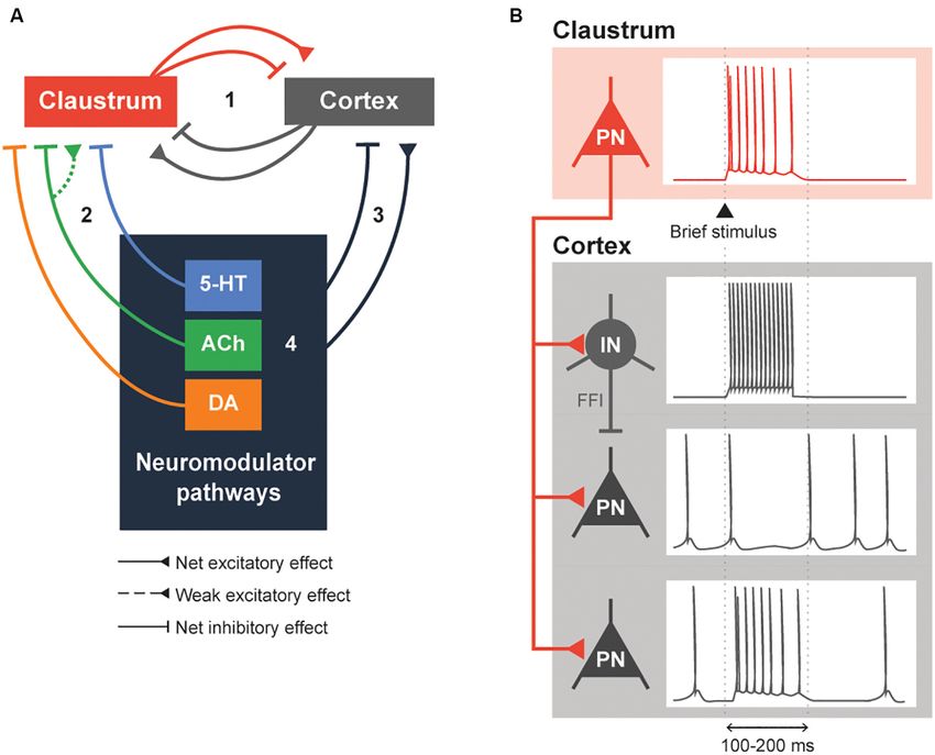
Frontiers in Neural Circuits | www.frontiersin.org 8 May 2021 | Volume 15 | Article 658228Wong et al. Neuromodulation of the Claustrum FIGURE 2 | Neuromodulation of the claustrocortical loop. (A) A simplified model consisting of the claustrum, the cortex and various neuromodulatory structures that release serotonin (5-HT), acetylcholine (ACh) and dopamine (DA). The claustrocortical loop (Pathway 1) consists of the claustrum and the cortex causing either a net excitation or inhibition of each other. Both the claustrum (Pathway 2) and cortex (Pathway 3) receive heavy input from the neuromodulator pathways. These neuromodulator pathways are also capable of regulating themselves (black box 4). (B) An illustration of claustrum input to the cortex. A claustral projection neuron (PN) receiving a brief stimulus will fire action potentials, which sends excitatory input into cortex. The cortical interneuron (IN) fires action potentials due to the excitation and subsequently inhibits the cortical PN. The cortical PN, receiving feed-forward inhibition through the cortical IN and excitation by the claustral PN, might initially fire action potentials and will remain silent during the period of inhibition by the cortical IN. Conversely, a subset of cortical PN will be directly excited by the claustral PN. These changes in cortical PN and IN activity would typically last for 100–200 ms or until claustral PN activity is diminished. of a variety of neuromodulators, including DA and 5-HT neurons also were shown to cause a brief net excitation of (Picciotto et al., 2012). cortical neurons (Fodoulian et al., 2020). Unlike the claustrum, Claustral projections to the cortex share some similarities with neuromodulators can exert their effects on either a short the neuromodulatory projections arising from the cholinergic timescale or a long timescale, depending on the postsynaptic basal forebrain, dopaminergic VTA and SNpc, and serotonergic receptors involved. Further, neuromodulatory systems heavily raphe nuclei: in all cases, multiple cortical regions are targeted, rely on volume transmission—diffusion of neuromodulators which enables widespread control of the cortex (Torgerson through the extracellular space—to modulate entire populations et al., 2015; Wang et al., 2017, 2019; Marriott et al., 2020; of spatially contiguous neurons in the central nervous system Narikiyo et al., 2020). Then what is the logic of having both (Fuxe et al., 2010). These differences distinguish claustral and types of pathway project to the cortex? One distinction appears neuromodulatory input. to be the time course of cortical regulation. Activation of Another difference between the claustrum and the claustrum can cause a brief inhibition of cortical activity neuromodulatory system is the degree of feedback from the that lasts 100–200 ms (Figure 2B; Cortimiglia et al., 1982; cortex. The paucity of afferents makes it unlikely that the Salerno et al., 1984; Jackson et al., 2018; Narikiyo et al., 2020). claustrum and cortex exert much direct feedback control This arises from strong FFI of cortical projection neurons on neuromodulatory brain areas (Peyron et al., 1998; Zhang by local interneurons, which serves to attenuate the initial et al., 2001; Polack et al., 2013; Ogawa et al., 2014; Beier et al., excitation provided by excitatory claustrum input (Jackson 2015). In contrast, it is well-established that the claustrum et al., 2018; Narikiyo et al., 2020). More recently, claustral is bilaterally and bidirectionally connected to most cortical Frontiers in Neural Circuits | www.frontiersin.org 9 May 2021 | Volume 15 | Article 658228
Wong et al.                                                                                                       Neuromodulation of the Claustrum

regions (Torgerson et al., 2015; Wang et al., 2017, 2019); any                  At a more macroscopic level, neuromodulation can select
changes in cortical output should swiftly influence claustral               for the activity of specific brain networks during different
activity. Therefore, as compared to neuromodulatory pathways,               brain states. This can be reflected in changes in intrinsic
the dynamics of the claustrocortical loop should allow for                  connectivity networks (ICNs): claustral functional connectivity
temporally precise recruitment of neuronal ensembles across                 to various brain regions can be altered in different brain states.
the cortex.                                                                 This has been established by many studies demonstrating that
    By virtue of the broad distribution of neuromodulatory axons            pharmacological interventions, anatomical lesions and sleep
throughout the brain, neuromodulators will simultaneously                   perturb both claustrum activity and claustrum connectivity to
affect many brain regions. How does neuromodulation affect                  various ICN nodes (Hong et al., 2009; Chau et al., 2015; Barrett
the claustrocortical loop? As illustrated in Figure 2A, we can              et al., 2020; Snider et al., 2020). However, the precise change
anticipate complex interactions between neuromodulatory                     in claustral functional connectivity relative to various ICN
structures, the cortex and the claustrum. The precise                       nodes in different brain states and neuromodulators remains an
effect of neuromodulation is likely dependent on the                        open question.
connectivity between the different cortical areas and                           We can take cues from the ICNs the claustrum is involved
the claustrum. For instance, neuromodulation of the                         in and from the responses of these ICNs to neuromodulators
claustrum-anterior cingulate cortex (ACC) loop should be very               to form a unified picture of neuromodulator control of the
different from that of the claustrum-retrosplenial cortex (RSC)             claustrum. The claustrum is thought to participate primarily in
connection: unlike the ACC, the RSC receives dense                          two ICNs: the salience network (SAN) and the default mode
monosynaptic excitatory input from the ipsilateral claustrum                network (DMN; Figure 3; Smith et al., 2019b). Specifically,
but does not send afferents to the claustrum (Wang et al., 2017;            the claustrum serves as a link between the anterior insula
Brennan et al., 2020; Marriott et al., 2020). Additionally, there           and the ACC (Wang et al., 2017; Chia et al., 2017, 2020;
may be differences in the types of postsynaptic neurons that are            Qadir et al., 2018; Zingg et al., 2018; Krimmel et al., 2019;
targeted by neuromodulator pathways, which could differentially             Rodríguez-Vidal et al., 2019; Smith et al., 2019a), two very
affect responses in the claustrum and elsewhere (Beier et al.,              prominent components of the SAN (Menon and Uddin, 2010).
2015; Huang et al., 2019; Nair et al., 2021). At present, most              As the claustrum also is connected within the DMN, it is
research has focused on a claustrum-centric model, where                    ideally positioned to process sensory-limbic information and
neuromodulators primarily act upon the claustrum, which then                use this information to toggle in and out of the DMN
inhibits the cortex (Martin and Nichols, 2016; Graf et al., 2020b;          like a switch; the claustrum is linked to the recruitment of
Norimoto et al., 2020; Terem et al., 2020; Nair et al., 2021). More         the SAN and disengagement of the DMN (Krimmel et al.,
research is required to understand whether neuromodulatory                  2019; Rodríguez-Vidal et al., 2019; Smith et al., 2019b). Such
input to the cortex, including presynaptic modulation (Nadim                toggling is likely a result of selection of different subsets of
and Bucher, 2014), affects claustral function similarly.                    claustral projection neurons with differing projection patterns
    Brain states, such as those occurring during sleep                      (Wang et al., 2019; Chia et al., 2020; Graf et al., 2020a;
and psychedelic-altered conditions, are characterized by                    Marriott et al., 2020) during a particular state. Thus, when
synchronized brain activity. These states apparently arise                  the claustrum is activated in the SAN, other task-positive
from the coordinated action of long-lasting neuromodulators                 networks such as the fronto-parietal network can be activated
(Kringelbach and Deco, 2020; McCormick et al., 2020). To better             (Menon and Uddin, 2010).
understand the mechanisms producing brain states, neuronal                      Which neuromodulators could permit the claustrum to
activity must be correlated with observable behaviors. While                switch from the DMN to the SAN? Several human and murine
still at an early stage, available studies of the claustrum have            studies have investigated how neuromodulators, particularly
demonstrated patterns of claustral activity associated with                 5-HT, DA and ACh, strengthen and weaken these ICNs. A
different brain states, including those of wake and sleep states.           serotonin-dopamine antagonism can be seen between the SAN
A majority of claustral neurons fire at a low rate during wake              and DMN; in humans, DA from the VTA strengthens the
states (Jankowski and O’Mara, 2015; Narikiyo et al., 2020; Reus-            SAN while 5-HT strengthens the DMN (Conio et al., 2020).
García et al., 2020). During tasks that requires attention and/or           Such antagonism is also apparent in the observation that the
cognition, such as the 5-CSRTT and attentional set-shifting task,           serotonergic psychedelic psilocybin strengthens DMN networks
claustral activity exhibits a gradual increase over a second before         and dampens DA-related networks in mice (Grandjean et al.,
a reward and scales with the cognitive load of the task (White              2021). A possible mechanism for 5-HT-associated strengthening
et al., 2018, 2020; Fodoulian et al., 2020). In comparison to wake          of the DMN could be 5-HT inhibition of the claustrum
states, claustrum activity increases during sleep (Narikiyo et al.,         (Wong and Augustine, 2019; Norimoto et al., 2020), which
2020; Norimoto et al., 2020). State-related differences could also          would attenuate claustral activity and connectivity with the
influence the effect of claustral output; cortical slow-wave ripples        DMN (Barrett et al., 2020), thereby strengthening the DMN
caused by claustral stimulation appear smaller in magnitude                 (Krimmel et al., 2019; Rodríguez-Vidal et al., 2019; Smith
during wake states than in sleep states (Narikiyo et al., 2020).            et al., 2019b). In contrast to 5-HT, DA from the VTA
These differences in claustral activity and output during distinct          strengthens the SAN. Perhaps VTA-related DA activation of the
brain states may arise from differences in neuromodulator                   claustrum via D1 is required for salience (Terem et al., 2020).
actions in the claustrum.                                                   However, this hypothesis is inconsistent with the observations

Frontiers in Neural Circuits | www.frontiersin.org                     10                                    May 2021 | Volume 15 | Article 658228
You can also read