Ayres Theories of Autism and Sensory Integration Revisited: What Contemporary Neuroscience Has to Say - MDPI
←
→
Page content transcription
If your browser does not render page correctly, please read the page content below
brain
           sciences
Review
Ayres Theories of Autism and Sensory Integration
Revisited: What Contemporary Neuroscience Has
to Say
Emily Kilroy 1,2, *, Lisa Aziz-Zadeh 1,2 and Sharon Cermak 1
 1    Mrs. T.H. Chan Division of Occupational Science and Occupational Therapy, University Southern California,
      Los Angeles, CA 90089, USA; lazizzad@usc.edu (L.A.-Z.); cermak@usc.edu (S.C.)
 2    Brain and Creativity Institute, University Southern California, Los Angeles, CA 90089, USA
 *    Correspondence: ekilroy@usc.edu

 Received: 1 March 2019; Accepted: 17 March 2019; Published: 21 March 2019

 Abstract: Abnormal sensory-based behaviors are a defining feature of autism spectrum disorders
 (ASD). Dr. A. Jean Ayres was the first occupational therapist to conceptualize Sensory Integration
 (SI) theories and therapies to address these deficits. Her work was based on neurological knowledge
 of the 1970’s. Since then, advancements in neuroimaging techniques make it possible to better
 understand the brain areas that may underlie sensory processing deficits in ASD. In this article,
 we explore the postulates proposed by Ayres (i.e., registration, modulation, motivation) through
 current neuroimaging literature. To this end, we review the neural underpinnings of sensory
 processing and integration in ASD by examining the literature on neurophysiological responses
 to sensory stimuli in individuals with ASD as well as structural and network organization using
 a variety of neuroimaging techniques. Many aspects of Ayres’ hypotheses about the nature of the
 disorder were found to be highly consistent with current literature on sensory processing in children
 with ASD but there are some discrepancies across various methodological techniques and ASD
 development. With additional characterization, neurophysiological profiles of sensory processing in
 ASD may serve as valuable biomarkers for diagnosis and monitoring of therapeutic interventions,
 such as SI therapy.

 Keywords: Autism Spectrum Disorder (ASD); Ayres Sensory Integration (ASI); sensory processing;
 functional magnetic resonance imaging (fMRI)

1. Introduction
       Autism Spectrum Disorder (ASD) is a neurological developmental disorder clinically
characterized by impairments in social interaction and communication and restricted or repetitive
patterns of behavior, interests or activities [1]. In 2018, the Centers for Disease Control and Prevention
reported that 1 in every 59 children in the United States has ASD [2]. Among those diagnosed with ASD,
it is estimated that over 90% show symptoms of sensory abnormalities [3,4]. Research in ASD sensory
processing has become increasingly prevalent since Kanner’s initial description of the condition in
1943 (Figure 1). Recently, in the Diagnostic Manual for Mental Disorders-Fifth Edition (DSM-5), hypo-
and hyper-sensory reactivity have been included as diagnostic criteria of ASD [1]. These criteria,
however, manifest differently across individuals with ASD; for example, some individuals seem
unaware of certain auditory, visual or tactile stimuli (hyposensitive), while others may avoid the same
stimuli altogether (hypersensitive). These behaviors can both contribute to and/or overlap with the
aforementioned core characteristics and hinder participation in everyday activities [5–7].

Brain Sci. 2019, 9, 68; doi:10.3390/brainsci9030068                              www.mdpi.com/journal/brainsci
Brain Sci. 2019, 9, 68 2 of 20
Brain Sci. 2019, 9, x FOR PEER REVIEW 2 of 20
Figure 1. PubMed publication search for Autism and Sensory modalities. Publications by decade for
Autism Spectrum
Spectrum Disorder
Disorder and
and sensory
sensory processing
processing from
from 1980
1980through
through2019.
2019.
Over
Over the the past
past 40 40 years,
years,research
researchononsensory sensory processing
processing in in those
those with with
andand without
without ASDASD has
has enhanced
enhanced our understanding
our understanding of how of how the brain
the brain processes
processes sensory sensory
input.input.
As early As asearly
1963, asAyres
1963,
Ayres
conducted conducted
some ofsome of studies
the first the first studies examining
examining sensory problems sensoryin problems
a wide range in aof wide range of
developmental
developmental disorders. Through her work, she conceptualized
disorders. Through her work, she conceptualized an intervention approach now referred to as Ayres an intervention approach now
referred to as Ayres(ASI)
Sensory Integration Sensory Integration
to treat (ASI) to treat
the sensorimotor the sensorimotor
foundations of academic foundations of academic
skills and other higher
skills and other higher order abilities (i.e., planning and organization)
order abilities (i.e., planning and organization) [8]. The therapy involves the client interacting [8]. The therapy involves
with a
the client interacting
combination of equipmentwith asuch
combination
as scooters of equipment
and swings, such as scooters
providing the and swings, providing
opportunity to obtain and the
opportunity
process enhanced to obtain and process
sensory input and enhanced
developsensorynormalinputlevelsand developand
of arousal normal
securitylevels of arousal
when and
interacting
security when interacting with their environment. Ayres’ intervention
with their environment. Ayres’ intervention approach was founded on the hypothesis that sensory approach was founded on
the hypothesis
integration (SI) that sensoryand
disturbance integration (SI) disturbance
other processing and other
abnormalities were,processing
in part, theabnormalities
result of abnormal were,
in part, the result of abnormal brain functioning [8]. When Ayres began to
brain functioning [8]. When Ayres began to study ASD in the late 1970’s, it was considered a rare and study ASD in the late 1970’s,
it wasstudied
little considered a rare and little
developmental studied
disorder (4.5developmental disorder (4.5 in
in 10,000) [9]. Nevertheless, 10,000)
she [9]. Nevertheless,
proposed a theoretical
she proposed a theoretical framework to describe the condition. Due
framework to describe the condition. Due to the current prevalence of the disorder, ASD research to the current prevalence of has
the
disorder, ASD research has significantly increased. In this paper we revisit
significantly increased. In this paper we revisit Ayres’ framework for using a sensory integration Ayres’ framework for using
aapproach
sensory integration
to address approach to address sensory
sensory impairment in autism impairment
and assess in autism and assess
its cogency its cogency
in light of current in
light of current
neuroscience research.neuroscience research.
Much
Much of of the
the technology
technology usedused today
today to to study
study brain
brain structure
structure and and function
function was was not
not available
available to to
Ayres
Ayres when
when she she developed
developed her her theoretical
theoretical explanations
explanations and interventions. Currently,
and interventions. Currently, researchers
researchers use use
methods
methods such such as structural and
as structural functional Magnetic
and functional Resonance Imaging
Magnetic Resonance Imaging (MRI), eye-tracking systems
(MRI), eye-tracking systems
and electroencephalogram (EEG) to generate information
and electroencephalogram (EEG) to generate information about brain processing. We about brain processing. We can
can nownow useuse
data collected from these new methodologies to examine their consistency
data collected from these new methodologies to examine their consistency with Ayres’ theories of with Ayres’ theories of ASD
and
ASDconsider their implications
and consider for SI. for
their implications TheSI.
first
The aim of aim
first this paper
of thisispaper
to review
is to three
review aspects
three of sensory
aspects of
processing deficits indeficits
sensory processing ASD thatin ASDAyresthat
discussed
Ayres in Sensory Integration
discussed in Sensory and the Childand
Integration [8]:the
registration,
Child [8]:
modulation
registration, and motivation.
modulation and The second aim
motivation. The issecond
to assess aimthe extent
is to assessto which
the extent current neuroscience
to which current
research supports Ayres’ postulates.
neuroscience research supports Ayres’ postulates.
2. Background
2. Background
Sensory processing involves perceiving, organizing and interpreting information received through
Sensory processing involves perceiving, organizing and interpreting information received
sensory systems (e.g., taste, touch, smell, sight, auditory, vestibular) in order to produce an adaptive
through sensory systems (e.g., taste, touch, smell, sight, auditory, vestibular) in order to produce an
response. The term “sensory integration” as used by Ayres [8] refers to the ability to produce
adaptive response. The term “sensory integration” as used by Ayres [8] refers to the ability to produce
appropriate motor and behavioral responses to stimuli. In her work Sensory Integration and the
appropriate motor and behavioral responses to stimuli. In her work Sensory Integration and the Child,
Child, Ayres [8] observed hyper- and hypo-responses to sensory stimuli in individuals with ASD.
Ayres [8] observed hyper- and hypo-responses to sensory stimuli in individuals with ASD.
Specifically, she noted that these individuals exhibited problems in registration (signal detection
Specifically, she noted that these individuals exhibited problems in registration (signal detection and
and interpretation), in modulation (signal inhibition or propagation), in interacting with certain
interpretation), in modulation (signal inhibition or propagation), in interacting with certain objects,
and/or in motivation. Shortly after publishing Sensory Integration and the Child [8], Ayres and Tickle [10]
investigated sensory disturbances in ASD and their responses to SI therapy specifically. In thisBrain Sci. 2019, 9, 68 3 of 20
objects, and/or in motivation. Shortly after publishing Sensory Integration and the Child [8], Ayres and
Tickle [10] investigated sensory disturbances in ASD and their responses to SI therapy specifically.
In this retrospective study, the authors found that individuals with hyper-reactivity (a disorder of
modulation) had better outcomes than those who were hypo-reactive and proposed that children who
register sensory input respond better to therapy than those who do not [10]. Although Ayres did
not identify the neural structures that underlie these disturbances in her publications, she implicated
two neural systems in registration and modulation: 1) the limbic system and 2) the vestibular and
proprioceptive systems. Her manuscript with Tickle [10] and the chapter on Autism in Sensory
Integration and the Child are the two main publications in which Ayres delineates her views of SI and
ASD. The theory that disruptions in the limbic system also contribute to motivation deficits in ASD is
further found in unpublished archival documentation of Ayres’ work and lectures preserved at the
University of Southern California (USC) library archives [11].
Components of impaired sensory integration in ASD according to Ayres.
Registration: Registration is the detection of sensory sensations within the central nervous system.
Ayres [8] used the term “registration of sensory information” which expands beyond the clinical
definition of the initial detection of a stimulus to also include the recognition of significant meaning of
sensory stimulation. Ayres [8] suggested that some children with ASD do not register sensory inputs
properly; and as a result, these children allocate attention differently from typically developing (TD)
children. For example, children with ASD may not register the presence of a salient stimulus (e.g.,
a person walking in the room, the appearance of a new toy or the sensation of a puff of air on their
necks) the way typical children do. Ayres [8] hypothesized that registration problems are located in
the limbic system (also known as emotion-related brain regions), which she described as responsible
for ‘deciding’ what is brought to consciousness and whether we will act on it (pp. 124–125). Ayres
also identified the vestibular nuclei as being involved in the registration of visual input and making it
“meaningful” to the child (p. 125).
Modulation: Modulation is the ability of the brain to regulate inhibition or propagation of neural
signaling. Sensory modulation reflects adjustments made in response to continual physiological
processes to ensure adaptation to new or changing sensory information. Ayres [8] defined modulation
as the “brain’s regulation of its own activity” (p. 182). She proposed that children with ASD not
only fail to register sensory input properly but also have trouble modulating input that they do
register. She suggested that over- or under-activity, especially in response to vestibular and tactile
sensations, may manifest in gravitational insecurity (fear of movement, especially when not in the
upright position), tactile defensiveness (fight, fright or flight reaction to light touch that most others
would consider non-noxious) or a combination of both.
Motivation: Ayres [8] described motivation as the desire or willingness to respond to a stimulus
that has been registered or to ignore it (pp. 127–128) and proposed that children with ASD have a
motivation deficit in children with ASD. More specifically, she observed that individuals with ASD
may have limited interest in doing purposeful or constructive activities. According to Ayres, although
children with ASD have the motor ability, they may not have enough motivation to actually carry out
certain activities. Ayres located this problem in “the ‘I want to do it’ function of the brain” (p. 127) but
she did not attribute it to a particular brain region in her published work. Lectures and notes written by
Ayres preserved at the USC library archives, however, indicate that Ayres’ implicated the amygdala—a
subcortical region of the limbic system—for this “do something” function. Ayres additionally identified
poor “inner drive” and environment/body precepts as contributing to motivation impairments but
did not delineate how these components interacted. Henceforth, a lack of motivation will be referred
to as motivation deficit, which is consistent with other researchers’ definitions of motivation [12,13].
Given the research tools available at the time, Ayres [8] established this basic framework for
conceptualizing sensory processing deficits in registration, modulation and motivation in children
with ASD. To engage in an updated discussion of her conceptualizations, this paper examines current
neuroscience research related to the aforementioned postulations, including studies investigatingBrain Sci. 2019, 9, 68 4 of 20
emotion-related brain regions, neural responses to sensory stimuli and value of stimuli in individuals
with ASD compared to TD individuals.
3. Current Neuroscience Evidence
3.1. Registration and Modulation: Emotion-Related Brain Regions (Previously Referred to as the Limbic System)
As previously stated, Ayres [8] suggested that a collective set of brain regions associated with
emotions, often called the limbic system, is responsible, in part, for sensory registration. She posited
that these regions of the brain are atypical in children with ASD and therefore, these children do not
register and value stimuli in the same way as TD children. Moreover, Ayres proposed that, “the more
poorly this part (of the brain) is working, the less the autistic child will respond to therapy” (p. 124).
In other words, the more abnormal the limbic system (she did not specify functional or structural
differences), the less likely SI therapy will effectively ameliorate sensory processing impairments.
Within the last decade, current neuroscience research has affirmed Ayres’ assertions that limbic
emotion-processing regions are impaired in individuals with ASD [14–18]. Findings from this research
provides evidence to support predictions of impairments of registration, as well as modulation of
sensory processing in ASD. However, to date, there is no research that specifically tests Ayres theories
regarding effectiveness of interventions using a SI approach as a function of neural functioning in ASD.
For a review of behavioral outcomes of ASI therapy see [19–22].
The term, limbic system, refers to a specific set of regions thought to encompass emotion-related
brain regions. The system is comprised of several subcortical nuclei and cortical structures including
the insula, hypothalamus, hippocampus, parahippocampal gyrus, amygdala, fornix, mammillary
body, septal nuclei, cingulate gyrus and dentate gyrus on both sides of thalamus [23]. These regions
are involved in emotion, motivation, learning, memory and certain aspects of sensory processing.
Additional regions outside of the limbic system, such as the prefrontal cortices and ventral and medial
sectors, are now known to also be important to emotion processing [24]. Here we will collectively refer
to these regions as emotion-related brain regions, a term that is currently preferred [25].
3.1.1. Brain Structure and Function
Consistent with Ayres’ theory, current research has thoroughly documented that individuals
with ASD have both structurally and functionally atypical emotion-related brain regions [17,18,26–28].
These regions have been observed to be abnormal in size and function across the lifespan [29], however,
it is still not clear if volume or responsivity are increased or decreased or how these abnormalities
underlie symptomology in ASD. Structurally, volume size trajectories across development for these
regions start out larger in children with ASD than in TD children [30,31]. It is thought that children
with ASD have an overgrowth of neurons that diminishes in adolescents [32]. In older adolescence
and adulthood, there are mixed findings regarding emotion-related structures. For example, it has
been observed that adults with ASD have reduced amygdala [33] and hippocampus volume compared
to typical individuals [34–36] while other studies have reported increased hippocampal volume [37] or
no differences [38]. In some instances, the cortical and subcortical volume of regions in this system
have been related to cognitive functioning (i.e., social processing, attention) [39–41]. In general,
increased volume in structures has been found to be related to ASD deficits [42]. However, it is not
well understood how structural components of this system relate to sensory processing specifically.
A few white matter studies have reported variation in tracts connecting emotion-related brain regions
to auditory and cognitive processes [43,44] and white matter volume related to motor skills [45]. Motor
impairments in ASD have also been found to be positively correlated with white matter volume in
regions of the brain stem, the central tegmental tract/medial lemniscus [46]. The lemniscus conveys
tactile information and proprioception to the cortex via the thalamus. This finding provides some
support for Ayres theory that the brainstem structures are related to sensory motor impairment inBrain Sci. 2019, 9, 68 5 of 20
ASD. However, more research is needed to fully understand the specificity of how brain structures are
related to sensory impairments in ASD.
Studies of neural activation (function) in emotion-related brain regions have similarly reported
abnormal functioning in ASD. The amygdala in particular has been consistently found to be
dysfunctional. Neuroscience research published at the time of Ayres’ career reported that the
amygdalae contain a larger proportion of neurons that signal valance than other emotional-related
brain regions and are responsive to motivationally significant stimuli [47]. Ayres predictions about the
connection between impaired detection and understanding of the meaning of stimuli (registration) and
the “limbic system” may indeed, in part, be due to functional disruptions in the amygdalae in ASD.
In Ayres’s lectures and notes, she identified the amygdala as being involved in sensory registration
(USC Archives) [11]. The amygdala has since continued to be implicated in recognizing valence in
stimuli [48] as well as encoding reward associations of visual stimuli and attention [49]. Moreover,
many studies report attenuated amygdala activity when individuals with ASD perform social tasks
compared to typical individuals [50–52]. Yet, other researchers have found that in individuals with
ASD that the amygdala over-activates with eye-gaze compared to typical controls and that in these
individuals amygdala activation is correlated to time spent gazing (i.e., looking at eyes) [53,54].
Hyperactivation in the amygdala to eye contact may indicate a modulation dysfunction in ASD
in which some have proposed an indication for why individuals with ASD avoid eye contact with
others [54,55]. This hypothesis suggests that avoiding eye contact is a motivational response [56],
which aligns with Ayres’ understanding of motivation (see Section 3.2). Alternatively, it also has been
posited that individuals with ASD are not aversive to eye-gaze but are indifferent to it; that is, they do
not perceive others’ eyes as informative or salient stimuli [57–59]. This lack of saliency detection may
be why other studies have observed amygdala hypoactivation. In regard to “registration,” Ayres
discussed individuals with ASD as having impairments in both the detection of a stimulus at the
level of the central nervous system (CNS) and in salience perception. Current findings support both
definitions. The amygdala has been frequently implicated in attention [49] and recognizing valence in
stimuli [48] as well as in encoding reward associations of visual stimuli [49]. In addition to “limbic
regions”, Ayres specifically hypothesized that the vestibular nuclei (located in the brain stem) were
involved in registering visual input and in helping to make it meaningful to the child [8] (p. 125).
To date, no neuroimaging studies have investigated the vestibular nuclei and “registration” in ASD
specifically, however, it is possible that abnormalities in the amygdala—which receives projections
from the medial vestibular nucleus via autonomic nuclei and parabrachial nucleus [60]—may play a
role in registration disturbances.
The insula is an important emotion-related region not identified by Ayres that is involved in
registration deficits in ASD [61]. The insula is important for attention and is a core node of the salience
network, which responds to novel and relevant sensory stimuli and is important for cognitive control
and switching between default mode networks (introspective functions) to task-based networks [62].
The insula also acts as an integration center for physiological and emotion perception [63]. This cortical
region has been repeatedly reported to be altered in ASD [64–66]. Insula abnormalities have been
observed in individuals with ASD while performing various cognitive tasks, such as social processing
tasks, emotion processing, spatial attention [67,68], set-shifting tasks [69] and executive function
tasks [64,70,71].
Together, current neuroimaging research supports Ayres’ framework by providing evidence that
emotion-related brain regions are structurally and functionally different in individuals with ASD
compared to TD individuals. Further, research findings suggest that these regions are important
for registration of sensory information and that they are disrupted in a way that impairs sensory
modulation. These differences, however, are multifaceted and may depend upon methodology (i.e.,
age, stimuli, instruction, etc.). Individual regions, such as the amygdala or insula, are part of a much
larger emotional processing network and are physically and functionally connected to other regions.
The connectivity between these regions and other brain regions also contribute to SI disruptions in ASDBrain Sci. 2019, 9, 68 6 of 20
and are discussed below. Functional task-based MRI studies that directly investigate how different
sensory experiences are processed in ASD are reviewed later.
3.1.2. Functional Connectivity
While characterization of brain regions provide insight into ASD dysfunction, a systems level
approach in the last decade has provided additional understanding into how sensory information is
communicated within and between networks. Recent research has examined network functioning of
specific regions, including emotion-related brain regions in those who are TD and individuals with
ASD. Although there are some inconsistencies [72], significant evidence to indicates that individuals
with ASD have atypical network connectivity. This has led some to characterize ASD as a disorder
of altered brain connectivity [73,74]. These functional connectivity studies identify areas of the brain
where activation patterns are synchronized across time. For example, Rudie et al. [75] observed that
children and adolescents with ASD displayed reduced functional connectivity between the amygdala
and secondary visual areas while passively viewing emotional expressions compared to the TD control
group. Moreover, the study found that increased ASD symptom severity correlated with decreased
connectivity, thereby suggesting that reduced communication between visual input and emotion
responsivity is related to ASD symptomatology. These and similar findings [73,76] also lend support
to Ayres’ postulation regarding the severity of abnormal “limbic” functioning and SI by demonstrating
that the degree of symptom severity corresponds with the degree of sensory registration disruptions.
Indeed, Ayres did predict that deficits in sensory processing hinder motor planning, which may
ultimately result in trouble with more complex behavior, including social and emotional cognition [8]
(p. 129). How this network responds to sensory stimulation is discussed later.
In addition to task-based connectivity networks, several functional networks are intrinsic to
neural functioning and are thought to be related to different aspects of sensory processing (i.e., default
mode network, salience network, motor network, auditory network, etc.). These networks closely
resemble functional networks that are active when performing tasks [77,78] and are often referred to as
resting state networks. Findings from current resting state studies suggest that individuals with ASD
have disrupted connectivity in many of these networks. Hypo- or hyper-connectivity findings suggest
that individuals with ASD have greater and/or weaker communication with networks compared
to typical peers, regardless of specific tasks or stimuli. Several of the studies correlate behavioral
symptomatology to the strength of these network connections, indicating that impairments in neural
modulation are not restricted to region specific impairments. In a recent study Maximo and Kana [79]
reported that individuals with ASD demonstrated network dysfunction in many sensory processing
networks, including hyperconnectivity in auditory-subcortical, motor-thalamic and lateral visual-basal
ganglia networks and hypoconnectivity in medial visual-subcortical networks. This and other studies
indicate that functional networks supporting primary sensory processing are impaired in ASD at the
intrinsic level [15,79–83] and may prevent appropriate integration of sensory information in higher
order sensory integration regions. Ayres [8] described individual’s impairments in registration as
“capricious” due to neural inefficiencies. Indeed, findings of reduced network efficiency are observed
in other types of network analysis, such as graph theory analysis [51,84].
3.1.3. Neural Responses to Aversive or Pleasant Sensory Stimulation
An increasing number of neuroimaging studies examine neural processing during exposure
to specific kinds of sensory input (i.e., tactile, auditory, visual) [85,86]. These studies demonstrate
that individuals with ASD have differences in “limbic system” responsivity to visual input (flashing
checkerboard), auditory input (loud noises) and tactile input (aversive or pleasant material) compared
to TD participants [87–89].
Cascio and colleagues [89] found tactile pleasantness (determined by a −100 to 100 hedonic
rating scale) varied in ASD compared to TD adults. Although psychophysical or perceived ratings
of roughness and pleasantness were largely similar across the two groups, the ASD group gaveBrain Sci. 2019, 9, 68 7 of 20
pleasant (i.e., cosmetic brush) and unpleasant (i.e., plastic mesh) textures more extreme ratings than
did controls. Moreover, ASD participants rated neutral textures with more variance than those in the
control group, thus indicating that ASD adults are less consistent when evaluating ambiguous stimulus.
It is possible that this variance in rating reflects the degree of sensitivity and discriminative ability
that some individuals with ASD may have. Further, changes in blood oxygenation level-dependent
(BOLD) signal in response to stimulation differed substantially between the groups; the ASD group
exhibited diminished responses in the posterior cingulate cortex and the insula, particularly for
pleasant and neutral textures, compared to the control group. For the most unpleasant textured
stimuli, the ASD group exhibited greater BOLD response than controls in affective somatosensory
processing areas, including the posterior cingulate cortex and the insula. The insula’s amplitude of
response to the unpleasant texture was positively correlated with social impairment as measured by
the Autism Diagnostic Interview-Revised (ADI-R [90]). According to these results, individuals with
ASD show diminished response to pleasant and neutral stimuli and exaggerated emotion-related
region responses to unpleasant stimuli, providing support for Ayres’ [8] theory of modulation deficits
in ASD, both behaviorally and neurologically.
In a set of complementary studies, high-functioning youth with ASD and age- and Intelligence
Quotient-equivalent TD youth were presented with adverse sensory stimuli during an functional
magnetic resonance imaging (fMRI) scan [86,91–93]. In response to the stimuli, ASD participants
displayed greater BOLD activation in primary sensory cortical areas and limbic structures including
the amygdala, hippocampus and orbital-frontal cortex [92]. In both groups, the level of activity in
these areas positively correlated with parent’s ratings of their child’s sensory responsivity on the
Sensory Over-Responsivity Scales (SOR) [92,94]. In a more recent study investigating functional
connectivity between emotion and sensory brain regions during sensory aversion of auditory and
tactile stimuli (white noise and scratchy texture), Green and colleagues [91] found that individuals
with ASD displayed aberrant modulation of connectivity between the thalamus (pulvinar nucleus)
and sensory-motor regions compared to their TD peers. Specifically, they posited that increased
amygdala-pulvinar connectivity may be related to selective attention in ASD since the amygdala signals
the brain to attend to distracting sensory stimuli. Taken together, these results demonstrate that youth
with ASD show neural hyper-responsivity to adverse sensory stimuli and that behavioral symptoms
characterized by SOR may be related to both heightened responsivity in primary sensory brain regions
and emotion-related brain regions at both the regional and network level. Again, these findings
provide support for Ayres theory of disrupted modulation of sensory stimuli in emotion-related
regions in ASD.
In a follow up study, Green and colleagues [86] investigated the effect of adverse sensory
distractions on the brain while participants performed a social cognition task involving the
interpretation of visual and audio cues (sarcasm task [95]). When the aversive stimuli (scratchy
material) was applied while participants completed the task, individuals with ASD showed decreased
activations in language and dorsolateral and dorsomedial prefrontal cortex compared to when no
aversive stimulus was applied. Activation in these regions was not correlated with SOR scores,
suggesting that the reduced activation was not related to sensory deficits. When the participants
were given instructions to focus on the face and voice during the social task, the addition of the
adverse stimulus did not decrease activation in the ASD group but increased activation in the medial
prefrontal cortex. This finding demonstrates that explicit instructions for attention can increase medial
prefrontal activation during this sarcasm task. The researchers of this study theorize that sensory
stimuli can disrupt neural networks processing social information in ASD and that instruction may
mitigate this effect. All of the fMRI sensory studies described above corroborate Ayres’ [8] postulate
that emotion-related brain regions have atypical modulation responses when experiencing different
sensory modalities in children with ASD compared to TD individuals.Brain Sci. 2019, 9, 68 8 of 20
3.2. Motivation: Attraction and Reward in the “I Want to Do It Part of the Brain”
Conscious registration of sensory stimuli results in one of two reactions: to respond to the stimuli
or to deliberately ignore it (unconscious registration will not be discussed here, for reviews see Fang,
Li, Chen, and Yang [96] and Morris, Öhman, and Dolan [97]. Ayres [8] suggested that a part of the
brain that motivates a response has an “energizing effect” that initiates behavior when encountering a
stimulus and triggers the desire to do something new or different (p.127). She postulated that the part
of the brain that decides, “I want to do something” is impaired in ASD and proposed that children
with ASD do not receive the same pleasure or get the same reward from activities as do TD children
when they register sensory stimuli. As such, they are not motivated to engage in the activities. Ayres
attributed an ASD child’s lack of acknowledgement or reward of important stimuli, in part, to the
child’s inability to register and/or to perceive the meaning or potential of things otherwise considered
meaningful by others. Ayres stated that children with ASD did not understand the meaning of stimuli
through observation alone; instead, they learned best through experiences and that ASI therapy works
with individuals to incentivize the registration of sensory sensations. In other words, Ayres postulated
that children with ASD do not generalize meaning from one sensory stimulus to another which hinders
their drive to do things. However, rewarding the child could help the child to register the stimuli
and motivate them engage with it. She gave an example of a child who knew how to ride a tricycle
but did not understand the meaning of riding a scooter (that it functions similar to the tricycle and
is something to ride) and therefore, was not motivated to try it [8] (p. 128). Ayres stated that simply
registering the image well enough to notice or pay attention to it is not always sufficient to motivate the
child’s inner drive to interact with it. Ayres suggested that the inner drive malfunctions in individuals
with ASD but did not state how. Simultaneously, Ayres acknowledged that some children with ASD
sought out and experienced much pleasure from self-selected sensory input. Therefore, a child with
ASD may be unmotivated by some stimuli because he or she is unable to imagine the ways in which
engagement with these stimuli would be satisfying or rewarding. Ayres hypothesized that this poor
understanding of the meaning of a stimulus was the result of poor ability to think abstractly as well as
poor environmental and bodily precept. Ayres described having a bodily precept (schema) as being an
important part of motor planning and motivation for purposeful activity.
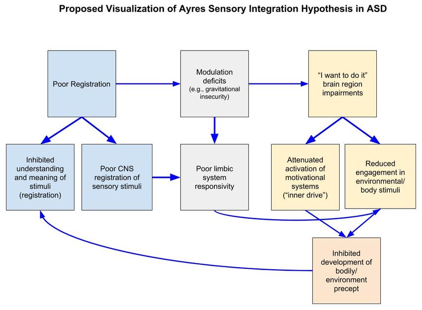
Ayres [8] outlined how multiple sensory processing impairments, including motivation deficits,
hinder motor development in ASD. She hypothesized that the lack of motivation to engage with
a stimulus inhibits the development of a precept which contributes to impaired understanding of
the potential meaning of the stimulus (poor registration), which also perpetuates the lack further
motivation to engage. This is in addition to other hindrances, such as modulation deficits, that motivate
a child to not engage with a stimulus (see Figure 2). While the specific motivation mechanisms that
Ayres proposed have not been tested (a relationships between bodily percept and motivation, “inner
drive” and “I want to do it” impairment), our current understanding of neural functioning suggest
that multiple brain regions and neural networks are responsible for motivating a response—in Ayres
own words—to “do it,” such as the reward system and the cerebellum [98,99]. These systems have
been tested and found to be disrupted in ASD as described later.motivate a child to not engage with a stimulus (see Figure 2). While the specific motivation
mechanisms that Ayres proposed have not been tested (a relationships between bodily percept and
motivation, “inner drive” and “I want to do it” impairment), our current understanding of neural
functioning suggest that multiple brain regions and neural networks are responsible for motivating
a response—in
Brain Sci. 2019, 9, 68Ayres own words—to “do it,” such as the reward system and the cerebellum [98,99].
9 of 20
These systems have been tested and found to be disrupted in ASD as described later.
Figure 2. A
Figure 2. A proposed
proposed visualization
visualization of
of Ayres
Ayres Sensory
Sensory Integration
Integration Hypothesis
Hypothesis in
inautism
autism spectrum
spectrum
disorders (ASD). CNS: Central nervous system.
disorders (ASD). CNS: Central nervous system.
Over the last few decades one prominent theory that has come forth and aligns with Ayres’
Over the last few decades one prominent theory that has come forth and aligns with Ayres’
understanding of impaired motivation in ASD is the social motivational theory of autism [13,100].
understanding of impaired motivation in ASD is the social motivational theory of autism [13,100].
This theory posits that individuals with ASD are not rewarded by social stimuli as are TD individuals.
In particular, studies have documented that children with ASD have reduced motivation for social
rewards, such as faces and human voices, compared to typical peers [13,101]. For example, one study
found that the sound of the human voice was less pleasing or rewarding to children with ASD than
other noises [102]. At the neurological level, in an fMRI study involving individuals with ASD, results
indicated reduced connectivity between the neural structures that process the human voice and the
neural regions that process reward [103]. Other researchers have reported similar findings utilizing
other social visual stimuli, such as faces and biological motion [104–107].
An important component of motivation and reward in both ASD and TD individuals is attraction.
Attraction can be defined as the action or power of evoking interest, pleasure or liking for someone
or something and can be measured through various signals or biomarkers [108–110]. One primary
behavioral biomarker is the amount of time an individual spends looking at an item, person or space.
In a lecture by Ayres given in 1981, she explained that she had not yet found a reliable procedure to
measure eye gaze (USC archives) [11]. Since that time, neuroimaging techniques in conjunction with
sophisticated eye-tracking studies have quantitatively shown that children with ASD are attracted to
different stimuli than TD children [13,111,112]. It has been well established that individuals with ASD
focus more on non-socially relevant information when scanning a face (i.e., mouth, nose) than do TD
individuals. As previously discussed, individuals with ASD look significantly less at core features of
the face such as the eyes [113]. In addition to abnormal amygdala responses indicating registration
and/or modulation deficits that may result in individuals with ASD being motivated to avoid eye
gaze, this lack of motivation has also been attributed to a decreased reward value for social stimuli.
Reward system deficits in individuals with ASD have been observed when viewing eyes [109], as well
as other social stimuli, including faces and biological motion [101,106,107,114–117].
Attenuated attraction and reward for various social stimuli in ASD has been observed across
development beginning in early infancy. Klin et al. [118] found that infants with autism failed to
recognize displays of biological motion in the form of a point light display technique—a projection
reflecting natural biological motion by limited points of light that produce the visual phenomena of a
moving animate object (i.e., walking). On the other hand, children with ASD were highly sensitive
to the presence of a non-social, physical contingency that occurred within the stimuli presentedBrain Sci. 2019, 9, 68 10 of 20
by chance [118]. These results empirically support Ayres supposition that children with ASD are
not attracted/have preference for stimuli that typical children find meaningful and have difficulty
generalizing meaningful information. While the above eye tracking studies demonstrate reduced
motivation for social stimuli, they do not elucidate the neurological underpinnings of this disruption.
In a more recent study, neural activity elicited in social motivation/reward regions (orbitofrontal cortex,
putamen, ventral striatum) while viewing biological motion point light display predicted outcome
in pivotal response treatment [119] in children with ASD [120]. This study is the first of its kind to
provide evidence that neural signatures in brain circuits implicated in social information processing
and social motivation/reward can predict treatment effectiveness at the individual level in children
with ASD.
Disturbances in motivation, especially social motivation, can be detrimental to social development
and participation in meaningful occupations across the lifespan. Ayres [8] suggested that this reduced
interest and salience of stimuli results in an underdeveloped precept and is the reason why individuals
with ASD are not motivated to “do things”. Ayres thought that this lack of reward leads to reduced
motivation, which she described as an important factor in ASD-related behavioral deficits. Although
the regions that Ayres had in mind when she wrote about the “’I want to do it’” part of the brain
are uncertain, motivational and reward systems are now better understood. Functionally, reward
processing also involves cortical activity in the anterior cingulate cortex, orbitofrontal cortex and ventral
striatum [121,122]. These areas, as well as other reward-related regions such as the insula [123,124],
underlie reward processing in humans for food rewards [125], monetary rewards [126,127] and social
rewards (e.g., viewing faces) [128]. Individuals with ASD have reduced activation in these regions for
both social and monetary rewards [101,106]. There is also evidence to suggest that abnormal motivation
in ASD extends to primary rewards, such as food. In a study by Cascio [89] researchers asked children
with ASD and typical peers to fast for four hours before they viewed images of high-calorie foods.
The researchers observed that the ASD group had a stronger response to food cues in reward areas
(bilateral insula along the anterior-posterior gradient and in the anterior cingulate cortex) compared to
the TD group.
Another core brain system for processing reward value is the mesolimbic reward pathway.
This pathway connects the ventral tegmental area (VTA) and the nucleus accumbens (NAC) [129]
and is involved in evaluating, regulating and reinforcing appetitive behaviors through dopaminergic
signaling [130]. This pathway is important for detecting and modulating responses to rewarding
stimuli and modulating seeking behaviors [131]. Reduced functional and structural integrity of
the mesolimbic pathway has been reported in individuals with ASD and related to parent-report
measures of social interactions [132]. Therefore, functional and structural abnormalities in the reward
systems of individuals with ASD may account for their lack of reward, reinforcement of behaviors and
subsequently reduced “energizing effect” that Ayres [8] described.
In addition to the reward system, other brain regions help drive and reinforce seeking behavior
which also may be consistent with the “energizing effects” Ayres [8] described. The lack of motivation
to explore and seek out novelties implicates atypical cerebellum processing in ASD. Pierce and
Courchesne [133] directly linked the likelihood of individuals with ASD to explore novel stimuli to the
magnitude of cerebellar hypoplasia of vermis lobules VI–VII. Other non-clinical experimental brain
imaging studies that have investigated the relationship between the cerebellum and motivation have
reported evidence that suggesting links between motivation, emotion and action and connections
with emotion-related brain regions [134,135]. The cerebellum possesses several somatotopic maps
of the body and connects to reward and emotion related regions such as the amygdala and ventral
tegmental area [136] via the fastigial nuclei of the deep cerebellar nuclei providing evidence that the
cerebellum is involved in motivation and emotion [135]. Some models of the cerebellum propose
that it monitors emotion related brain regions and provides feedback that directs behavior [137,138].
Likewise, functional and structural imaging studies have found neuroanatomical abnormalities in the
cerebellum in individuals with ASD [139,140]. Taken together, this growing body of research providesBrain Sci. 2019, 9, 68 11 of 20
evidence consistent with Ayres’ [8] postulate that individuals with ASD have deficits in motivation that
result from abnormalities in the brain. Moreover, they also provide evidence that “limbic” structures,
such as the amygdala and insula, are related to motivation deficits observed in ASD.
4. Discussion
This paper revisits Ayres’ [8] primary postulates regarding sensory processing deficits in
registration, modulation and motivation in individuals with ASD and examines them in light of
current neuroscience research. To this end, we reviewed studies of sensory processing and sensory
integration that used a variety of modern neuroimaging technologies and techniques to examine
components related to sensory processing in ASD. Findings from these studies provide preliminary
evidence to support Ayres’ postulates and expand upon her original theories of sensory processing in
individuals with ASD.
While registration has not explicitly been tested at the neurological level, Ayres’ [8] framework
is corroborated by findings of abnormal structure and function of regions important for identifying
relevant information and reduced eye-gaze to salient information in ASD. The emotion-related
brain regions implicated in her theories of registration are commonly found to be disrupted
in this group across multiple levels of neurobiology (structure, function, network organization).
Emotion-related regions such as the amygdala and insula play important roles in identifying salient
information; in ASD these regions respond abnormally when attending to relevant social and sensory
information [50,52,75,93]. Reduced activation to primary sensory (e.g., touch, sounds) and secondary
sensory (socially relevant) stimuli in these regions support Ayres’ theories that they are not detecting
the information in the same way as TD peers. This disruption contributes to impairments in network
functioning as well. Connectivity networks involving these regions also are altered in ASD [15,79].
Current resting state findings suggest that individuals with ASD, in general, have less efficient
network connectivity and trouble switching from passive internal thoughts to functional tasks [66,141].
Overall, research confirms that registration functions involved in the detection of stimuli and in the
understanding of the stimulus meaning are impaired in ASD.
In addition to “limbic” regions, Ayres’ hypothesized that the vestibular nuclei are involved
in sensory registration. In the 1970’s, several important papers were published implicating the
vestibular system in ASD [142–145]. To our knowledge, no significant research published to date
uses neuroimaging techniques to directly investigate vestibular processing in ASD. Main factors
contributing to the paucity of neuroimaging research related to vestibular functions is twofold:
(1) current MRI techniques are highly sensitive to motion, which distorts the rendering process
and introduces artifacts in the data; and, (2) the vestibular nuclei are located in the brainstem,
which is notoriously difficult to collect quality data from due to motion artifacts produced by
blood boluses. Nevertheless, a few recent studies have indirectly investigated vestibular pathways
and have implicated components of vestibular processing in ASD, such as impaired thalamus
functioning [146–149]). Non-imaging studies have continued to examine vestibular function
behaviorally and have found that atypical responses of the rotational vestibulo-ocular reflex (rVOR;
which functions to maintain stable vision during head movements) indicating alterations in cerebellar
and brainstem circuitry [150]. Others, however, reported typical rVOR function when measuring the
head tilt-suppression mechanism of rVOR [151].
Research providing evidence for registration impairments also provides evidence for modulation
deficits in ASD. Hypo- and hyper-responsiveness to sensory stimuli (now listed as diagnostic criteria
in DSM-5 [152] and its correlation with sensory responsivity supports Ayres’ second postulate about
abnormal modulation of sensory input in ASD. Ayres [8] hypothesized that poor sensory stimulus
registration and modulation impairments were closely linked. While some eye-tracking research
submits that individuals with ASD do not register certain visual stimuli (i.e., eye contact), increased
activation in the amygdala when gazing at the eyes indicates modulation problems as well. Current
imaging research has demonstrated that many emotion-related brain regions do not respond to sensoryBrain Sci. 2019, 9, 68 12 of 20
stimulation in the same way in ASD compared to TD individuals [85,86,89,91,92]. Moreover, in the
ASD groups, correlations of SOR scores with activity elicited during aversive stimuli experiences
further indicate that individuals with ASD have abnormal neural responses as a function of their
sensory impairments. Again, these differences may be due to variation and abnormality in both
structural and functional activity and connectivity. According to a meta-analysis, which reviewed
fourteen distinct studies, individuals with ASD demonstrated both under- and over-responsivity to
sensory stimuli [153].
Ayres’ third postulate regarding motivation, is her least cultivated. Ayres provides little theory
regarding the neurological mechanisms that are involved in “wanting to do something”. However,
she did describe several symptoms in individuals with ASD that may contribute to their reluctance to
respond to sensory stimuli or to do new and different things (Figure 2). These components include
malfunctions in the “I want to do it” part of the brain and inner drive, an individual’s inability to
have abstract thought and register meaning of sensory stimuli, modulation impairments motivating
aversion to stimuli and reduced quality of environment and body precept. Several theories and neural
mechanisms have since been implicated and align with Ayres’ proposed components. The social
motivation theory [100] is congruent with Ayres statements that children with ASD do not register the
potential meaning of things and are, therefore, not inclined to “do” anything in response. Imaging
research on social reward processing has demonstrated that children with ASD do not recruit the
same reward processing regions for socially salient stimuli, as well as other rewards (monetary, food)
compared to TD children [101,107,125]. The theory attributes reduced attraction to social stimuli to a
lack of reward system activation. It is also possible that these same social reward processes are linked to
reward processing abnormalities implicated in sensory processing in general. Functional connectivity
research findings demonstrate an overall reduction in primary sensory regions and reward system
connectivity in ASD [154,155]. Studies using sensory stimuli as a reward would provide additional
data to support this theory. It is still important to clarify whether individuals are unmotivated by
sensory sensations or find it aversive. Given the heterogeneity in ASD symptomatology, this may
vary across individuals. Furthermore, cognitive abilities, such as abstract thinking and intelligence,
must be considered because deficits in motivation and reward may be due to poor understanding of
the meaning of verbal and visual commands or intent, as Ayres suggested. This may be too difficult to
investigate because eligibility to participate in neuroimaging studies is typically restricted to those
with an IQ over 80 in order to ensure participant safety and data quality, which poses a particular
limitation on studies involving persons with ASD. Finally, the cortical and subcortical connections
within the reward network and with the cerebellum are promising directions for future neuroimaging
research. While Ayres did not name the brain region responsible for “wanting to do it”, research on
reward circuitry and the cerebellum is warranted given their respective roles in motivation, as well
as the reported structural and functional abnormalities of this region in ASD [156]. Exploring the
connections between these regions may help elucidate specific neural circuits and sensory pathways
that are impaired in individuals with ASD.
5. Conclusions
With the advancement of neuroimaging and other innovative technologies, scientists have begun
to map the structure and function of the brain areas that may underlie sensory processing deficits
in ASD. Ayres’ predictions about sensory registration, modulation and motivation are strongly
supported by the findings of various studies. Ayres observed sensory heterogeneity in ASD and
predicted that it would have implications for therapy. She theorized from her own work that
individuals with modulation but not registration deficits would respond better to SI therapy. Stratifying
sensory phenotypes of ASD with neurological markers may lead to improved individualized therapy.
However, today very little research has linked ASD neurosignatures to therapeutic outcomes. Further
research is necessary to better understand the relationship between neural abnormalities in ASD and
therapeutic approaches intended to ameliorate sensory impairment symptoms and to promote easierBrain Sci. 2019, 9, 68 13 of 20
participation in everyday life activities. To our knowledge, no published studies have specifically
investigated the neural response to Ayres sensory integration therapy in individuals with ASD.
Research is needed to examine whether intervention using a sensory integration approach will help
improve sensory registration and/or modulation impairments in ASD by developing a more efficient
network connectivity.
Author Contributions: The authors contributed in the following ways: Conceptualization, E.K., L.A.-Z., S.C.;
writing—original draft preparation, E.K.; writing—review and editing, L.A.-Z., S.C.; funding acquisition, L.A.-Z.
Funding: Research reported in this publication was supported by the Eunice Kennedy Shriver National Institute of
Child Health and Human Development of the National Institutes of Health under Award Number R01HD079432.
The content is solely the responsibility of the authors and does not necessarily represent the official views of the
National Institutes of Health.
Acknowledgments: We would like to thank Florence Clark, Sharada Krishnan and Christiana Butera for
thoughtful comments, edits and discussion during the development of this manuscript.
Conflicts of Interest: The authors declare no conflict of interest.
References
1. American Psychiatric Association. Diagnostic and Statistical Manual of Mental Disorders, 5th ed.; DSM-5;
American Psychiatric Association: Philadelphia, PA, USA, 2013; ISBN 978-0-89-042555-8.
2. Baio, J.; Wiggins, L.; Christensen, D.L.; Maenner, M.J.; Daniels, J.; Warren, Z.; Kurzius-Spencer, M.;
Zahorodny, W.; Rosenberg, C.R.; White, T.; et al. Prevalence of Autism Spectrum Disorder among Children
Aged 8 Years—Autism and Developmental Disabilities Monitoring Network, 11 Sites, United States, 2014.
MMWR Surveill. Summ. 2018, 67, 1–23. [CrossRef]
3. Geschwind, D.H. Advances in Autism. Annu. Rev. Med. 2009, 60, 367–380. [CrossRef] [PubMed]
4. Marco, E.J.; Hinkley, L.B.N.; Hill, S.S.; Nagarajan, S.S. Sensory Processing in Autism: A Review of
Neurophysiologic Findings. Pediatr. Res. 2011, 69, 48R–54R. [CrossRef] [PubMed]
5. Jasmin, E.; Couture, M.; McKinley, P.; Reid, G.; Fombonne, E.; Gisel, E. Sensori-Motor and Daily Living Skills
of Preschool Children with Autism Spectrum Disorders. J. Autism Dev. Disord. 2009, 39, 231–241. [CrossRef]
[PubMed]
6. MacDonald, M.; Lord, C.; Ulrich, D. The Relationship of Motor Skills and Adaptive Behavior Skills in Young
Children with Autism Spectrum Disorders. Res. Autism Spectr. Disord. 2013, 7, 1383–1390. [CrossRef]
7. Matsushima, K.; Kato, T. Social Interaction and Atypical Sensory Processing in Children with Autism
Spectrum Disorders. Hong Kong J. Occup. Ther. 2013, 23, 89–96. [CrossRef]
8. Ayres, J. Sensory integration therapy. In Sensory Integration and the Child; Western Psychological Services:
Los Angeles, CA, USA, 1979; p. 1352156.
9. Wing, L.; Yeates, S.R.; Brierley, L.M.; Gould, J. The Prevalence of Early Childhood Autism: Comparison of
Administrative and Epidemiological Studies. Psychol. Med. 1976, 6, 89–100. [CrossRef] [PubMed]
10. Ayres, A.J.; Tickle, L.S. Hyper-Responsivity to Touch and Vestibular Stimuli as a Predictor of Positive
Response to Sensory Integration Procedures by Autistic Children. Am. J. Occup. Ther. 1980, 34, 375–381.
[CrossRef] [PubMed]
11. Lectures and Speeches (1964–1985), Dr. A. Jean Ayres Archive, Collection no. 0317, Special Collections,
USC Libraries, University of Southern California. Available online: https://archives.usc.edu/repositories/
3/resources/2321 (accessed on 20 March 2019).
12. Koegel, R.L.; Mentis, M. Motivation in Childhood Autism: Can They or Won’t They? J. Child Psychol.
Psychiatry 1985, 26, 185–191. [CrossRef] [PubMed]
13. Dawson, G.; Webb, S.J.; McPartland, J. Understanding the Nature of Face Processing Impairment in Autism:
Insights from Behavioral and Electrophysiological Studies. Dev. Neuropsychol. 2005, 27, 403–424. [CrossRef]
[PubMed]
14. Harms, M.B.; Martin, A.; Wallace, G.L. Facial Emotion Recognition in Autism Spectrum Disorders: A Review
of Behavioral and Neuroimaging Studies. Neuropsychol. Rev. 2010, 20, 290–322. [CrossRef] [PubMed]You can also read